Note: Descriptions are shown in the official language in which they were submitted.
CA 02324304 2000-09-18
WO 99/49"193 PCT/US99/07115
0 CATHETERS, SYSTEMS AND METHODS FOR
PERCUTANEOUS IN SITU ARTERIO-VENOUS BYPASS
Related Application
This application claims priority to United States Provisional Patent
Application Serial No. 601080,196 entitled Methods and Apparatus for
Percutaneous In Situ Coronary Artery Bypass, filed March 31, 1998.
Field of the Invention
The present invention relates generally to medical devices and
methods, and more particularly to catheter devices and methods that are
useable to form channels (e.g., penetration tracts) between vessels such as
arteries and veins as well as between vessels and other anatomical
structures, in furtherance of a therapeutic purpose such as bypassing an
arterial blockage, delivering therapuetic agents, or performing other
interventional procedures.
Background of the Invention
Applicant has invented several new interventional procedures
wherein channels (e.g., bloodflow passageway(s)) are formed between
blood vessels, and between blood vessels and othertarget structures, usicg
transluminally advanceable catheters. These new procedures include novel
percutaneous, transluminal techniques for bypassing obstructions in
coronary or peripheral arteries through the use of the adjacent veins) as in
sifu bypass conduit(s), and other means of revascularizing oxygen starved
tissues or delivering therapuetic substances to vessels, tissue and other
organs. These procedures are fully described in United States Patent
5,830,222 and in United States Patent Applications 08/730,496, 09/048,147
and 09/048,147. Some of these procedures may be performed by a venous
-1-
CA 02324304 2000-09-18
WO 99/49793 PC'fNS99/07115
0 approach, such as vein-to-artery wherein a tissue penetrating catheter is
inserted into a vein and the desired arterio-venous passageway is initially
formed by passing a tissue penetrating element (e.g., a flow of energy or an
elongate penetration member) from a catheter, through the wall of the vein
in which the catheter is positioned, and into the lumen of an adjacent artery.
Alternatively, some of these procedures may be performed by an artery-to-
vein approach wherein the catheter is inserted into an artery and the desired
arterio-venous passageway is initially formed by passing a tissue
penetrating element {e.g., a flow of energy or elongate penetration member
from the catheter, through the wall of the artery in which the catheter is
positioned, and into the lumen of an adjacent vein. Both approaches have
been previously described in United States Patent Application 08/730,327.
In addition, it may be advantageous to direct a penetrating element directly
into other anatomical structures such as the myocardium, pericardium,
chamber of the heart or other organs as described in United States Patent
I S Application 09/048,147.
Different considerations and limitations may apply, depending upon
which ofthese approaches (the "vein-to-artery approach, the "artery-to-vein"
approach, or vessel to other anatomical structure) is being used or, more
generally, the size and contour of the blood vessel lumen in which the
operative catheters are to be placed, and the distance and/or angle between
the vessels or other target. This is due in part to the fact that, in the
heart
as well as in other areas of the body, adjacent arteries and veins ri~ay be of
significantly different diameter and significantly different dilatory
capability.
In addition, depending on the procedure to be performed, for example, such
as the desired angle of channel creation between blood vessels, one
approach may be preferred over the other, to promote, among other things,
blood flow channels that encourage non-turbulent blood flow. Also, the
consequences associated with causing temporary complete obstruction of
-2-
CA 02324304 2000-09-18
WO 99/49793 PCTNS99/07115
0 a vein may be significantly less than the consequences of causing
temporary complete obstruction of an artery. Thus, it is desirable to devise
tissue penetrating catheters of the above-described type that are sized,
configured and/or equipped differently for use in blood vessels of different
sizes, shapes and in connection with different types of pathology.
Moreover, it is desirable fortissue penetrating catheters of the above-
described type to be constructed and equipped for precise aiming and
control of the tissue penetrating element as the tissue penetrating element
passes from the catheter, through at least the wall of the blood vessel in
which the catheter is located, and to the target location. Such aiming and
control of the tissue penetrating element ensures that it will create the
desired penetration tract at the intended location with minimal or no damage
to surrounding tissues or other structures.
Summary of the Invention
The present invention provides methods and apparatus for
performing the percutaneous in situ coronary arterio-venous bypass
procedures generally described in United States Patent No. 5,830,222 and
United States Patent Application Serial No. 08/730,327, and other
procedures requiring the use of accurately placed catheter elements.
A. Devices and System:
In accordance with the invention, there is provided a system for
forming an initial penetration tract from the lumen of a blood vessel in which
the catheter is positioned to a target location (such as another blood vessel,
organ or myocardial tissue). This system generally comprises:
a) a coronary sinus guide catheter which is insertable
within the venous system of the body and into the coronary
sinus of the heart;
b) a tissue penetrating catheter which is advanceable
to a position within a coronary vein, such tissue-penetrating
-3-
CA 02324304 2000-09-18
WO 99/49793 PCTNS99/07115
0 catheter comprising i) a flexible catheter body, ii) a tissue
penetrating element (e.g., a needle member, electrode orflow
of energy) which is passable from the catheter body, through
the wall of the coronary vein in which the catheter body is
positioned and into the lumen of an adjacent coronary artery,
or other targeted structure, iii) an imaging lumen through
which an imaging catheter (e.g., an intravascular ultrasound
imaging (iVUS) catheter} may be passed; and,
c) a separate imaging catheter (e.g., an intravascuiar
ultrasound (IVUS) catheter) that is advanceable through the
imaging lumen of the tissue-penetrating catheter.
In addition to components a-c above, this catheter
system may include a subselective sheath and introducer.
The subselective sheath comprises a flexible tubular sheath
that has a proximal end, a distal end and a lumen extending
therethrough. The introducer is insertable through the lumen
of the sheath and has a tapered, non-traumatizing distal
portion that protrudes out of and beyond the distal end of the
sheath as well as a guidewire lumen extending longitudinally
therethrough. The tapered, non-traumatic distal portion of the
introducer serves to dilate the blood vessel lumens or
openings through which the sheath is inserted, thereby
facilitating advancement and positioning of the sheath at a
desired location within the body. After the sheath has been
advanced to its desired position within the body, the
introducer is extracted and various channel modifying
catheters, connectordelivery catheters and/orblockerdelivery
catheters may be advanced through the subselective sheath.
-4-
CA 02324304 2000-09-18
WO 99/49793 PCTNS99/07115
0 The coronary sinus guide catheter may incorporate a
hemostatic valve to prevent backflow or leakage of blood from the
proximal end thereof. Also, the coronary sinus guide catheter may
include an introduces that is initially insertable through the guide
catheter lumen. This introduces has a tapered, non-traumatizing distal
portion that protrudes out of and beyond the distal end of the guide
catheter, and a guidewire lumen extending longitudinally
therethrough. The tapered, non-traumatizing distal portion of the
introduces served to dilate the blood vessel lumens through which the
guide catheter is inserted, thereby facilitating advancement and
positioning of the coronary sinus guide catheter within the coronary
venous sinus.
The tissue-penetrating catheter may incorporate one or more
of the following elements to facilitate precise aiming and control of the
tissue-penetration element and the formation of the passageway at
x 5 the desired location:
a) Orientation Structure: An orientation structure may
be positioned or formed on the distal end of the tissue
penetrating catheter. This orientation structure has i) a hollow
cavity or space formed therewithin in alignment with the
z0 catheter's imaging lumen and ii) a marker member positioned
in direct alignment with the opening in the catheter through
which the tissue penetrating element emerges (or otherwise
in some known spacial relationship to the path that will be
followed by the tissue penetrating element as it passes from
~5 the tissue penetrating catheter). The separate imaging
catheter may be advanced through the tissue penetrating
catheter's imaging lumen and into the receiving space of the
orientation structure. Thereafter, the imaging catheter is
-5-
CA 02324304 2000-09-18
WO 99/49793 PCT/US99/07115
0 useable to image the target location as well as the marker.
The image of the marker provides a path indication that is
indicative of the path that will be followed by the tissue
penetrating element as it passes from the tissue penetrating
catheter. The operator may then adjust the rotational
orientation of the tissue penetrating catheter as necessary to
cause the path indication to be aligned with or aimed at the
target location, thereby indicating that when the tissue
penetrating member is subsequently passed from the catheter
body, it will advance into the target location and not to some
other location. In this manner the imaging lumen, separate
imaging catheter and orientation structure that are
incorporated into the catheter system of this invention
operate, in combination with each other, to facilitate precise
rotational orientation of the tissue penetrating catheter and
aiming of the tissue penetrating element before the tissue
penetrating element is advanced, thereby ensuring that the
tissue penetrating element will enter the desired target at the
desired location. In particular, the orientation structure may
comprise a plurality (e.g., three) of longitudinal struts, such
longitudinal struts being, disposed about a central space into
which the' IVUS catheter may be advanced. One of such
longitudinal struts may be aligned or specifically positioned in
relation to the path that will be followed by the tissue
penetrating element as it passes from the catheter, thereby
providing on the display of the image received from the IVUS
catheter, an artifact of other indication delineating the path or
direction in which the tissue penetrating element will pass.
The tissue-penetrating catheter may then be selectively
-6-
CA 02324304 2000-09-18
WO 99/49793 PCTNS99/07115
0 rotated to aim the tissue penetrating element into the lumen
of the artery or other target anatomical structure into which it
is intended to pass.
b) Soft Distal Tip Member: The catheter may
incorporate a soft distal tip member that is formed or mounted
on the distal end of the tissue-penetrating catheter (e.g., on
the distal aspect of the above-described orientation structure).
Such soft tip member is preferably formed of material which is
soft enough to avoid trauma to the walls of the blood vessels
through which the tissue-penetrating catheter is passed. A
lumen may extend longitudinally through the soft tip member,
to allow the operator to selectively advance the IVUS catheter
or other device beyond the distal end of the tissue-penetrating
catheter when it is desired to image blood vessels or other
structures located distal to the then-current position of the
tissue-penetrating catheter or perform other diagnostic
functions with said IVUS catheter or other device.
c} Tissue Penetratin4 Member Stabilizer: In
embodiments wherein the tissue penetrating element is a
needle or other elongate member that is advanceable laterally
from the catheter body, the tissue penetrating catheter may
incorporate a stabilizer to prevent or deter the tissue
penetrating member from rotating or deviating from a
predetermined acceptable penetration zone (APZ) (hereinafter
sometimes referred to as the "stabilizer"). As used herein, the
:25 term stabilizer shall mean any structural or functional
attributes of the catheter andlor tissue penetrating member
that deter or prevent the tissue penetrating member from
rotating or otherwise deviating from its intended path of
-7_
CA 02324304 2000-09-18
WO 99149793 PC'f/US99/07t 15
0 advancement within a predetermined acceptable penetration
zone (APZ). Examples of such structural andlor functional
attributes include but are not limited to; curved
distal housing
formed to mirror the curve or form of the tissue penetrating
element, engagement projections or elements for frictional
engagement between the tissue penetrating member and
the
catheter body, bushings or narrowed/reduced diameter
regions of the tissue penetration member lumen that
serve to
constrain the tissue penetrating member preventing
side-to-
side play or movement thereof, permanent magnets or
electromagnets that create a magnetic field that prevents
or
deters lateral or rotational movement of the tissue
penetrating
member, etc. More specifically, for example, this
stabilizer
may comprise one or more of the following:
i) a curved needle housing which mates
1'.5 (i.e. has the same direction of curvature) with a
preformed curvature formed in the needle. This
mating of the curvatures of the needle and
needle housing serves to deter unwanted
rotation and resultant lateral deviation (flopping
or wagging) of the portion of the needle which
extends out of the catheter body;
ii) frictionally engaged surfaces formed
on the needle member and surrounding
catheter body (e.g., the wall of the lumen in
25 which the needle member is disposed) to lock
or deter rotation of the needle member relative
to the catheter body;
_g_
CA 02324304 2000-09-18
WO 99/49793 PCT/US99/07115
0 iii) a steering mechanism for causing
the distal portion of the catheter body to
become curved in the direction in which the
needle member is intended to advance so as to
cause the preformed curve of the needle
member to mate with the induced curvature of
the surrounding catheter body; and,
iv) A laterally deployable needle guide
member (e.g., a balloon or rigid annular
structure) that is deployable from side of the
tissue penetrating catheter adjacent to the
outlet opening through which the tissue
penetrating member passes to support and
prevent unwanted lateral "play" or movement of
the tissue penetrating member as it is advanced
from the catheter. This outwardly deployable
needle guide member is initially disposed in
a
"stowed" position wherein it does not protrude
(or only minimally protrudes) from the catheter
body, and is subsequently deployable to an
"active" position wherein it protrudes laterally
from the catheter body, in the area of the
needle outlet aperture, to provide support '
andlor guidance for the advancing tissue
penetrating element (e.g., needle member or
.25 flow of energy) as the tissue penetrating
element passes from the catheter body to the
target location. This laterally deployable needle
guide member may comprise a tubular cuff that
_9_
CA 02324304 2000-09-18
WO 99/49793 PCTNS99/07115
0 has a lumen. The lumen of such tubular cuff
may form, in combination with the catheter
lumen in which the tissue penetrating element
is positioned, a curvature that mates with or
conforms to the preformed or intended
curvature of the path of the tissue penetrating
element as it passes from the catheter to the
target location. In cases where the tissue
penetrating element is a curved needle, the
curvature of the laterally deployable needle
guide member and/or catheter lumen may mate
with or be the same as the curvature of the
needle member.
d) Needle Member Locking A~naratus: The tissue
penetrating catheter may incorporate an apparatus
that
prevents or deters rotation of the tissue penetration
member
within the catheter body prior to its advancement
out of the
catheter. Such rotational locking of the tissue penetrating
member while it is in its retracted position serves
i) to
maintain the desired rotational orientation of the
needle
member and ii) to enhance or couple the transfer of
torque
from the proximal end of the catheter to the distal
end of the
needle, without the addition of mass or cross-sectional
dimension to the catheter body.
e) Catheter Body Construction: The tissue
~5 penetrating device may comprise an elongate catheter
body
12 with proximal, medial and distal segments of varying
flexibility and torque strength as described more fully in United
States Patent Application 08/837,294, incorporated herein by
-10-
CA 02324304 2000-09-18
WO 99/49793 PCTNS99/07115
0 reference. Said catheter body may incorporate reinforcement
members such as a reinforcement braid member which
imparts structural integrity/stability as well as enhancing the
ability of the catheter body to transmit torque along its length.
In addition, it may be important for said reinforcement member
to maintain the longitudinal integrity of said catheter body, and
to minimize any variability of the catheter components during
operation in the body.
B. Methods:
Further in accordance with the invention, there are provided methods
for using the above-summarized catheter system to bypass an obstruction
in a coronary artery by forming one or more arterio-venous passageways.
Examples of these methods are the Percutaneous In Situ Coronary Artery
Bypass (PICAS), as well as the Percutaneous Coronary Venous
Arterialization (PICVA). It is understood that the same orientation steps and
procedures may be used to access various targets and anatomical
structures from placement of a tissue penetrating catheter within a blood
vessel and orienting said catheter in accordance with this invention.
i. Percutaneous In Situ Coronary Artery Bypass (PICAB)
The PICAS procedure generally comprises the following steps:
1. Introduce a coronary sinus guide catheter into the coronary sinus;
2. Pass a tissue-penetrating catheter of the above-described type
through the guide catheter and into the coronary vein;
3. Position an iVUS catheter or ultrasound transducer within the
orientation structure of the tissue-penetrating catheter, and utilize the IVUS
catheter or ultrasound transducer to view the artery into which the arterio-
venous passageway is to extend as well as the marker that denotes the
path that will be followed by the tissue penetrating member as it is advanced
from the catheter body;
-11-
CA 02324304 2000-09-18
WO 99/49793 PCT/US99/07115
0 4. Rotate or move the tissue-penetrating catheter, as necessary, to
cause the needle path indicator generated by the marker to become aligned
with the lumen of the artery; and,
5. Pass the tissue penetrating element from the catheter, through the
wall of the vein in which the catheter is positioned, and into the lumen of
the
artery, thereby forming an initial arterio-venous passageway distal to the
arterial obstruction. In some embodiments, the tissue penetrating element
has a lumen extending longitudinally therethrough for passage of a
guidewire from vessel to vessel.
6. Move the catheter to a second location and repeat steps 4-6 to
form an initial arterio-venous passageway proximal to the arterial
obstruction.
7. Enlarge the proximal and distal arterio-venous passageways, if
necessary, to permit the desired volume of blood flow through such
passageways.
8. Piaceconnector(s), stent(s), liner(s)orotherstentingorconnecting
devices within the proximal and/or distal passageways, if necessary, to
maintain the patency of the passageways; and,
9. Optionally, if necessary, place one or more blocker(s) within the
coronary vein, or otherwise fully or partially block blood flow through the
coronary vein, at locations) that urge arterial blood to flow from the artery,
through the first passageway and into the vein, through a segment of the
vein, through the second passageway, and back into the artery
(downstream of the blockage), thereby restoring arterial blood flow to the
ischemic myocardium.
-12-
CA 02324304 2000-09-18
WO 99/49793 PCTNS99/07115
0 ii. Percutaneous Coronary Venous Arterialfzation (PlCVA)
Further, in accordance with the present invention, there is provided
a method for Percutaneous In Situ Coronary Venous Arterialization (PICVA)
procedure, using a catheter system of the foregoing character. This
preferred PICVA procedure generally comprises the steps of:
1. Introduce a coronary sinus guide catheter into the coronary
sinus;
2. Pass a tissue-penetrating catheter of the above-described
type through the guide catheter and into the coronary vein;
3. Position an IVUS catheter or ultrasound transducer within the
orientation structure of the tissue-penetrating catheter, and
utilize the IVUS catheter or ultrasound transducer to view the
artery into which the arterio-venous passageway is to extend
as well as the marker that denotes the path that will be
followed by the tissue penetrating member as it is advanced
from the catheter body;
4. Rotate or move the tissue-penetrating catheter, as necessary,
to cause the needle path indicator generated by the marker to
become aligned with the lumen of the artery; and,
5. Pass the tissue penetrating element from the catheter,
through the wall of the vein in which the catheter is positioned,
and into the lumen of the artery, thereby forming an initial
arterio-venous passageway distal to the arterial obstruction.
. In some embodiments, the tissue penetrating element has
a lumen extending longitudinally therethrough for passage of
a guidewire from vessel to vessel.
6. Enlarge the initial arterio-venous passageways, if necessary,
to permit the desired volume of blood flow through such
passageway.
-13-
CA 02324304 2000-09-18
WO 99/49793 PCTNS99/07115
0 7. Place connector(s), stent(s), liners) or other stenting or
connecting devices within the arterio-venous passageway, if
necessary, to maintain the patency of the passageway; and,
8. Optionally, if necessary, place one or more blocker(s) within
the coronary vein, or otherwise fully or partially block blood
flow through the coronary vein, at locations) that urge arterial
blood to flow from the artery, through the arterio-venous
passageway and into the vein, such that the arterial blood will
flow through the vein in a direction opposite normal venous
flow, thereby retro-perfusing the ischemic myocardium by
arteralization of the coronary vein.
Further aspects and advantages of the present invention will become
apparent to those of skill in the art upon reading and understanding the
detailed description of preferred embodiments set forth herebelow and the
accompanying drawings.
Brief Description of the Drawings
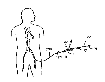
Figure 1 is a schematic showing of a human being having a tissue-
penetrating catheter system of the present invention percutaneously
inserted via a femoral entry site.
Figure 1 a is a broken, side elevational view of a first embodiment of
a tissue-penetrating catheter of the present invention.
Figure 1 b is an enlarged view of the distal end of the catheter of
Figure 1 b.
Figure 1 b' is a broken, side view of the catheter body construction of
the catheter shaft of a tissue penetrating catheter of the present invention.
Figure 1 b" is a detailed view of the braided construction of the
catheter shaft of Figure 1 b'.
Figure 1 c is a cross sectional view through line 1 c-1 c of Figure 1 a.
Figure 1 d is a cross sectional view through line 1 d-1 d of Figure 1 a.
-14-
CA 02324304 2000-09-18
WO 99!49793 PCTNS99/07115
0 Figure 1 a is an enlarged, side elevational view of the needle
housing/stabilizer assembly of the catheter of Figure 1 a.
Figure 1f is a cross sectional view through line 1f 1f of Figure 1e.
Figure 1 f is a cross sectional view through line 1 f-1 f of Figure 1 f.
Figure 2 is a representation of the intravascular ultrasound image that
is obtained when the tissue-penetrating catheter of Figure 1 a is positioned
within a coronary vein and properly oriented/aimed such that deployment of
its tissue penetrating member will form a penetration tract (i.e., a
passageway) from the coronary vein to an adjacent coronary artery.
Figure 3 is a representation of the intravascular ultrasound image
which is obtained when the tissue-penetrating catheter of Figure 1 a is
positioned within coronary vein and improperly oriented/aimed such that
deployment of its tissue penetrating member will not form a passageway
from the coronary vein to the adjacent coronary artery.
Figure 4 is a side elevational view of a subselective sheath and
i 5 accompanying introducer that are useable in combination with the tissue-
penetrating catheter of the present invention.
Figure 4 a is a side elevational view of a dilator that is insertable
through and useable in conjunction with the subselective sheath of Figure
4.
Figure 5 is a partial longitudinal sectional view of the subselective
sheath of Figure 4 having the dilator of Figure 4 a operatively inserted
therein.
Figure 5a is an enlarged, cross sectional view through line 5a-5a of
Figure 5.
'~5 Figure 6 is an enlarged, longitudinal sectional view of the distal
portion of the subseiective sheath of Figure 4.
Figure 7 is a side elevational view of the tissue puncturing needle
member of the tissue-penetrating catheter of Figure 1 a.
-15-
CA 02324304 2000-09-18
WO 99/49793 PCTNS99/07115
0 Figures 8a is an enlarged, side elevational view of the distal end of
the needle member of Figure 7.
Figure 8b is an enlarged top view of the of the distal end of the
needle member of Figure 7.
Figures 9 & 9a show the hand piecelneedle controller and distal end,
respectively, of tissue-penetrating catheter of Figure 1 a with its tissue
penetrating needle member in its retracted position.
Figures 10 & 10a show the handpiece/needle controller and distal
end, respectively, of tissue-penetrating catheter of Figure 1 a with its
tissue
penetrating needle member in its fully advanced position.
Figure 10 d is a side elevational view of an optional rotation-inhibiting
key insert and corresponding keyed needle member which may be
incorporated into the tissue-penetrating catheters of the present invention
to prevent the tissue-penetrating needle memberfrom rotating relative to the
body of the catheter.
Figure 10 d' is a cross sectional view through line 10 d'-10 d' of
Figure 10 d.
Figure 10 d" is a cross sectional view through line 10 d"-10 d" of
Figure 10 d.
Figure 10 a is a side elevational view of an optional rotation-inhibiting
:ZO oval insert and corresponding oval shaped needle member which may be
incorporated into the tissue-penetrating catheters of the present invention
to prevent the tissue-penetrating needle memberfrom rotating relative to the
body of the catheter.
Figure 10 e' is a cross sectional view through line 10 e'-10 e' of
'~5 Figure 10 e.
Figure 10 e" is a cross sectional view through line 10 a"-10 e" of
Figure 10 e.
-16-
CA 02324304 2000-09-18
WO 99/49793 PCT/US99/07115
0 Figure 10 f is a partial longitudinal sectional view of a tissue-
penetrating catheter device of the present invention incorporating an
optional locking collar apparatus for preventing the tissue penetrating needle
member from rotating relative to the catheter body when the needle member
is in its retracted position.
Figure 10 f ' is an enlarged view of region 10 f ' of Figure 10 f.
Figure 10 g is a side elevational view of a tissue-penetrating catheter
of the present invention having a laterally deployable needle stabilizer
disposed in its "stowed' position.
Figure 10 g' is a side elevational view of a tissue-penetrating
catheter of the present invention having a laterally deployable needle
stabilizer disposed in its "active" position.
Figure 11 is a side elevational view of a coronary sinus guide
catheter/introducer assembly of~the present invention.
Figure 11 a is a cross-sectional view through line 11 a-11 a of Figure
t5 11.
Figure 11 b is an enlarged, longitudinal sectional view of the proximal
end/hemostatic valve of the coronary sinus guide catheter shown in Figure
11.
Figure 11 c is broken, side elevational view of the introducer of the
coronary sinus guide catheter/introducer assembly.
Figure 12 is an enlarged, cross-sectional view through a coronary
artery and adjacent coronary vein, showing the typical difference in diameter
of the artery and vein, and delineating a preferred Acceptable penetration
zone (APZ) wherein the arterio-venous bloodflow passageways of the
present invention are formed.
Figures 13a-13x are schematic, step-by-step showings of a preferred
method for performing a percutaneous, in situ coronary arterio-venous
-17-
CA 02324304 2000-09-18
WO 99/49793 PCT/US99/07115
0 bypass (P1CAB) procedure to bypass a blockage in the proximal Left
Anterior Descending coronary artery, using a a vein-to-artery approach.
Figures 14 a-14m are schematic, step-by-step showings of a
preferred method for performing a percutaneous coronary venous
arterialization (PICVA) procedure to provide retrograde arterial bloodflow
through a coronary vein.
Detailed Description of Preferred Embodiments
The following detailed description, and the drawings to which it refers,
are provided for the purpose of describing and illustrating certain preferred
embodiments or examples of the invention only, and no attempt has been
made to exhaustively describe all possible embodiments or examples of the
invention. For example, the tissue penetrating catheter of this invention may
be utilized is numerous locations in the body to reliably access organs,
tissue or other structures to deliver therapeutic substances or procedures.
Thus, the following detailed description and the accompanying drawings are
not intended to limit, in any way, the scope of the claims recited in this
patent application and any patents) issuing therefrom.
A. The Catheter Sysfem:
Referring generally to Figures 1-12, a presently preferred catheter
system of the present invention generally comprises i) a tissue-penetrating
catheter component 10 (Figures 1-3 and 7-10 a), ii) a subselective
sheathlintroducer component 100 (Figures 4-6) and iii) a coronary sinus
guide catheter/introducer component 200 (Figures 11-11 c). Each of these
components is described in substantial detail herebelow. These
components of the catheter system may be packaged together in a single
kit, or may be provided in separate packages to permit the operator to mix
and match component sizes in accordance with the particular anatomy of
the patient, the size of the channels to be formed, the types of connectors
and/or stents andlor blockers to be used, etc.
-18-
CA 02324304 2000-09-18
WO 99/49793 PCTNS99/07115
0 I, The Tissue-Penetratingi Catheter Component of the Catheter
System:
Referring to Figures 1-3 and 7-10 there is shown a tissue-penetrating
catheter device 10 which is insertable into the vasculature of a mammalian
patient and useable to form passageways (e.g., puncture tracts) between
the blood vessel in which the distal end of the catheter device 10 is situated
and another blood vessel or other anatomical structure. This catheter
device 10 generally comprises an elongate, flexible catheter body 12 having
a proximal portion 12P of a first diameter D, and a distal portion 120 of a
second diameter D2 which is smaller than the first diameter D,. The catheter
body 12 has two (2) lumens 14,16 which extend longitudinally therethrough.
The first lumen 14 is sized and configured to permit a standard commercially
available IVUS catheter (e.g., those available from Endosonics of Rancho
Cordova, CA; CVIS of Natick, MA or Hewlett-Packard of Andover, MA) to be
inserted therethrough and slidably disposed therewith. The second lumen
t 5 16 is sized and configured to house a tissue penetrating needle member 30
(see Figures 7, 9 and 10) which is alternately moveable between i) a
retracted position (Figures 9-9a) wherein the distal end DE of the needle
member 30 is contained within the catheter body 12, and ii) an extended
position (Figures 10-10a) wherein the needle member 30 is advanced out
of the catheter body 12 so as to penetrate through the walls of the blood
vessels and through any intervening tissue located between the blood
vessels. '
a. Orientation Structure:
An orientation structure 36 and tip member 38 are formed integrally
with or mounted on the distal end of the catheter body 12, as shown in
Figures 1 b, 9a and 1 Oa. The orientation cage 36 comprises first 40, second
42 and third 44 strut members which extend longitudinally between the
distal end of the catheter body 12 and the proximal end of the distal tip
-19-
CA 02324304 2000-09-18
WO 99/49793 PCT/US99/07115
0 member 38. The first strut member 40 is in direct longitudinal alignment
with
a needle outlet opening 46 formed in the side of the catheter body 12
through which the tissue penetrating needle member 30 is advanced. The
second and third strut members 42, 44 are located at equally spaced
distances from the first strut rriember 40, while the distance between the
second and third strut members 42,44 is less than the distance between
either of those second and third strut members 42, 44 and the first strut
member 40. Such disparate (e.g., unequal) radial spacing of these strut
members 40, 44 and 46 allows the operatorto easily identify and distinguish
the first strut member 40 from the other two strut members 42, 44 by way of
the image received from an IVUS catheter positioned within the orientation
structure 36. Thus, in this manner, the operator may selectively rotate the
catheter body 12 until the first strut member 40 is directly aligned or
juxtapositioned with the target blood vessel into which the needle member
30 is to be advanced. An illustration of this technique is shown in Figures
2 and 3. Figure 2 shows the IVUS image which is obtained when the tissue-
penetrating catheter 10 is properly rotated such that the first strut member
40 is aligned with the target artery A and the needle member 30 will
advance into such target artery A. Figure 3 shows another situation where
the tissue-penetrating catheter 10 is not properly rotated, the first strut
member 40 is not aligned with the target artery A and the needle member
30, if advanced, would not enter the target artery A.
It will be appreciated that the disparate distancing of the strut
members 40, 42, 44 is only one possible way of rendering the first strut
member 40 distinguishable from the other two strut members 42, 44.
Alternatively, the size or configuration of the first strut member could be
different so as to produce a distinguishable ultrasound image orthe material
or surface characteristics of the first strut member 40 could be made
different from the other two strut members 42, 44 such that the first strut
-20-
CA 02324304 2000-09-18
WO 99149793 PCTNS99/07115
0 member 40 would reflect more or less ultrasound than the other two strut
members 42, 44 thus producing an ultrasound image which is
distinguishable from the images produced by the other two strut members
42, 44. It will also be appreciated that only one strut member may be
required to provide a distinguishable element to aid catheter orientation, or
alternatively two strut members may be positioned to delineate a zone within
which the tissue penetrating member may be deployed, or other procedure
conducted.
b. Distal Tlp Member:
The distal tip member 38 is preferably of blunt tipped configuration
and is formed of smooth soft material (e.g., PEBAX having a durometer
hardness of 35D) so as to minimize trauma to the vasculature as the tissue
penetrating catheterdevice 10 is advanced or otherwise manipulated about.
A hollow lumen 39 may extend longitudinally through the tip member 38, in
alignment with the first lumen 14 of the catheter body 12, such that an IVUS
catheter or other device.such as a guidewire may be advanced from the first
lumen 14, through the orientation structure 36, through the distal tip lumen
38 and distally beyond the catheter device 10. This permits the operator to
use the IVUS catheter to explore areas which are ahead of the distal end of
the tissue-penetrating catheter without having to advance the tissue-
penetrating catheter from its then-present position. It also permits the
catheter device 10 to be introduced to the vasculature in the preferred Nover
the wire" manner.
c_. Tissue Penetrating! Needle Member'
The tissue penetrating element of the tissue penetrating catheter may
comprise a sharp tipped needle 30 as shown in Figures 7, 8a and 8b. This
needle 30 includes a proximal shaft 30p formed of stainless steel
hypotubing and a resilient, curved distal portion 30d formed of a resilient
material or, more preferably, a material such as NiTi alloy. Preferably a
-21-
CA 02324304 2000-09-18
WO 99/49793 PCTNS99/07115
0 lumen 31 extends longitudinally through the proximal shaft 30p and the
curved distal portion 30d.
The particular radius of curvature of the curved distal portion 30d may
be an important factor in determining the trajectory and path of the needle
tip as it advances and the point at which the needle tip will stop when in its
fully advanced position.
The distal tip of the needle member 30 is preferably sharpened so as
to easily penetrate through the walls of the blood vessels and any
intervening tissue located therebetween. One preferred needle tip
configuration is the lancet type bevel 36 shown in Figures 8a and 8b. This
~0 lancet type bevel comprises a first radial surface 36a and a second radial
surface 36b. Such lancet type tip 36 provides excellent tissue-penetrability
and retains its sharpness after multiple retractions into/advancements from
the catheter. In practice it may be important for the material surrounding the
lumen of the needle, particularly at the distal tip of the needle, and
1.5 particularly the heel of the needle lumen 36c, to be smooth and free of
rough edges or burrs. This allows smooth passage of devices, such as
guidewires, through the needle lumen.
In many applications, the controllability and aiming of the needle
member 30 may be enhanced by constraining the needle member 30 such
that it will remain in a preferred plane or acceptable penetration zone APZ
as shown in Figure 12, as it is advanced from the catheter. In embodiments
where a curved needle member 30 is advanced out of a side aperture in the
catheter ( e.g., the embodiment shown in Figure 10a), any rotation of the
needle member 30 prior to, during or after advancement of the needle
~5 member 30 can cause the distal end of the curved needle member to
deviate from or move out of the intended plane or acceptable penetration
zone APZ. In this regard, the potential for such unwanted lateral movement
of the distal end of the needle member 30 may be prevented or substantially
-22-
CA 02324304 2000-09-18
WO 99/49793 PCT/US99/07115
0 limited by providing a stabilizer to prevent or substantially limit the
amount
of rotation that the needle member 30 may undergo relative to the catheter
body 12 or to otherwise prevent or deter the needle member from deviating
from a predetermined acceptable acceptable penetration zone APZ (Figure
12) as it is advanced from the catheter 10. In particular, by preventing or
limiting the rotation of the needle member 30 within the needle lumen 16,
the curved distal portion of the needle member will be deterred from
deviating from its intended path of advancement as it is extended laterally
from the catheter body 12 (see Figure 10a). Such prevention or limitation
of the potential for rotation or Lateral movement of the needle member 30
l0 may be accomplished in any suitable way. As described in detail
herebelow, specific apparatus which may be incorporated into the catheter
device 10 to prevent or deter rotation or lateral movement (i:e., "wagging"
or "flopping") ofthe needle member 30 during or after its advancement from
the catheter body 12, include:
t5 a) a curved needle housing 60 which has a curve at its distal end
which mates with the preformed curvature of the needle member 30 to deter
rotation (see Figures 9-10f);
b) engaged surfaces 76, 77 formed on the needle member 30 and
surrounding catheter body 12 to lock or deter rotation of the needle member
20 30, examples of such engaged surfaces 76, 77 including but not necessarily
being limited to i) a tongue in groove or key in key-way arrangement (see
Figures 10d-10d") or ii) an oval to oval arrangement (see Figures 1 Oe-10"),
etc;
c) a steering mechanism for causing the distal portion of the
~5 catheter body 12 to curve in the lateral direction in which the needle
member 30 is intended to advance so as to cause the preformed curve of
the needle member 30 to mate with the induced curvature of the catheter
body 12; and,
-23-
CA 02324304 2000-09-18
WO 99/49793 PGTNS99/07115
0 d) a needle guide member 500 which is laterally projectable from the
catheter body 12 in the area of the needle outlet aperture 46 to support the
needle member 30 andJor to form a lateral extension of the needle lumen
16 so as to create a lateral curve in the needle lumen which mates with the
preformed curvature of the needle member 30 (see Figure 10 g).
i. Curved Needle Housincr to Deter RotationlLateraJ Deviation of
Extended Needle
An example of a preferred curved needle housing 60 mountable
within the needle lumen 16 is specifically shown in Figures 1b-1f. Such
needle housing 60 comprises a curved, rigid tube. A tubular liner 61 may
be disposed within, and may extend from either end of, the curved needle
housing 60. Such tubular liner 61 may be formed of a three-layer composite
wherein the inner layer is a lubricious polymer material (e.g.,
polytetrafluoroethylene (PTFE)), the middle layer is a structural polymer
material (e.g., polyimide) and the outer layer is an adhesive material which
will band to the inner surface of the curved needle housing 60 and to the
inner surface of the needle lumen 16 at either end of the housing 60 (e.g.,
polyurethane adhesive). When the needle member 30 is in its retracted
position (Figures 9 and 9a), and during advancement, the portion of the
needle member which resides within the needle housing 60 will remain in a
slightly curved state in conformance to the slightly curved configuration of
the needle housing 60. This serves to deter the needle member 30 from
rotating relative to the catheter body 12 and/orfrom undergoing uncontrolled
movement (i.e., "flopping") out of the intended acceptable penetration zone
APZ, during or after advancement from the catheter. This prevention or
deterrence from rotation of the needle member 60 allows the operator to
control the orientation of the lancet type or other bevel formed in the needle
tip, and also enhances the operator's ability to predict the precise position
of the needle tip by eliminating or minimizing the uncontrolled side-to-side
-2a-
CA 02324304 2000-09-18
WO 99/49793 PCTNS99/07115
0 movement of the needle. To facilitate the desired positioning and
orientation of the curved needle housing 60 during manufacture of the
catheter 10, a locator member 62 may be attached to the needle housing 60
and incorporated into the catheter body 12 as shown in Figures 1 b, 1 e, 1 f,
1 f, 9a and 10a. This locator member 62 comprises a rigid disc 64 which is
transversely positionable within the catheter body, having a first bore 66 and
a second bore 68 extending longitudinally therethrough. A chamfered edge
69 is formed about the proximal, end of the first bore 66, as shown in Figures
1f and 1f. During manufacture of the catheter body 12, a rod or mandrel is
inserted through the first bore 66 of the locator and into the first lumen 14
of the proximal catheter body portion 12P and the curved needle housing 60
having a tubular liner 61 extending therethrough and protruding for either
end, are inserted through the second bore 68 and into the second lumen
16 of the proximal catheter body portion 12P. Thereafter, a distal plastic
tube is advanced about the locator, a tubular polymer skin 73 is applied, and
the composite is then heated to form the distal portion of the catheter body
12, as shown.
ii. Fractionally Enaacred Surfaces of Needle Member and Cafheter to
Deter RotationlLateral Deviation of Extended Needle:
As an alternative to, or in addition to, the use of the curved needle
housing 60 as a means for preventing rotation of the needle member 30 and
for providing more accurate and stable deployment of the needle member
30, the needle member 30 and at least a portion of the second lumen 14
may incorporate engaged surfaces which are fractionally engaged to one
another so as to prevent or deter rotation of the needle member 30 within
the needle lumen 16. Examples of such engaged surfaces 76, 77 include
a keylkey-way design shown in Figures 10d-10d" or an oval/oval design
such as that depicted in Figures 10e-10e".
-25-
CA 02324304 2000-09-18
WO 99/49793 PCTNS99/07115
0 With specific reference to the showings of Figures 10d-10d", the
key/keyway method of preventing independent rotation of the needle
member 30 may be effected by use of a key-way element 76 in combination
with a keyed needle 30,~,~,. The key-way element 76 comprises a tubular
member which has a key-way shaped lumen 77 with a key portion 79
extending longitudinally therethrough. The keyed needle 30k,Y comprises a
hollow needle of the type described hereabove and shown in Figures 7-8b
having a longitudinally extending rail or key member 33 formed upon a
segment thereof. The key member 33 may be formed as a portion of the
needle wall or may alternatively comprise a separate member, such as a
section of hypotube, affixed to the side of the needle wall. The keyed
needle 30k~, is sized and configured to be advanced and retracted through
the lumen 77 of the key-way housing, with the key member 31 being
disposed within the key portion 79 of the lumen 77. In this manner the
keyed needle member 30,~y is longitudinally advanceable and retractable,
but can not be rotated within the lumen 77 due to the engagement of the
needle key member 31 with the key portion 79 of the lumen 77. The key-
way element 76 is provided with a stabilizer 78 which is substantially the
same as the needle housing stabilizer 62 described above and shown in
Figures 1e-1f, and the key-way element 761stabilizer 78 assembly may be
2.0 installed and mounted within the catheter body at the time of manufacture
in the same manner as described hereabove with respect to the needle
housing 60/stabilizer 62 assembly shown in Figures 1e-1f. This key-way
element 76/stabilizer 78 assembly is typically installed and mounted in the
catheter body 12 proximal to the location of the needle housing 60/locator
62 assembly shown in Figures 1 e-1 f but near enough to the distal end of
the catheter device 10 to prevent the portion of the needle adjacent its
distal
end from undergoing untoward rotation within the catheter body 12 during
the catheter insertion procedure.
-26-
CA 02324304 2000-09-18
WO 99/49793 PCTNS99/07115
0 With specific reference to the oval/oval arrangement shown in
Figures 10e-10e", the device 10 may incorporate an oval shaped needle
housing 76an in combination with an oval shaped needle 300. The oval
shaped needle housing 76~ comprises a tubular member positioned within
the needle lumen 16 and having an oval shaped lumen 778n extending
longitudinally therethrough. The oval shaped needle 30~, comprises a
hollow needle of the type described hereabove and shown in Figures 7-8b
having an oval, ovoid or other non-round cross-sectional configuration. The
oval shaped needle 30~, is sized and configured to be advanced and
retracted through the lumen 77s;, of the oval shaped needle housing 76~,
but can not be rotated within the lumen 77~ due to the engagement of the
oval shaped needle member 30~, with the oval shaped wall of the housing
lumen 77a". The oval shaped needle housing 76an is provided with a locator
78 which is substantially the same as the needle housing locator 62
described above and shown in Figures 1e-1f, and the oval shaped needle
housing 768~/locator 78 assembly may be installed and mounted within the
catheter body 12 at the time of manufacture, in the same manner as
described hereabove with respect to the needle housing 60/locator 62
assembly shown in Figures 1e-1f. This oval shaped needle housing
76a~llocator 78 assembly will .typically be installed and mounted in the
catheter body 12 proximal to the location of the needle housing 60/locator
62 assembly shown in Figures 1e-1f, but near enough to the distal end of
the catheter device 10 to prevent the portion of the needle 30~, adjacent its
distal end from undergoing untoward rotation within the catheter body 12
during the catheter insertion procedure.
iii. Laterally Deployable Needle Guide to Deter RotationlLateral
Deviation of Extended Needle:
Figures 10g and 10g' show an example of a needle guide member
500 which may be caused to project or extend laterally from the catheter
-27-
CA 02324304 2000-09-18
WO 99/49793 PCTNS99/07115
0 body 12 in the area of the needle outlet aperture 46 to stabilize and guide
the advancing needle member, thereby deterring lateral or side-to-side
movement of the needle member 30 and further constraining the path which
will be followed by the advancing needle. The deployment of such needle
guide member 500 may also give rise to a lateral extension of the needle
lumen 16 which mates with the preformed curve of the needle member 30
to prevent rotation of the needle member 30 in essentially the same manner
as the curved needle housing 60 described above.
The particular laterally deployable guide member 500 shown in
Figures 10g and 10g' is an inflatable annular member that is connected to
an inflation fluid lumen 502 that extends through the catheter body 12 to
permit inflation fluid to be infused and withdrawn from the inflatable guide
member 500. When deflated (Figure 10g) the guide member 500 will nest
within a depression or cut out region in the catheter's outer wall thereby
assuming a configuration that is substantially flush with the outer surface
504 of the catheter body 12. When inflated (Figure 1 Og') the guide member
500 will form an annular support collar around the tissue penetrating
member 30 as it advances laterally from the catheter body. The surfaces)
of the inflatable guide member 500 that may be brushed against or
contacted by the tip of the tissue penetrating member as it advances out of
the outlet opening 46 may be armored or coated with a metal foil or other
material that will resist puncture by the tip of the tissue penetrating member
30.
iv. Steerable Cathefer Body to Defer RofafionlLateral Deviafion of
fhe Extended Needle Member:
~5 The catheter body 12 may be provided with a mechanism for inducing
a curve or bend in the region of the catheter body 12 proximal to the needle
outlet aperture 46 to cause the portion of the needle lumen 16 proximal to
the outlet aperture 46 to assume a curvature which mates with the curved
-28-
CA 02324304 2000-09-18
WO 99/49793 PC"f/US99/07115
0 shape to which the needle member 30 is biased, thereby deterring rotation
of the needle member 30 within the catheter in the same manner described
above with respect to the curved needle housing 60. The mechanism by
which the catheter body 16 may be induced to curve may be any suitable
catheter steering apparatus known in the art, such as an internal pull wire
or spine member formed on shape memory alloy which is alternately
transitionable between a straight configuration and a curved configuration.
v. Rotational Lockincr ofNeedle Member When Refracted to Maintain
Correct Orientation and Enhance Tor~qme Transfer
It is desirable for the proximal shaft of the tissue-penetrating catheter
10 to be endowed with enough structural integrity to transmit torque to the
distal end of the catheter, as necessary for precise rotational orientation
and
aiming of the catheter device 10 before advancement of the needle member
30 therefrom. Also, in many applications, it is desirable for the needle
member 30 to be maintained in a predetermined rotational orientation within
the catheter body 12 prior to advancement of the needle member 30 from
the catheter 10 (i.e., while the needle member 30 is still in its retracted
state). In many applications, it is also desirable to minimize the diameter of
the catheter body 12 to allow it to pass through small blood vessel lumens.
Each of these three (3) objectives may be achieved by rotational locking of
the needle member 30 within the catheter body prior to its advancement
from the catheter, as such rotational locking i) prevents unwanted needle
rotation, ii) enhances the efficiency of torque transfer to the distal end of
the
catheter body 12 and thereby the needle, and iii) does not add any mass or
additional diameter to the catheter body 12.
2.5 Figures 10f 10f show a needle locking collar assembly 520, which
comprises an enlarged region 522 formed within the needle lumen 16,
wherein a first locking collar member 524 and second locking collar member
526 are located. The first locking collar member 524 is stationarily affixed
-29-
CA 02324304 2000-09-18
WO 99/49793 PCT/US99/07115
0 to the catheter body 12 and has cavities or grooves 528 formed in the distal
surface thereof and a central aperture through which the needle member
30~, may be advanced and retracted. The second locking collar member
526 is affixed to the needle member 30~, and has projections 530 extending
from the proximal surface thereof. The projections 530 are sized, located
and configured to be received within the grooves 528 of the first collar
member 524 when the needle member 30~, is in its retracted position,
thereby frictionally locking the needle member 30~, to prevent its rotation
relative to the catheter body 12. However, when the needle member 30~,
is in its extended position, the projections 530 will not be inserted within
the
grooves 528, and the collar assembly 520 will not prevent the needle
member 30~, from rotating within the catheter body 12. It will be
appreciated that these stabilizing devices may be employed at various
points along the length of the catheter body, including the proximal, medial,
distal or needle housing portion.
I S d. HandplecelNeedle Controller:
A handpiece/needle controller 75 is mounted on the proximal end of
the Catheter body 12, and is useable to control the rotational orientation of
the catheter body 12 and the advancement/retraction of the needle member
30.
Also this handpiece/needle controller 15 has a proximal port 27
formed on its proximal end through which a small guidewire (e.g., a 0.0010-
0.016 inch diameter wire) may be advanced through the lumen 31 of the
needle member 30, a first side port 21 through which a large guidewire (e.g.,
a 0.030-0.040 inch diameter wire) may be advanced through the first lumen
14 when that first lumen 14 is not occupied by an IVUS catheter, and a
second side port 23 through which a flush solution maybe infused into the
catheter's second lumen 16 outside of the needle member 30 disposed
therein.
-30-
CA 02324304 2000-09-18
WO 99/49793 PG"T/US99/07115
0 e. Catheter Bodv:
The catheter body 12 includes a relatively stiff proximal section 12a,
a medial section 12b, and a distal section 12cshown in Figs 1A and 1 B. The
catheter body exhibits varying flexibility and torque strength along its
length,
and may incorporate reinforcement members such as a reinforcement braid
member which imparts structural integrity as well as enhancing the ability of
the catheter body to transmit torque. A hand piece 15 is attached to the
proximal end of the proximal section 12a, as shown. In the preferred
embodiment the hand piece 15 and proximal section 12a are approximately
115cm in length. The medial section extends approximately 25cm
terminating approximately 2cm from the distal section 12c. The proximal
and medial sections of the catheter contain a braided component 50 as
shown in Figs. 1 B' and 1 B", encased in a polymer material (e.g. Pebax,
nylon, polyurethane, polyester or PVC) extruded to form the inner lumen
50b and out jacket 50a of catheter body 12.
It has been determined that material expansion and changes in the
physical properties of certain materials may occur after the catheter 10 is
inserted into the patient's body and warmed from room temperature to body
temperature. This material expansion and changes in the physical
properties of certain materials can result in variation in the tolerances and
sizing of the catheter 10 (e.g. elongation or shrinking) and can thus give
rise
to an unwanted modification of the position of the tissue penetrating
member 30. This could, in at least some cases, interfere with the precise
aiming and advancement of the tissue penetrating member as desired. Fig.
1 B" illustrates the braid angle A and pick count PC of the catheter braid 50.
The "pick count" PC of the braid is, as is well known in the art, a function
of
the braid angle A (i.e., the greater the braid angle the more picks per inch).
-31-
CA 02324304 2000-09-18
WO 99/49793 PC'f/US99/0~115
0 Also, the torque transmission and stiffness of the braided section 50
is a function of the braid angle (i.e., a braid angle of 90 degrees provides
maximum torque transfer and a braid angel of 0 degrees provides minimum
torque transfer). Catheters used in the present invention that have exhibited
this phenomenon have braid angles A that result in a pick count of 50 - 70
picks per inch. However, applicant has determined that by decreasing the
braid angle A of the braid 50 within the proximal and medial sections of the
catheter 10 to result in a pick count of 20 - 30 picks per inch, it is
possible
to minimize oreliminate the unwanted longitudinal expansion ofthe catheter
and/or its components, while retaining sufficient torque transmission and
10 acceptable stiffness to accomplish the procedures for which the catheter 10
is intended (examples of such procedures are illustrated in Figures 13a-14m
herebelow). This variation in braid angle or picks per inch may vary
depending on the material of construction of the catheter and/or the braid
fiber, and the diameter of the catheter body.
II. The Coronay Slnus Guide Com~~oonent of the Catheter
S s em:
Figures 11-11c show a preferred coronary sinus guide
catheter/introducer assembly 200, which comprises a) a flexible coronary
sinus guide catheter 203 that has a curved distal portion 204, a proximal
assembly 214 mounted on the proximal end of the flexible catheter body
203, and a hollow lumen 202 extending longitudinally therethrough and b)
an introducer 213 that has a tapered, soft distal portion 213d that protrudes
out of and beyond the distal end DE of the guide catheter 203 and a
guidewire lumen 215 that extends longitudinally through the introducer 213
to permit the guide catheter/introducer assembly to be advanced over a
guidewire GW as described more fully herebelow in connection with a
preferred method of using the catheter system 10.
CA 02324304 2000-09-18
WO 99/49793 PGTNS99/07115
0 A reinforcement braid 212, such as a wire braid, is formed within a
portion of the catheter body 203 but terminates approximately 2 to 5
centimeters from the distal end DE. In this manner, the reinforcement braid
212 will prevent kinking and improve torque strength of the proximal portion
of the catheter body 203, and the curved portion thereof, up to a location at
about 2 to 5 centimeters from its distal end DE.
The proximal assembly comprises a rigid body 248 through which the
lumen 202 extends, and upon which a proximal port 250 is formed to permit
the guide introducer 213, subselective sheath 100 (Figures 4-6), tissue-
penetrating catheter device 10 (Figures 1 and 9-10), or other catheters,
:d 0 guidewires and/or devices (e.g., blocker delivery catheter, channel
connector delivery catheter, channel enlarging device, etc...) to be inserted
through the lumen 202 of the coronary sinus guide catheter 200. A
hemostatic valve 244, such as a cross-cut resilient membrane, a slit-cut
resilient membrane, or a flapper valve) is positioned transversely within the
1.5 lumen 202 of the proximal assembly 214 to prevent blood from backflowing
out of the proximal port 250 when no catheter or other device is inserted
therethrough and to prevent or minimize the amount of blood which may
leak out of the proximal port 250 when a catheter or other device is inserted
therethrough. A side port 246 is formed on the proximal assembly 214 to
~0 permit preparation fluid to be infused or injected into or through the
lumen
202. A plurality of side apertures 210 are formed in the wall of the catheter
body 203 near its distal end to allow pressure relief in the event that a
radiographic contrast medium or other fluid is injected.
III. The Subselective Sheath Com~~onent of the Catheter Svstem
~5 As shown in Figures 4-6, a preferred subselective sheath 100 of the
present invention comprises a .flexible sheath body 102 having a proximal
hub 104 and a lumen 106 extending longitudinally therethrough. A
reinforcement braid 108 is formed within the catheter body 102 to prevent
-33-
CA 02324304 2000-09-18
WO 99/49793 PCT/US99/07115
0 kinking and improve torque strength. Such reinforcement braid terminates
distally at 0.1-1.0 centimeter from the distal end of the catheter body 102.
A gradual taper 110 is formed about the distal end of the sheath body's
outer surface to such that the sheath 100 will taper to a flush transition
with
the distally protruding portion 111 d of its introduces 111. The lumen 202 has
an inner diameter D1 which is substantially the same as the outer diameter
of the introduces 111 that is initially inserted through the lumen 106. The
introduces 111 has a guidewire lumen 109 that extends longitudinally
therethrough to permit the sub~selective sheath/introducer assembly to be
advanced over a previously inserted guidewire GW (e.g., a 0.035 inch
guidewire). The outer diameter of the sheath 100 is sized to be advanced
and retracted through the lumen 202 of the coronary sinus guide catheter
200 (Figures 11-11b). The preferred method of using this subselective
sheath 100 and introduces 111 are described in detail herebelow with
respect to the methods of the present invention.
B. Preferred Methods for Using the Catheter System:
The present invention also includes methods for using this catheter
system described hereabove (or any other catheter system or devices that
may be suitable to carry out the desired purpose), in conjunction with other
apparatus such as guidewires, channel enlarging catheters/devices, channel
connecting catheters/devices and vessel blocking catheters/devices to
perform percutaneous, in situ coronary arterio-venous bypass procedures
by way of a vein-to-artery approach, such method being fully described
herebelow and shown in step-by-step fashion in Figures 13a-13x and 14a-
14m.
The catheter system described hereabove and shown in Figures 1-
11 b is useable in conjunction with a fluoroscope, an IVUS imaging catheter,
a coronary sinus access catheter (e.g., a standard angiographic catheter),
a channel-enlarging catheterdevice, lumen-blocking device(s), a 0.035 inch
-34-
CA 02324304 2000-09-18
WO 99/49793 PCT/US99/07115
0 diameter guidewire, and one or more 0.014 inch diameter guidewire(s) to
perform various revascularization procedures including, as described in
detail herebelow, a Percutaneous In Situ Coronary Artery Bypass (PICAS)
procedure as well as a Percutaneous In Situ Coronary Venous
Arterialization (PICVA) procedure. It will be appreciated that, in addition to
the particular PICAS and PICVA examples described in detail herebelow,
the catheter system of the present invention may also be useable to perform
various other procedures such as directed drug delivery procedures of the
type described in co-pending United States Patent Application SN
09/048,147 and other revascularization procedures.
I. A Preferred Method for Performing the PICAB Procedure:
Figures 13a-13x show, in step-by-step fashion, an example of a
PICAS procedure wherein the catheter system 10 of the present invention
is used for the purpose of bypassing a blockage located in the proximal
portion of the Anterior Descending Coronary Artery (LAD) of a human
patient. In this PICAS procedure, a coronary sinus access catheter (e.g.,
a standard angiographic catheter such as the modified Simmons-type
angiographic catheter available from Cook Cardiology, Bloomington,
Indiana) is initially inserted through a femoral vein or external jugular vein
approach, using standard percutaneous catheter insertion technique. After
such initial percutaneous catheter insertion has been accomplished, the
PICAS procedure proceeds as follows:
First Ste~~ ~ Coronary Sinus Access l Introduction of First Guidewire
As shown in Figure 13a, an arterial blockage AB to be bypassed is
located in the left anterior descending coronary artery (LAD). The coronary
sinus access catheter 500 is advanced into the coronary sinus CS, as
shown in Figure 13b, to assist in the placement of a 0.035 inch diameter
guidewire GW, into the great cardiac vein (GCV) and anterior interventricular
-35-
CA 02324304 2000-09-18
WO 99/49793 PCTNS99/07115
0 vein (AIV). This guidewire GW, can be pre-loaded in the lumen of the
coronary sinus access catheter 500 or can be advanced through the lumen
of the coronary sinus access catheter 500 after it has been positioned inj the
coronary sinus, as a separate step. Thereafter, the coronary sinus access
catheter 500 is removed, leaving the 0.035 inch guidewire GW, in place.
Second Ste~~~ Infroducton of Coronary Sinus Guide Catheter / AlV
Access:
As shown in Figure 13c-13d, the coronary sinus guide catheter 200
with introduces sheath 100 disposed within or through its lumen 202, is
advanced over the 0.035 inch ,guidewire GW, until the tip of the coronary
sinus guide catheter 200 is past the "mouth" of the coronary sinus. The
introduces sheath 100 is then removed, leaving the coronary sinus guide
catheter 200 in place, in the manner shown in Figure 13d.
Third Step- Introduction & Aiminqi of Tissue ~~enetratina Catheter.
As shown in Figure 13e, the tissue-penetrating catheter 10 is then
inserted over the pre-positioned 0.035 inch guidewire GW, , through the
lumen 202 of the coronary sinus guide catheter 200, and is advanced using
fluoroscopy to a position distal to the arterial blockage AB being bypassed.
The 0.035 inch guidewire GW, is then extracted and removed from the first
lumen 14 of the tissue-penetrating catheter 10 and an IVUS imaging
catheter (not shown) is then advanced through that first lumen 14 until the
IVUS transducer resides within the hollow interior space of the orientation
structure 36. The IVUS catheter is then used to receive a 360 degree
ultrasound image from a vantage point within the interior space of the
orientation structure 36. Such image enables the operator to see both the
resident vessel (the AIV) and the target vessel (the LAD), as well as the
reflections or artifacts from the three strut members 40, 42 &44 of the
orientation structure 36. Because of the disparate distancing between the
strut members 40, 42 &44, the reflections or artifacts produced by the strut
-36-
CA 02324304 2000-09-18
WO 99/49793 PGT/US99/07115
0 members will form a generally °Y" shaped image as illustrated in
Figures 2
and 3 of this patent application. The reflection 40R~, produced by the first
strut member 40 is clearly distinguishable from the reflections 42Re,, 44,x,
produced by the second and third strut members 42, 43, and provides an
indication of the particular direction in which the needle member 30 will
travel when advanced from the needle outlet opening 46 in the side of the
catheter body 12. Thus, if the first strut member reflection 4ORer observed
on the IVUS image does not extend directly toward or into the lumen of the
LAD (as illustrated in Figure 3), the operatorwill rotate the tissue-
penetrating
catheter 10 until such first strut member reflection 40R~ observed on the
IVUS image does extend directly toward or into the lumen of the LAD (as
illustrated in Figure 2). This will ensure that the needle member 30 is
properly aimed to enter the LAD when advanced.
Fourth Stea: Formation of Initial Arferio-Venous Penetration Tracf
Distal to Blockage:
1 S As shown in Figure 13f 13h, the tissue penetrating needle member
30 is then advanced in the distal direction to its extended position such that
it punctures through the wall of the resident vessel (the AIV), through any
tissue which may exist between the resident vessel (the AIV) and the target
vessel (the LAD) and into the lumen of the target vessel (the LAD) at a
2.0 location downstream of the arterial blockage AB. This maneuver results in
the formation of an initial arterio-venous penetration tract PT. With the
needle member 30 in its extended position and its distal tip in the lumen of
the target vessel (the LAD), a 0.014 inch diameter guidewire GW2 is inserted
through the proximal port 2? of the tissue-penetrating catheter
25 handpiece/needle controller 15 and advanced through the lumen 31 of the
needle member 30 into the target vessel (the LAD), as shown in Figure 14h.
After the 0.014 inch diameter guidewire GW2 has been introduced into the
target vessel (the LAD) the needle member 30 is withdrawn to its retracted
-37-
CA 02324304 2000-09-18
WO 99/49793 PCT/US99/07115
0 position, leaving the 0.014 inch.diameter guidewire GW2 extending through
the initially formed interstitial passageway into the target vessel (the LAD)
as shown in Figure 14h. After the needle member 30 is withdrawn to its
retracted position, the tissue-penetrating catheter 10 is withdrawn and
removed, leaving the 0.014 inch guidewire in place (i.e., extending through
the newly formed arterio-venous penetration tract PT).
Fifth Stem: Deployment of Blocker into Vein Lumen Distal to
Blockage:
As shown in Figures 13 i-k, the subselective sheath 100 with its
introduces 111 inserted therethrough is advanced through the coronary
l0 sinus guide 200 over the large guide wire GW,. Thereafter, the introduces
111 and guidewire GW, are removed and one or more embolic blocker
members BM are introduced into the proximal end of the subselective
sheath, pushed through the lumen of the subselective sheath 100 using a
pusher rod (not shown) and expelled into the lumen of the coronary vein
(the AIV) where such embolic blocker(s) expand and engage the wall of the
vein to cause substantial occlusion and blockage of bloodflw through the
vein ath that location. Examples of such blocker members BM and their
methods of implantation are described in United States Patent Application
Serial No. 09/117,156 The 0.035 inch diameter guidewire GW, is then
removed, and an embolic blo~ker member BM is inserted into the proximal
end of the subselective sheath. A push rod is then advanced through the
lumen of the subselective sheath to push the embolic blocker member BM
out of the distal end of the subselective sheath and into its desired position
within the lumen of the coronary vein (the AIV). It is to be noted that this
blocker deployment step may be performed at this point in the procedure,
or alternatively may be delayed until a later time in the procedure. After the
distal blocker member BM has been implanted at its desired location, the
0.035 inch diameter guidewire GW, is reinserted through the subselective
-38-
CA 02324304 2000-09-18
WO 99!49793 PCTNS99/07115
0 sheath 100 and the subselective sheath 100 is then withdrawn and removed
as shown in Figure 13k.
Sixth Step: Formation of Initial Arterio-Vinous Penetration Tract
Proximal to Blockagie:
As shown in Figures 131-13n, the tissue-penetrating catheter 10 is
then once again advanced over the 0.035 inch diameter guidewire GW,,
under fluoroscopy, to a position that is proximal to the previously-formed
distal penetration tract PT. The above-described fourth step is then
repeated to form another initial arterio-venous penetration tract PT proximal
to the blockage, and to pass a second 0.014 inch guidewire GW3 through
that second arterio-venous penetration tract PT. The tissue-penetrating
catheter 10 is then withdrawn and removed, leaving both 0.014 inch
guidewires GW, and GW3 in place, in the manner shown in Figure 13n.
Seventh Ste~~: Enlarr~ement of Distal Penetration Tract to Fom~
Arterio-Venous Bloodflow Passaaewav:
As shown in Figure 130, the subselective sheath 100 and its
introducer 111 are advanced through the guide catheter 203, over the
second guidewire GW2 to a location where the distal end of the subselective
sheath 100 is within the AIV immediately adjacentthe distal penetration tract
PT. Thereafter, the introducer 111 is withdrawn and a channel enlarging
catheter device CEC, of the type described in United States Patent
Application 09/056,589, is advanced over the 0.014 inch guidewire GW2
which extends through the distal arterio-venous penetration tract PT,
thereby the dilating or enlarging that tract to form an arterio-venous
bloodflow passageway PW. This step of the procedure provides control
over the diameter or size of the arterio-venous bloodflow passageways PW
and helps to ensure that the passageways PW will remain patent and
functional following completion of the procedure. After such enlargement
of the penetration tract to form the intended passageway PW, the channel
-39-
CA 02324304 2000-09-18
WO 99/49793 PCTNS99/07115
0 enlarging catheter device CEC is withdrawn. and removed along with the
subselective sheath 100, leaving both 0.014 inch guidewires GW, and GW3
in place, in the manner shown in Figure 13p.
Eighth Sten: Placement of Connector Device in Distal Arterio-Venous
Bloodflow Passa eq war:
As an optional step, a connection device may be deployed in the
passageway PW. As shown in Figures 13q-13s, the subselective sheath
100 and its introducer 111 are then advanced over the distal channel
guidewire GW2 to a position where the distal end of the subselective sheath
100 is in the AIV immediately adjuacent the distal bloodflow passageway
PW. Thereafter, the introducer 111 is removed and a connector device
delivery catheter CDC, of the type described in United States Patent
Application Serial No. 08/970,694, is advanced over through the
subselective sheath 100 and over the 0.014 inch guidewire GW2 which
extends through the distal arterio-venous passageway PW, to implant a
connector device CD within that passageway PW. The connector delivery
catheter device CDC is then removed, along with the subselective sheath
100 and the distal 0.014 inch guidewire GW2that had extended through the
distal arterio-venous passageway PW, leaving the distal connector device
CD in place within the distal arterio-venous passageway PW in the manner
shown in Figure 13s.
Ninth Sten: Enlaroemenf of Proximal Penetration Tract fo form the
Proximal Arterio-Venous Bloodflow Passa eway~
As shown in Figures 13t-13u, the subselective sheath 100 and its
introducer 111 are then advanced over the distal channel guidewire GW2
to a position where the distal end of the subselective sheath 100 is in the
AIV immediately adjuacent the distal bloodflow passageway PW.
Thereafter, the introducer 111 is removed and a and a channel enlarging
catheter device CEC, of the' type described in United States Patent
-40-
CA 02324304 2000-09-18
WO 99/49793 PCTNS99/07115
0 Application 09/056,589, is advanced over the 0.014 inch guidewire GW3
that extends through the proximal arterio-venous penetration tract PT,
thereby the dilating or enlarging that tract to form a proximal arterio-venous
bloodflow passageway PW. This step of the procedure provides control
over the diameter or size of the arterio-venous bloodflow passageways PW
and helps to ensure that the passageways PW will remain patent and
functional following completion of the procedure. After such enlargement
of the proximal penetration tract to form the intended passageway PW, the
channel enlarging catheter device CEC is withdrawn and removed leaving
the subselective sheath 100 and proximal 0.014 inch guidewire GW3 in
place, as shown in Figure 13u:
Tenfh Sfeo: Placement of Connector Device in Proximal Arterio-
Venous Passageway:
As an optional step, a connection device may be deployed in the
passageway PW. As shown in Figures 13v , a connector device delivery
i 5 catheter CDC,. of the type described in United States Patent Application
Serial No. 08/970,694, is then advanced through the subselective sheath
100 and over the 0.014 inch guidewire GW3 which extends through the
proximal arterio-venous passageway PW, to implant a connector device CD
within that passageway PW. The connector delivery catheter device is then
removed, and the subselective, sheath 100 and 0.014 inch guidewire GW3
are then retracted to a position within the Great Cardiac Vein GCV, proximal
to the proximal passageway PW, as shown in Figure 13w, leaving the
proximal connector device CD in place within the proximal arterio-venous
passageway.
2:5 Elevenfh Step: De,~lo,Yment of Blockerinto Vein Lumen Proximal fo
BlockycLe.~,
As shown in Figure 13w, the above-described fifth step is then
repeated to implant a second blocker device BD within the lumen of the
-41-
CA 02324304 2000-09-18
WO 99149793 PCTNS99/07115
0 Great Cardiac Vein (GCV), proximal to the proximal arterio-venous
passageway PW. This completes the procedure, and results in the flow of
arterial blood from the Circumflex Artery (CX), through the proximal arterio-
venous passageway PW, through the Great Cardiac Vein GCV and Anterior
Interventricular Vein in the retrograde direction, through the distal arterio-
venous passageway PW, and into the Left Anterior Descending coronary
artery LAD, downstream of the blockage AB, as illustrated by the flow-
indicating arrows on Figure 13x.
II. A Preferred Method for Performing the PJCVA Procedure:
Figures 14a-14m show, in step-by-step fashion, an example of a
PICVA procedure wherein the catheter system 10 of the present invention
is used forthe purpose causing arterial blood to be rerouted into the Anterior
Interventricular Vein and caused to subsequently flow through the AIV in
retrograde fashion (i.e., in a direction opposite normal venous return)
thereby bypassing an extensive blockage within the patient's Anterior
Descending Coronary Artery (LAD) and perfusing the region of myocardium
that had been rendered ischemic due to the extensive blockage in the LAD.
In this PIVA procedure, a coronary sinus access catheter (e.g., a standard
angiographic catheter such as the modified Simmons-type angiographic
catheter available from Cook Cardiology, Bloomington, Indiana) is initially
inserted through a femoral vein or external jugular vein approach, using
standard percutaneous catheter insertion technique. After such initial
percutaneous catheter insertion has been accomplished, the PICAS
procedure proceeds as follows:
First Sten: Coronary Sinus Access l Introduction of First Guidewire
As shown in Figure 14a, an extensive arterial blockage AB extends
though substantially the entire length of the left anterior descending
coronary artery (LAD), thereby rendering this patient an unlikely candidate
for the above-described PICAB procedure because no patent distal portion
-42-
CA 02324304 2000-09-18
WO 99/49793 PCT/US99/07115
0 of the LAD remains available to receive the bypass arterial bloodflow. It is
appreciated that in cases where the disease AB does not extend into the
proximal portion of the LAD, a connection may be established between the
LAD and the AIV proximal to the blockage, but there would be no
opportunity to make a distal connection as required by the PICAB
procedure. As shown in Figure 14b, a coronary sinus access catheter 500
is advanced into the coronary sinus CS to assist in the placement of a 0.035
inch diameter guidewire GW, into the great cardiac vein (GCV). This
guidewire GW, can be pre-loaded in the lumen of the coronary sinus
access catheter 500 or can be advanced through the lumen of the coronary
sinus access catheter 500 after it has been positioned in the coronary sinus,
as a separate step. Thereafter, the coronary sinus access catheter 500 is
removed, leaving the 0.035 inch guidewire GW, in place.
Second Stew Infroducfion of Coronary Sinus Guide CatheterlAIV
Access:
I S As shown in Figure 14c-14d, the coronary sinus guide catheter 200
with introducer sheath 100 disposed within or through its lumen 202, is
advanced over the 0.035 inch guidewire GW, until the tip of the coronary
sinus guide catheter 200 is past the "mouth" of the coronary sinus. The
introducer sheath 100 is then removed, leaving the coronary sinus guide
catheter 200 in place, in the manner shown in Figure 14d.
Third Stea: Introduction,& Aiming of Tissue ~~enetrating Catheter
As shown in Figure 14e, the tissue-penetrating catheter 10 is then
inserted over the pre-positioned 0.035 inch guidewire GW, , through the
lumen 202 of the coronary sinus guide catheter 200, and is advanced using
fluoroscopy to a position proximal to the arterial blockage AB being
bypassed. The 0.035 inch guidewire GW, is then extracted and removed
from the first lumen 14 of the tissue-penetrating catheter 10 and an IVUS
imaging catheter (not shown) is then advanced through that first lumen 14
-43-
CA 02324304 2000-09-18
WO 99/49793 PCTNS99/07115
0 until the IVUS transducer resides within the imaging catheter-receiving
space of the orientation structure 36. The IVUS catheter is then used to
receive a 360 degree ultrasound image from a vantage point within the
interior space of the orientation structure 36. Such image enables the
operator to see both the resident vessel (the GCV) and the target vessel
(the CX), as well as the reflections or artifacts from the three strut members
40, 42 &44 of the orientation structure 36. Because of the disparate
distancing between the strut members 40, 42 &44, the reflections or artifacts
produced by the strut members will form a generally "Y" shaped image as
illustrated in Figures 2 and 3 of this patent application. The reflection
40,x,
produced by the first strut member 40 is clearly distinguishable from the
reflections 42~,, 44R8, produced by the second and third strut members 42,
43, and provides an indication of the particular direction in which the needle
member 30 will travel when advanced from the needle outlet opening 46 in
the side of the catheter body 12. Thus, if the first strut member reflection
l:i 40Rer observed on the IVUS image does not extend directly toward or into
the lumen of the CX (as illustrated in Figure 3), the operator will rotate the
tissue-penetrating catheter 10 until such first strut member reflection 4ORef
observed on the IVUS image does extend directly toward or into the lumen
of the CX (as illustrated in Figure 2). This will ensure that the needle
member 30 is properly aimed to enter the CX when advanced.
Fourth Sten: Formation of Inifial Arterio-Venous Penetration Tract
Distal to Blockaoe:
As shown in Figure 14f-14h, the tissue penetrating needle member
is then advanced in the distal direction to its extended position such that
25~ it punctures through the wall of the resident vessel (the GCV), through
any
tissue which may exist between the resident vessel (the GCV) and the
target vessel (the CX) and into the lumen of the target vessel {the CX) at a
location downstream of the arterial blockage AB. This maneuver results in
-44-
CA 02324304 2000-09-18
WO 99/49793 PCT/US99/07115
n the formation of an initial arterio-venous penetration tract PT. With the
needle member 30 in its extended position and its distal tip in the lumen of
the target vessel (the CX), a 0.014 inch diameter guidewire GWZ is inserted
through the proximal port 27 of the tissue-penetrating catheter
handpiece/needle controller 15 and advanced through the lumen 31 of the
needle member 30 into the target vessel (the CX), as shown in Figure 14h.
After the 0.014 inch diameter guidewire GW2 has been introduced into the
target vessel (the LAD) the needle member 30 is withdrawn to its retracted
position, leaving the 0.014 inch diameter guidewire GW2 extending through
the initially formed interstitial passageway into the target vessel (the CX)
as
I (> shown in Figure 14h. Thereafter, the needle member 30 is withdrawn to its
retracted position and the tissue-penetrating catheter 10 is withdrawn and
removed, leaving the 0.014 inch guidewire in place (i.e., extending through
the newly formed arterio-venous penetration tract PT), as shown in Figure
14h.
Fifth Step: Enlar~aement oiPenetrafion Tracf to Form Arterio-Venous
Bioodflow Passagew~v:
As shown in Figure 14i, the subselective sheath 100 and its
introduces 111 are advanced through the guide catheter 203, over the
second guidewire GW2 to a locatiojn where the distal end of the
20~ subselective sheath 100 is within the AIV immediately adjacent the distal
penetration tract PT. Thereafter, the intoducer 111 is withdrawn and a
channel enlarging catheter device CEC, of the type described iri United
States Patent Application 09/05fi,589, is advanced over the 0.014 inch
guidewire GW2 which extends through the arterio-venous penetration tract
25 PT, thereby the dilating or enlarging that tract to form an arterio-venous
bloodflow passageway PW. This step of the procedure provides control
over the diameter or size of the arterio-venous bloodflow passageways PW
and helps to ensure that the passageways PW will remain patent and
-45-
CA 02324304 2000-09-18
WO 99/49793 PCTNS99/07115
0 functional following completion of the procedure. After such enlargement
of the penetration tract to form the intended passageway PW, the channel
enlarging catheter device CEC is withdrawn and removed, leaving the
subselective sheath 100 and second guidewire GW2 in place.
Sixfh Sten: Placement of Connector Device in Arterio Venous_
Bloodflow Passagrewa~
It may be desirable, in an optional step as shown in Figures 14j-14k,
to place a connector device within the passageway PW. A connectordevice
delivery catheter CDC, of the type described in United States Patent
Application Serial No. 08/970,fi94, is advanced over through the
subselective sheath 100 and over the 0.014 inch guidewire GW2 which
extends through the arterio-venous passageway PW, to implant a connector
device CD within that passageway PW. The connector delivery catheter
device CDC is then removed, and the subselective sheath 100 and the
0.014 inch guidewire GW2 that had extended through the distal arterio-
1.5 venous passageway PW are then retracted to a position proximal to the
passageway PW..
Seventh Sfev: Denlovment of Blocker into Vein Lumen Proximal to
Blockage:
As shown in Figures 141-14m, the guidewire GW2 is then removed
and one or more embolic blocker members BM are introduced into the
proximal end of the subselective sheath 100, pushed through the lumen of
the subselective sheath 100 using a pusher rod (not shown) and expelled
into the lumen of the Great Cardiac Vein {GCV) proximal to the bloodflow
passageway PW where such embolic blocker(s) expand and engage the
wall of the vein to cause substantial occlusion and blockage of bloodflow
through the vein at that location. Examples of such blocker members BM
and their methods of implantation are described in United States Patent
Application Serial No. 09/117,516. The 0.035 inch diameterguidewire GW,
-46-
CA 02324304 2000-09-18
WO 99/49793 PCT/US99/07115
0 is then removed, and an embolic blocker member BM is inserted into the
proximal end of the subselective sheath. A push rod is then advanced
through the lumen of the blocker delivery catheter to push the embolic
blocker member BM out of the distal end of the subselective sheathand into
its desired position within the lumen of the coronary vein (the GCV). It is to
be noted that this blocker deployment step may be performed at this point
in the procedure, or alternatively may be delayed until a later time in the
procedure. This completes the procedure, and results in the flow of arterial
blood from the Circumflex Artery (CX), through the arterio-venous
passageway PW, through the Great Cardiac Vein GCV and Anterior
Interventricular Vein in the retrograde direction so as to perfuse the
myicardium that has been rendered ischemic due to the blockage of the Left
Anterior Decending coronary arter (LAD) as illustrated by the flow indicating
arrows on Figure 14m.
It is to be understood and appreciated that the invention has been
described herein with reference to certain presently preferred embodiments
and examples only, and no effort has been made to exhaustively describe
all possible embodiments and examples of the invention. Indeed, as those
killed in the art will appreciate, various additions, deletions, modifications
and variations may be made to the particular embodiments and examples
described hereabove without departing from the intended spirit and scope
of the invention. For example, where this patent application has listed the
steps of a method or procedure in a specific order, it may be possible (or
even expedient in certain circumstances) to change the order in which some
steps are performed, and it is intended that the particular steps of the
method or procedure claims set forth herebelow not be construed as being
order- specific unless such order specificity is expressly stated in the
claim.
Another example is that, although the specific procedures described in detail
in this application involve penetrating through tissue located within an
-47-
CA 02324304 2000-09-18
WO 99/49793 PCTNS99/07115
0 "acceptable penetration zone," such acceptable penetration zone need not
be occupied by tissue but rather such acceptable penetration zone may fully
or partially comprise an open . space such as a body cavity or void.
Accordingly, it is intended that all such additions, deletions, modifications
and variations be included within the scope of the following claims.
_~8_