Note: Descriptions are shown in the official language in which they were submitted.
CA 02327060 2000-11-29
. P-4807
SELF VENTING REAGENT VESSEL AND METHOD OF
DELIVERING A REAGENT TO AN ANALYZING INSTRUMENT
OR OTHER APPARATUS
Field of the Invention
The present invention is directed to a self venting reagent vessel.
More particularly, the invention is directed to an apparatus and
method for supplying at least one reagent to a testing and analyzing
instrument or other instrument.
Background of the Invention
Many types of instruments exist for analyzing cells of a
biological sample, such as a blood sample, to detect the presence of
pathogens or other abnormalities. A flow cytometer, for example,
photo-optically analyzes the fluorescent and light transmissive
properties of reagent-stained cells contained in a sheath sample
material stream to detect the presence of designated pathogens or
other abnormalities in the cells.
A flow cytometer typically includes a sample reservoir for
receiving a fluid sample, such as a blood sample, and a sheath
reservoir containing a sheath fluid. The flow cytometer transports the
cells in the fluid sample as a cell stream to a flow cell, while also
directing the sheath fluid to the flow cell.
Within the flow cell, a liquid sheath is formed around the cell
stream to impart a substantially uniform velocity to the cell stream.
The flow cell hydrodynamically focuses the cells within the stream to
pass through the center of a laser beam. The point at which the cells
intersect the laser beam, commonly known as the interrogation point,
CA 02327060 2000-11-29
-2-
can be inside or outside the flow cell. As a cell moves through the
interrogation point, it causes the laser light to scatter. The laser light
also excites components in the cell stream that have fluorescent
properties, such as fluorescent markers that have been added to the
fluid sample and adhered to certain cells of interest, or fluorescent
beads mixed into the stream.
The flow cytometer further includes an appropriate detection
system consisting of photomultiplier tubes, photodiodes or other light
detecting devices, which are focused at the intersection point. The
flow cytometer analyzes the detected light to measure physical and
fluorescent properties of the cell. The flow cytometer can also sort the
cells based on these measured properties.
Prior to introducing a cell sample into a flow cytometer or other
type of instrument for analysis, it is generally necessary to treat the
cell sample with specific dyes, lysing agents, diluents, or other types of
reagents. The reagents are stored in a separate container or vessel
and are typically mixed with the biological sample shortly before being
analyzed. Generally, the reagents are supplied to the analyzing
instrument or, in the case of a flow cytometer, to a sample processing
apparatus used in conjunction with the analyzing instrument, from a
suitable storage vessel. Transferring the reagents to the analyzing
instrument or other apparatus can be difficult under some conditions,
and can expose the operator to various substances or result in
contamination of the reagents.
Various dispensing devices are known in the art for liquid
reagents. One example is disclosed in U.S. Patent No. 4,997,768 to
Uffenheimer. This device includes a housing having flexible side walls
and a pair of breakable capsules contained within the housing. A
mechanical force is applied to the side walls to break the capsules and
CA 02327060 2000-11-29
-3-
release the contents. The device is intended for use in a centrifuge to
force the liquid contents through a liquid supply channel.
Accordingly, there is a continuing need in the industry for an
improved apparatus for supplying a reagent to an analytical
instrument or other instrument.
Summary of the Invention
The present invention is directed to a method and an apparatus
for supplying a reagent to an instrument, particularly an instrument
for preparing and/or analyzing a test sample. More particularly, the
invention is directed to a reagent vessel assembly for supplying a
reagent to an analyzing instrument or other instrument.
A primary object of the invention is to provide a reagent vessel
for quickly and easily supplying a reagent to an analyzing instrument,
sample preparation device, or other instrument.
Another object of the invention is to provide a drug delivery
device having at least two components that can be mixed before
delivering to a patient or instrument.
A further object of the invention is to provide a reagent vessel
that is self venting so that a reagent can be transferred at ambient
pressure, or under pressure, to an analyzing instrument or other
instrument.
Another object of the invention is to provide a reagent vessel
with a porous hydrophobic closure member to allow venting of the
vessel without leakage of aqueous media contained in the vessel.
A further object of the invention is to provide a reagent vessel
having a breakable ampoule containing a first reagent, and a second
reagent in the vessel outside the ampoule, where the reagents can be
mixed prior to being dispensed from the vessel.
CA 02327060 2000-11-29
-4-
Still another object of the invention is to provide a reagent vessel
that is inexpensive to manufacture and can be used without the risk
of contamination of the reagent or exposure of the operator to the
reagent while dispensing the contents of the vessel.
Another aspect of the invention is to provide a reagent vessel
having a breakable ampoule containing a reagent and a filter to
prevent pieces of the ampoule and other solids from exiting the vessel.
A further aspect of the invention is to provide a reagent vessel
having a self-sealing septum that can be pierced to deliver the reagent
to an analyzing instrument or other instrument.
Still another aspect of the invention is t:o provide a reagent
vessel having a removable closure member on a coupling end of the
vessel, wherein the coupling end can be coupled to the inlet of an
analyzing instrument or other instrument.
The various aspects of the invention are: substantially attained
by providing an apparatus for supplying a reagent to an analyzing
instrument or other instrument. The reagent supplying apparatus
comprises a vessel having at least one first reagent contained therein.
The vessel has a side wall, a first open end and a second open end.
The reagent supply apparatus also comprises a first closure member
coupled to the first open end of the vessel closing the first open end.
The first closure member has a hydrophobic permeable membrane for
allowing air to enter the vessel and for substantially preventing the
passage of aqueous liquid. The reagent supply apparatus also
comprises a second closure member coupled r_o the second end of the
vessel for closing the vessel.
Other objects, advantages and other salient features of the
invention will become apparent form the following detailed description
CA 02327060 2000-11-29
-5-
of the invention which, taken in conjunction with the annexed
drawings, discloses preferred embodiments o:f the invention.
Brief Description of the Drawings
Referring to the drawings, which form a part of this original
disclosure:
Figure 1 is an exploded cross-sectional view of the reagent
vessel assembly in a first embodiment of the invention;
Figure 2 is a cross-sectional view of the assembled reagent
vessel of Figure 1;
Figure 3 is a partial cross-sectional view of the inlet of an
analyzing instrument or other apparatus and the reagent vessel;
Figure 4 is a cross-sectional view of the analyzing instrument or
other apparatus of Figure 3 showing the cannula piercing the septum
of the reagent vessel;
Figure 4A is a partial cross-sectional view of the reagent vessel
having a molded one-piece filter element;
Figure 5 is a top view of the venting closure of the reagent vessel
in a preferred embodiment of the invention;
Figure 6 is a schematic diagram of the feed system of an
analyzing instrument or other apparatus in o:ne embodiment of the
invention;
Figure 7 is a cross-sectional view of the reagent vessel and inlet
of an analyzing instrument or other apparatus in a second
embodiment of the invention;
Figure 8 is a cross-sectional view of the reagent vessel coupled
to the inlet of the analyzing instrument or other apparatus of Figure 7;
Figure 9 is a cross-sectional view of a reagent vessel in a third
embodiment of the invention;
CA 02327060 2000-11-29
-6-
Figure 10 is a cross-sectional view of the reagent vessel coupled
to the analyzing instrument or other apparatus;
Figure 11 is a top view of a cracking device for rupturing the
ampoule in one embodiment of the invention; and
Figure 12 is a cross-sectional view showing the cracking device
breaking the ampoule.
Detailed Description of the Invention
The present invention is directed to an assembly containing a
reagent or drug and to a method for delivering a reagent or drug to a
patient, an analyzing instrument, sample preparation device or other
instrument. More particularly, the invention is directed to a reagent-
containing vessel for supplying a reagent to an analyzing instrument
or other instrument.
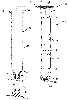
Referring to Figures 1-4, a first embodiment of the invention
includes a reagent vessel assembly 10. In this embodiment, the
assembly 10 includes a reagent vessel 12 and a reagent containing
ampoule 14. In the embodiment illustrated, t:he reagent vessel 12 is a
plastic syringe barrel. In further embodiments, the reagent vessel 12
can have a variety of shapes and is not limited to the shape or
structure of a syringe barrel. For example, the vessel can be a tubular
or square member with at least one outlet. The vessel can be of a
suitable size to supply a required volume of a reagent. In one
embodiment of the invention, the vessel contains about 3-10 ml of a
liquid reagent.
The reagent vessel 12 is preferably made of a suitable plastic
material such as polyethylene or polypropylene by various molding
processes as known in the art. In the illustrated embodiment, the
reagent vessel 12 includes a cylindrical side wall 16 having an open
CA 02327060 2000-11-29
_7_
top end 18 and a flange 20 surrounding the open top end 18. The
side wall is preferably sufficiently flexible to allow the ampoule 14 to
be rupturable when ready for use. The side wall 16 has a tapered
bottom wall 22 converging toward a nozzle 24 forming an outlet
passage 26. A collar 28 encircles the nozzle ;?4 and has an axial
length slightly shorter than the axial length of the nozzle 24. The
collar 28 includes internal threads 30 to define a threaded luer-type
fitting 32.
A tip cap 34 is coupled to the nozzle 24 to close the bottom end
of the reagent vessel 12. The tip cap defines a septum and is
preferably made of a flexible rubber like material capable of deforming
and forming a seal for the bottom end of the reagent vessel 12. As
shown in Figure 1, the tip cap 34 includes a cylindrical sleeve 36
having external threads 38 and an end portion 40. The sleeve 36 is
dimensioned to fit over the nozzle 24 and engage the threads 30 of the
luer fitting 32 as shown in Figure 2. Preferably, the tip cap is made of
a resilient material that can be pierced by a cannula and can self seal
after the cannula is removed.
A filter member 42 is positioned in the bottom end of the
reagent vessel 12 at the outlet end. The filter material can be a mesh
or screen of woven or non-woven material, an expanded material or a
frit as known in the art. In the embodiment illustrated, the filter
member 42 is a porous filter membrane. The filter member 42 is a
disk shaped member dimensioned to engage t:he side wall 16 of the
reagent vessel 12 to retain the filter member 42 in place.
Alternatively, the filter membrane can be a one-piece, integrally
formed member 43 molded or shaped to conform to the bottom
surfaces of the vessel 12 as shown in Figure 4A. In the embodiment
illustrated in Figures 1-4, the filter member 42 is coupled to a filter
CA 02327060 2000-11-29
_ g _
holder 44 having a shape and dimensions similar to a standard
syringe plunger tip. The filter holder 44 has a diameter corresponding
to the diameter of the side wall 16 with a tap<~red bottom wall 46 and
a central opening 48 extending axially through the holder 44. A
recess 50 is provided to receive the filter member 42.
In the embodiment of Figures 1-4, the ampoule 14 contains a
reagent 52. The reagent is generally a liquid, such as an aqueous
reagent, although in further embodiments, the reagent can be a non-
polar solvent, a solid or dried powder. In some embodiments, the
ampoule may contain a lyophilized reagent that can be reconstituted
prior to use. The ampoule is preferably made of glass or other
breakable material that will not interact with the reagents. The
ampoule 14 includes a substantially cylindrical side wall having a
dimension to fit within the side wall 16 of the reagent vessel 12. The
ends 56 and 58 of the ampoule 14 are sealed to contain the reagent.
The ampoule is dimensioned to fit within the side wall of the vessel 12
and to contain a desired amount of a reagent.
A closure member 60 is provided to close the top open end 18 of
the reagent vessel 12. The closure member 60 preferably contains a
hydrophobic porous member that allows air to pass through the
closure while preventing aqueous media from passing through the
closure member. In the embodiment illustrated, the closure member
60 includes a substrate 62 having a foil layer 64 and a layer 66 of a
heat sealable polymeric material. The heat sealable layer 66 can be,
for example, polyethylene or polypropylene that is compatible with the
reagent vessel 12 to form a seal and prevent leakage from the reagent
vessel. The closure member 60 can be welded to the flange 20 of the
vessel 12 by heat or ultrasonic energy. Alternatively, an adhesive can
be used to attach the closure member. In further embodiments, the
CA 02327060 2000-11-29
-9-
closure member can be positioned in the vessel spaced from the flange
20.
A central aperture 68 is provided through the substrate 62 as
shown in Figure 1. A breathable, semi-permeable membrane 70 is
attached to the substrate 62 on the bottom face of the aperture 68.
The breathable membrane 70 preferably is a porous hydrophobic
member having a pore size sufficiently small 1:o allow air to pass
through the membrane while preventing the passage of aqueous
solutions. A suitable breathable membrane 70 can be made from
polytetrafluoroethylene as known in the art. Suitable hydrophobic
membranes are disclosed in U.S. Patent No. 4,997,768 to
Uffenheimer; U.S. Patent No. 5,600,358 to Baldwin; and U.S. Patent
No. 5,863,499 to Kralovic, which are hereby incorporated by reference
in their entirety.
The assembly 10 is formed by coupling the tip cap 34 to the luer
fitting 32 and positioning the filter 42 member and filter holder 44 in
the bottom portion of the reagent vessel 12. The ampoule 14
containing the reagent is then placed in the reagent vessel. In
embodiments of the invention, a second reagent 72 can be placed in
the reagent vessel 12 between the ampoule 14 and the side wall 16.
Alternatively, a second reagent can be placed in a space below the
ampoule adjacent the surface of the filter member 42. The second
reagent can be a liquid reagent, such as a dye, or a dried or
lyophilized reagent. Generally, about 1 ml of the second reagent is
provided in the vessel 12 between the ampoule 14 and the side wall
16. The closure 60 is then attached to the flange 20 of the reagent
vessel to close the vessel. The assembly is generally suitable, for most
two-component systems where it is desirable to separate the
components until ready for use.
CA 02327060 2000-11-29
- 10-
It is desirable to separate the second reagent from the reagent
within the ampoule until ready for use when the combination of the
reagents forms an unstable solution or suspension. For example,
lyophilized reagent can be provided in the vessel 12 with a
reconstituting solvent contained in the ampoule 14. Another example
is a dye that is stable in non-polar solvent, but must be delivered in
polar solvent. In this example, the ampoule c:an contain a buffered
saline solution and a second reagent, such as a dye dissolved or
dispersed in DMSO, is provided in the vessel outside the ampoule.
The ampoule can be broken to disperse the dye in the saline solution.
The reagents can be a number of differ<~nt reagents depending
on the particular analytic tests being performed. In one embodiment
of the invention, the ampoule contains a buffered saline solution
containing up to about 25% by volume dimethylsulfoxide. The saline
solution can also contain other reagents as known in the art. The
second reagent in one embodiment of the invc;ntion is a dried or
solution of a hydrophobic dye for staining the nuclei of cells in DNA
analysis. Various cytodyes, probes or markers can be contained in
the vessel for treating the test sample.
The assembly 10 provides a convenient and efficient system for
supplying one or more reagents to an analytical testing device or other
apparatus without contaminating the reagents or exposing the
operator to the reagents. In the embodiment shown in Figure 3, the
analytical testing or preparation instrument 7 4 includes a sleeve 76
having an open end 78 for receiving the assembly 10. The sleeve 76 is
dimensioned to guide and support the reagent vessel 12. A cannula
80 extends through a collar 82 into the sleeve 76. The cannula 80
includes a blunt or pointed tip 84 and an opening 86 in the side of the
cannula 80 that is offset from the tip and communicates with a hollow
CA 02327060 2000-11-29
-11-
passage 88 for carrying the reagents to the analytical instrument.
Preferably, the cannula 80 is of a blunt tip of a non-coring design to
prevent coring of the tip cap 34 and be self-sealing when the cannula
is removed. In this manner, the cannula can pierce the tip cap and
then be withdrawn without the remaining contents of the vessel
leaking through the pierced hole in the tip cap.
The assembly 10 is positioned and guided by the sleeve 76 as
shown in Figure 4 and pressed downwardly until the cannula 80
pierces the tip cap 34. Generally, hand pressure by the operator is
sufficient to cause the cannula 80 to pierce tree tip cap 34. The
cannula 80 has a length to extend through the tip cap 34 without
penetrating the filter member 42. The ampoule 14 is broken by
compressing the flexible side walls 16 of the reagent vessel 12 until
the ampoule ruptures and releases the reagent 52 contained therein.
The ampoule can be broken by hand or using a suitable tool. The
ampoule can be broken before, during or after the reagent vessel 12 is
pressed into the sleeve 76 of the instrument T4. The reagent solution
52 flows downwardly through the filter member 42 into the nozzle 24
and is carried through the cannula 80 to the analyzing instrument.
The filter member 42 prevents glass fragments from the ampoule and
other solid materials from exiting the reagent vessel 12.
The breathable membrane 70 allows air to pass through the
aperture 68 so that the reagent solution can be fed by gravity to the
cannula 80 under atmospheric pressure. Generally, the instrument
74 provides a suction to draw the reagent from the vessel 12. In this
manner, it is not necessary to apply a positive pressure to the interior
of the reagent vessel 12 or apply a negative pressure to the needle to
withdraw the reagent solution. In alternative embodiments, the vessel
can be coupled to a device for applying a positive pressure to the
CA 02327060 2000-11-29
- 12-
breathable membrane to force the reagent solution through the
cannula 80 at a desired rate. After the reagent has drained into the
analyzing instrument, the vessel is removed by pulling the vessel from
the sleeve. The resilient tip cap 34 and non-coring cannula 80 allow
the puncture hale to close and prevent leakage of any remaining
contents. The vessel 12 can be discarded or refilled as desired.
In the embodiment illustrated, a single ampoule containing a
reagent, diluent or solvent is shown. In further embodiments, two or
more ampoules can be included in the vessel 12. The ampoules can
be arranged end-to-end or side-by-side depending on dimensions of
the ampoules. Alternatively, a second ampoule can be contained
within a larger first ampoule. This allows the various reagents to be
separated until ready for use. Generally, the ampoules are ruptured
and the vessel gently shaken to mix the reagents together. The vessel
containing the mixed reagents can then be placed in the sleeve of the
analyzing instrument to deliver the reagents to the instrument.
Referring to Figure 6, an analyzing instrument 90 is illustrated
schematically for use in conjunction with the reagent vessel. A
reagent as indicated by block 92 and a test sample indicated by block
94 are supplied to the analyzing instrument 90 for analysis. Data
from the analyzing instrument 90 can be fed t:o a suitable recording
device 96. The analyzing instrument 90 can be any suitable
instrument as known in the art capable of analyzing liquid or gaseous
samples such as a blood analyzing instrument. For example, the
instrument can be able to detect the presence of pathogens in blood
samples or airborne pathogens in a gas sample. In one embodiment
of the invention, the analyzing instrument is a sample preparation
device for a flow cytometer as known in the art. Examples of flow
cytometers are disclosed in U.S. Patent Nos. 3,960,449; 4,347,935;
CA 02327060 2000-11-29
- 13-
4,667,830; 5,464,581; 5,438,469; 5,602,039; 5,643,796 and
5,700,692; the entire contents of which are incorporated by reference
in their entirety. In other embodiments, the r. eagent 92 and sample
94 can be supplied to a sample processing apparatus (not shown) that
is used in conjunction with the analyzing instrument 90, with the
processed sample then being supplied to the analyzing instrument 90.
An example of such a sample processing app<~ratus is a cell lysing and
washing apparatus of the type disclosed in a copending U. S. patent
application of Kenneth F. Uffenheimer and Pierre Bierre, filed on
November 23, 1999 and entitled "Apparatus and Method for
Processing Sample Materials Contained in a Plurality of Sample
Tubes", also incorporated by reference herein.
Referring to Figures 7 and 8, a second embodiment of the
invention is illustrated. The reagent containing assembly 10' is
identical to the assembly 10 of the embodiment of Figures 1-6 so that
identical components are identified by the same reference numerals
with the addition of a prime.
As in the previous embodiment, the reagent vessel 12' includes
an ampoule 14' containing a liquid reagent. The analyzing instrument
98 or other apparatus includes a cylindrical sleeve 100 for receiving
the reagent vessel 12. A threaded collar 102 having a central passage
104 is provided in the bottom of the sleeve 100. The collar 102
includes external threads 106 for coupling with the luer fitting 32' of
the reagent vessel 12'. The passage 104 carries the liquid reagents to
the analyzing instrument or other apparatus as in the previous
embodiment.
In this embodiment, the tip cap 34' is removed from the luer
fitting 32' of the reagent vessel 12' prior to inserting the vessel 12' into
the sleeve 100. To prevent the reagent solution 52' from flowing
CA 02327060 2000-11-29
- 14-
outwardly through the nozzle 24', it is desirable to close the aperture
68' to cover the breathable membrane 70' to prevent air from entering
the reagent vessel 12'. A convenient manner of closing the aperture
68' is for the operator to place a finger over the aperture 68 until the
S reagent vessel 12' is threaded onto the collar 102. At that time, the
operator's finger can be removed from the aperture 68' to allow the
reagent solution to flow through the passage 104. In fi~.rther
embodiments, a tape or other peelable strip (not shown) can be
applied to cover the porous membrane to prevent air from entering or
liquid from evaporating until the reagent is ready for delivery. At that
time, the tape or strip is removed to dispense the reagent. As in the
previous embodiment, the ampoule 14' can b~° ruptured before, during
or after the reagent vessel 12' is threaded onto the collar 104. In still
further embodiments, a piston or plunger can be provided to dispense
the contents of the vessel.
Referring to Figures 9 and 10, a third embodiment of the
invention is illustrated. In this embodiment, the reagent assembly 10"
is similar to the assembly 10 of Figures 1 and 2 except that the
breakable ampoule is not included. In this embodiment, a reagent
solution 108 is contained within the reagent vessel 12". As in the
previous embodiments, the open top end 18" is closed by the
breathable closure member 60" and a tip cap 34".
As in the previous embodiment, the analyzing instrument 74" or
other apparatus includes a sleeve 76" and a cannula 80". The reagent
vessel 12" is placed in the sleeve 76" and pushed downwardly until
the cannula 80" penetrates the tip cap 34". The reagent solution 108
is then able to flow downwardly through the needle to the analyzing
instrument or other apparatus. In this embodiment, the filter member
is not necessary in the reagent vessel 12" since there are no glass
CA 02327060 2000-11-29
- 15-
ampoule fragments or solid materials which c:an flow through the
nozzle 24" and interfere with the feeding of the reagent solution.
In the disclosed embodiments of the invention, the reagent
solution is contained in a rupturable ampoule that is preferably made
of glass. The ampoule can be broken by hand or by using standard
methods as known in the art. For example, t:he ampoule can be
broken using a cracking device 110 as shown in Figure 11. The
cracking device 110 is a substantially flat member having a generally
square configuration. One side edge 112 of the cracking device 110
includes a recess 114 having a curved bottom side 116 and tapered
corners 118. The recess 114 has a dimension slightly less than the
diameter of the side wall of the reagent vessel 12. The cracking device
110 is forced around the side wall of the reagent vessel to compress
the side walls inwardly to break the ampoule as shown in Figure 12.
The cracking device 110 is then removed and the reagent vessel
placed in the analyzing instrument or other instrument.
Various embodiments have been chosen to illustrate the present
Invention. In each of the illustrated embodiments, a breathable
closure member is provided on the top end of the reaction vessel to
allow air to enter the vessel and prevent the reagent solution from
passing through the closure member. Thus, in each of the
embodiments, the reagent vessel is coupled to the analyzing
instrument or other instrument so that the reagent solution can flow
through the outlet nozzle by gravity without the need for a
pressurizing or pumping system to withdraw the reagent solution from
the vessel. It will be apparent to those skilled in the art that various
modifications can be made to the reagent vessel without departing
from the scope of the invention as set forth herein.