Language selection

Search

Patent 2328237 Summary

Third-party information liability

Some of the information on this Web page has been provided by external sources. The Government of Canada is not responsible for the accuracy, reliability or currency of the information supplied by external sources. Users wishing to rely upon this information should consult directly with the source of the information. Content provided by external sources is not subject to official languages, privacy and accessibility requirements.

Claims and Abstract availability

Any discrepancies in the text and image of the Claims and Abstract are due to differing posting times. Text of the Claims and Abstract are posted:

  • At the time the application is open to public inspection;
  • At the time of issue of the patent (grant).
(12) Patent Application: (11) CA 2328237
(54) English Title: BONDABLE EXPANSION PLUG FOR SOFT TISSUE FIXATION
(54) French Title: TAMPON DILATABLE ADHESIF DESTINE A LA FIXATION DE TISSUS MOUS
Status: Deemed Abandoned and Beyond the Period of Reinstatement - Pending Response to Notice of Disregarded Communication
Bibliographic Data
(51) International Patent Classification (IPC):
  • A61B 17/04 (2006.01)
  • A61F 2/08 (2006.01)
(72) Inventors :
  • EGAN, THOMAS D. (United States of America)
(73) Owners :
  • AXYA MEDICAL, INC.
(71) Applicants :
  • AXYA MEDICAL, INC. (United States of America)
(74) Agent: RICHES, MCKENZIE & HERBERT LLP
(74) Associate agent:
(45) Issued:
(86) PCT Filing Date: 1999-04-13
(87) Open to Public Inspection: 1999-10-21
Availability of licence: N/A
Dedicated to the Public: N/A
(25) Language of filing: English

Patent Cooperation Treaty (PCT): Yes
(86) PCT Filing Number: PCT/US1999/008045
(87) International Publication Number: WO 1999052414
(85) National Entry: 2000-10-13

(30) Application Priority Data:
Application No. Country/Territory Date
09/061,604 (United States of America) 1998-04-16

Abstracts

English Abstract


This invention is an expandable soft tissue fixation assembly (10) for use in
anchoring soft tissue to bone. The assembly includes a tab (16) connected to
an anchor (18), a sleeve (20) adapted to surround the anchor (18), and a
flange (19) adapted to hold a soft tissue segment next to a bone. The sleeve
(20) is inserted into a blind hole (12) in a bone, and a section of soft
tissue is placed over the hole (12) next to the bone. Energy is applied to the
flange (19) while a predetermined axial tension is applied to the tab (16) to
compress a flared portion of the anchor against the sleeve (20). An upper tube
portion of the anchor and the flange (19) are bonded together. The applied
axial force on the tab separates it from the anchor, leaving the assembly
anchored in the bone, the soft tissue section anchored in place between the
flange (19), and the bone.


French Abstract

L'invention concerne un ensemble dilatable de fixation de tissus mous destiné à s'utiliser pour l'ancrage de tissus mous aux os. L'ensemble comprend une attache reliée à une ancre, un manchon adapté de manière à entourer l'ancre et un rebord adapté pour retenir un segment de tissu mou à côté d'un os. Le manchon est introduit dans un trou borgne d'un os et une section de tissu mou est placée au-dessus du trou à côté de l'os. On applique une énergie sur le rebord et une tension axiale prédéterminée sur l'attache pour comprimer une partie biseautée de l'ancre contre le manchon. On fait adhérer une partie de tube supérieur de l'ancre au rebord et la force axiale appliquée sur l'attache la sépare de l'ancre, laissant l'ensemble ancré à l'os et la section de tissu mou en place entre le rebord et l'os.

Claims

Note: Claims are shown in the official language in which they were submitted.

CLAIMS
1. An expandable soft tissue fixation assembly, comprising:
a tab extending along an axis and connected, via a connecting region, to an
anchor having a tube portion and a flared portion, the tube portion having a
diameter
D;
a sleeve adapted to surround the anchor and including multiple sections which
can be expanded radially outward upon application of a force thereto; and
a tubular flange having an inner bore of a diameter which is smaller than D;
wherein the flange is adapted to hold and distribute pressure over a section
of
soft tissue between the flange and a bone surface, wherein the walls of the
bore of the
tubular flange interferingly engage with the walls of the tube portion of the
anchor,
so that the bore walls and tube portion walls form a shear weld upon
application of
sufficient energy to the tubular flange, wherein concurrent application of a
predetermined tensile force to the tab along the axis of the tab compresses
the flared
portion of the anchor against the sleeve, thereby radially extending the
multiple
sections of the sleeve, anchoring the flared portion and the sleeve in the
bone,
holding the soft tissue section between the flange and the bone, and causing
the tab to
separate from the tube portion of the anchor.
2. An assembly according to claim 1, wherein at least the flange and the tube
portion of the anchor are made of a bondable thermoplastic material.
3. An assembly according to claim 2, wherein ultrasonic energy is applied to
the
flange to bond the flange to the tube portion of the anchor.

Description

Note: Descriptions are shown in the official language in which they were submitted.


CA 02328237 2000-10-13
WO 99/52414 PCT/US99/08045
BONDABLE EXPANSION PLUG FOR SOFT TISSUE FIXATION
Field of the Invention
The present invention relates to surgical soft tissue fixation devices, and
more particularly to expandable soft tissue fixation devices which are
suitable for
use in attaching soft tissue to a blind hole in a bone.
Background of the Invention
U.S. Patent No. 5,593,425 to Bonutti et al. discloses a heat-bondable,
expandable surgical anchor for fastening soft tissue to a blind hole in a
bone. One
embodiment of the device, shown in FIGS. 9A-9D of Bonutti et al., includes an
expandable anchor portion, a suture extending from the anchor, and a heat
bondable fastener portion which is bonded to the suture to lock the anchor in
place.
Another embodiment, shown in FIGS. 11A-11C, is a rivet fastener including a
sleeve and a headed mandrel which extend into a blind hole in a bone through
the
tissue to be anchored. The mandrel is pulled through the sleeve, which is held
stationary, so that the head spreads the rear portion of the sleeve to lock it
in place
in the bone. The end of the mandrel is cut off and heat bonded to fix the
anchor in
place.
The Bonutti et al. devices are deployable in a blind hole in a bone but
require a final heat bonding step to lock the devices in place. It would be
advantageous to provide an expandable soft tissue anchoring device which is
fully
deployable in a single step.
Summary of the Invention
According to the invention, there is provided an expandable soft tissue
fixation assembly. The assembly comprises:
a tab extending along an axis and connected, via a connecting region, to an
anchor having a tube portion and a flared portion, the tube portion having a
diameter D;
-1-

CA 02328237 2000-10-13
WO 99/52414 PCTIUS99/08045
a sleeve adapted to surround the anchor and including multiple sections
which can be expanded radially outward upon application of a force thereto;
and
a tubular flange having an inner bore of a diameter which is smaller than D.
The flange is adapted to hold and distribute pressure over a section of soft
tissue
between the flange and a bone surface. The walls of the bore of the tubular
flange
interferingly engage with the walls of the tube portion of the anchor, so that
the
bore walls and tube portion walls form a shear weld upon application of
sufficient
energy to the tubular flange. Concurrent application of a predetermined
tensile
force to the tab along the axis of the tab compresses the flared portion of
the anchor
against the sleeve, thereby radially extending the multiple sections of the
sleeve.
The compression force anchors the flared portion and the sleeve in the bone
and
holds the soft tissue section between the flange portion and the bone. It also
causes
the tab to separate from the flared portion of the anchor.
At least the flange and the flared portion of the anchor are made of a
bondable thermoplastic material. In a preferred embodiment, ultrasonic energy
is
applied to the flange to bond the flange to the flared portion of the anchor.
These and other features of the invention will be more fully appreciated
with reference to the following detailed description which is to be read in
conjunction with the attached drawings.
Brief Description of the Drawinlzs
The invention is further described by the following description and figures,
in which:
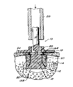
FIG. 1 is a sectional view of an assembly according to the invention, in
which the assembly is installed in a blind hole in a bone in preparation for
welding
of the flange and flared portion of the anchor;
FIG. 2 is a sectional view of the assembly after ultrasonic welding of the
flange and flared portion of the anchor;
FIG. 3 is a sectional view of the assembly after the tab has been separated
-2-

CA 02328237 2000-10-13
WO 99/52414 PCT/US99/08045
from the flared portion of the anchor;
FIG. 4 is a perspective view of the sleeve portion of the assembly; and
FIG. 5 is a sectional view of the sleeve along section lines V-V.
Like elements in the respective FIGURES have the same reference
numbers.
Detailed Description of the Preferred Embodiment
The assembly 10 of the present invention comprises a bondable, expandable
device which can be installed in a blind hole 12 in a bone 14 to anchor it in
the
bone. Such an anchor can be used to fasten soft tissue, such as ligaments and
tendons, to bone. The assembly includes three separate pieces, two of which
are
bonded together in situ in a single step to form a unitary soft tissue anchor.
The assembly 10 includes a main tab 16 connected to an anchor 18 having a
tube portion 18A and a flared portion 18B, a separate flange 19, and a
separate
sleeve 20 which fits around the anchor, as shown in FIG. 1. The tab 16 is
connected to the tube portion 18A of the anchor by a connecting region 22
which,
as detailed more fully below, is designed to separate under an applied
predetermined tensile force after the sleeve 20 and anchor 18 are joined
together.
The flange 19 rests on the section of soft tissue 24 placed adjacent to the
surface of
the bone 14 and distributes force against the bone over the surface area of
the
flange.
The assembly is designed to be ultrasonically welded in situ upon
application of ultrasonic energy to the anchor by a tubular ultrasonic welding
horn
26. Prior to welding, the sleeve 20 is inserted into a predrilled hole 12 in
the bone
14. The tissue 24 to be held in place next to the bone is placed over the hole
with
the sleeve in it, and the flange 19 is placed over the tissue section 24 and
aligned
with the underlying hole. The tubular ultrasonic weld horn 26 is brought into
contact with the flange 19 and compressed against the flange, thereby
compressing
-3-

CA 02328237 2000-10-13
WO 99/52414 PCT/US99/08045
the tissue 24 to the bone surface. As shown most clearly in FIG. 1, the outer
diameter of the tube portion 18A of the anchor is sufficiently greater than
the inner
diameter of the flange 19 to establish an interference between the walls of
the
flange and the tube portion of the anchor. The flange 19 receives ultrasonic
energy
from the horn 26 and vibrates relative to the tube portion 18A of the anchor,
which
abuts the bone and thus serves as a stationary anvil. The tube portion 18A and
the
flange 19 melt locally at their respective interference points and bond
together in a
shear weld, as shown in FIG. 2.
During ultrasonic welding of the tube portion and flange, the tab 16 is
grasped by a non-ultrasonically activated grasper (not shown) and pulled in
the
direction of arrow 28 to compress the tube portion and flange together,
clamping
the tissue section 24 between them. This compression pulls the anchor up,
thereby
radially distending the multipart sections of the sleeve 20 and lodging the
flared
portion 18B of the anchor in the sleeve 20 against the sides of the hole 12 in
the
bone 14. At some predetermined tensile force the connecting region 22 between
the tab 16 and the tube portion 18A fails, causing the tab 16 to separate from
the
welded tube/flange. The connecting region 22 is designed to fail in tension at
a
predetermined force and is accordingly necked down, scored or otherwise
constructed to accomplish this objective. The fully deployed assembly 40,
shown
in FIG. 3, includes the welded tube/tlange and the distended sleeve 20. The
tissue
section 24 is firmly held between the flange 19 and the bone 14.
The sleeve 20 is shown in greater detail in FIGS. 4 and 5. A bottom
portion 30 of the sleeve is separable into multiple flared sections 33 which
can be
distended radially out from the bore 32 upon application of a force against
the
sections. The bore 32 of the sleeve is sized to accommodate the flared portion
18B
of the anchor, as shown in FIG. 1, and the axial force applied to the tab 16
causes
the flared portion 18B to press radially against the sleeve 20, as shown by
arrows
34 in FIG. 2.
-4-

CA 02328237 2000-10-13
WO 99/52414 PCT/US99/08045
At least the tube portion 18B and the flange 19 are made of a material, such
as a thermoplastic, which can be bonded together upon the application of
ultrasonic
or thermal energy thereto. The sleeve 20 can be, but need not be, made of a
material which can be ultrasonically welded, as it is joined to the anchor 18
by
virtue of a compression fit and not a thermal bond.
The invention may be embodied in other specific forms without departing
from the spirit or essential characteristics thereof. The present embodiments
are
therefore to be considered in all respects as illustrative and not
restrictive, the
scope of the invention being indicated by the appended claims rather than by
the
foregoing description. All changes that come within the meaning and range of
the
equivalency of the claims are therefore intended to be embraced therein.
-5-

Representative Drawing
A single figure which represents the drawing illustrating the invention.
Administrative Status

2024-08-01:As part of the Next Generation Patents (NGP) transition, the Canadian Patents Database (CPD) now contains a more detailed Event History, which replicates the Event Log of our new back-office solution.

Please note that "Inactive:" events refers to events no longer in use in our new back-office solution.

For a clearer understanding of the status of the application/patent presented on this page, the site Disclaimer , as well as the definitions for Patent , Event History , Maintenance Fee  and Payment History  should be consulted.

Event History

Description Date
Inactive: IPC expired 2016-01-01
Inactive: IPC from MCD 2006-03-12
Application Not Reinstated by Deadline 2005-04-13
Time Limit for Reversal Expired 2005-04-13
Deemed Abandoned - Failure to Respond to Maintenance Fee Notice 2004-04-13
Inactive: Abandon-RFE+Late fee unpaid-Correspondence sent 2004-04-13
Letter Sent 2001-03-20
Inactive: Single transfer 2001-02-20
Amendment Received - Voluntary Amendment 2001-02-20
Inactive: Cover page published 2001-02-06
Inactive: First IPC assigned 2001-01-31
Inactive: Courtesy letter - Evidence 2001-01-30
Inactive: Notice - National entry - No RFE 2001-01-24
Application Received - PCT 2001-01-22
Application Published (Open to Public Inspection) 1999-10-21

Abandonment History

Abandonment Date Reason Reinstatement Date
2004-04-13

Maintenance Fee

The last payment was received on 2003-04-11

Note : If the full payment has not been received on or before the date indicated, a further fee may be required which may be one of the following

  • the reinstatement fee;
  • the late payment fee; or
  • additional fee to reverse deemed expiry.

Please refer to the CIPO Patent Fees web page to see all current fee amounts.

Fee History

Fee Type Anniversary Year Due Date Paid Date
MF (application, 2nd anniv.) - small 02 2001-04-17 2000-10-13
Registration of a document 2000-10-13
Basic national fee - small 2000-10-13
MF (application, 3rd anniv.) - small 03 2002-04-15 2002-03-11
MF (application, 4th anniv.) - small 04 2003-04-14 2003-04-11
Owners on Record

Note: Records showing the ownership history in alphabetical order.

Current Owners on Record
AXYA MEDICAL, INC.
Past Owners on Record
THOMAS D. EGAN
Past Owners that do not appear in the "Owners on Record" listing will appear in other documentation within the application.
Documents

To view selected files, please enter reCAPTCHA code :


To view images, click a link in the Document Description column. To download the documents, select one or more checkboxes in the first column and then click the "Download Selected in PDF format (Zip Archive)" or the "Download Selected as Single PDF" button.

List of published and non-published patent-specific documents on the CPD .

If you have any difficulty accessing content, you can call the Client Service Centre at 1-866-997-1936 or send them an e-mail at CIPO Client Service Centre.


Document
Description 
Date
(yyyy-mm-dd) 
Number of pages   Size of Image (KB) 
Representative drawing 2001-02-06 1 12
Abstract 2000-10-13 1 55
Description 2000-10-13 5 206
Claims 2000-10-13 1 41
Drawings 2000-10-13 2 67
Cover Page 2001-02-06 1 55
Notice of National Entry 2001-01-24 1 194
Courtesy - Certificate of registration (related document(s)) 2001-03-20 1 113
Reminder - Request for Examination 2003-12-16 1 123
Courtesy - Abandonment Letter (Request for Examination) 2004-06-22 1 167
Courtesy - Abandonment Letter (Maintenance Fee) 2004-06-08 1 175
Correspondence 2001-01-24 1 24
PCT 2000-10-13 7 294
Fees 2003-04-11 1 36
Fees 2002-03-11 1 40