Note: Descriptions are shown in the official language in which they were submitted.
CA 02331337 2000-12-14
WO 99/67291 PCT/US99/13953
TITLE
SITE SPECIFIC PROTEIN MODIFICATION BY MUTAGENESIS
BACKGROUND OF THE INVENTION
Field of the Invention
The present invention relates to processes for modifying proteins. More
particularly, the present invention involves processes for linking
polyethylene glycol to
proteins in a manner which provides advantages associated with polyethylene
glycol
conjugated proteins while maintaining a desired protein bioactivity.
Description of Related Art
Processes and reagents for chemically modifying proteins have been used
extensively for decades. Traditionally, protein chemical modifications were
carried out in
order to study their functional properties and structural characteristics.
With the
emergence of recombinant DNA techniques and interest in protein therapeutics,
researchers have chemically modified proteins to improve their clinical
performance. In
particular, processes for conjugating proteins with polyethylene glycol have
gained
widespread use within the pharmaceutical and biochemical communities as a
result of
numerous improved pharmacological and biological properties associated with
polyethylene glycol conjugated proteins. For example, polyethylene glycol
conjugated
proteins are known to have significantly enhanced plasma half life, and thus
have
substantially improved the clinical usefulness. Additionally, polyethylene
glycol
conjugated proteins generally have reduced antigenicity and immunogenicity,
thereby are
less prone to causing life-threatening anaphylaxis.
Another benefit associated with polyethylene glycol conjugated proteins is
that of
water solubility which is increased as a result of the high water solubility
of polyethylene
glycol. The increased water solubility can improve the protein's formulation
characteristics at physiological pH's and can decrease complications
associated with
aggregation of low solubility proteins.
Additionally, polyethylene glycol conjugated proteins have found use in
bioindustrial applications such as enzyme based reactions in which the
reaction
environment is not optimal for the enzyme's activity. For example, some
polyethylene
CA 02331337 2000-12-14
WO 99/67291 PCT/US99/13953
glycol conjugated enzymes demonstrate a wider optimum pH activity and reduced
optimum activity temperature. Moreover, enzymes having reduced activity in
many
organic solvents have been successfully conjugated with polyethylene glycol to
a degree
that renders them useful for catalyzing reactions in organic solvents. For
example,
polyethylene glycol has been conjugated with horseradish peroxidase which then
becomes
soluble and active in chloroform and toluene (Urrotigoity et al.,
Biocatalysis, 2:145 - 149,
1989).
Polyethylene glycol conjugated proteins vary in the extent to which plasma
circulation half life is increased, immunogenicity is reduced, water
solubility is enhanced,
and enzymatic activity is improved. Factors responsible for these variations
are numerous
and include the degree to which the protein is substituted with polyethylene
glycol, the
chemistries used to attach the polyethylene glycol to the protein, and the
locations of the
polyethylene glycol sites on the protein.
The most common methods for attaching polyethylene glycol to proteins involve
activating at least one of the hydroxyl groups on the polyethylene glycol with
a
functionality susceptible to nucleophilic attack by the nitrogen of amino
groups on the
protein. These methods generally result in loss of biological activity due to
the
nonspecific attachment of polyethylene glycol
Alternative approaches to conjugating proteins with polyethylene glycol
include
controlling the conjugation reactants and conditions so that the conjugation
site is
confined to the N-terminus (Kinstler et al. Pharm. Res. 13:996, 1996);
attaching
polyethylene glycol to protein carbohydrate functionalities (Urrutigoity et
al. Biocatalysis
2:145, 1989); attaching polyethylene glycol at protein cysteine residues
(Goodson et al.
Biotechnology 8:343, 1990); attaching polyethylene glycol during solid phase
and
solution phase peptide synthesis (Felix, ACS Symposium Series 680 ch 16, 1997)
and,
selectively replacing protein arginine residues with lysine residues that
provide an
polyethylene glycol attachment site (Hershfield et al. Proc. Natl. Acad. Sci.
88:7185,
1991). While these offer some degree of control of the reaction site, there is
a continuing
need for improved methods for providing polyethylene glycol conjugated
proteins. In
particular, it would be desirable to provide methods for conjugating proteins
with
polyethylene glycol that result in modified proteins having enhanced
bioactivity or little
loss in bioactivity while maintaining the benefits of polyethylene glycol
conjugation,
including substantially decreased immunogenicity, increased solubility, and
prolonged
circulation half lives characteristic of modified proteins.
2
CA 02331337 2007-07-30
72249-104
SUMMARY OF THE INVENTION
The present invention provides protein modification processes that result in
modified proteins having little or no decrease in an activity associated with
the protein.
More particularly, the invention described herein includes processes for
modifying a
protein by first deleting one or more amino acid sites on the protein that is
suitable for
polyethylene glycol conjugation and then contacting the protein with
polyethylene glycol
under conditions suitable for conjugating the polyethylene glycol to the
protein.
Preferably, the deleted amino acid residue is replaced with an amino acid
residue that
does not react with polyethylene glycol. The resulting polyethylene glycol
conjugated
protein has improved characteristics over proteins modified according to prior
art
procedures. An advantageous retention of activity is attributed to the
availability of one
or more protein binding sites which is unaltered in the protein modification
process and
thus remains free to interact with a binding partner subsequent to the
modification
process. Within the present invention are proteins useful for polyethylene
glycol
conjugation and polyethylene glycol conjugated proteins prepared by processes
described
herein.
3
CA 02331337 2007-07-30
72249-104
According to one aspect of the present invention,
there is provided a polypeptide comprising an amino acid
sequence as set forth in SEQ ID NO:2.
According to another aspect of the present invention,
there is provided a polypeptide comprising an amino acid
sequence as set forth in SEQ ID NO:6.
According to still another aspect of the present
invention, there is provided a polynucleotide comprising a
nucleic acid encoding the polypeptide as described above.
According to yet another aspect of the present
invention, there is provided a process for conjugating a
protein with polyethylene glycol, said process comprising the
steps of preparing the polypeptide as described above; and
contacting the polypeptide with polyethylene glycol under
conditions sufficient to conjugate polyethylene glycol to the
polypeptide.
BRIEF DESCRIPTION OF THE DRAWINGS
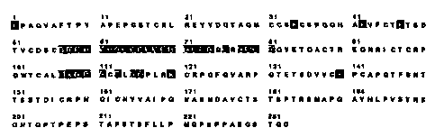
Fig. 1 illustrates lysine residues within p75 TNF
receptor extracellular domain that are polyethylene glycol
conjugation sites and lysine residues that make contact with
TNFa.
DETAILED DESCRIPTION OF THE INVENTION
The present invention provides processes and reagents
for conjugating proteins or polypeptides with polyethylene
glycol in a manner that results in polyethylene glycol
conjugated proteins having little or no reduction in a desired
activity. More specifically, the present invention provides
processes for conjugating polyethylene glycol with proteins
under conditions which preclude polyethylene glycol conjugation
at one or more selected sites on the protein. When the one or
3a
CA 02331337 2007-07-30
72249-104
more selected sites is active in a protein binding domain,
preventing polyethylene glycol conjugation at the site in
accordance with the present invention contributes to
maintaining a desired bioactivity while providing benefits
associated with polyethylene glycol conjugation.
3b
CA 02331337 2000-12-14
WO 99/67291 PCT/US99/13953
The processes of the present invention are based upon the discovery that by
deleting one or more selected amino acid residues that are capable of reacting
with
polyethylene glycol sites, and then conjugating the protein with polyethylene
glycol, the
resulting polyethylene glycol modified protein does not demonstrate a
significant
reduction in a desired activity. In one embodiment, the selected amino acid
residue is a
lysine residue that, if reacted with a polyethylene glycol, interferes with
the ability of the
resulting conjugated protein to bind with its binding partner, substrate, or
receptor. It is
believed that the selected amino acid residues are associated with binding
sites, and, if
modified, interfere with the conjugated protein's structural elements that
determine
protein conformation and function. By deleting the selected amino acid
residue,
polyethylene glycol does not modify the protein at the site of the selected
amino acid
residue during a subsequent polyethylene glycol modification reaction.
Preferably, in
order to preserve the number of amino acid residues and maintain the optimum
protein
conformation, the deleted amino acid residue is replaced with an amino acid
residue that
is not reactive with polyethylene glycol under the reaction conditions. For
example,
lysine can be deleted and replaced with an arginine residue. Arginine has the
same
structure as lysine, with the exception of the polyethylene glycol reactive F--
NHZ
functionality on lysine which is absent in arginine.
Any protein is suitable for polyethylene glycol modification in accordance
with
the present invention including but not limited to protein ligands, receptors,
antigens,
antibodies, enzymes, protein fragments, peptides, and polypeptides.
Particularly desirable
protein candidates for polyethylene glycol modification as described herein
are those
which, subsequent to their modification by prior art methods, demonstrate a
reduction in a
desired activity. Other proteins which are suitable for modification in
accordance with
the present invention are those having multiple binding sites. In this
embodiment, a
protein may be conjugated with polyethylene glycol so that an activity
associated with
one or more of the multiple binding sites can be reduced while maintaining an
activity
associated with one or more different binding sites. This is accomplished by
deleting one
or more selected amino acid residues that are associated with binding sites
for which
activity is to be maintained and which are capable of reacting with
polyethylene glycol,
and leaving amino acid residues associated with binding sites for which
activity is to be
reduced for subsequent polyethylene glycol conjugation. Preferably, the
deleted amino
acid residue or residues is replaced with an amino acid residue that is not
reactive with
4
CA 02331337 2000-12-14
WO 99/67291 PCT/US99/13953
polyethylene glycol under the reaction conditions. Additionally, the deleted
amino acid
residue or residues is replaced with an amino acid residue that does not
significantly
diminish the activity of the native protein. The resulting polyethylene glycol
conjugated
protein will have an activity associated with selected binding sites and,
depending upon
the degree to which additional sites are involved in the conjugation process,
will have a
diminished, or no activity, associated with such additional sites. This
approach is useful
in cases in which cognate or substrate binding to one or more protein binding
sites is
desirably suppressed in certain clinical, diagnostic or industrial
applications.
Proteins that may be modified in accordance with the present invention include
those having utility in clinical and diagnostics applications and those used
in the
biotechnology industry, such as enzymes in bioreactors. Receptors which may be
modified as taught herein include cytokine receptors, for example, TNFR, IL-
4R, IL-1R,
IL-17R, IL-15R, p55 TNFR:Fc and p75 TNFR:Fc. Candidate antibodies for
conjugation
include but are not limited to OKT3 (anti-T-Cell), AVAKINETM (anti-TNF) and
anti
Her2/Neu. Enzymes of interest for conjugation include CD39, tPA, and DNAse.
Many
proteins have multimeric binding sites and require more than one association
for activity.
Such proteins are particularly desirable for modification since loss of one
binding site
leaves the whole protein inactive. Members of the group of multimeric proteins
include
TNF, hGH, CD40L, and FasL. Other candidate protein ligands are known to bind
multiple receptor subunits and include IL-2, IL-15, GM-CSF, and G-CSF.
In accordance with the present invention, a selected amino acid residue is one
that
is associated with a site on a protein which contributes to a specific
function of that
protein, and which is reactive with polyethylene glycol under the protein
modification
reaction conditions. The selected amino acid residue may be directly involved
with a
binding association with a protein binding partner. Alternatively, the amino
acid may be
sufficiently central to the spatial configuration of the protein that
modifying the protein
with polyethylene glycol results in a significant loss of desirable properties
even though
the amino acid residue is neither within a binding site nor directly or
physical involved
with the protein's interaction with a binding partner. Sites include, but are
not limited to,
cognate sites or substrate binding sites which are associated with a protein
activity.
Amino acid residues that are reactive with polyethylene glycol under
conditions
known in the art include those having residues having nucleophilic moieties
that are
available for reaction with polyethylene glycol or an activated polyethylene
glycol. For
5
CA 02331337 2000-12-14
WO 99/67291 PCT/US99/13953
example, lysine is reactive with polyethylene glycol through its E-NH,;
aspartic acid and
glutamic acid are reactive with polyethylene glycol through their COOH
(carboxyl)
functionalities; serine and threonine are potentially reactive through their
OH (hydroxyl)
sites; and, cysteine with available SH (sulfhydryl) groups may also react with
polyethylene glycol. Conditions suitable for reactions between polyethylene
glycol or
activated polyethylene glycols and specific amino acid residues in proteins
are known and
those skilled in the art are charged with knowledge such reactions. It is
known in the art
that lysine residues react with activated polyethylene glycol under favorable
reaction
conditions and with minimum side reactions. Thus, in accordance with the
present
invention, lysine residues are typically the targeted residue and the reaction
conditions are
controlled to maximize the reaction between polyethylene glycol and lysine.
Determining a suitable amino acid residue to select for deletion and,
preferably,
replacement, can be accomplished using a number of different techniques. In
cases where
the three dimensional structure and epitopes or structural elements that
determine protein
function are not known, one method involves using site directed mutagenesis
techniques
to empirically determine amino acid residues that are associated with a site
on a protein
which contribute to a specific function of that protein. More particularly,
one or more
predetermined polyethylene glycol reactive amino acid residues on the protein
can be
deleted and preferably replaced with non reactive amino acid residues using
mutagenesis
and recombinant DNA methodologies. Conjugating the thus modified protein with
polyethylene glycol and then testing the resulting polyethylene glycol
conjugated protein
for activity and other relevant properties provides valuable information
relating to the
suitability of the predetermined amino acid residue or residues for deletion
and
replacement. Sequentially repeating the above described process for different
polyethylene glycol reactive amino acid residues will provide more complete
information
relating to the role of the deleted amino acid residue in determining the
function and
activity of the protein. For example, if a protein has 8 lysine residues, DNA
encoding the
protein can be mutated in a site directed manner to produce a number of
different mutants
with one or more of the codons coding for the lysine residues replaced with
codons
coding for an arginine residue. The specific lysine coding codons that are
mutated can
include one selected codon, all of the lysine coding codons, are any
permutation of the
lysine coding codons, including the simultaneous mutagenesis of DNA coding
lysine
residues that are adjacent to each other.
6
CA 02331337 2000-12-14
WO 99/67291 PCT/US99/13953
After expressing, collecting and purifying the engineered proteins encoded by
the
mutated DNA, the expressed proteins can be reacted with polyethylene glycol to
form a
conjugated protein. Then the conjugated protein can be tested for functional
activity and
other characteristics such as immunogenicity, physiological clearance, and
solubility.
The polyethylene glycol conjugated proteins that have the desired activity and
most
favorable clearance, solubility and immunogenicity properties also contain the
desired
selected lysine residues i.e., the residues that had been deleted and replaced
prior to
reacting the protein with polyethylene glycol.
For many proteins, the location of polyethylene glycol reactive amino acid
residues and their conformational contribution to the structure and function
of the protein
are known. Among these proteins, are those for which the crystalline structure
of the
protein is known, and, in some cases, the crystalline structure of the protein-
binding
partner complex is known. For these proteins, determining a selected amino
acid residue
typically requires only identifying the residues that are within the protein's
binding
domain or in close spatial proximity to the protein's binding region and
identifying those
residues that are reactive with polyethylene glycol under the contemplated
polyethylene
glycol reaction conditions.
In accordance with the present invention, deleting a selected amino acid
residue on
the protein can be accomplished with a variety of suitable procedures that
provide
modified proteins. In the context of the present invention, such procedures
include, but
are not limited to, site directed mutagenesis techniques and direct protein
synthesis
methods in which the protein lacking one or more selected amino acid residues
is
synthesized using standard protein synthesis procedures known in the art. As
noted
above, preferably the process of deleting a selected amino acid residue
additionally
involves replacing the selected amino acid residue with an amino acid residue
that is not
reactive with polyethylene glycol.
Proteins may be prepared by any of a number of conventional techniques. A
desired DNA sequence may be chemically synthesized using techniques known per
se.
DNA fragments also may be produced by restriction endonuclease digestion of a
full
length cloned DNA sequence, and isolated by electrophoresis on agarose gels.
Linkers
containing restriction endonuclease cleavage site(s) may be employed to insert
the desired
DNA fragment into an expression vector, or the fragment may be digested at
cleavage
sites naturally present therein.
7
CA 02331337 2007-07-30
72249-104
Alterations of amino acid sequence, including deleting selected amino acid
residues and replacing the deleted residues with a different residue, may be
accomplished
by any of a number of conventional methods. Mutations can be introduced at
particular
loci by synthesizing oligonucleotides containing a mutant sequence, flanked by
restriction
sites enabling ligation to fragments of the native sequence. Following
ligation, the
resulting reconstructed sequence encodes an analog having the desired amino
acid
insertion, substitution, or deletion.
Alternatively, oligonucleotide-directed site-specific mutagenesis procedures
can
be employed to provide an altered gene wherein predetermined codons can be
altered by
substitution, deletion or insertion. Exemplary methods of making the
alterations set forth
above are disclosed by Walder et al. (Gene 42:133, 1986); Bauer et al. (Gene
37:73,
1985); Craik (BioTechniques, January 1985, 12-19); Smith et al. (Genetic
Engineering.=
Principles and Methods, Plenum Press; 1981); Kunkel (Proc. Nati. Acad. Sci.
USA
82:488, 1985); Kunkel et al. (Methods in Eizzynzol. 154:367, 1987); and U.S.
Patent Nos.
4,518,584 and 4,737,462.
Similarly, the present invention provides methodologies for preventing
multimeric
association of proteins. For example, polyethylene glycol can be selectively
conjugated
onto sites in or around the multimeric association interface, while preserving
the binding
of the protein for its natural cognate through "site protected" polyethylene
glycol
conjugation as taught herein, thus preventing receptor multimerization.
After preparing an altered protein having at least one selected amino acid
residue
that is deleted and preferably replaced with an amino acid residue that does
not react with
polyethylene glycol under the chosen reaction conditions, the altered protein
is conjugated
with polyethylene glycol. Reagents and procedures for forming polyethylene
glycol-
protein conjugates are known in the art per se and are generally applicable to
the practice
of the present invention. Typically, these procedures involve first providing
an activated
polyethylene glycol in which one or both hydroxyl groups on a polyethylene
glycol are
activated, and reacting the activated polyethylene glycol with active sites on
a protein
selected for polyethylene glycol conjugation. As mentioned above, the most
widely
utilized procedures for conjugating a protein with polyethylene glycol are
based upon a
nucleophilic reaction between protein amino sites (the E-amine nitrogen of
lysine or the a-
amino terminal amine) and an activated hydroxyl of polyethylene glycol. Since
sulfhydryls are also nucleophiles, cysteine sulfhydryls that are not part of a
disulfide
8
CA 02331337 2007-07-30
72249-104
bridge are also potential reaction sites on the protein. The general
principles of
polyethylene glycol conjugation with protei.ns, and common .activating
reagents are
described by Delgado et al. in The Uses and Properties of PEG-Linked Proteins,
from
Critical Reviews in Therapeutic Drug Carrier Systenzs, 9(3,4):249-304 (1992)
and the
ACS Symposium Series 680 ed. y Harris et al., Poly(ethylene glycol) Chernistry
and
Biological Applications 1997.
Activated forms of polyethylene glycol and monomethoxypolyethylene glycol are
commercially available and may be used in processes of the present invention.
Most
notably, Shearwater Polymers, Inc of Huntsville, AL provides a number of
polyethylene
glycol polymers and polyethylene glycol derivatives. The Shearwater Polymers,
Inc
Catalog (Shearwater Polymers, Inc. Catalog Functionalized Biocompatible
Polymers for
Research, 1997-1998) describes and makes available a
wide variety of activated polyethylene glycols suitable for coupling with
proteins under a
wide range of reaction conditions. This catalog additionally provides
preferred reaction
conditions for their derivatized polyethylene glycol reagents. Those skilled
in the art
having been made aware of the numerous reagents suitable for conjugating
proteins with
polyethylene glycol will appreciate the v.ariety of reagent choices in view of
the nature of
the protein selected, the nature of the reactive amino groups or sulfhydryl
groups on the
protein and the end use of the conjugated protein. For example, to provide
conjugated
proteins having improved solubility, activity characteristics and delivery
properties but
not necessarily increased clinical clearance time, a succinimidyl succinate
activated
polyethylene glycol (SS-PEG) can be used in the conjugation reaction. The
ester link to
the protein is less stable and will hydrolyze in vivo, releasing the
polyethylene glycol
from the protein. Activated polyethylene glycols are available which will more
preferentially react with amino groups as opposed to sulfhydryl groups and
vice versa.
Commonly selected activated polyethylene glycols include succinimidyl
carbonate
activated polyethylene glycols, succini,midyl succinate activated polyethylene
glycol and
succimidyl propionic acid polyethylene glycols.
As an alternative to selecting commercially available activated polyethylene
glycols, a polyethylene glycol of interest may be activated using reagents
which react
with hydroxyl functionalities to form a site reactive with a site on a protein
of interest.
Typically, the protein reactive site is an amino group but can be a sulfhydryl
or hydroxyl
and the activated polyethylene glycol typically is _an active ester or
imidizole (See pgs 274
- 285 ibid.) Preferably, only one hydroxyl functionality of the polyethylene
glycol is
9
CA 02331337 2000-12-14
WO 99/67291 PCTIUS99/13953
activated which can be accomplished by utilizing a monomethoxypolyethylene
glycol in
an activating reaction. However, processes in which two hydroxyls are
activated are
within the scope of the present invention. Depending upon the nature of the
activating
group and the nucleophilic attack, the activating moiety may or may not become
incorporated into the protein following the nucleophilic reaction.
The polyethylene glycol may be of any molecular weight but is preferably in
the
range of about 500 to about 100,000 and more preferably in the range of 2,000
to 20,000.
The criteria for selecting a specific polyethylene glycol molecular weight
include, but are
not limited to, the molecular weight of the protein selected for modification,
the charge on
the protein, type of protein and the number and location of potential sites
for conjugation.
Immunological and plasma half-life characteristics of proteins conjugated with
different
molecular polyethylene glycols molecular weight are discussed in Delgado et
al, Critical
Reviews in Therapeutic Drug Carrier Systems, 9:249, 1992 and the ACS Symposium
Series 680, Harris et al. Poly(ethylene glycol) Chemistry and Biological
Applications,
1997. As known in the art, in general, the greater the amount of polyethylene
glycol
conjugated to the protein, the longer the plasma half-life and the greater the
protein
solubility. Since the molecular weight cut-off for glomerular filtration is
roughly 70kDa,
proteins having molecular weights less than about 70kDa will experience
lengthened
plasma half-life. For proteins larger than 70kDa, the effects of the
polyethylene glycol
and its molecular weight will vary with its clearance mechanism.
In general, using a polyethylene glycol having a high molecular weight in the
processes of the present invention results in conjugated proteins having more
polyethylene glycol per molecule of protein than using polyethylene glycol
having a
lower molecular weight. Thus, when a high amount of polyethylene glycol per
protein
molecule is desirable, the molecular weight of the polyethylene glycol is
preferably up to
20,000. However, smaller molecular weight polyethylene glycols, because of
their greater
solution mobility, may conjugate to more sites on the protein than a higher
molecular
protein. Thus, when a protein has a number of desired conjugation sites it may
be
preferable to use a polyethylene glycol having a lower molecular weight to
assure that an
optimum number of sites is conjugated. This may be a particularly desirable
approach
when the potential conjugation sites or reaction site on the protein are in
close proximity
to each other. Another consideration used in selecting a polyethylene glycol
molecular
weight is that even though proteins treated in accordance with the present
invention have
protected sites, larger molecular weight polyethylene glycols may be so large
that, once
CA 02331337 2000-12-14
WO 99/67291 PCT/US99/13953
conjugated, their molecular size causes them to extend their spacial or steric
influence so
that binding or receptor sites have reduced accessibility. It is within the
knowledge of
those skilled in the art to determine an optimum polyethylene glycol molecular
weight for
any selected protein and benefits desired from the polyethylene glycol
conjugation.
While the above described polyethylene glycol conjugation procedures are those
in which the result is polyethylene glycol conjugated to protein via a
covalent bond, it is
within the scope of the present invention to include procedures in which the
conjugation
is via a different association. In the context of the present invention,
proteins may be
modified by conjugating them to polyethylene glycol using a variety of
different linking
or conjugating mechanisms. For example, a protein selected for conjugation can
be
derivatized at an amino group or other suitably reactive functionality with a
polyA
oligonucleotide and then conjugated with a polyethylene glycol derivatized
with a polyT
oligonucleotide. Another approach involves derivatizing the protein with a
functionality
having a known specific binding partner and then conjugating the protein with
polyethylene glycol which has been derivatized with the binding partner for
the
functionality. For example, a protein can be derivatized with biotin and the
polyethylene
glycol derivatized with streptavidin or avidin (or vice versa). This results
in the specific
binding of polyethylene glycol to those protein sites having the biotin. A
number of
reagents for modifying proteins for the purpose of introducing certain
functionalities are
commercially available. For example, the Pierce ImmunoTechnology catalogue
identifies
and provides access to a variety of reagents associated with protein
modification. Among
these are Traut's Reagents and SATA (Pierce ImmunoTechnology Catalogue, Vol I,
pg
E-14) which can introduce active groups at N-terminal amines and lysine amino
functionalities. These active groups provide sites for further introducing
functionalities
for reacting more specifically with polyethylene glycol. Those skilled in the
art will also
recognize that ionic interactions between polyethylene glycol and a protein of
interest are
also possible. For example, an association between an ionic moiety on the
protein and its
counter ion on polyethylene glycol can be utilized if the association is
sufficiently strong
to remain associated under physiological conditions.
Further embodiments of the present invention which may utilize prior modified
proteins include those processes in which the protein selected for conjugation
has too few
potential polyethylene glycol conjugation sites or no potential polyethylene
glycol
conjugation sites outside the protected amino acid region. By modifying the
selected
protein to introduce amino and sulfhydryl sites on the protein sufficient
polyethylene
11
CA 02331337 2007-07-30
72249-104
glycol may be conjugated to the selected protein to provide the desired
benefits.
Modifying the selected protein can be achieved using genetic engineering
methodologies
or chemical modification. As mentioned above, processes and reagents
formod'ifying
proteins to achieve a large variety of desired results are well known in the
art. In
particular, in Wong, Chemistry of Proteirz Conjugation and Ci-oss-linking, CRC
Press,
1993, provides information relating to conjugation
reagents and process conditions.
While polyethylene glycol is a preferred protein conjugating reactant, a
variety of
additional polymer modifiers have been used to modify proteins. These include
modified
polyethylene glycols, branched polyethylene glycols, crosslinked polyethylene
glycols,
dextrans, polyvinylpyrrolidone, polyvinylalcohol, polyamino acids,.albumin and
gelatins.
Those skilled in the art will appreciate, once having an understanding of the
present
invention, that theprinciples and methods described herein can be applied to
processes for
modifying proteins. with any of these additional reagents.
Proteins modified according to the procedures described herein have benefits
associated with polyethylene glycol conjugation without the expected
significant loss in
activity. By merely applying known testing procedures to establish post
conjugation
activity, the benefits to proteins conjugated in accordance with the present
invention can
be demonstrated. Activity tests are specific for the protein and should be
selected
according to the protein of interest. Many proteins have more than one site
associated
with one or more activities The choice of activity for measurement for such
proteins
depends upon the activity of interest and the site which is specifically
selected for the
amino acid residue deletion and subsequent conjugation reaction. In addition
to
evaluating polyethylene glycol conjugated proteins for their activity, they
can be analyzed
for the degree of polyethylene glycol substitution, molecular weight, and
sites of
conjugation. Techniques for performing these analytical procedures are well
known and
some are described with respect to polyethylene glycol conjugated proteins in
Critical
Reviews in Therapeutic Drug Carrier Systems, 9(3:4):285 - 291, 1992. Example 4-
6
describe exemplary methods for characterizing polyethylene glycol conjugated
proteins.
In addition to providing compounds having improved bioactivity
characteristics,
the processes of the present invention provide polyethylene glycol conjugated
molecule
product that is more homogeneous and in higher yields. Because conjugation
will not
take place at amino acid residues that are critical to the molecule's
bioactivity, the
reaction product neednot be purified by cutting out numerous unwanted product
12
CA 02331337 2007-07-30
72249-104
fractions. Because the polyethylene glycol reaction can be taken to completion
and all the
available polyethylene glycol sites can be fully reacted, the final product is
more
homogeneous than prior art products which are prepared under conditions that
favor
reaction at specific sites.
The following examples are presented in order to provide a more detailed
description of specific embodiments of the present invention and are not to be
construed
as limiting the scope of the invention.
EXAMPLE 1
Selecting a Protein Modification Site
The following describes a procedure for identifying amino acid residues of p75
TNF receptor for deletion and substitution in accordance with the present
invention.
Because the expected polyethylene glycol modification reaction conditions were
to be
those that favor modification of the E-amino group of lysine residues and the
N-terminal
amine, the amino acids identified were lysine residues that make contact
between the TNF
receptor and the ligand in the TNF receptor-ligand complex.
The p75 TNF receptor is from a-family of structurally homologous receptors
which includes the p55 TNF receptor. TNFa and TNFO (TNF ligands) compete for
binding to the p55 and p75 TNF receptors. The x-ray crystal structure of the
complex
formed by the extracellular domain of the human p55 TNF receptor and TNFO has
been
determined (Banner et al. Cell 73,431, 1993). This
crystallography work confirmed that the complex of p55 TNF receptor and TNFD
has
three p55 TNF receptor molecules bound symmetrically to one TNF(3 trimer. The
studies
further demonstrated that the receptor binds in a groove between two adjacent
TNFO
subunits. Advantageously, the crystal structure of the complex provides a
model for TNF
receptor structure and activation and can be used to identify amino acid
domains within
the ligand and in the receptor that make contact to for the complex.
A sequence alignment of the p55 TNF receptor amino acid sequence and the p75
TNF receptor amino acid sequence reveals that p75 TNF receptor residues K34,
K42,
K47, K108, K120, and K140 are closely aligned with p55 TNF receptor residues
K32,
Y40, G45, S 108, L119 and T138. (See Banner et al. Ce1173:431, 1993). Based
upon this
alignment information and molecular modeling that illustrates the spatial
positions of
13
CA 02331337 2000-12-14
WO 99/67291 PCT/US99/13953
lysine residues on p75 TNF receptor, it can be seen that two lysine residues
on the p75
receptor make contact between the p75 receptor and ligand. These lysine
residues are
K108 and K120 (the lysine at position 108 and the lysine at position 120).
Fig. 1
provides an amino acid sequence of the extracellular domain of p75 TNF
receptor
(without the signal sequence) and illustrates lysine residues that are
polyethylene glycol
conjugation sites and lysine residues that make contact with TNFa. Thus, the
lysine
residues at positions 108 and 120 were selected for deletion and substitution
in
accordance with this invention.
EXAMPLE 2
Preparing Wildtype p75TNF Receptor and Mutant p75 TNF Receptor
The following describes processes for preparing a wildtype soluble p75 TNF
Receptor molecule (extracellular domain of p75 TNF receptor) and three mutant
soluble
TNF receptor molecules. The wildtype soluble p75 TNF Receptor has the
nucleotide and
amino acid sequences described in SEQ ID NO:7 and SEQ ID NO:8. The wildtype
and
mutant TNF receptor molecules utilized in the following experiments were the
extracellular domains without the signal peptide.
The soluble p75 TNF receptor in the form of a covalently dimerized fusion
construct of two extracellular, ligand binding portions of the human p75 TNF
receptor
fused together by an IgGlFc moiety (TNFR:Fc) (Mohler et al. J. Immunol.
151:1548 -
1561, 1993) was prepared by expressing the protein in CHO cells using the
dihydrofolate
reductase selectable amplifiable marker. Suspension cells were centrifuged and
resuspended into serum-free medium in a controlled bioreactor. The product was
collected after 7 days and the TNFR:Fc molecule was purified using protein A
affinity
chromatography followed by an ion-exchange chromatography step.
For each of the three mutant soluble TNF receptor molecules a specific lysine,
K,
was deleted and an arginine, R, was engineered in the same position. More
specifically,
the lysine at position 108 and/or the lysine at position 120 were mutated
individually so
that two single mutants (K108R or K120R) and one double mutant (K108R, K120R)
were
prepared in which the K at position 108 and/or position 120 was replaced by an
R at the
same position. SEQ ID NO:I provides the nucleotide sequence for the K108R
mutant
and SEQ ID NO:2 describes the amino acid sequence encoded by SEQ ID NO: 1. SEQ
ID
NO:3 provides the nucleotide sequence for the K120R mutant and SEQ ID NO:4
describes the amino acid sequences encoded by SEQ ID NO:3. SEQ ID NO:5
provides
14
CA 02331337 2007-07-30
72249-104
the nucleotide sequence for the K108R, K120R mutant and SEQ ID NO:6 describes
the
amino acid sequences encoded by SEQ ID NO:5.
Briefly, the mutants were prepared using site directed mutagenesis of K108
and/or
K120 in the human p75 TNF receptor using PCR mutagenesis of the Sfrl-Notl
fragment
of hTNF receptor and Fc fusion protein (hTNFR:Fc). The mutant TNF receptor
fragments were ligated in frame with a human Fc fragment in the mammalian
expression
vector sf Haveo409. Several of the prepared clones were sequence to confirm
that the
desired nucleic acid changes were incorporated into the mutein nucleotide
sequences.
More particularly, PCR mutagenesis was used to generate mutated 430 base pair
Sal/Sfrl -fragments. The PCR mutagenesis procedures utilized wild type TNFR
cDNA
(SEQ ID NO:7) used as the template for the PCR reactions. The oligonucleotide
sequences used in the PCR reactions to generate the 3 mutant Sall-Srfl DNA
fragments
were as follows:
For the TNF receptor (K108R) mutant the 3' oligonucleotide contained an A to G
substitution at position 389 and a Srfl site at the 3'end. For the TNF
receptor (K120R)
mutant the 3' oligonucleotide contained an A to G substitution at position 425
and a Srfl
site at the 3'end. For the TNF receptor (K108R,K120R) mutant the
oligonucleotide
contained an A to G substitution at position 389 and 425 and a Srfl site at
the 3' end. The
5' oligonucleotide used to generate the mutant PCR DNA fragments had no
nucleotide
changes in the TNFR coding nucleotides and contained the 5' Sall site.
For the PCR Reactions the Boehringer Mannheim Expand High Fidelity PCR kit
and reagents were used according to manufacturer's directions. The PCR cycling
protocol involved the following conditions: 94 C for 2 minutes;94 C for 30
seconds;
50 C for 15 seconds, 72 C for 1 minute. 25 cycle reaction.
The DNA fragments generated in the PCR reactions were separated on a 1%
agarose gel and the 430 base pair TNFR fragments were isolated using
GeneCleari reagent
from BIO101. The isolated fragments were restriction digested with Sall and
Srfl from
NEB in Universal Restriction Buffer from Stratagene. The DNA was then
repurified
using the GeneClean reagents from BIOIO1.
Each of the mutant Sal1/Srfl DNA 430 fragments generated above (and
corresponding to the 5'end of the TNF receptor) was individually ligated with
the 1065
basepair Srfl/Notl DNA fragment corresponding to the 3' TNF receptor and human
Fc
cDNA and the 7730 basepair Sa11/Not1 pDC409 expression. 20ng of the pDC409
vector
*Trade-mark
CA 02331337 2000-12-14
WO 99/67291 PCT/US99/13953
was used for each ligated reaction and the TNF receptor fragments were present
at a 3-
fold higher molar concentration. The ligation reaction was done in Boehringer
Mannheim
ligation mix with 500 units of ligase enzyme at room temperature for 3 hours.
The ligation reaction mixtures were dialyzed and 1/10 of the reaction mixture
was
electroporated into E. coli DH10B cells. 10 colonies from each construction
were grown
in liquid culture and the expression vector constructs was confirmed using
restriction
enzyme analysis. The TNF receptor cDNA insert in one construct of each of the
3 mutants
was analyzed by nucleotide sequencing to confirm the desired nucleotide
mutations.
The three mutant fusion cDNA constructs were transfected into CV 1/EBNA cells.
The transfected cells were cultured at 37 C for 7 days and then conditioned
media from
these cells was harvested and monitored for TNFR:Fc expression using an Fc
ELISA
assay. The conditioned media was also monitored for TNF receptor bioactivity
using an
A375 cell growth bioassay that is based upon measuring inhibition of TNF
activity. The
three TNFR:Fc mutants and the TNFR:Fc wildtype construct demonstrated similar
receptor molecule expression levels.
In order to collect and purify the mutant TNF receptor proteins, supernatants
from
the transfected CV 1/EBNA cells were collected 7 days post transfection and
clarified by
centrifugation and filtration through a 0.45 m filter. Purification of the
collected and
filtered wild type protein and the mutant proteins was carried out using
protein A affinity
chromatography. A protein A sepharose column was used to capture the Fc
portion of the
fusion proteins. Once bound, the protein was washed with 3 column volumes of
25 mM
TRIS/140 mM NaCI at pH7.4 and eluted with 3 columns volumes of 50 mM sodium
acetate/100 mM NaCI at pH 4Ø Each eluted fusion protein was dialyzed against
20 mM
NaZHPO4 at pH 7.4 and diluted to approximately 1 mg/mL. The final collected
products
were purified soluble p75 TNFR:Fc mutants as described above. SEQ ID NO:1
provides
the nucleotide sequence for the K108R mutant and SEQ ID NO:2 describes the
amino
acid sequence encoded by SEQ ID NO:1. SEQ ID NO:3 provides the nucleotide
sequence for the K120R mutant and SEQ ID NO:4 describes the amino acid
sequences
encoded by SEQ ID NO:3. SEQ ID NO:5 provides the nucleotide sequence for the
K108R, K120R mutant and SEQ ID NO:6 describes the amino acid sequences encoded
by
SEQ ID NO:5.
16
CA 02331337 2007-07-30
72249-104
EXA1VIPLE 3
Conjugating Wildtype and Mutant p75 TNF:Fc Receptors with Polyethylene Glycol
The following describes a process for preparing polyethylene glycol conjugated
wildtype TNFR:Fc molecules and polyethylene glycol conjugated mutant TNFR:Fc
molecules. For each polyethylene glycol conjugation reaction, a one hundred
micrograms
(100 g) portion of wildtype TNFR:Fc, or mutant TNFR:Fc, prepared in Example 2
was
dissolved in 400 L of 50 mM Na,HPO4 at pH 8.5 and allowed to react with SPA-
PEG
5000 at different molar ratios of polyethylene glycol to protein (calculated
as number of
lysine residues in TNFR:Fc) overnight at 4 C. The molar ratios of protein to
lysine
residues 1:1 and 10:1. SPA-PEG is a 5,000 MW succinimidyl carbonate activated
monomethoxypolyethylene glycol purchased from Shearwater Polymers, Birmingham,
AL. The protein and polyethylene glycol solutions were allowed to react
overnight at 2-
8 C.
Each of the polyethylene glycol conjugated 3NFR:Fc molecules was purified by
ion exchange chromatography using SP Sepharose Fast Flow resin (Pharmacia)
equilibrated with 20 mM sodium phosphate, pH 7.4. Polyethylene glycol
conjugated
TNFR:Fc bound to the resin under these conditions. Unreacted polyethylene
glycol and
reaction byproducts were rinsed from the column with 5 column volumes of the
equilibration buffer. The polyethylene glycol conjugated TNFR:Fc was eluted
from the
column with five column volumes of 20 mM sodium phosphate, 200 mM NaCI, pH
7.4.
The eluted fractions were pooled and concentrated to approximately 1-5 mg/mL.
The following indicates the designation given each of the TNFR:Fc molecules
conjugated with polyethylene glycol (PEG) by the above described procedure:
1. PEG-TNFR:Fc(K108R, K120R);
2. PEG-TNFR:Fc(K108R);
3. PEG-TNFR:Fc(K120R);
4. PEG-TNFR:Fc.
EXAMPLE 4
Characterization of Conjugated TNFR:Fc
The following describes the characterization of polyethylene.glycol conjugated
wildtype polyethylene glycol conjugated mutant TNFR:Fc molecules prepared in
Example 3 and a control characterization of unconjugated wildtype and mutant
'1NFR:Fc
*Trade-mark 17
CA 02331337 2007-07-30
72249-104
molecules prepared in Example 2. The characterization analyses included SDS-
polyacrylamide gel electrophoresis, size exclusion chromatography, ELISA and
in vir,-o
bioassay testing.
SDS-PAGE gradient gels of 4-20% acrylamide (Novex, San Diego) were run with
1Ag of each polyethylene glycol conjugated mutant TNFR:Fc molecule and
polyethylene
glycol conjugated wildtype TNFR:Fc. The gels were stained with Novex fast
stain
according to manufacturer's instructions. The gradient gels showed that the
degree of
polyethylene glycol conjugation was similar for each of the polyethylene
glycol
conjugated mutant TNFR:Fc molecules and the polyethylene glycol conjugated
wildtype
TNFR:Fc molecule.
Size exclusion chromatography was performed on each of the molecules
conjugated with polyethylene glycol as described in Example 3. The size
exclusion
characterization was performed using a Waters HPLC system from Millipore Corp.
Milford, MA that was equipped with a 300 x 8 mm SEC-400 Biosil "column from
BioRad.
Sample injection sizes were 50-100 gg and the mobile phase was phosphate
;buffered
saline at 1 mIlmin. The results confirmed that the polyethylene glycol
conjugated
mutants and the polyethylene glycol conjugated wildtype TNFR:Fc had
substantial
increases in overall size. More particularly, depending upon the ratio of
polyethylene
glycol to lysine used in the conjugation reaction, the polyethylene glycol
conjugated
molecules were 2-3 times larger than the unconjugated molecules.
The polyethylene glycol conjugated mutant TNFR:Fc molecules, the polyethylene
glycol conjugated wildtype TNFR:Fc molecule and unconjugated forms of TNFR:Fc
were subjected to ELISA testing that involved coating 96 well microtiter
plates with anti-
IgGl-Fc monoclonal antibodies, applying the polyethylene glycol modified
molecules to
the microtiter plates and allowing them to bind with the anti-IgGl-Fc
antibodies. A
secondary polyclonal anti-TNFR antibody was used to detect the quantity of
polyethylene
glycol conjugated molecules and the quantity of unconjugated TNFR:Fc bound to
the
plate. The results of these studies demonstrated that the polyethylene glycol
conjugated
mutant TNFR:Fc and polyethylene conjugated wildtype TNFR:Fc reduced or
eliminated
binding with anti-IgGl-Fc and /or anti-TNFR antibodies. The results suggest
that
polyethylene glycol conjugation shields epitopes that are active in antibody
binding.
*Trade-mark
18
CA 02331337 2000-12-14
WO 99/67291 PCT/US99/13953
Example 5
Pharmacokinetics of Wildtype and Mutant TNFR:Fc Molecules
The following describes experiments designed to compare the pharmacokinetics
of wildtype TNFR:Fc with the polyethylene glycol conjugated TNFR:Fc mutant
molecule
K108R,K120R (the lysine at 108 and 120 substituted with arginine). The mutant
molecule had been conjugated with a polyethylene glycol:lysine ration of 10:1.
Groups of 2 10-12 week old female BALB/c mice were injected intravenously
with 10 g of wildtype TNFR:Fc or conjugated mutant TNFR:Fc in a total volume
of
100 L. Following the injection, mice were sacrificed and blood samples were
collected
at 5 minutes, 1 hour, 8 hours, 24 hours, 48 hours and 72 hours via cardiac
puncture.
Plasma samples were analyzed by A375 bioassay. The elimination half lives,
t1/2, of the
polyethylene conjugated mutant and the wildtype TNFR:Fc were determined. The
half-
life values are presented as t'/a +/- S.E. were S.E. indicates the standard
error in fitting the
log linear line to the data points. The t'/z of wildtype TNFR:Fc was
determined to be 16.5
+/- 1.0 hours and that of the polyethylene glycol mutant was determined to be
36.5 +/-8.5
hours.
The results of the above experiments demonstrate that polyethylene glycol
conjugated TNF receptor prepared in accordance with the present invention has
a
significantly enhanced circulation half life compared to a TNF receptor that
is not
polyethylene glycol conjugated.
Example 6
Bioactivity of Polyethylene Conjugated Wild type TNFR:Fc and Polyethylene
Conjugated Mutant TNFR:Fc
The bioactivities of the polyethylene glycol conjugated TNFR:Fc molecules
prepared in Example 3 were measured by in vitro A375 bioassays. This assay is
generally
described in Onozaki et al. J. Immunology 135:3962 (1985) and Nakai et al.
Biochem.
Biophys. Res. Comm. 154:1189 (1988). The bioassay is based upon the inhibitory
response of the A375 human malignant melanoma adherent cell line to TNFa.
Soluble
TNFR:Fc can specifically neutralize the inhibitory activity of TNFa in a dose
dependent
manner. To perform the bioassay, 375 cell line (ATCC CRL 1872) was harvested
using a
trypsin-EDTA solution to remove the cell monolayer from flasks. The harvested
cells
were washed with an assay medium of Dulbeccos' Modified Eagles Medium with
added
19
CA 02331337 2000-12-14
WO 99/67291 PCT/US99/13953
fetal bovine serum, non-essential amino acids, and sodium pyruvate (all
purchased from
GIBCO).
Ninety-six well plates were prepared with serial dilutions of working
solutions of
the polyethylene glycol conjugated mutant TNFR:Fc described in Example 3.
Then,
equal amounts of TNFa (R & D Systems, Cat. No. #210-CA TF) in the assay medium
described above were added to wells in 96 well plates followed by adding an
equal
volume of about 4 X 10⁵ harvested cell suspension to each well.
The plates were placed in a humidity chamber at 37 C and 10% COZ and the cells
were allowed to incubate for 72 hours. Then the plates were removed from the
chamber
and the cells were washed with PBS solution, blotted, and fixed with ethyl
alcohol.
Viable cells were made visible by staining the fixed cells with 0.1% aqueous
crystal violet
solution. After washing the plates with water and blotting the cells, 2%
sodium
deoxycholate solution was added to each well and the wells of each plate were
read for
optical density at 570 nm on a plate reader using Delta Soft microplate
analysis software.
Standard bioactivity units were assigned for each sample and adjusted to take
into account
the concentration of TNFR:Fc in the wells. Wells containing blanks were
assigned a
bioactivity of zero.
The results of the A375 bioassays demonstrated the following order of activity
for
the polyethylene glycol conjugated molecules:
PEG-TNFR:Fc(K108R,K120R,) > PEG-TNFR:Fc(K108R) >> PEG-TNFR:Fc(K120R) _
PEG-TNFR:Fc (PEG =>polyethylene glycol conjugated)
The results indicate that the polyethylene glycol conjugated TNFR:Fc molecules
retain
significant biological activity as determined by in vitro methods. Because the
TNFR:Fc
mutein PEG-TNFR:Fc(108R), in which the lysine at position 108 was mutated to
arginine, retains much greater activity than the mutein in which the lysine at
120 is
mutated to arginine, it is suggested the polyethylene glycol conjugated to
K108 interferes
with TNF binding. When this residue is mutated to R108, polyethylene
conjugation at the
108 position is prevented and does not significantly reduce TNF binding
activity.
CA 02331337 2000-12-14
WO 99/67291 PCTIUS99/13953
SEQUENCE LISTING
(1) GENERAL INFORMATION:
(i) APPLICANT: Pettit, Dean
(ii) TITLE OF INVENTION: Site Specific Protein Modification
(iii) NUMBER OF SEQUENCES: 8
(iv) CORRESPONDENCE ADDRESS:
(A) ADDRESSEE: Janis C Henry
(B) STREET: 51 University
(C) CITY: Seattle
(D) STATE: WA
(E) COUNTRY: US
(F) ZIP: 98101
(v) COMPUTER READABLE FORM:
(A) MEDIUM TYPE: Floppy disk
(B) COMPUTER: IBM PC compatible
(C) OPERATING SYSTEM: PC-DOS/MS-DOS
(D) SOFTWARE: PatentIn Release #1.0, Version #1.30
(vi) CURRENT APPLICATION DATA:
(A) APPLICATION NUMBER:
(B) FILING DATE: 18 June 1999
(C) CLASSIFICATION:
(viii) ATTORNEY/AGENT INFORMATION:
(A) NAME: Henry, Janis C
(B) REGISTRATION NUMBER: 34,347
(C) REFERENCE/DOCKET NUMBER: 2637-WO
(ix) TELECOMMUNICATION INFORMATION:
(A) TELEPHONE: (206)470-4189
(2) INFORMATION FOR SEQ ID N0:1:
(i) SEQUENCE CHARACTERISTICS:
(A) LENGTH: 705 base pairs
(B) TYPE: nucleic acid
(C) STRANDEDNESS: single
(D) TOPOLOGY: linear
(ii) MOLECULE TYPE: DNA (genomic)
(iii) HYPOTHETICAL: NO
(iv) ANTI-SENSE: NO
(v) FRAGMENT TYPE: N-terminal
(ix) FEATURE:
(A) NAME/KEY: CDS
(B) LOCATION: 1..705
1
CA 02331337 2000-12-14
WO 99/67291 PCT/US99/13953
(xi) SEQUENCE DESCRIPTION: SEQ ID N0:1:
TTG CCC GCC CAG GTG GCA TTT ACA CCC TAC GCC CCG GAG CCC GGG AGC 48
Leu Pro Ala Gin Val Ala Phe Thr Pro Tyr Ala Pro Glu Pro Gly Ser
1 5 10 15
ACA TGC CGG CTC AGA GAA TAC TAT GAC CAG ACA GCT CAG ATG TGC TGC 96
Thr Cys Arg Leu Arg Glu Tyr Tyr Asp Gln Thr Ala Gln Met Cys Cys
20 25 30
AGC AAA TGC TCG CCG GGC CAA CAT GCA AAA GTC TTC TGT ACC AAG ACC 144
Ser Lys Cys Ser Pro Gly Gln His Ala Lys Val Phe Cys Thr Lys Thr
35 40 45
TCG GAC ACC GTG TGT GAC TCC TGT GAG GAC AGC ACA TAC ACC CAG CTC 192
Ser Asp Thr Val Cys Asp Ser Cys Glu Asp Ser Thr Tyr Thr Gln Leu
50 55 60
TGG AAC TGG GTT CCC GAG TGC TTG AGC TGT GGC TCC CGC TGT AGC TCT 240
Trp Asn Trp Val Pro Glu Cys Leu Ser Cys Gly Ser Arg Cys Ser Ser
65 70 75 80
GAC CAG GTG GAA ACT CAA GCC TGC ACT CGG GAA CAG AAC CGC ATC TGC 288
Asp Gln Val Glu Thr Gln Ala Cys Thr Arg Glu Gln Asn Arg Ile Cys
85 90 95
ACC TGC AGG CCC GGC TGG TAC TGC GCG CTG AGC AGG CAG GAG GGG TGC 336
Thr Cys Arg Pro Gly Trp Tyr Cys Ala Leu Ser Arg Gln Glu Gly Cys
100 105 110
CGG CTG TGC GCG CCG CTG CGC AAG TGC CGC CCG GGC TTC GGC GTG GCC 384
Arg Leu Cys Ala Pro Leu Arg Lys Cys Arg Pro Gly Phe Gly Val Ala
115 120 125
AGA CCA GGA ACT GAA ACA TCA GAC GTG GTG TGC AAG CCC TGT GCC CCG 432
Arg Pro Gly Thr Glu Thr Ser Asp Val Val Cys Lys Pro Cys Ala Pro
130 135 140
GGG ACG TTC TCC AAC ACG ACT TCA TCC ACG GAT ATT TGC AGG CCC CAC 480
Gly Thr Phe Ser Asn Thr Thr Ser Ser Thr Asp Ile Cys Arg Pro His
145 150 155 160
CAG ATC TGT AAC GTG GTG GCC ATC CCT GGG AAT GCA AGC ATG GAT GCA 528
Gln Ile Cys Asn Val Val Ala Ile Pro Gly Asn Ala Ser Met Asp Ala
165 170 175
GTC TGC ACG TCC ACG TCC CCC ACC CGG AGT ATG GCC CCA GGG GCA GTA 576
Val Cys Thr Ser Thr Ser Pro Thr Arg Ser Met Ala Pro Gly Ala Val
180 185 190
CAC TTA CCC CAG CCA GTG TCC ACA CGA TCC CAA CAC ACG CAG CCA ACT 624
His Leu Pro Gln Pro Val Ser Thr Arg Ser Gln His Thr Gln Pro Thr
195 200 205
CCA GAA CCC AGC ACT GCT CCA AGC ACC TCC TTC CTG CTC CCA ATG GGC 672
Pro Glu Pro Ser Thr Ala Pro Ser Thr Ser Phe Leu Leu Pro Met Gly
210 215 220
CCC AGC CCC CCA GCT GAA GGG AGC ACT GGC GAC 705
Pro Ser Pro Pro Ala Glu Gly Ser Thr Gly Asp
225 230 235
2
CA 02331337 2000-12-14
WO 99/67291 PCT/US99/13953
(2) INFORMATION FOR SEQ ID NO:2:
(i) SEQUENCE CHARACTERISTICS:
(A) LENGTH: 235 amino acids
(B) TYPE: amino acid
(D) TOPOLOGY: linear
(ii) MOLECULE TYPE: protein
(xi) SEQUENCE DESCRIPTION: SEQ ID NO:2:
Leu Pro Ala Gln Val Ala Phe Thr Pro Tyr Ala Pro Glu Pro Gly Ser
1 5 10 15
Thr Cys Arg Leu Arg Glu Tyr Tyr Asp Gln Thr Ala Gln Met Cys Cys
20 25 30
Ser Lys Cys Ser Pro Gly Gln His Ala Lys Val Phe Cys Thr Lys Thr
35 40 45
Ser Asp Thr Val Cys Asp Ser Cys Glu Asp Ser Thr Tyr Thr Gln Leu
50 55 60
Trp Asn Trp Val Pro Glu Cys Leu Ser Cys Gly Ser Arg Cys Ser Ser
65 70 75 80
Asp Gln Val Glu Thr Gln Ala Cys Thr Arg Glu Gln Asn Arg Ile Cys
85 90 95
Thr Cys Arg Pro Gly Trp Tyr Cys Ala Leu Ser Arg Gln Glu Gly Cys
100 105 110
Arg Leu Cys Ala Pro Leu Arg Lys Cys Arg Pro Gly Phe Gly Val Ala
115 120 125
Arg Pro Gly Thr Glu Thr Ser Asp Val Val Cys Lys Pro Cys Ala Pro
130 135 140
Gly Thr Phe Ser Asn Thr Thr Ser Ser Thr Asp Ile Cys Arg Pro His
145 150 155 160
Gln Ile Cys Asn Val Val Ala Ile Pro Gly Asn Ala Ser Met Asp Ala
165 170 175
Val Cys Thr Ser Thr Ser Pro Thr Arg Ser Met Ala Pro Gly Ala Val
180 185 190
His Leu Pro Gln Pro Val Ser Thr Arg Ser Gln His Thr Gln Pro Thr
195 200 205
Pro Glu Pro Ser Thr Ala Pro Ser Thr Ser Phe Leu Leu Pro Met Gly
210 215 220
Pro Ser Pro Pro Ala Glu Gly Ser Thr Gly Asp
225 230 235
(2) INFORMATION FOR SEQ ID NO:3:
(i) SEQUENCE CHARACTERISTICS:
(A) LENGTH: 705 base pairs
(B) TYPE: nucleic acid
(C) STRANDEDNESS: single
(D) TOPOLOGY: linear
(ii) MOLECULE TYPE: DNA (genomic)
3
CA 02331337 2000-12-14
WO 99/67291 PCTIUS99/13953
(iii) HYPOTHETICAL: NO
(iv) ANTI-SENSE: NO
(v) FRAGMENT TYPE: N-terminal
(ix) FEATURE:
(A) NAME/KEY: CDS
(B) LOCATION: 1..705
(xi) SEQUENCE DESCRIPTION: SEQ ID NO:3:
TTG CCC GCC CAG GTG GCA TTT ACA CCC TAC GCC CCG GAG CCC GGG AGC 48
Leu Pro Ala Gln Val Ala Phe Thr Pro Tyr Ala Pro Glu Pro Gly Ser
1 5 10 15
ACA TGC CGG CTC AGA GAA TAC TAT GAC CAG ACA GCT CAG ATG TGC TGC 96
Thr Cys Arg Leu Arg Glu Tyr Tyr Asp Gln Thr Ala Gln Met Cys Cys
20 25 30
AGC AAA TGC TCG CCG GGC CAA CAT GCA AAA GTC TTC TGT ACC AAG ACC 144
Ser Lys Cys Ser Pro Gly Gln His Ala Lys Val Phe Cys Thr Lys Thr
35 40 45
TCG GAC ACC GTG TGT GAC TCC TGT GAG GAC AGC ACA TAC ACC CAG CTC 192
Ser Asp Thr Val Cys Asp Ser Cys Glu Asp Ser Thr Tyr Thr Gln Leu
50 55 60
TGG AAC TGG GTT CCC GAG TGC TTG AGC TGT GGC TCC CGC TGT AGC TCT 240
Trp Asn Trp Val Pro Glu Cys Leu Ser Cys Gly Ser Arg Cys Ser Ser
65 70 75 80
GAC CAG GTG GAA ACT CAA GCC TGC ACT CGG GAA CAG AAC CGC ATC TGC 288
Asp Gln Val Glu Thr Gln Ala Cys Thr Arg Glu Gln Asn Arg Ile Cys
85 90 95
ACC TGC AGG CCC GGC TGG TAC TGC GCG CTG AGC AAG CAG GAG GGG TGC 336
Thr Cys Arg Pro Gly Trp Tyr Cys Ala Leu Ser Lys Gln Glu Gly Cys
100 105 110
CGG CTG TGC GCG CCG CTG CGC AGG TGC CGC CCG GGC TTC GGC GTG GCC 384
Arg Leu Cys Ala Pro Leu Arg Arg Cys Arg Pro Gly Phe Gly Val Ala
115 120 125
AGA CCA GGA ACT GAA ACA TCA GAC GTG GTG TGC AAG CCC TGT GCC CCG 432
Arg Pro Gly Thr Glu Thr Ser Asp Val Val Cys Lys Pro Cys Ala Pro
130 135 140
GGG ACG TTC TCC AAC ACG ACT TCA TCC ACG GAT ATT TGC AGG CCC CAC 480
Gly Thr Phe Ser Asn Thr Thr Ser Ser Thr Asp Ile Cys Arg Pro His
145 150 155 160
CAG ATC TGT AAC GTG GTG GCC ATC CCT GGG AAT GCA AGC ATG GAT GCA 528
Gln Ile Cys Asn Val Val Ala Ile Pro Gly Asn Ala Ser Met Asp Ala
165 170 175
GTC TGC ACG TCC ACG TCC CCC ACC CGG AGT ATG GCC CCA GGG GCA GTA 576
Val Cys Thr Ser Thr Ser Pro Thr Arg Ser Met Ala Pro Gly Ala Val
180 185 190
CAC TTA CCC CAG CCA GTG TCC ACA CGA TCC CAA CAC ACG CAG CCA ACT 624
His Leu Pro Gln Pro Val Ser Thr Arg Ser Gln His Thr Gln Pro Thr
195 200 205
4
CA 02331337 2000-12-14
WO 99/67291 PCT/US99/13953
CCA GAA CCC AGC ACT GCT CCA AGC ACC TCC TTC CTG CTC CCA ATG GGC 672
Pro Glu Pro Ser Thr Ala Pro Ser Thr Ser Phe Leu Leu Pro Met Gly
210 215 220
CCC AGC CCC CCA GCT GAA GGG AGC ACT GGC GAC 705
Pro Ser Pro Pro Ala Glu Gly Ser Thr Gly Asp
225 230 235
(2) INFORMATION FOR SEQ ID NO:4:
(i) SEQUENCE CHARACTERISTICS:
(A) LENGTH: 235 amino acids
(B) TYPE: amino acid
(D) TOPOLOGY: linear
(ii) MOLECULE TYPE: protein
(xi) SEQUENCE DESCRIPTION: SEQ ID NO:4:
Leu Pro Ala Gln Val Ala Phe Thr Pro Tyr Ala Pro Glu Pro Gly Ser
1 5 10 15
Thr Cys Arg Leu Arg Glu Tyr Tyr Asp Gln Thr Ala Gln Met Cys Cys
20 25 30
Ser Lys Cys Ser Pro Gly Gln His Ala Lys Val Phe Cys Thr Lys Thr
35 40 45
Ser Asp Thr Val Cys Asp Ser Cys Glu Asp Ser Thr Tyr Thr Gin Leu
50 55 60
Trp Asn Trp Val Pro Glu Cys Leu Ser Cys Gly Ser Arg Cys Ser Ser
65 70 75 80
Asp Gln Val Glu Thr Gln Ala Cys Thr Arg Glu Gln Asn Arg I1e Cys
85 90 95
Thr Cys Arg Pro Gly Trp Tyr Cys Ala Leu Ser Lys Gln Glu Gly Cys
100 105 110
Arg Leu Cys Ala Pro Leu Arg Arg Cys Arg Pro Gly Phe Gly Val Ala
115 120 125
Arg Pro Gly Thr Glu Thr Ser Asp Val Val Cys Lys Pro Cys Ala Pro
130 135 140
Gly Thr Phe Ser Asn Thr Thr Ser Ser Thr Asp Ile Cys Arg Pro His
145 150 155 160
Gln Ile Cys Asn Val Val Ala Ile Pro Gly Asn Ala Ser Met Asp Ala
165 170 175
Val Cys Thr Ser Thr Ser Pro Thr Arg Ser Met Ala Pro Gly Ala Val
180 185 190
His Leu Pro Gin Pro Val Ser Thr Arg Ser Gln His Thr Gln Pro Thr
195 200 205
Pro Glu Pro Ser Thr Ala Pro Ser Thr Ser Phe Leu Leu Pro Met Gly
210 215 220
Pro Ser Pro Pro Ala Glu Gly Ser Thr Gly Asp
225 230 235
CA 02331337 2000-12-14
WO 99/67291 PCT/US99/13953
(2) INFORMATION FOR SEQ ID NO:5:
(i) SEQUENCE CHARACTERISTICS:
(A) LENGTH: 705 base pairs
(B) TYPE: nucleic acid
(C) STRANDEDNESS: double
(D) TOPOLOGY: linear
(ii) MOLECULE TYPE: DNA (genomic)
(iii) HYPOTHETICAL: NO
(iv) ANTI-SENSE: NO
(v) FRAGMENT TYPE: N-terminal
(ix) FEATURE:
(A) NAME/KEY: CDS
(B) LOCATION: 1..705
(xi) SEQUENCE DESCRIPTION: SEQ ID NO:5:
TTG CCC GCC CAG GTG GCA TTT ACA CCC TAC GCC CCG GAG CCC GGG AGC 48
Leu Pro Ala Gin Val Ala Phe Thr Pro Tyr Ala Pro Glu Pro Gly Ser
1 5 10 15
ACA TGC CGG CTC AGA GAA TAC TAT GAC CAG ACA GCT CAG ATG TGC TGC 96
Thr Cys Arg Leu Arg Glu Tyr Tyr Asp Gln Thr Ala Gln Met Cys Cys
20 25 30
AGC AAA TGC TCG CCG GGC CAA CAT GCA AAA GTC TTC TGT ACC AAG ACC 144
Ser Lys Cys Ser Pro Gly Gln His Ala Lys Val Phe Cys Thr Lys Thr
35 40 45
TCG GAC ACC GTG TGT GAC TCC TGT GAG GAC AGC ACA TAC ACC CAG CTC 192
Ser Asp Thr Val Cys Asp Ser Cys Glu Asp Ser Thr Tyr Thr Gln Leu
50 55 60
TGG AAC TGG GTT CCC GAG TGC TTG AGC TGT GGC TCC CGC TGT AGC TCT 240
Trp Asn Trp Val Pro Glu Cys Leu Ser Cys Gly Ser Arg Cys Ser Ser
65 70 75 80
GAC CAG GTG GAA ACT CAA GCC TGC ACT CGG GAA CAG AAC CGC ATC TGC 288
Asp Gln Val Glu Thr Gln Ala Cys Thr Arg Glu Gln Asn Arg Ile Cys
85 90 95
ACC TGC AGG CCC GGC TGG TAC TGC GCG CTG AGC AGG CAG GAG GGG TGC 336
Thr Cys Arg Pro Gly Trp Tyr Cys Ala Leu Ser Arg Gln Glu Gly Cys
100 105 110
CGG CTG TGC GCG CCG CTG CGC AGG TGC CGC CCG GGC TTC GGC GTG GCC 384
Arg Leu Cys Ala Pro Leu Arg Arg Cys Arg Pro Gly Phe Gly Val Ala
115 120 125
AGA CCA GGA ACT GAA ACA TCA GAC GTG GTG TGC AAG CCC TGT GCC CCG 432
Arg Pro Gly Thr Glu Thr Ser Asp Val Val Cys Lys Pro Cys Ala Pro
130 135 140
GGG ACG TTC TCC AAC ACG ACT TCA TCC ACG GAT ATT TGC AGG CCC CAC 480
Gly Thr Phe Ser Asn Thr Thr Ser Ser Thr Asp Ile Cys Arg Pro His
145 150 155 160
CAG ATC TGT AAC GTG GTG GCC ATC CCT GGG AAT GCA AGC ATG GAT GCA 528
Gln Ile Cys Asn Val Val Ala Ile Pro Gly Asn Ala Ser Met Asp Ala
165 170 175
6
CA 02331337 2000-12-14
WO 99/67291 PCTIUS99/13953
GTC TGC ACG TCC ACG TCC CCC ACC CGG AGT ATG GCC CCA GGG GCA GTA 576
Val Cys Thr Ser Thr Ser Pro Thr Arg Ser Met Ala Pro Gly Ala Val
180 185 190
CAC TTA CCC CAG CCA GTG TCC ACA CGA TCC CAA CAC ACG CAG CCA ACT 624
His Leu Pro Gln Pro Val Ser Thr Arg Ser Gln His Thr Gin Pro Thr
195 200 205
CCA GAA CCC AGC ACT GCT CCA AGC ACC TCC TTC CTG CTC CCA ATG GGC 672
Pro Glu Pro Ser Thr Ala Pro Ser Thr Ser Phe Leu Leu Pro Met Gly
210 215 220
CCC AGC CCC CCA GCT GAA GGG AGC ACT GGC GAC 705
Pro Ser Pro Pro Ala Glu Gly Ser Thr Gly Asp
225 230 235
(2) INFORMATION FOR SEQ ID NO:6:
(i) SEQUENCE CHARACTERISTICS:
(A) LENGTH: 235 amino acids
(B) TYPE: amino acid
(D) TOPOLOGY: linear
(ii) MOLECULE TYPE: protein
(xi) SEQUENCE DESCRIPTION: SEQ ID NO:6:
Leu Pro Ala Gln Val Ala Phe Thr Pro Tyr Ala Pro Glu Pro Gly Ser
1 5 10 15
Thr Cys Arg Leu Arg Glu Tyr Tyr Asp Gln Thr Ala Gln Met Cys Cys
20 25 30
Ser Lys Cys Ser Pro Gly Gln His Ala Lys Val Phe Cys Thr Lys Thr
35 40 45
Ser Asp Thr Val Cys Asp Ser Cys Glu Asp Ser Thr Tyr Thr Gln Leu
50 55 60
Trp Asn Trp Val Pro Glu Cys Leu Ser Cys Gly Ser Arg Cys Ser Ser
65 70 75 80
Asp Gln Val Glu Thr Gln Ala Cys Thr Arg Glu Gln Asn Arg Ile Cys
85 90 95
Thr Cys Arg Pro Gly Trp Tyr Cys Ala Leu Ser Arg Gln Glu Gly Cys
100 105 110
Arg Leu Cys Ala Pro Leu Arg Arg Cys Arg Pro Gly Phe Gly Val Ala
115 120 125
Arg Pro Gly Thr Glu Thr Ser Asp Val Val Cys Lys Pro Cys Ala Pro
130 135 140
Gly Thr Phe Ser Asn Thr Thr Ser Ser Thr Asp Ile Cys Arg Pro His
145 150 155 160
Gin Ile Cys Asn Val Val Ala Ile Pro Gly Asn Ala Ser Met Asp Ala
165 170 175
Val Cys Thr Ser Thr Ser Pro Thr Arg Ser Met Ala Pro Gly Ala Val
180 185 190
His Leu Pro Gln Pro Val Ser Thr Arg Ser Gln His Thr Gln Pro Thr
195 200 205
7
CA 02331337 2000-12-14
WO 99/67291 PCTIUS99/13953
Pro Glu Pro Ser Thr Ala Pro Ser Thr Ser Phe Leu Leu Pro Met Gly
210 215 220
Pro Ser Pro Pro Ala Glu Gly Ser Thr Gly Asp
225 230 235
(2) INFORMATION FOR SEQ ID NO:7:
(i) SEQUENCE CHARACTERISTICS:
(A) LENGTH: 705 base pairs
(B) TYPE: nucleic acid
(C) STRANDEDNESS: single
(D) TOPOLOGY: linear
(ii) MOLECULE TYPE: DNA (genomic)
(iii) HYPOTHETICAL: NO
(iv) ANTI-SENSE: NO
(v) FRAGMENT TYPE: N-terminal
(ix) FEATURE:
(A) NAME/KEY: CDS
(B) LOCATION: 1..705
(xi) SEQUENCE DESCRIPTION: SEQ ID NO:7:
TTG CCC GCC CAG GTG GCA TTT ACA CCC TAC GCC CCG GAG CCC GGG AGC 48
Leu Pro Ala Gin Val Ala Phe Thr Pro Tyr Ala Pro Glu Pro Gly Ser
1 5 10 15
ACA TGC CGG CTC AGA GAA TAC TAT GAC CAG ACA GCT CAG ATG TGC TGC 96
Thr Cys Arg Leu Arg Glu Tyr Tyr Asp Gln Thr Ala Gln Met Cys Cys
20 25 30
AGC AAA TGC TCG CCG GGC CAA CAT GCA AAA GTC TTC TGT ACC AAG ACC 144
Ser Lys Cys Ser Pro Gly Gln His Ala Lys Val Phe Cys Thr Lys Thr
35 40 45
TCG GAC ACC GTG TGT GAC TCC TGT GAG GAC AGC ACA TAC ACC CAG CTC 192
Ser Asp Thr Val Cys Asp Ser Cys Glu Asp Ser Thr Tyr Thr Gln Leu
50 55 60
TGG AAC TGG GTT CCC GAG TGC TTG AGC TGT GGC TCC CGC TGT AGC TCT 240
Trp Asn Trp Val Pro Glu Cys Leu Ser Cys Gly Ser Arg Cys Ser Ser
65 70 75 80
GAC CAG GTG GAA ACT CAA GCC TGC ACT CGG GAA CAG AAC CGC ATC TGC 288
Asp Gln Val Glu Thr Gln Ala Cys Thr Arg Glu Gln Asn Arg Ile Cys
85 90 95
ACC TGC AGG CCC GGC TGG TAC TGC GCG CTG AGC AAG CAG GAG GGG TGC 336
Thr Cys Arg Pro Gly Trp Tyr Cys Ala Leu Ser Lys Gln Glu Gly Cys
100 105 110
CGG CTG TGC GCG CCG CTG CGC AAG TGC CGC CCG GGC TTC GGC GTG GCC 384
Arg Leu Cys Ala Pro Leu Arg Lys Cys Arg Pro Gly Phe Gly Val Ala
115 120 125
AGA CCA GGA ACT GAA ACA TCA GAC GTG GTG TGC AAG CCC TGT GCC CCG 432
Arg Pro Gly Thr Glu Thr Ser Asp Val Val Cys Lys Pro Cys Ala Pro
130 135 140
8
CA 02331337 2000-12-14
WO 99/67291 PCT/US99/13953
GGG ACG TTC TCC AAC ACG ACT TCA TCC ACG GAT ATT TGC AGG CCC CAC 480
Gly Thr Phe Ser Asn Thr Thr Ser Ser Thr Asp Ile Cys Arg Pro His
145 150 155 160
CAG ATC TGT AAC GTG GTG GCC ATC CCT GGG AAT GCA AGC ATG GAT GCA 528
Gln Ile Cys Asn Val Val Ala Ile Pro Gly Asn Ala Ser Met Asp Ala
165 170 175
GTC TGC ACG TCC ACG TCC CCC ACC CGG AGT ATG GCC CCA GGG GCA GTA 576
Val Cys Thr Ser Thr Ser Pro Thr Arg Ser Met Ala Pro Gly Ala Val
180 185 190
CAC TTA CCC CAG CCA GTG TCC ACA CGA TCC CAA CAC ACG CAG CCA ACT 624
His Leu Pro Gln Pro Val Ser Thr Arg Ser Gln His Thr Gln Pro Thr
195 200 205
CCA GAA CCC AGC ACT GCT CCA AGC ACC TCC TTC CTG CTC CCA ATG GGC 672
Pro Glu Pro Ser Thr Ala Pro Ser Thr Ser Phe Leu Leu Pro Met Gly
210 215 220
CCC AGC CCC CCA GCT GAA GGG AGC ACT GGC GAC 705
Pro Ser Pro Pro Ala Glu Gly Ser Thr Gly Asp
225 230 235
(2) INFORMATION FOR SEQ ID NO:8:
(i) SEQUENCE CHARACTERISTICS:
(A) LENGTH: 235 amino acids
(B) TYPE: amino acid
(D) TOPOLOGY: linear
(ii) MOLECULE TYPE: protein
(xi) SEQUENCE DESCRIPTION: SEQ ID NO:8:
Leu Pro Ala Gln Val Ala Phe Thr Pro Tyr Ala Pro Glu Pro Gly Ser
1 5 10 15
Thr Cys Arg Leu Arg Glu Tyr Tyr Asp Gln Thr Ala Gln Met Cys Cys
20 25 30
Ser Lys Cys Ser Pro Gly Gln His Ala Lys Val Phe Cys Thr Lys Thr
35 40 45
Ser Asp Thr Val Cys Asp Ser Cys Glu Asp Ser Thr Tyr Thr Gln Leu
50 55 60
Trp Asn Trp Val Pro Glu Cys Leu Ser Cys Gly Ser Arg Cys Ser Ser
65 70 75 80
Asp Gln Val Glu Thr Gln Ala Cys Thr Arg Glu Gln Asn Arg Ile Cys
85 90 95
Thr Cys Arg Pro Gly Trp Tyr Cys Ala Leu Ser Lys Gln Glu Gly Cys
100 105 110
Arg Leu Cys Ala Pro Leu Arg Lys Cys Arg Pro Gly Phe Gly Val Ala
115 120 125
Arg Pro Gly Thr Glu Thr Ser Asp Val Val Cys Lys Pro Cys Ala Pro
130 135 140
Gly Thr Phe Ser Asn Thr Thr Ser Ser Thr Asp Ile Cys Arg Pro His
145 150 155 160
9
CA 02331337 2000-12-14
WO 99/67291 PCT/US99/13953
Gln Ile Cys Asn Val Val Ala Ile Pro Gly Asn Ala Ser Met Asp Ala
165 170 175
Val Cys Thr Ser Thr Ser Pro Thr Arg Ser Met Ala Pro Gly Ala Val
180 185 190
His Leu Pro Gln Pro Val Ser Thr Arg Ser Gln His Thr Gln Pro Thr
195 200 205
Pro Glu Pro Ser Thr Ala Pro Ser Thr Ser Phe Leu Leu Pro Met Gly
210 215 220
Pro Ser Pro Pro Ala Glu Gly Ser Thr Gly Asp
225 230 235