Note: Descriptions are shown in the official language in which they were submitted.
CA 02333734 2007-06-07
- 1 ~
ARTIFICIAL LENS INCLUDING A MULTIFOCAL LENS SYSTEM HAVING
ECCENTRIC AXIS AND_METHOD
BACKGROUND OF THE INVENTION
This invention relates to the field of ophthalmic optics and
artificia'= lens adapted to be affixed to an eye and more
specificallv relates to an artificial lens adapted to be located
in an eye having a macula wherein the artificial lens comprises a
multitocal optical lens system wherein each principal axis is
eccentric to each other for directing light rays from each image
of each lens onto the macula of an.eye_ In the preferred
embodiment a fi-rst lens system having a prism directs
paracentral light rays from a near object onto the macula and a
second lens system having a prism positioned in a cooperating
relationship to the first lens system directs central light rays
from a distant object onto the macula of an eye.
This invention also relates to method for producing multiple
images of ari object for an eye using a multifocal optical lens
system wherein the principal axis of each lens system is
eccentric to each other.
It is known in the art that when the optical power of the
natural eye is emmetropic, the eye is naturally focused for
distance with the ciliary body at rest. The natural.eye has the
ability to change (increase or decrease) the converging power of
the naturai (crystalline) lens for near vision and for
intermediate vision, that is vision in the range of about 10" to
about 18" or 20".
With aging, the eye's'natural (crystalline) lens los.es its
ability to adequately increase its converging power. In order to
CA 02333734 2007-06-07
2
provide for a sharp focus near vision, it is known in the art to
make use of artificial lens system. It is also known in the art
to utilize a plurality of artificial lens systems such as glasses
or spectacles, contact lens, intraocular lens, corneal lens and
intracorneal lens, all o'L which are utilized to produce a focused
near vision_ Such lens systems are designed to use concentric
lens system for distant and near images and the images are passed
through the natural round pupil as the only entrance of light to
the ret-ina.
Glasses and spectacles are well known in the art and are
selected to have a diopter power to produce the correction
required to focus near vision. Also, it is known in the art that
such glasses or spectacles comprise bifocal lens for near and
distant vision correction or trifocal glasses for near,
intermediate and distant correction vision, all of which have
concentric principal axes.
Contact lens likewise are well known in the art_ Typicai of
the known prior art which describes contact lens are U.S. Patent
3,034,403 relating to a contact lens of apparent variable light
absorption characteristics; U.S. Patent 3,270,099 which relates
to a method for making multi-focal length, concentric contact
lens and U.S. Patent 4,402,579 which discloses and teaches
various concentric axes contact lens structures.
Typically, contact lens are positioned over the anterior
surface of the pupil. The natural crystalline lens and iris
remain in place and perform their natural functions and cooperate
with the contact lens to focus the appropriate images on the
macula.
CA 02333734 2007-06-07
3
It is also knowr. in the art to utilize prisms in glasses and
spectacles both located along the same axis to improve the image
focused on the natural crystalline lens.
It is also known in the art to utilize intraocular lens to
replace the natural crystalline lens in a cataracts operation.
Intraocular lens are implanted into either the anterior chamber
or posterior chamber of the eye and are utilized in place of the
natural crystalline lens. Typical of such intraocular lens are
U.S. Patents 4,010,496 which discloses a bifocal lens which is
positioned within the anterior chamber; U.S. Patent 4,244,060
which discloses an intraocular lens having a lens body and a
plurality of lens-centering filaments extending outwardly in a
common plane from spaced rim portions of the lens body; U.S.
Patent 4,485,499 which discloses intraocular posterior chamber
lens and U.S. Patent 4,976,732 which discloses an optical lens
wherein the lens body has integral therewith a predetermined area
which is adapted to selectively intercept and pass light through
the lens body in a manner to obtain an optical effect for
substitution of the loss of accommodation of a phakic, aphakic
and pseudophakic eye.
U.S_ Patent 4,994,080 discloses an optical lens having
stenopaeic openings located in the central area thereof which
produces parallel light transmitting paths for passing light rays
along a path defining the visual axis of the eye and forwarded
onto the fovea centralis in a manner to obtain an optical effect
by increasing the depth of focus.of the eye in order to
substitute for the loss of at least one of the focusing power and
the accommodatior_ of the eye.
CA 02333734 2007-06-07
4
Artificial lens are also known in the art which are capable
of being implanted into the cornea of an eye and which become
encapsulated by growth of the corneal epithelium of the cornea of
the eye over the anterior surface of lens implanting the same.
One such artificial lens fabricated from a collagen-hydrogel
material is disclosed in U.S. Patent 5,112,350.
The natural (crystalline) lens degrades as the age of an
individual approaches the 40-to-SO-year-age range such that the
natural lens can no longer adequately change shape due to a loss
of elasticity of the lens of the eye causing defective
accommodation and inability to focus sharply for near vision.
This condition is referred to as a presbyopia.
When this occurs, an individual requires additional
converging power (plus) for near vision. This is commonly
supplied by the lower lens in a bifocal artificial lens. As the
individual approaches the age range of 65-to-70-years,
substantially all of the natural converging powers of the lens is
lost and additional convergence for near requirement must be made
stronger. In such instances, the bifocal lens of the glasses,
contact lens or artificial lens must supply all the convergence
of light for near vision.
Following cataract extraction and intraocular lens
implantation, there remains the need for additional convergence
of light for near vision. With monofocal intraocular lens
("IOL") focused for distance, the near vision convergence must be
completely supplied by the bifocal glasses or asingle vision
reading glasses.
CA 02333734 2007-06-07
Multiple lens IOLs are known in the art and typically create
multiple light rays which are directed on the macula. The
artificial lens disclosed in U.S. Patents 3,034,403 and 4,976,732
described above produce multiple light rays for the eye.
Typically, the multiple lens IOLs do not have provisions for
restricting the light from near and far and spontaneously flood
the macula with excess light. Also, light passing through
multiple lens IOLs enters the eye through each of the optical
systems resulting in both a sharp image and a blurred image of
the same image impinging upon the macula. This results in: (a)
loss of color purity; (b) loss of contrast; and (c) inability of
the retina to adapt since the brain perceives the flooding and
receipt of extraneous light as too much light.
An intraocular lens that functions as a regular intraocular
lens and, in tandem with or concentric with a high plus spectacle
lens, as a Galilean telescope, was described in an article
entitled "The Telescopic Intraocular Lens" by Jeffrey Koziol,
M.D_, which appeared at pages 43 and 44 of a compilation of
papers presented at the Eleventh National Science Writers Seminar
in Ophthalmology, September 16 - September 19, 1990 at Universal
City, California (the -Koziol Reference"). The Koziol Reference
describes the telescopic intraocular lens as a teledioptic lens
having a peripheral convex and central concave (minus) portion
which have concentric axes. A full range of visual field and
normal image size is achieved with the teledioptic lens_ A
magnified image is obtained when an image in_a visual field is
viewed through the minus portion of the lens and a high-plus
spectacle.
CA 02333734 2007-06-07
6
SUMMARY OF THE INVENTION
None of the prior art discloses, teaches or suggests an
artificial lens system adapted to be affixed to an eye involving
the separation of retinal images and directing light rays from
both near and far images such that simultaneously different light
rays of the same object strike the macula. In the preferred
embodiment portions of the light rays'are directed to locations
superior and inferior to the macula.
The known glasses or spectacles having.a prism do not place
the prism on a selected surface of a lens to produce and direct
disparate images to the macula.
The intraocular lens of the prior art utilized in the eye
function to pass light rays of both near and far vision images
onto the macula. Under certain light conditions, the macula is
flooded with excess' light thereby making it more difficult for
the brain to interpret the image due to the presence of excess
light.
In multiple lens IOLs, numerous light rays are presented to
the macula through the multiple optical systems resulting in both
a sharp image and a blurred image of the same object. As a
result, the retina is unable to adapt to the multiple images
since the brain perceives the flooding of extraneous light and
the blurred image as additional light making interpretation
thereof difficult.
The telescopic intraocular lens of the Koziol Reference
requires use with a high plus, concentric spectacle to develop a
magnified image.
CA 02333734 2007-06-07
7
The present invention relates to a novel, new and unique
lens which is in the form of an artificial lens including a
multifocal optical lens system having eccentric axes which is
affixed to an eye. The lens of the present invention overcomes
each of the above problems associated with the prior art while
concurrently producing a system for developing specific light
rays from near and distant images of objects which are focused on
the macula.
The artificial lens of the present invention is adapted for
use in an eye and comprises means adapted to be affixed to an eye
having multifocal optical lens system wherein the principal axis
of each lens is eccentric to each other for directing light rays
from each image of each of the multifocal lens onto a macula of
an eye. In the preferred embodiment, the artificial lens
includes an image producing means comprising a first lens having
a predetermined diopter power for receiving a near image and a
prism having a preselected diopter power. The prism is
positioned on a selected surface of the first lens and directs
paracentral light rays from a'near object onto the macula of the
eye and central light rays of the near object superior of the
macula. The artificial lens includes a second lens having a
predetermined diopter power positioned eccentrically inferior of
the first lens for receiving light rays from a distant object.
The second lens includes a second prism having a preselected
diopter power. The second prism is positioned on a selected
surface of the second lens and directs paracentral light rays
from the distant object onto a macula of the eye and central
light rays from the distant object inferior of the macula. Also,
CA 02333734 2007-06-07
8
a method is disclosed herein for producing multiple images for an
eye comprising the step of affixing to an eye an artificial lens
having a multifocal optical lens system wherein the principal
axis of each lens is eccentric to each other for directing light
rays from each image of each lens of the multifocal optical lens
onto a macula of an eye.
Although it is known in the prior art to utilize prisms in
glasses, the prior art does not disclose, teach, suggest
utilizing an artifici=al lens within the eye having a multifocal
optical lens system wherein the principal axis of each lens
system is eccentric to each other for directing light rays from
each image of each lens of the multifocal optical lens system
onto a macula of an eye. The artificial lens of the present
invention maintains a separation of light rays from images of the
two lens svstems such that the macula will not be simultaneously
presented with a fuzzy image and a clear image of the same
object.
Thus, one advantage of the present invention is that the
artificial lens system in the preferred embodiment is arranged
such that the first lens system located superiorly in the eye,
when in use, permi-ts light to pass therethrough onto the macula
thereby directing paracentral light rays of a near object onto
the macula and central light rays of the same object superior of
the fovea onto the macula.
Another advantage of the present invention is that the
multifocal optical system provides for near and distant
correction of refractive. error that does not use glas'ses or other
similar external eye devices.
CA 02333734 2007-06-07
9
Another advantage of the present invention is that the two
lens system in the multifocal lens optical system are eccentric
and direct light rays from the same image onto the macula of an
eye.
Another advantage of the present system is that the imaging
producing means can be so arranged that when one lens system is
in use, the light allowed to go through the other or unused lens
system is minimized or completely eliminated. By placing the
"near=optica' vision system".superiorly on the artificial lens,
the uppe= eyelid position can be varied and thereby be utilized
to cover up the nearest system while primarily using the "distant
optical vision system" to pass selected paracentral light rays
from an image onto the macula.
Another advantage of the present invention is that the pupil
size can be altered or reconfigured by making the pupil larger
and preferab?v an elongated vertically shaped elliptical natural
pupil. By altering the pupil size or configuration, the quantity
of a_vailable light is increased to 150% to 175% of the light that
would have traversed the untreated or unaltered pupil. This is a
marked improvement over the prior art lens system where the
transmitted light is divided between the two lens system.
Therefore, approximately 65% to 75% light (compared to the
quantity of the light passing through the pupil before treatment)
would be available for the lens system of the present invention
to use to focus light rays from the images on the macula. If the
pupil is not aitered, only approximately 40% of the light is
available for each optical system. This is typical of the
numerous lens design of the prior art described above.
CA 02333734 2007-06-07
Another advantage of the present invention is that the
artificial lens of the present invention can have'one or both ol'
the imaging lens system configured with an extended objective
lens to function as a light gathering means.
P.nothe= advantage of the present invention is that eccentric
location of the near system in a superior position can be
util'L2ed ip. an unaltered pupil.
Anothe-r advantage of the present invention is that further
eccentricity c= the near lens svstem is achievable by altering
the natura'_ aupil by vertical elongation of the natura'l pup_l or
by use of an accessory pupil.
Accordingly, in one aspect, the invention provides an
artificial lens system adapted for use in an eye having a
macula, the system comprising a first optical lens system
and a second optical lens system wherein the principal axis
of each optical lens system is eccentric to each other for
directing light rays from each image of each of the first
optical lens system and the second optical lens system onto
a macula of an eye, and wherein the first optical lens
system has a predetermined diopter power for receiving
light rays from a near object, further including a prism
having a preselected diopter power, the prism being
positioned on a selected surface of the first optical lens
system for directing a portion of the light rays from the
near object onto a macula of an eye and the light rays of a
different object superior of the macula.
In another aspect, the invention provides an
artificial lens system adapted for use in an eye, the
system comprising means adapted to be affixed to an eye for
producing disparate near and distant macular images, the
CA 02333734 2007-06-07
11
image producing means including a first lens having a
predetermined diopter power for receiving light rays from a
near object, and a first prism having a preselected diopter
power, the first prism being positioned on a selected
surface of the first lens for directing paracentral light
rays from a near object onto a macula of an eye and central
light rays from a near object superior of the macula.
In another aspect, the invention provides an
artificial lens system adapted for use in an eye having a
macula, the system comprising means adapted to be affixed
to an eye having a first optical lens system and a second
optical lens system wherein the principal axis of each
optical lens system is eccentric to each other for
directing light rays from each image of each of the first
optical lens system and second optical lens system onto a
macula of an eye, the first optical lens system and second
optical lens system being adapted to produce at least one
of disparate near and distant macular images of an object
and two similar images of an object wherein at least a
portion of light rays from each image is directed upon a
macula of an eye and wherein each of the first optical lens
system and second optical lens system respectively include
a first and second prism for passing different light rays
from an object.
In another aspect, the invention provides an
artificial lens system adapted to be located in an eye
having a macula, the system comprising a first lens system
for receiving and directing light rays from a near object,
and a second lens system positioned inferior in an
eccentric arrangement to the first lens system for
receiving and directing light rays from a distant object,
and wherein at least one of the first lens system and
second lens system includes a prism for passing light rays
CA 02333734 2007-06-07
12,
from its applicable object onto the macula of an eye and to
at least one of a location and superior of the macula.
In another aspect, the invention provides an optical
lens system for a human eye having a macula, the system
comprising a lens body having an anterior surface and a
posterior surface, the lens body including multifocal
eccentrically arranged lens systems for forming at least
two images of an object which are adapted to be transmitted
from the anterior surface of the lens body, through the
lens body and beyond the posterior surface of the lens, the
multifocal lens system including means for directing
selected paracentral light rays from each object onto a
macula of an eye and central light rays from each object
being directed to at least one of superior to and inferior
to the macula of the eye in a manner to obtain an optical
effect for substitution of the loss of accommodation of an
eye.
In another aspect, the invention provides a lens
adapted for use in an eye comprising means adapted to be
affixed to an eye for producing disparate near and distant
macular images.
In another aspect, the invention provides a method for
making a lens comprising the steps of forming a lens with a
multifocal optical lens system, the principal axis of each
lens system being eccentric to each other.
In yet another aspect, the invention provides an
artificial lens system adapted for use in an eye, the
system comprising:
means adapted to be affixed to an eye for producing
disparate near and distant macular images, said images
producing means including:
a first lens having a predetermined diopter power for
receiving light rays from a near object;
CA 02333734 2007-06-07
13
a first prism having a preselected diopter power, said
first prism being positioned on a selected surface of said
first lens for directing paracentral light rays from a near
object onto a macula of an eye and central light rays from
a near object superior of the macula; and
a second lens having a predetermined diopter power
positioned inferior of said first lens for receiving light
rays from a distant object and including an optical member
to produce disparate near and distant macular images.
In yet another aspect, the invention provides an
optical lens system for a human eye having a macula, the
system comprising:
a lens body having an anterior surface and a posterior
surface, said lens body including a multifocal
eccentrically arranged lens systems for forming two images
of an object which are adapted to be transmitted from the
anterior surface of the lens body, through the lens body
and beyond the posterior surface of the lens body, said
multifocal lens systems including means at least one prism
having a preselected diopter power being positioned on a
selected surface of one of the multifocal eccentrically
arranged lens systems for directing selected paracentral
light rays from each object onto a macula of an eye and
central light rays from each object being directed to at
least one superior to and inferior to the macula of the eye
in a manner to obtain an optical. effect for substitution of
the loss of accommodation of an eye.
In yet another aspect, the invention provides a lens
adapted for use in an eye comprising means adapted to be
affixed to an eye for producing disparate near and distant
macular images, said lens including:
a first lens having a predetermined diopter power for
receiving light rays from a near object;
CA 02333734 2007-06-07
14
a first prism having a preselected diopter power, said
first prism being positioned on a selected surface of said
first lens for directing paracentral light rays from a near
object onto a macula of an eye and central light rays from
a near object superior of the macula; and
a second lens having a predetermined diopter power
positioned inferior of said first lens for receiving light
rays from a distant object and including an optical member
to produce disparate near and distant macular images.
In yet another aspect, the invention provides a method
for making a lens comprising the steps of forming a lens
with a multifocal optical lens system, the principal axis
of each system being eccentric to each other and wherein
said multifocal optical lens system includes:
a first lens having a predetermined diopter power for
receiving light rays from a near object;
a first prism having a preselected diopter power, said
first prism being positioned on a selected surface of said
first lens for directing paracentral light rays from a near
object onto a macula of an eye and central light rays from
a near object superior of the macula; and
a second lens having a predetermined diopter power
positioned inferior of said first lens for receiving light
rays from a distant object and including an optical member
to produce disparate near and distant macular images.
In yet another aspect, the invention provides an
artificial lens system adapted for use in an eye having a
macula comprising a first optical lens system and a second
optical lens system wherein the principal axis of each
optical lens system is eccentric to each other for
directing light rays from each image of each of the first
optical lens system and the second optical lens system has
a predetermined diopter power for receiving light rays from
CA 02333734 2007-06-07
a near object, further including a prism, the prism having
a preselected diopter power, said prism being positioned on
a selected surface of said first optical lens system for
directing the light rays from the near object onto a macula
of an eye and light rays of a different near object
superior of the macula.
BRIEF DESCRIPTION OF THE DRAWINGS
These and other advantages of this invention will be readily
apparent when considered in light of the detailed description
hereinafter Gr the preferred embodiment and when considered in
ligh-L of the drawings set forth herein which include the
following figures:
Figs. ia, lb and lc are pictorial represen-Lations of the eye
illustrating rotation positions of the eye about its rotational
axis showing the positional relationship between the natural
crystalline lens and the macula;
Fig. 2 is a front view of an eye having an artificial lens
in the form of an intraocular lens having an eccentric lens
system for producing near and distant macular images;
Fig. 3 is a pictorial representation of an image producing
means comprising a first lens having a predetermined diopter
powe= and a second lens having a preselected diopter power
CA 02333734 2007-06-07
16
eccentric to the first lens for focusing similar images onto the
macula in an eye; "
Fig. 4 is a pictorial representation of an image producing
means having a first lens having a prism and a second lens having
a prism for directing light rays from near and far objects onto
the eye, with the near image N, directed superior to the macula
and the distant image D, inferior of the macula;
Fig. 5 is a pictorial representation of an artificial lens
of the present invention formed as an intraocular '].ens located in
the anterior chamber of an eye;
Fig. 6 is a pictorial representation of an artificial lens
of the present invention formed as an intraocular lens located in
the posterior chamber of an eye;
Fig. 7 is a pictorial representation of an artificial lens
of the nresent invention affixed to the cornea of an eye
subepithelially;
Fig. 8 is a pictorial representation of an artificial lens
of the present invention which is implanted as an intracorneal
lens intrastromal;
Fig. 9 is a pictorial representation of an artificial lens
of the present invention having a near lens system superior and a
distant lens system inferiorly, in -an eccentric arrangement, with
the position of both lens system being below the upper eyelid;
Fig. 10 is a pictorial representation of the position of the
image producing means of Fig. 9 observing images below the
eyelid;
CA 02333734 2007-06-07
17
Fig. 11 is a pictorial representation of an eye having an
artificial lens of the present invention wherein the image
producing means includes a first lens system and a second lens
system wherein the near lens system is covered by the upper
eyelid resulting in only the second lens system passing light
rays from a distant object to the macula of the eye;
Fig. 12 is a pictorial representation of an eye having image
producing means wherein the near lens system is occluded by the
upper eyelid resulting in only the light rays from the distant
object being passed by an artificial lens of this invention to
the macula or an eye;
Fig. 13 is a pictorial representation of a pupil having an
accessory pupil formed therein wherein a first lens system is
located posteriorly to the accessory pupil and the second lens
system is located posteriorly to the natural. pupil;
Fig_ 14 is a pictorial representation of an eye showing the
front view of the eye having an accessory pupil formed therein
for cooperating with the first lens system and wherein the
natural pupil cooperates with the second lens system;
Fig. 15 is a pictorial representation of an eye having an
altered pupil to form the same into a vertical ellipitically
shaped pupil for cooperating with an image producing means havina
a first lens system and a second lens system eccentrically
arranged;
Fig_ 16 is a pictorial representation of an image producing
means having a first lens system having a first lens and a prism,
and a second lens svstem having a second lens located in the
accessory pupil and natural pupil, respectively;
CA 02333734 2007-06-07
18
Fig. 17a is a pictorial representation of a bi-convex lens;
Fig. 17b is a pictorial representation of a double convex
lens having a prism operatively connected there between adapted
for use as a lens system;
Fig. 17c is a pictorial representation of a first lens
system-having a prism and a second lens system having a prism;
Fig. 18 is a pictorial representation of an image producing
means having a pair of extended objective lens having a lens
system including a prism located at the distal end thereof for
producing disparate macular images;
Fig. 19 is a pictorial representation of the distal section
of the lens system illustrated in Fig. 18 showing another
embodiment oi an image producing means;
Fig. 20 is a pictorial representation of an artificial lens
of the present invention having an extended objective lens and a
prism in the superior location in an altered elongated natural
pupi~ and a plano-convex lens and a prism in the normal natural
pupil;
Fig. 21 is a front plan view of the artificial lens of Fig.
20;
Figs. 22a, 22b and 22c are pictorial representations of:
(i) an artificial lens system having an extended objective lens
in accessory pupil; (ii) an artificial lens having an extended
objective lens in both the accessory pupil and natural pupil with
a third extended objective lens alternative; and (iii) an
artificial lens having an extended objective lens in the naturalpupil;
CA 02333734 2007-06-07
19
Fig. 23 is a front plan view of an artificial lens in the
form of an intraocular lens having an extended objective lens and
a prism in the superior location on the lens and an extended
objective lens located inferior on the lens;
Fig. 24 is an elevational end view of the intraocular lens
of Fig. 23;
Fig. 25 is a pictorial representation of the eye showing the
natural puD?l and an accessory pupil having the intraocular lens
of Fig. 23 implanted in the eye;
Fig. 26 is a pictorial representation of the eye showing the
natural pupil being formed into a vertically extending
ellipitical shape forming an enlarged pupil which is in lieu oJE
an accessory pupil and having the intraocular lens of Fig. 23
implanted in the eye;
Fig. 27 is another embodiment of an artificial lens in the
form of an intraocular lens having a lens with an extended
objective lens and a prism located superiorly on the lens and a
plano-convex lens in the natural pupil;
Fig. 28 is a pictorial representation of an eye having a
natural pupil which is formed into an enlarged pupil with the
intraocular lens of Fig. 27 implanted therein and showing the
various positions of the upper eyelid to control passing of light
ravs from a near image through the extended objective lens;
Fig. 29a shows a pictorial representation of the eye having
a natural lens and an intrastromal lens having a plano-convex
lens and a "base up" prism located superiorly within the cornea
of an eye to form an image through the natural pupil;
CA 02333734 2007-06-07
Figs. 29b and 29c are pictorial representations of a near
lens system having a "base up" and "base down" prism,
respectively; and
Fig. 30 is a pictorial representation of an eye having a
partial (no superior cut) radial keratotomy and a vertically
elongated natural pupil for receiving light rays from an
intracorneal lens located superiorly in the stroma in front of
the pupil for passing a separate image through the enlarged
natural pupil.
DESCRIPTION O: THE PREFERRED EMBODIMENT
Before beginning with the description of the preferred
embodiment, the following background information is provided for
a better understanding of the present invention.
The anatomical center of the human eye is not necessarily
the ontical center of the human eye. The anatomical center of
the human eye is calculated or derived from measurement of the
diameter of the cornea, and this dimension can be obtained bv
using techniques well known in the art. However, the optical
center of the human eye is generally slightly nasal and downward
relative to the anatomical center.
The angular difference between the optical center and the
anatomical center is generally known in the art as the angle
kappa (k) . For examnle, the optical center may be 3 and 1.5
inferior to the anatomical center. It is known in the art that
the above angular differences could be as much as about 6 to
about 7 or more.
CA 02333734 2007-06-07
21
In addition, the term "fovea centralis" refers to the small,
rodless depression of the retina in line with the visual axis
which affords acute vision. The term "fovea vision" refers to
vision being accomplished by looking directly at objects in
daylight so that the image falls on or near the fovea centralis.
This is also known as photopic vision. The term "macula" refers
to the anatomical structure ol' the eye having the form of a spot
as differentiated from surrounding tissue.
The fovea centralis is located in the macula of the eye,
which, iri turn, is a component of the retina oT the eye.
Sometimes the fovea centralis is the area referred to as the
macula upon which the image is actually focused. A location
referred to herein as superior of the macula descr-ibes a location
position situated generally above the macula while a location
referred to herein as "inferior" to the macula describes a
location position situated generally below the macula.
The term "accommodation" describes the following
characteristics of..the-eye_ When the brain perceives that
attention o'L the person is required for near, enervation is
initiated to the ciliary body, which is a circular, sphincter
type, muscle located just behind the iris for 360 degrees; by
means of the occulomoter nerve. The muscle contracts and in so
doing brinas about relative relaxation o'L the zonules. Slackened
zonules result in decreased lateral traction on the capsule of
the crvstalline lens. As a result, the elastic quality of the
capsule causes the lens to seek the shape of greatest volume
which is that which is most spherical. This in turn results in
an increase in the anterior-posterior diameter of the lens. This
CA 02333734 2007-06-07
22
results in an increase in plus dioptic power of the lens_ As a
consequence, the focal point of the optical system of the eye
moves anteriorly, that is closer to the front of the eyes.
Divergent rays from an object at near which would have come into
focus behind the retina are thereby brought to focus on the
macula of the retina.
The term "eccentric" means situated to one side with
reference to a center as contrasted to the word concentric w;ich
pertains to the relationship between two different sized
circular, cv-1indrical or spherical shapes when the smaller on=- is
exactlv (or substantially) centered with the larger one.
Referring now to Figs. la, lb and lc, the human eye is shown
generally as 30 with the retina being shown generally as 32. The
macula including the fovea centralis is shown generally as 34.
The pupil 40 is spaced a predetermined distance from the macula
34. As illust-rated in Fig. la, the lens has a central rotational
axis 36 about which the eyeball rotates_
Fig. la shows the eye of the human wherein-the eyeball is
positioned such that the pupil looks straight ahead to an object.
The image of an object observed by the eye passes through the
pupil 40 onto the macula 34.
Fig. lb illustrates how the eyeball rotates when a person
looks upward in the direction as shown by arrow 44: The pup== 40
moves upward in the same direction as the arrow 44 while the
macula 34 moves in an opposite direction. Thus, the image oT an
object is passed through the pupil 40 and is directed onto the
macula 34.
CA 02333734 2007-06-07
23
In a similar manner, Fig. ic shows the rotation of the eye
when a person looks downward as illustrated by arrow 46. The
image perceived by the user from an object passes through the
pupil 40 and onto the macula 34.
Fig. 2 illustrates pictorially an eye 50 having a posterior
capsule shown by dashed line 52. An artificial lens of the
present invention, shown generally as 54, is in the form of an
intraocular lens having a near lens system 58 located superiorly
of a distant lens system 60 supported in the eye by three haptics
56. The artificial lens 54 is adapted for use in the human eve.
The artificial lens 54 is a multifocal optical lens system
wherein the principal axis of each lens is eccentric to each
other for directing light rays from each image lens of the
multifocal optical lens system onto a macula of an eye. In the
preTerred embodiment as illustrated Fig. 2, the artificial lens
54 includes a near lens vision system 58 and a distant lens
vision system. In this embodiment, the multifocal optical lens
system includes a first lens system which is adapted for
receiving light rays from a near object and a second lens system -
which is adapted for receiving light rays from a distant object.
The principal axis of each lens is eccentric to each other.
Fig. 3 shows one embodiment of the present invention wherein
the artificial lens 62 is adapted for producing similar images
from the same object from lens in an eccentric arrangement
wherein light rays from each object are directed upon the macula
34. In Fig. 3, the first lens system includes a first lens 64
having a predetermined diopter power for receiving light ravs
from a near object shown as N, and the light rays illustrated by
CA 02333734 2007-06-07
24
line 94 are directed onto the macula. The first lens 64 has a
selected surface 66 located on the anterior surface thereof.
In the embodiment illustrated in Fig. 3, a second lens
system includes a second lens 74 having a second selected surface
76. The second lens 74 is in a form of a plano-convex lens
adapted to pass light rays from a distant object shown as D, and
for directing the light rays 92 from a distant object onto the
macula 34 of the eye. The two lens systems have an eccentric
relationship.
Thus, light rays Nlfrom a near object passes along a path
shown by line 94 through the first lens 64 and is directed to the
macula 34 shown as N,.
In the second lens system, light rays from the distant
object shown as D, are passed along a path shown by line 92
through the selected surface 76 of the lens 74 and then is
directed along a path shown by line 92 to the macula of the eye
34 as shown by Di.
Fig. 4 is an alternative embodiment of the artificial lens
62 adapted for=use in the present invention. In Fig. 4, the first
lens 64 includes a prism 68 having a-preselected diopter power
which is positioned with its base 70 in a "base up" position such
that the wedge-shaped edge 66 is positioned adjacent the edge of
the second lens 74. Referring to Figure 4, the first lens 64 has
a first prism 68 mounted on a surface of the first lens 64. The
first prism 68 is wedge-shaped and has a wedge-shaped edge 69
which is situated adjacent the proximal edge 71 of the first lens
64. The second lens 74 has a second prism 102 mounted on a
surface of the second lens 74-. The second prism 102 is also
wedge-shaped and has a wedge-shaped edge 73 which is situated
adjacent the proximal edge 75 of the second lens 74. As shown in
Figure 4, the wedge-shaped edges 69, 73 of the first and second
prisms 68, 102 are located adjacent each other and in proximity
to the proximal edges 71, 75 of the first and second lenses 64,
74. As illustrated in Figure 4, the prism 68 is positioned
against the selected surface 66 of the first lens 64 of the first
lens system.
CA 02333734 2007-06-07
In the second lens system, the second lens 74 includes
a second prism 102 having a preselected diopter power which
is positioned with the base 104 in a "base down" position
such that wedge-shaped edge 106 is positioned adjacent the
edge 72 of the prism 68 affixed to the selected surface 66
of the first lens 64.
The light rays from the near objects are passed by the
first lens 64 and prism 68 and light rays N1 and N2 from the
near objects transverse the paths shown by dashed lines 80
and 82 for N1 and solid lines 86 and 88 for N2. The light
rays shown by dashed line 80 pass through the first lens 64
and are directed by prism 68, by deflection towards the
base 70, to a location superior of the macula shown by
dashed line 82.
However, the path traversed by the light rays from the
distant objects are different. As illustrated in Fig. 4,
the light rays from the distant objects shown as D1 pass
along a path shown by dashed line 90 through the second
lens 74 and through the prism 102 wherein the prism 102
directs the light rays from the distant object along a path
shown by dashed line 92 to a location inferior of the
macula 34 as shown by D1. The light rays D2 from the
distant object are passed along a path shown by solid line
108, through the second lens 74-and, through the prism 102
where the image is deflected towards the base 104. The
prism 102 directs the light rays from the distant image
along the path shown by thesolid line 110 to the macula as
shown by DZ .
Fig. 4 shows that by utilizing the two prisms 68 and
102, the prisms function to separate the light rays from
different objects into separate light ray paths wherein the
light rays of some of the objects, the paracentral light
rays, are directed onto the macula and the remainder of the
light rays, the central light rays, of some of the objects
are directed to a location at least one of superior to the
macula for near and inferior to the
CA 02333734 2007-06-07
26
macula for distant objects. Thus, paracentral rays are directed
to the macula from distant and near objects.
Fig. 5 illustrates the implantation of an artificial lens in
the form of an intraocular lens shown generally as 132 into an
eye shown generally as 116. The intraocular lens 132 is located
in the anterior chamber of eye 116 and is spaced from the cornea
118. The iris 120 and ciliary processe's 124 define the
irdiocapsular cleft 122 which is located in the posterior chamber
of the eye 116. The hyaloid membrane 126 has an end 130 which is
attached to the ciliary processes 64. The hyaloid membrane 126
maintains the vitreous humor 128 within the eye.
As illustrated in Fig. 5, an artificial lens of the present
invention in the form of intraocular lens 132 has a near lens
system 136 and distant lens system 138. Resilient support members
shown generally as 140, which may be four equally spaced haptic
members, and its associated annular-shaped guide and support
elements are located forward of the pupil 120. The resilient
support members 140 and their associated annular-shaped guide and
support elements support the intraocular lens 132 having the
first lens system and the second lens system formed therein in
the anterior chamber of the eye 116.
Fig. 6 illustrates an alternate location of the intraocular
lens in the eye 116. In Fig. 6, an artificial lens 132 utilizing
the teachings of this invention is implanted in the posterior
chamber of the eye 116. Typically, the resilience support means
140 and their associated annular-shaped guides and support
elements which formed part of the intraocular lens 132 are
CA 02333734 2007-06-07
27
located within the capsular bag shown by dashed lines 150 of the
original natural crystalline lens.
The intraocular lens utilizing the artificial lens of the
present invention could be located with the resilient support
means 140 of the lens 132 being positioned in the ciliary sulcus
which is located between the iris 120 and the ciliary processes
124 or in the capsular bag 150 of the natural crystalline lens
after the natural crystalline lens is removed by using known
surgical procedures. The resilient support means 140 of lens 132
can comprise two to four haptic members which are equally spaced
around the outer peripheral surface and the plane substantiallv
coplaner, or with 5 to 10 angulation which is deemed to be
substantially coplanar, with the lens body. In the alternative,
the resilient support beams could comprise three haptic members
(similar to Fig. 2) or more, such as four haptic members (Fig. 5)
equally spaced thereon the outer peripheral surface of the lens
body and in a plane substantially copianer, or with 5 to 10
angulation which is deemed to be substantiallv coplanar, with the
lens body. The reference to a resilient support means 140 as
illustrated Figs. 5 and 6 includes a two haptic member, three
haptic member or four haptic member resilient support.
Fig. 7 illustrates another embodiment of an artificial lens
which utilizes the teachings of the present invention in the form
of a corneal overlay lens which is adapted to be affixed to the
surface of the cornea 118 of eye 116 subepithelially. The
artificial lens shown generally as 142 includes a near lens
syste~ 144 and a distant lens system 148. The artificial lens
142 .is positioned centrally within a lens body 152.
CA 02333734 2007-06-07
28
It is envisioned that the corneal lens body 152 forming the
artificial lens 142 can be implanted using known surgical
techniques for affixing an artificial lens to the cornea of an
eye with a patient's epithelium cove'ring the anterior surface of
the lens.
Fig. 8 is another embodiment of an artificial lens of the
present invention in the form of an intracorneal lens shown as
artificial lens 142. Artificial lens 142 has a near lens system
144 and the far lens system 148 with an eccentric relationship.
The artificial lens 142 is implanted within the stroma, or
intrastromally, of the cornea 118 using known surgical
implantation techniques. The structure of the artificial lens
142 is the same as that illustrated on Fig. 7. In the case of
myopia, a concave (negative) lens could be used for distance in
place of lens system 148 and if necessary for near in place of
lens system 144.
A similar arrangement for eccentrically arranged lens
without prism, similar to Fig. 3, can be used in a similar lens.
Figs. 9, 10, 11 and 12'illustrates the lens of Fig. 2
positioned within an eye 156 having an upper eyelid 158 wherein
the eyelid has the edge thereof defined by dashed line 160. The
artificial lens 50 is positioned on the eye as described herein
before and when the user directs the eye to look generally
downward in a direction as shown in Fig. 10, the near vision
system 58 and the distant vision system 60 are both positioned
below the edge 160 of eyelid 158. However, the distant vision
system is blocked by the lower eyelid 162 by edge 164 shown by
CA 02333734 2007-06-07
29
dashed line being interrupted by the lower eyelid 162, and the
near system is the only system positioned to receive light.
Fig. 10 shows the relationship between the eye 156, the
eyelid 158 including edge 160 thereof and the artificial lens 54
thereof supporting the near vision system 58 and the far vision
system 60 in a position below the eyelid edge 160.
Figs. 11 and 12 depict the same relationship except that the
eyeball has been adjusted into a position similar to that
depicted by Fig. lb hereinabove or the upper eyelid has been
lowered. In that position, the near image syst.em 58 is moved
past the eyelid edge 160 and under the eyelid 158. Thus, the
distant vision system 60 is the only portion of the image
producing means which is adapted to receive light.
Fig. 12 illustrates the relationship between the artificial
lens 50 and edge 160 of the eyelid 150. The near vision is
blocked. This illustrated by the dashed line being interrupted
by the upper evelid 158.
This selective coverage of the near lens system is possible
because of the eccentric arrangement of the lens system.
In Fig. 10, the user receives light rays from both a near
image and a distant object, and selected paracentral light rays
are directed onto the macula as described hereinbefore. in Fig.
12, only iight rays from the distant vision system are received
by the macula through the distant vision system 60.
Fig. 13 discloses another embodiment of the present
invention wherein the artificial lens is posterior to and is
adapted to cooperate with a pupil 170 of eye 156 which has been
CA 02333734 2007-06-07
altered and reconfigured. In Fig. 13, the iris has been altered
to form an auxiliary pupil 178 located superiorly.'
There are two ways for accomplishing the alteration and
reconfiguration of the iris. Fig. 14 illustrates one method
wherein an accessory pupil 178 is formed in a location superior
to the natural pupil 170. Thus, the iris would have two distinct
pupils, a natural pupil 170 and an accessory pupil 176. This has
the advantage of cooperating with the separation or eccentricity
between the principal axes of the near lens system and distant
lens system, implanted or affixed to the eye even greater. Also,
there is no diffraction of the light of the interface between the
two lens systems.
Fig. 15 shows another method for altering and reconfiguring
the pupil 170 to make the same larger. As illustrated Fig. 15,
the equivalent to an-accessory pupil, area 182, is formed by
enlarging the natural pupil 170 to make the same into an
elongated vertically elliptical shape pupil.
Referring again to Fig. 13, the artificial lens 172 would
then be positioned with the near imaging system 174 located in
the accessory pupil 178 and the.distant imaging portion 176 would
be located in the natural pupil 170.
By altering the size of pupil 170 and reconfiguring the same
or by making an accessory pupil, the quantity of available ligh-L.
is increased to about 150% to about 175% of the light that would
have been passed by the untreated or unaltered pupil 170. The
altered pupil is adapted to cooperate with a first lens system
and a second lens system eccentrically arranged. This-represents
a significant improvement with respect to the transmitted light
CA 02333734 2007-06-07
31
being divided equally between the near image system 174 and the
distant image svstem 176. The path of the light r'ays are shown
generally by dashed lines 186 for the near vision and dashed line
188 for the distant vision. Again, the disparate images are
directed onto macula 190 of eye 15'6.
Typically, the diameter of a lens to be located in the
accessorv pupil or the enlarged portion of an elongated
vertically elliptically shaped pupil would be in the order of 2.0
mm to 4 mm.
Fig. 16 depicts that the artificial lens system 54 of Fig. 3
could likewise be used in the eye having the altered and
reconfigured principal as illustrated in Fig. 16. In Fig. 16, the
macula 190 would receive light ray N, from near objects and
light ray D, from far objects. Since the near lens 58 has a
prism 68, prism 68 directs light rays from a near object onto a
location superior to the macula 190 as illustrated by N, in Fig.
16. Light ravs N; from a different near object would be
transmitted to the macula.
Fig. 17a, Fig. 17b and Fig. l7c depict different embodiments
of lens systems adapted for use in either the near or distant
lens system in an artificial lens for practicing this invention.
Fig. 17a depicts a lens structure for either one of the near
vision system or distant vision system. The image producing
means is depicted by lens system 200 having a bi-convex lens
formed by a pair of plano-convex lens 202 and 204. Similarly a
plano-convex lens could be used. In Fig. 17a, the bi-convex lens
formed by lens 202 and 204 are joined or fused together forming a
homogenous lens. In this embodiment, light rays D, from a
CA 02333734 2007-06-07
32
distant object would pass through the lens system and be directed
onto the macula. Thus, light rays from similar macular images of
the same object would be developed by two eccentric, independent
bi-convex lens system or plano-convex lens system.
Fig. 17b shows another embodiment of an artificial lens
image system of the present invention showing that one of the
imaging lens could be in the form of a bi-convex lens 210 having
a first plano-convex lens 212, a second plano-convex lens 214 and
a prism 216 positioned therebetween. In practice, these lenes
would fused to make a homogenous lens. By controlling the ratio
of the length of the base to the angle of the edge of the prism,
the angle of incidence of the light ratio shown by D, can be
controlled'to direct the light rays from a near object onto the
macula or to a position superior to the macula. A second lens
system in the form of that of Fig. 17b could be reversed placing
the base of the prism 216 in a position opposite to that
illuszrated in Fig. 17b to cause one of the images to be formed
at a location inferior to the macula whether involving the near
vision system or the distant vision system.
Fig. 17c shows another embodiment of the lens system
illustrated in Fig. 4 and the lens body has been modified using
prisms having a larger base. The artificial lens system 220
includes a first plano-convex lens 222 and a second plano-convex
lens 224. Plano-convex lens 222 has a prism lens 226
incorporated in the back or posterior surface thereof wherein the
length of the base 228 is selected to control the angle of
incidence such that the light rays from a near object is directed
at sufficiently superior of the macula to avoid placing similar
CA 02333734 2007-06-07
33
blurred images on the macula. Light rays N2 from a differen-L
object would be projected on the macula resulting'in disparaLe
macula images.
In a similar manner, plano-convex lens 224 has a prism lens
232 affixed to the posterior surface thereof wherein the base 234
of the prism 232 being positioned in an opposed relationship to
that of the base 228 affixed to the first plano-convex lens 222.
Again, the length of the base 234 of prism 232 is selected to be
of a length to cause light rays D; from a far object to be
directed at a tiredetermined location inferior of the macula to
avoid placing a similar blurred distant image onto the macula.
Light rays D2 from a different object would be projected on the
macu2a resulting in disparate macula image from the near vision
system and the distant vision system.
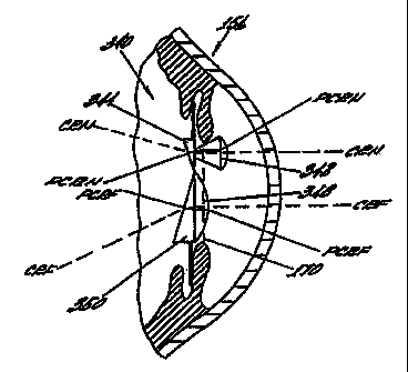
Figs. 18 and 19 illustrate an alternative of an artificial
lens for practicing the invention wherein the imaging producing
mear.s defines a first lens system and second lens system which
each include an extended objective lens to increase the amount of
light collection by the artificial lens and passed to the
posterior segment of the eye. Fig. 18 illustrates that the eye
156 has the artificial lens system shown generally as 300
extending through iris opening into the anterior chamber thereof.
The arti-fficia'_ lens system 300 includes a first extended
objective lens 302 and a second extended objective lens 304. The
objective lens 302 extend into the anterior chamber of the eve
156. As shown by Fig. 18, the distant end of each objective lens
302 and 304 cerminates in a surface as illustrated at. the d_stal
lens 310 of extended objective lens 302 and distant lens 314- of
CA 02333734 2007-06-07
34
the extended objective lens 304. The distal lens 310 includes a
shaped lens/prism member 312 with the base of the prism in a
"base up" position. The distant end of the distant extended
objective lens 304 has a shaped lens/prism member 316 with the
base of the prism being located in a "base down" position.
Although, for purposes of this disclosure, the lenes are
described as separate and opposed; in p'ractical application, the
lens are fused together and homogenous. The effect of the prism
is to change the angle of the ocular lens (posterior lens) in
relationship 1-o the longitudinal axis of the lens svstem. The
prisms are positioned in an opposed spaced relationship to each
other.
In the event that the length of the extended objective lens
is of a length which extends through the posterior capsule, a
procedure referred to as capsulorhexis can be performed on the
posterior capsule to form opening in the posterior capsule. In
such event, the posterior end of the lens system would extend
into the vitreous humor.
Fig. 18 illustrates that the paracentral ray near ("PCRN")
passes through the objective lens 302, the midsection 310, the
shaped lens/prism member 312 and the PCRN is focused onto the
macula 318. In a similar manner, the central ray near ("CRN")
passes through the extended objective lens 302 to the distal end
310 where the image is deflected by the prism to position the CRN
superior of the macula 318.
The far extended objective lens 304 receives the paracentral
ray far ("PCRr") and passes the same through the midsection 314
CA 02333734 2007-06-07
where the prism 320 then directs the PCRF ray through shaped.
lens/prism member 316 onto the macula 318.
Similarlv, the extended objective lens 304 receives the
central ray far ("CRF") and passes the same to the midsection 314
where the prism directs the CRF to a location inferior of the
macula 318.
Fig. 19 shows another embodiment of the extended objective
lens system of Fig. 19 wherein the midsections 310 and 314 are
terminated by a different lens system. Specifically, midsection
310 of the extended obiective lens 302 and midsection 314 of the
extended objective lens 304 are each terminated posteriorly in a
prism 330 at the respective midsections 310 and 314. The bases
of the prisms 330 are positioned in a"base up"P'base down"
relationshia as shown in Fig. 19. The prisms each have a
posterior surface 332 for supporting a negative lens 332.
The light rays pass through the midsection 310 and are
deflected by the prism 330 through the-negative lens 334 such
that- the light,ray CRN is directed superior of the macula and the
light ray PCRN is directed onto the macula. By allowing an
extension of the lens systems from the posterior chamber into the
anterior chamber as illustrated in Fig. 18, the following
advantages are obtained. The CRF and PCRF light rays passing
through the extended objective lens 304 are directed such that
the PCRF light rays go to the macula and the CRF light rays
inferior to the macula.
The lens system 300 provides a greater collection of
possible light. Due to the objective lens in the extension,
there is an increase in the field of vision. Further, by
CA 02333734 2007-06-07
36
utilizing the extended objective lens, there is a decrease in the
problems of centering the lens.
The combination of a plus power objective lens in the
anterior chamber and a minus power ocular lens in the posterior
chamber or vitreous constitutes a totally intraocular galelian
telescope. The purpose of this light gathering and magnification
(enlargement) oT the image is for use i'n patients with macular
degeneration.
By utilizing different lens structure in Figs. 18 and 19, it
is possible zhzt specific lens structures could be developed for
special applications for macular degeneration wherein the retinal
image can be spread over more of the retina to stimulate more of
the sending neurons to the brain thereby improving the ability of
the brain to interpret the image.
By utilizing extended objective lens, the overall size of the
artificial lens base could be made smaller resulting in smaller
incisions needed for insertion.
In Fig. 20, the artificial lens 340 in the form of an
intraocular lens is implanted in an altered pupil.within the eye
156. The artificial lens 340 includes an extended objective lens
342 and a "base up" prism 344 which are adapted to be located to
be in the superior location of the enlarged pupil, such as
superior in the enlarged vertically extending ellipitical shaped
area of the,natural pupil 170 as illustrated in Fig. 15 which is
functionally equivalent to the accessory pupil. The artificial
lens 340 also includes a plano-convex lens 348 and a "base down"
prism 350 which are adapted to be located in the natural pupil
CA 02333734 2007-06-07
37
= 170. A similar lens system without prisms for similar macular
image is a variation of this novel concept.
The artificial lens 340 illustrated in Fig. 20, the PCRN
passes through the extended objective lens 342 and is deflected
by the "base up" prism onto the macula and the CRN is directed to
a location superior of the macula. In this structure, the
objective lens collects more light for-near vision due to its
extension into the anterior chamber. The optical surface of the
objective lens can be made I-arger to create a larger field of
vision.
In the lower section of the artificial lens, the PCRF rays
pass through the plano-convex lens 348 and are directed by the
"base down" prism 350 onto the macula. The CRF rays are passed
through the plano-convex lens 348 and are deflected by the "base
down" prism 350 inferior of the macula and the PCRF is directed
onto the macula.
Fig. 21 illustrates in a front plan view artificial lens 340
of Fig. 20. The extended objective lens 342 is positioned on the
plano-convex lens 348 in a superior position on lens 348
(eccentrically arranged). The "base up" prism is located on the
reverse surface of lens 342. The central body lens 348 likewise
has its prism 350 located "base down" on the reverse surface.
The artificial lens 340 includes three haptic members 352 spaced
substantially equal to hold the intraocular lens in the eye as
described hereinbelow.
Ir, the pictorial representation of Figs. 22a, 22b and 22c,
various other possible configurations for intraocular lens
utilizing the teaching of this invention are shown. Fig. 22a
CA 02333734 2007-06-07
38
illustrates an artificial lens system implanted in an eye
156 wherein the artificial lens has an extended objective
lens 360 which is adapted to be located in the accessory
pupil 178 and any other suitable lens may be used in the
natural pupil 170. This arrangement can utilize prisms for
disparate macular images and without prisms for similar
macular images.
Fig. 22b illustrates an artificial lens system
implanted in an eye 156 wherein the artificial lens has
extended objective lens 360 and 370 wherein objective lens
360 is adapted to be located in the accessory pupil and
extended objective lens 370 is adapted to be located in=the
natural pupil 170. In addition, for a trifocal lens
equivalent, a third extended objective lens 372 can be,
located within the natural pupil 170.
The concept of a trifocal structure illustrated in
Fig. 22b is exemplary, and any artificial lens of the
invention can utilize the trifocal concept.
Fig. 22c illustrates an artificial lens system
implanted in an eye 156 wherein the artificial lens has an
extended objective lens 370 which is adapted to be located
in the natural pupil 170 and any other suitable lens may be
used in the accessory pupil 178. These are all variations
of eccentric lens systems.
Figs. 23 and 24 illustrate an artificial lens in the
form of an intraocular lens 378 attached to haptic members
380, the intraocular lens 378 having a piano-convex lens on
the surface and a "base up" prism 382 in the superior
location of the lens and a larger extended objective lens
376 having a plano-convex lens on the surface located in
the inferior location on the lens 378. The diameter of
lens 374 could be in the order of about 2.5 millimeters and
CA 02333734 2007-06-07
39
the diameter of lens 376 could be in the order of about 3.0
millimeters.
The structure or the intraocular lens in Figs. 23 and 24
permit an additional quantity of light rays to be is directed
onto the macula which counteracts the decreased amount of light
available by using two lens systems.
Fig. 25 is a pictorial representation of the eye showing the
natural pupil 170 and an accessory pupil 178 having the
intraocular lens 378 of Fig. 23 implanted in the eye. The
intraocular lens 378 of Fig. 23 is implanted in the eye with lens
374 being located posterior to the accessory pupil 178 and lens
376 located posterior to the natural pupil 170. Again, a prism
is used for disparate macular images and no prism for similar
images.
Fig. 26 is a pictorial representation of the eye showing the
natural pupil 170 being formed into a vertically extending
ellipitically shaped pupil forming an enlarged area 170' which is
in Fig. 25. The intraocular lens 378 of Fig. 23 represented by
dashed lines is implanted in the eye with lens 374 being located
in the enlarged pupil 170' and lens 376 located in the natural
pupil 170.
Referring now to Fig. 27, the embodiment of an intraocular
lens of Fig. 27 is in the form of plano-convex lens 388 having
with an extended objective lens 392 and a "base up" prism 394
located superiorly on the lens. A plano-convex lens 390 is used
for a distant image. This embodiment produces separate light
rays from another object which is directed onto the macula 34
(disparate macular image). Similarly, the lens system
CA 02333734 2007-06-07
arrangement can be used without prisms for similar macular
images.
Fig. 28 is a pictorial representation of an eye having a
natural pupil 170 which is formed into an enlarged pupil 178
having a vertically extending ellipitical shape with the
intraocular lens of Fig. 27 implanted therein_ Fig. 28 also
shows the various positions of the upper eyelid shown in the
open position represented by dashed line 160 to pass an image
through the extended objective lens 392. The upper eyelid is
also shown in the blocking position as represented by dashed line
162 wherein light rays from a near image is a blocked from
passing through the extended objective lens 392. The distant
image is passed by lens 390. A similar effect would be obtained
with an accessory pupil used with the lens system with or without
a prism.
Fig. 29a shows a pictorial representation of the eye having
a natural lens 400 in the eye. An intracorneal lens having a
plano-convex lens 402 is located superiorly within the cornea oi
the eye to pass light rays from an object through the superior
part of the natural lens 400 and directs the paracentral light
rays from the near object onto the macula 34. The intracorneal
lens having the plano-convex lens 402 is eccentric to the natural
lens 400.
Figs. 29b and 29c show pictorially alternative arrangements
of the plano-convex lens 402 having a prism 404 or 404' . In Fig.
29b, the prism 404 is mounted "base up" and in Fig. 29c, the
prism 404' i-s mounted "base down"_
CA 02333734 2007-06-07
41
In all of these instances, the lens of Figs. 29a, 29b, and
29c are all arranged eccentrically to the natural lens 400.
Fig. 30 is a pictorial representation of an eye having a
partial (no superior cut) radial keratotomy having formed in the
cornea thereof seven (7) elongated angularly disposed slits or
cuts 406 spaced over less than 360 of the eye (approximately
316 as shown in Fig. 30) leaving the superior location of the
eye untreated with elongated slits or cuts. This untreated area
of the cornea of the eye then has the natural pupil er,larged to
from a vertically extending elongated ellipitically shaped pupil.
Near lens 402 with or without prisms 404 and 404' is implanted in
the enlarged area pupil area for passing a light ray from a near
object through the accessory pupil to the macula.
These principles apply also to a four (4) cut radial
keratotomv with oblique cuts (at 1:30; 4:30; 7:30 and 10:30
positions having no superior cuts).
One of the important teachings of the present invention is
that the size and/or shape of a natural pupil can be altered to
accommodate means adapted to be affixed to an eye having
multifocal lens system wherein the principal axes are eccentric,
such as for example, by implantation, intracorneal insertion or
corneal overlay.
It is envisioned that the natural pupil can be altered using
known techniques such as for example, Yag laser, Argon laser or
other known surgical techniques.
A Yag laser is typically used for cutting and care must be
taken to insure that the Yag laser does not hit, damage or
perforate the natural crystalline lens.
CA 02333734 2007-06-07
42
An Argon laser is essentially a coagulation device. It is
known that the Argon laser, when directed to the iris distorts
the pupil. This is generally referred to as "puckering".
Other surgical techniques includes performing a sector
iridectomy which forms a keyhole pupil.
One method for practising this invention includes premarking
of the cornea with a corneal marking device of approximately the
same size as the multiple lens system to be affixed to the eye.
After the cornea is so marked, the lens is inserted under the
marker. The marker should be of sufficient dimension to mark the
cornea sufficiently superior to the natural pupil to insure that
the multiple lens system to be located in the altered pupil will
be located at the desired location in the altered pupil.
Thereafter, the pupil can be further altered as desired using the
selected techniaue to allow entrance of light into the posterior
segment of the eye from the near lens system located superior to
the natural pupil.
It is also envisioned that the artificial lens implanted
into the eye having an altered natural pupil (either an accessory
pupil or enlarged pupil) may be a multiple optical system having
two identical optical or lens systems in an eccentric
arrangement. The superiorly positioned optical system is adapted
to be preferably located in the altered portion of the pupil and
the second optical system would be located in the natural pupil.
As such, the above described method has specific utility for
altering the size andlor shape of a natural pupil to accommodate
an artificial lens having a near lens located superiorly to
CA 02333734 2007-06-07
43
direct light rays to a macula including specifically, but without
limitation, the artificial lens described herein.
By utilizing the teaching of the present invention, the
preferred embodiment uses prisms within the eccentrically
arranged lens system to create light rays for disparate macular
images which are directed onto the macula of the retina by the
lens system at any given time while coricurrently diverting
blurred or otherwise uninterruptable light rays of the images to
a location which is at least one of inferior to or superior to
the macula. Also, the positioning of the lens system within the
pupillary zone may allow for a partial or a complete elimination
of one of the optical systems by adjacent structures such as the
eyelids and/or eyelashes. Several examples are shown herein
including, for example, the illustrations in Figs. 3 and 16.
Thus, the use of a prism in the optical systems for near
vision optically separates the light rays of the distant lens
system of the optical systems in the intraocular lens or other
artificial lens. The use of a prism creates disparity of the
highest order by producing two completely different light ray
paths from eccentric lens system. This is different than
simultaneous vision which is produced by two almost identical
images (difference in size) of the same object passed by
concentric lens systems. Eccentricity without prisms also
creates two almost identical images by creates the possibility of
covering one of the lens systems with eyelids or eyelashes_
The use of a prism in the optical system for far vision
optically separates light rays for the retinal images of the
optical systems in the same manner thereby creating a disparity
CA 02333734 2007-06-07
44
of the highest order in the form of two completely different
retinal images from different objects.
It is envisioned that the artificial lens of the present
invention can be incorporated into an optical lens system having
a lens body wherein the lens body including the imaging systems
are implanted onto the cornea or intracorneal of the eye and are
formed of a on-lay material which is compatible with the
.epithelial cells growing thereacross to implant the optical lens
systems in a subepithelial location.
By utilizing certain teachings of the present invention, it
is possible to make an extremely small intraocular lens which can
be folded or manipulated in such a manner that the same can be
passed through a very small incision in the eye and implanted
into the anterior or posterior chamber of the eye through the
small incision.
Further, by proper training of the patient or user, the user
can utilize the eyelid motion to minimize or eliminate use of one
of the lens systems as desired. As a result, the retina would be
able to dark adapt more easily and thereby become more sensitive
to the available light.
The artificial lens illustrated herein utilizes several
discreet lens systems elements to define each of the imaging
systems. However, using known techniques, the lens systems could
be molded to be an integral artificial lens. Composite lens
system having a predetermined shape so that the same can be
positioned'within the eye. For example, the lens system could be
molded to form the extended objective lens as illustrated in Fig.
18 by using known techniques such that the lens of form integral
CA 02333734 2007-06-07
with the lens body. Various types of material having different
selected angles of incident and angles of refraction could be
utilized for the lens system.
As discussed in connection with the description of Figs. 13,
14 and 15 hereinbefore, the pupil of the eye is altered and
configured into preferably a generally elongated vertically
extending elliptical shape. The alteration and reconstruction of
the pupil can be formed in one of two ways. The pupil alteration
can have its size, shape, position or configuration altered
(which is covered generically by the word "altered" as used
herein) to improve or perfect the optical systems by performing a
surgical alteration, such as an iridectomy. The surgical
alteration would be accomplished in the usual way for performing
intraocular surgery. This could involve either a sphincterotomy
or excision of a portion of iris to form ar. accessory pupil.
Also, the alteration could be formed with a laser. An Argon
laser could be utilized to cause contraction of the iris tissue
peripheral to the pupil resulting in vertical oval shaped pupil.
Another type of laser that can be utilized for performing a
laser alteration is a Yag laser. By utilizing a Yag laser, the
laser beam actually cuts the iris sphincter, thereby enlarging
the overal? size as well as configuration of the pupil. This
allows for selectively enlarging the pupil the one direction, but
not significantly shifting the overall pupil. By utilizing the
amount of tissue actually cut by the Yag laser, the pupil size
can be determined.
Another surgical step that could be utilized is that the
recipient's cornea could be marked with a marking ring to assure
CA 02333734 2007-06-07
46
proper location of the artificial lens within the stroma. By
marking the cornea surface with an indentation ring, the cornea
can be precisely marked to divide the optical zone of the cornea
such that one portion of the optical zone can be used for the
near focus optical system while the other portion is to be used
for the distance focusing system.
In the present invention, when an image is directed onto the
retina at a location superior of the macula, the brain perceives
the image as in the down position. The user would spontaneouslv
turn the eye downward to look through the near lens system. This
movement would "tuck" the distant lens system behind the lower
lid.
In the alternative, when an image is directed onto the
retina at a location inferior to the macula as would be the case
in the distance lens system with the base down prism, the brain
perceives the image as in the up position. The user would
spontaneously turn the eye upward to look through the distant
lens system and the upward movement would "tuck" the near lens
svstem behind the upper lid.
Using these characteristics, the artificial lens can be
specifically designed for a patient's special requirements. The
typical dimension of an artificial lens would be in the range o:
S mm to 6 mm diameter, also, the lens could be oblong with a
minor diameter of about 3.5 mm to about 4 mm and a major diameter
of about 6 mm.
Also, the distant lens could have a diameter of about 5 mm
to 6 mm with the near lens being smaller, say in the order of 2
CA 02333734 2007-06-07
47
mm to 3 mm and be located superiorly in the distant lens in an
eccentric relationship.
An intraocular lens could have the central body functioning
as the distant lens system with a diameter in the order of 3.5 mm
and the near lens system in an eccentric arrangement having a
diameter of about 1.5 mm to about 2.0 mm located superiorly in
the lens body.
Materials used in artificial lens for producing this
invention require a high index of refraction to obtain the plus
power in the lens for near vision. The curvature of the front
surface of the cornea could be changed to obtain more plus power.
Changing the curvature of the front surface of the cornea is an
alternate method that could be used to effect more plus power.
Suitable materials would include those materials that are
bio-compatible and which do have a high index of refraction,
examnles of such material are Polysulfone, Polycarbonate,
Fluorinated Silicone-PMMA Lens combination and other suitable
bio-compatible materials.