Note: Descriptions are shown in the official language in which they were submitted.
CA 02336571 2001-O1-03
WO 00/01367 PCTlUS99/14013
1
DESCRIPTION
Method And Apparatus For Performing Surgery
Inside The Human Retina Using Fluidic Internal
Limiting Membrane (Ilm) Separation (Films)
Background Of The Invention
1. Field Of The Invention
The present invention is directed generally to medical
procedures and, more particularly to a medical procedure for
removal of the innermost layer of the human retina (internal
limiting membrane) from the underlying neural retina at the
center of vision (macula).
2. Background Art
The rays of light entering the eye (EIG. 1) and bearing
the pattern of the object being looked upon pass through the
cornea 32, the aqueous humor, the pupil, the lens 34, and
the vitreous humor, then fall upon the retina 26. The
retina is the light sensitive film lining the back two-
thirds of the eye. Its appearance is similar to that of wet
tissue paper. Its layers consist of the internal limiting
membrane (ILM), the neurosensory retina, and the retinal
pigment epithelium; the ILM, being innermost, is the retinal
border with the vitreous gel cavity 42. If the parts of the
eye are normal and the lens is properly adjusted, the image
will be focused upon the retina. This condition results in
clear vision. At the back of the eye or, more specifically,
the back part of the retina is the macula lutea 36 having at
its center the fovea centralis. The macula is a small
orange-yellow, oval area (about 3 mm by 5 mm) of the retina
adjacent to the optic nerve 38. Vision in which the image
of the object looked upon falls upon the macula is the
sharpest vision and is called macular vision or central
vision, as opposed to gross, peripheral vision.
CA 02336571 2001-O1-03
WO 00/01367 PCT/US99/14013
2
A wrinkling of the internal limiting membrane and the
neural retina is called macular pucker. This can cause loss
of fine vision to the level of legal blindness. The
wrinkling is caused by contractile cells or fibrocellular
membranes (epimacular proliferation or EMP) and is usually a
process associated with aging.
Macular distortion and macular edema, with resultant
macular dysfunction, axe recognized sequelae of EMP. Often,
the macula will have a "wrinkled cellophane" appearance.
According to one theory, this appearance represents internal
limiting membrane (ILM) distortion by surface proliferative
cells without a distinct epimacular proliferative membrane
overlying the ILM, which might be surgically removed. This
ILM cellophaning may persist or occur months after seemingly
successful removal of EMP, limiting visual recovery.
Specimens analyzed after vitrectomy (the surgical
removal of a portion of the vitreous body and/or associated
epiretinal or fibrous membranes) using a microscope for
epimacular membrane removal often contain retinal ILM
fragments that have been intentionally or unintentionally
removed to treat "traction maculopathy," a term introduced
by Morris, R., Kuhn, F., Witherspoon, C.D., ("Retinal folds
and hemorrhagic macular cysts in Terson's syndrome,"
Ophthalmology (1994) 101:1). Written reports differ on
whether the presence of ILM fragments correlate with the
visual outcome. (Trese, M. et al., "Macular pucker
Ultrastructure," Graefe's Arch Clin Exp Ophthamol. (1983)
221:16-26; De Bustros, S. et al., "Vitrectomy for Macular
Pucker: Use after treatment of retinal tears or retinal
detachment," Arch Ophthalmol. (1988) 106:758-760;
Sivalingam, A. et al., "Visual prognosis correlated with the
presence of internal limiting membrane in histophathologic
specimens obtained from epiretinal membrane surgery,"
Ophthalmology. (1990) 97:1549-1552). More recently, William
Hutton and others have implicated even relatively small
amounts of traction as exacerbating diabetic macular edema.
CA 02336571 2001-O1-03
WO 00/01367 PCT/US99/14013
3
Additionally, Logan Brooks and Tom Rice have advocated
the intentional removal of the macular ILM in macular hole
surgery. (Brooks, L., "ILM peeling in full thickness
macular hole surgery," Vitreoretinal Surgery and Technology.
(1995) 7:2; Rice, T.A., "Technique of removal of the inner
retinal surface in macular hole surgery," Retina Society 2gth
Annual Meeting. Santa Fe, New Mexico, 1995). A macular hole
is thought to occur as a result of tangential traction on
the retina at the macula, usually leading to legal blindness
Thus, there are many advocates of the importance of ILM
removal in macular hole surgery. Previous methods as shown
in FIG.2, developed over a period of about twenty years,
have removed the macular ILM 59 and EMP 50 utilizing the
manual, mechanical method with grasping forceps 52. This
forceps procedure is the most delicate surgical maneuver
performed on the human body. The procedure requires ideal
surgical conditions and expert skill. Ideally, cataracts
and any other opacity obscuring surgical view will have been
eliminated for safe and predictable EMP/ILM removal.
Electron microscopy of surgical specimens frequently
demonstrates cellular proliferation contracting the ILM. It
is believed that the increased mobility of an ILM denuded
macula contributes to successful hole closure.
Furthermore, the results with ILM maculorhexis in
macular hole surgery were encouraging. In a consecutive
series of 32 idiopathic holes with less than two years
duration, a 97~ closure was achieved. Previous macular hole
edges were rarely discernible. Visual acuity improved at
least two Snellen lines in 91~ of eyes, and 41~ of eyes
achieved 20/40 or better visual acuity at the last follow-up
(Morris, R., Witherspoon, C.D., "Internal Limiting Membrane
Maculorhexis for Traction Maculopathy," Vitreoretinal
Surgery and Technology (1997) 8(4):1).
All of the above-described conditions may be considered
forms of traction maculopathy as first described by Morris
et al. The ultimate goal of all surgery to cure traction
CA 02336571 2001-O1-03
WO 00/01367 PCT/US99/14013
4
maculopathy is to return the neural retina to its normally
smooth contour, allowing resumption of fine vision and
relief from distorted vision.
In a very rare disease called Terson's syndrome, blood
under pressure from a ruptured vein or capillary
spontaneously lifts the ILM, resulting in what is called a
hemorrhagic macular cyst (HMC) (Morris, R., Kuhn, F.,
Witherspoon, C.D., American Academy of Opthalmology, 1990).
The hemorrhage usually then breaks through the ILM into the
vitreous. Vitreous and subinternal limiting membrane
hemorrhage occurs as a result of abrupt intracranial
hemorrhage from an aneurysm or closed head trauma. Although
the exact mechanism for these hemorrhages is unknown, it is
thought that the sudden increased intracranial pressure is
transmitted via the optic nerve to retinal venules and
capillaries, rupturing them. If bleeding has occurred at
the macula, it will appear as a circular or boat shaped cyst
(HMC) on the surface of the retina. The HMC is usually
encircling the macula. Its diameter and height vary, as
does its color, depending on the longevity of the
hemorrhage. Early intervention (i.e., for amblyopia
prevention in infants) finds a reddish cyst. A few months
after the incident, the surgeon encounters a yellow lesion
(degenerated blood products), a clear membrane spanning an
optically empty cavity, or a collapsed membrane. A
perimacular fold may form along the edge of the separation
of the ILM from the neurosensory retina at the cyst margin.
Sub ILM hemorrhagic macular cysts are almost
pathognomic to Terson's syndrome. Fourteen cases of retinal
folds from shaken baby syndrome or consequent to direct head
trauma were analyzed from various literature reports, each
had intracranial hemorrhage and various forms of intraocular
hemorrhage, including HMC. The HMC's occur not only in
traumatically induced cases of Terson's syndrome but also in
patients with spontaneous subarachnoid hemorrhage.
Accordingly, it has been proposed that intracranial
CA 02336571 2001-O1-03
WO 00/01367 PCTNS99/14013
hemorrhage, from whatever source, is the common denominator
in the formation -of both HMC's and their accompanying
perimacular folds.
In the series originally presented at the Annual
5 Meeting of the American Academy of Ophthalmology in 1990, it
was found that of 25 eyes undergoing vitrectomy for Terson's
syndrome, 8 (32~) demonstrated HMC's (Morris, R., Kuhn, F.,
Witherspoon, C.D., "Hemorrhagic Macular Cysts in Terson's
Syndrome and its Implications for Macular Surgery,"
Developments in Opthalmology (1997) 29:44). After careful
clinical examinations and light or electron microscopic
evaluations, it was concluded that the ILM had formed the
anterior cyst wall in five eyes. While several literature
reports have characterized these hemorrhagic lesions as
being subvitreous or under a proliferative membrane, it is
believed by Morris et al. (see above) that the majority of
hemorrhagic macular cysts in Terson's syndrome are in fact
submembranous (beneath the ILM) rather than subvitreous
(preretinal).
Although rare, submembranous HMC's in Terson's syndrome
are the most frequent lesion in which the macular ILM is
spontaneously lifted from the underlying neurosensory retina
as a result of a disease process. Thus, it was postulated
that if the denuded macula retains good function without
reparative surface proliferation developing, similar non-
traumatic surgical removal of the ILM during vitrectomy in
certain cases of traction maculopathy might be endorsed.
(Morris, R., Kuhn, F., Witherspoon, C.D., "Retinal folds and
hemorrhagic macular cysts in Terson's syndrome,"
Ophthalmology (1994) 101:1). For example, in none of the
five Te.rson's eyes in a series evaluated by the inventor and
colleagues did reparative proliferation develop during an
average follow up of 32 months (range: 6-70 months), and all
adult eyes reached and maintained excellent 20/25 visual
acuity.
CA 02336571 2001-O1-03
WO 00/01367 PCT/US99/14013
6 '
TY~erefore, the desirability of developing procedures
for the atraumatic surgical removal of the macular ILM in
certain forms of traction maculopathy was suggested.
("Hemorrhagic Macular Cysts in Terson's Syndrome and its
Implications for Macular Surgery," Developments in
Ophthalmology (1997) 29:44). Even minimal ILM surface
traction has been increasingly implicated in many forms of
maculopathy and often the EMP/ILM layers become, in effect,
fused together, not allowing surgical removal of EMP alone.
Additionally, long-term macular function appears to be
stable or improved even without the ILM. ("Hemorrhagic
Macular Cysts in Terson's Syndrome and its Implications for
Macular Surgery," Developments in Ophthalmology (1997)
29:44). Thus, ILM removal is an important technique in the
treatment of all forms of traction maculopathy because only
the removal of the ILM with all cellular and membrane
proliferation on its surface ensures total relief from all
traction on the underlying nerve fibers at the center of
vision. However, the methods thus far developed for such
ILM removal have certain deficiencies. The method of
mechanical pulling tearing away the macular ILM with forceps
can cause severe trauma to the macula and the resultant
injury can cause ocular damage of equal severity to the
problem the surgery is meant to correct.
The current method employed for removal of both EMP and
the macular ILM consists of cutting and then grasping, or
directly grasping, the macular EMP/ILM with specially
designed micro-forceps, 1 mm in maximum diameter, and slowly
pulling it apart from the neural retina. This is done with
great care in order to avoid engaging the neurosensory
retina.
One problem with the current method of tearing and
peeling away the macular ILM is the physical trauma
associated with pulling on the ILM until it separates
thereby unavoidably stressing the underlying nerve tissue,
sometimes causing irreparable nerve damage with worsened
CA 02336571 2001-O1-03
WO 00/01367 PCT/US99/14013
7
vision than may have been present preoperatively.
Accordingly, the surgeon may proceed slowly and carefully
but if too slowly the retina may be injured from light
toxicity coming from the fiberoptic probe inside the eyeball
enabling the surgeon's view. If the surgeon grasps too
shallow then his movements are ineffectual, adding to the
time of surgery and the chance of light toxicity. If the
surgeon grasps too deep, permanent nerve damage and
hemorrhage results. The difference is usually a matter of
microns of forceps movement, causing the surgeon's mindset
to be what has justly been described as "nerve-wracking."
The mass of the forceps, although ever so small, often
obscures the surgeon's view, further adding to the chance of
surgical damage to the retina. As a result of the above
factors, complete traction release is the exception rather
than the rule. Finally, even in the unusual case of
complete traction release, the nerve tissue will usually
require several months to resume a smooth contour with best
vision returning. Thus, for some twenty years, the removal
of epimacular proliferation so as to restore central vision
in the eyes that are approaching legal blindness has
remained a vexing problem for vitreoretinal surgeons
worldwide. The potential surgical risks and the uncertain
benefits, as well as the high level of skill required to
perform such surgery has caused many surgeons to be
reluctant to intervene until vision is substantially lost.
This has been true, despite the knowledge that persistence
of EMP causes permanent destruction of nerve function at the
center of vision, such that visual acuity is only partially
restorable, and progressively less so, as the EMP is allowed
to persist.
Solutions have been diligently sought over this twenty-
year period, including progressively smaller and finer
forceps. Finally, as an illustration of surgeons'
frustration, in 1997, a new concept was introduced by Tano
to surgically rub or scrape the surface of the retina at or
CA 02336571 2001-O1-03
WO 00!01367 PCTIUS99/14013
8
near the macula with a flexible, rubber instrument upon
which has been glued innumerable diamond chips so as to
allow the device to purchase a hold on these barely visible
membranes and/or ILM. This device was introduced and has
been substantially used, despite the obvious risk of damage
to the neural retina which underlays these thin membranes by
rubbing or scraping the retinal surface with an
intentionally roughened instrument, as well as the risk of
diamond chips dislodging and permanently remaining on the
retinal. surface within the eye. These risks have been
tolerated in the more or less desperate search for effective
remedies for traction maculopathy because the device adds an
additional means to gain a surgical edge against the all too
frequent need to conclude the operation before achieving
complete release of traction.
Wang U.S. Patent No. 5,066,276, described injecting
viscous material into the eye using a standard glue
injector. Wang, however, did not apply this surgical
procedure within the retina itself. Rather, Wang described
injecting the viscous material between a glia cell membrane
and the retina. Wang described his procedure as one in
which the pressure applied to the retina is very diffuse and
not localized in one small area in order to reduce stresses
on the retina.
There remains a need for improved methods for removal
of both epimacular proliferations and the abnormal ILM of
the retina to completely relieve all forms of traction
maculopathy in which the ILM is contributory. Such a method
must be based upon minimizing surgical traction on the
underlying nerve tissue at the center of vision (fovea).
The method and apparatus described herein overcomes the
above noted problems.
Summary Of The Invention
The present invention provides a novel method for
performing surgery inside the human retina using fluidic ILM
CA 02336571 2001-O1-03
WO 00/01367 PCT/US99/14013
9
separation (FILMS) and simultaneous retinal smoothing to
overcome the disadvantages of the previously known methods.
Briefly, the present invention is directed to a method
of separating the ILM layer of the retina from the neural
layer of the retina in order to remove the macular internal
limiting membrane and all EMP on its surface. The method
comprises inserting a hollow microcannula, considerably
smaller than any such cannula heretofore, which is shaped at
its distal end to conform tangentially to the surface of the
retina, between the retinal ILM and the neural retina.
After the microcannula is inserted, a sterile fluid is
injected at a pressure of about 25 mm Hg through the
microcannula between the ILM layer of the retina and the
neural layer of the retina. The fluid pressure lifts the
macular internal limiting membrane layer away from the
neural layer of the retina, separating it in the process of
lifting away and allowing for its easy forceps removal from
the eye without inflicting any physical trauma upon the
neural retina. The lifted macular internal limiting
membrane is removed by grasping the free-floating macular
internal limiting membrane with forceps and extending the
macular internal limiting membrane separation as distant
from the fovea as desired before tearing circumferentially
about t:he fovea and removing from the eye. The present
invention allows for the removal of the macular internal
limiting membrane without mechanically peeling or tearing it
away from the fovea, so as to minimize foveal traction and
the resultant physical trauma to the fovea. Moreover, the
present invention simultaneously actively smoothes the
underlying distorted and wrinkled neural retina by an
intentional build-up of localized pressure within the
confines of the developing FILMS cyst of which it is the
posterior border. Thus, visual recovery, the ultimate
surgical goal, is substantially accelerated as compared to
the months needed for passive, spontaneous retinal smoothing
after forceps traction removal. The preferred substance for
CA 02336571 2001-O1-03
WO 00/01367 PCTNS99/14013
use in practicing the invention and achieving complete
removal of the macular internal limiting membrane is sodium
hyaluronate (HealonC3~), as manufactured by Pharmacia & Upjohn
Inc. or chondroitin sodium hyaluronate (Viscoat~) as
5 manufactured by Alcon, Inc.
The present invention allows surgeons to operate, for
the first time, inside the human retina (intraretinal)
rather than above its surface. In so doing, it enables the
surgeon to gently, predictably, and rapidly remove the ILM
10 and all EMP adhered to the neural retina. The gentleness of
the invented method eliminates risk of mechanical traction
from pulling on the nerve fibers. The speed of the method,
typically 4 minutes as opposed to 15 minutes, substantially
reduces the risk of light toxicity. The predictability of
the method allows for a more certain benefit from the
surgery. Moreover, the method affords a significant
decrease in the surgical skill level needed to treat
traction maculopathy and makes visual recovery more rapid,
more certain, and more complete. The sum effect is to
enable patients suffering visual loss due to traction
maculopathy of any type to seek and find earlier and more
certain relief from distorted and reduced visual acuity,
while the associated neural retinal abnormality is still
reversible.
It is the principle object of the present invention to
provide an improved method for macular ILM removal and
retinal smoothing. Other objects and advantages of the
present invention will become readily apparent from the
following detailed description taken in conjunction with the
accompanying drawings.
Brief Description Of The Drawings
A better understanding of the present invention will be
had upon reference to the following detailed description
when read in conjunction with the accompanying drawings.
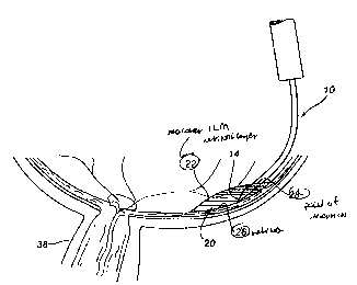
FIG. 3 is a representation of the microcannula apparatus to
CA 02336571 2001-O1-03
WO 00/01367 PCT/US99/14013
11
be used to perform the new method for fluidic ILM separation
(FILMS). FIG. 4 is the interior of the posterior half of
the left eye as viewed through an ophthalmoscope. The area
of most acute vision, the macula, is shown with the fovea
centralis at its center. FIG. 5 is a cross-sectional view
of the eye undergoing a macular FILMS procedure in
accordance with the method of the present invention.
Detailed Description Of The Preferred Embodiments
Definitions
"Maculorhexis" is the removal of the macular internal
limiting membrane (ILM) by the production of a circular,
360° ILM tear concentric with the fovea, while minimizing
foveal traction. This procedure is used to relieve all
forms of traction maculopathy in which the ILM is
contributory, as a result of its innate inelasticity or its
action as a scaffold for fibrocellular proliferation (EMP).
"Traction Maculopathy" is a pathological dysfunction of
the macula partially or entirely secondary to abnormal
tangential or anteroposterior forces (e. g. macular hole,
epeimacular proliferation, vitreomacular traction syndrome,
diffuse diabetic macular edema, cellophane maculopathy).
"Fluid" is a substance whose molecules move easily
across one another; a liquid or a gas.
"Neural Retina" is the middle layer of the retina,
between the ILM and the pigment epithelial layers, which is
composed of nerve tissue and which generates and transmits
the electrical signals ultimately recognized as vision.
Preferred Embodiments
Disclosed herein is a novel procedure allowing surgery
within the human retina to remove the internal limiting
membrane utilizing fluidic separation. With reference to
FIG. 4, to begin the process of ILM removal, an optimal
starting point is chosen within the arcade vessels but
remote from the fovea. Additionally, the chosen starting
CA 02336571 2001-O1-03
WO 00/01367 PCT/US99/14013
12
point site should not overlie the papillomacular bundle.
Furthermore, the starting point is selected on the basis of
the appearance of the ILM, and for surgical convenience. In
a preferred embodiment, referring to FIG. 3, the specially
designed hollow microcannula (microcannula and fluid
injector) has a proximal end 18 and a distal end 12. The
microcannula has an outside diameter of approximately 800
microns (0.8 mm) at its proximal end 18 stepwise tapering to
an outside diameter of approximately 100 microns (0.1 mm) at
its distal end 12. The microcannula is shaped at its distal
end 12 to conform tangentially 10 to the surface of the
retina 26. The micracannula is beveled at the distal tip 14
to promote an effective entry through the macular ILM
retinal layer 22 (FIG. 5) at the surface of the retina 26
and is adapted at the distal tip 14 to discharge a substance
which is contained in a reservoir 16 attached to the
microcannula of FIG. 3. The point of insertion 28 of the
microcannula is at the surface of the retina 26 and through
the macular ILM retinal layer 22. A sterile substance
stored in the reservoir 16 attached to the microcannula is
injected at a pressure of about 25 mm Hg through the
microcannula so as to discharge from the distal tip 14
beneath the macular ILM 22 within the retinal tissue. The
actual injection pressure is selected by the surgeon
immediately prior to microcannula introduction into the eye,
so that said injection pressure moves the injectate fluid
through said FILMS microcannula at the desired rate of flow
for effective but non-traumatic cleavage between the ILM and
neural layers of the retina. The substance 20 then cleaves
the human retina by lifting the macular ILM 22 away from the
neural retina, allowing for its subsequent forceps removal
without inflicting any physical trauma upon the neural
retina due to adhesion of the macular ILM 22 to the surface
of the neural retina. The separated macular ILM 22 is
removed by grasping said macular ILM 22 with forceps and
extending its separation by gentle traction beyond the
CA 02336571 2001-O1-03
WO 00/01367 PCT/US99/14013
13
macula, then tearing said macular ILM 22 in a circular, 360°
fashion concentric with the fovea.
The preferred substance 20 to discharge from the distal
tip of the microcannula 14 and achieve complete fluidic
separation of the macular ILM 22 from the neural retina is a
thick clear fluid such as sodium hyaluronate (Healon~) or
chondroitin sodium hyaluronate (Viscoat~). These are
preferred because their thickness helps form the FILMS cyst,
lifting the macular ILM 22 and simultaneously smoothing the
neural retina without detrimental leakage at the FILMS
microcannula insertion site. Furthermore, sodium
hyaluranate creates a clear field of vision thereby
facilitating intra-operative inspection of the retina.
However, it is possible that a different fluid, such as
sterile saline, or a gas could be used.
The free-floating raised macular ILM 22 is then grasped
by forceps. When the forceps are then used to grasp the
macular ILM 22 separated from the surface of the retina 26,
the maneuver becomes predictable and non-traumatic because
there is no need to tear or peel the macular ILM 22 away
from any direct adhesion to the fovea and surrounding
macular surface of the retina 26, eliminating any vertical
or tangential force vectors as placed upon the fovea by
forceps ILM removal, substituting in its stead a very gentle
and precisely controlled tamponad pressure. Then, a
(smooth-edged continuous tear) "rhexis" is created by slowly
tearing the ILM in a circular pattern concentric with the
fovea, at a distance from the fovea as selected by the
surgeon after further mechanical stripping of the ILM beyond
the macular FILMS cyst. A surgeon can often create the
complete 360° rhexis in one motion. However, if the tear is
incomplete, the TLM is simply regrasped at the new edge, and
the rhexis is resumed.
The problem of adhesion and the necessity to tear and
peel the macular ILM from the surface of the retina (fovea
and macular center of vision) and the resultant physical
CA 02336571 2001-O1-03
WO 00/01367 PCT/US99/14013
14
trauma impressed upon these areas is completely avoided. By
injecting a fluid substance within the retina tissue, the
macular ILM is separated from contact with the neural retina
prior to grasping and removing it from the retina.
Therefore, the macular ILM is never actually torn or peeled
away from the retina and there is no physical trauma caused
by adhesion of the macular ILM to the neural retina because
the injected fluid lifts the membrane away from the surface
of the retina before it is removed using the forceps.
While embodiments and applications of this invention
have been here shown and described, it would be apparent to
those skilled in the art that many more modifications are
possible without departing from the inventive concepts
herein. The invention, therefore, is not to be restricted
except in the spirit of the appended claims.
Examples
It: must be noted that as used herein and in the
appended claims, the singular forms "a" "and" and "the"
include the plural references unless the context clearly
dictates otherwise. Thus, for example, reference to "a
formulation" includes mixtures of different formulations and
reference to "the method of treatment" includes reference to
equivalent steps and methods known to those skilled in the
art, and so forth.
Unless defined otherwise, all technical and scientific
terms used herein have the same meaning as commonly
understood by one of ordinary skill in the art to which this
invention belongs. Although any methods and materials
similar or equivalent to those described herein can be used
in the practice or testing of the invention, the preferred
methods and materials are now described. All publications
mentioned herein are fully incorporated herein by reference.