Note: Descriptions are shown in the official language in which they were submitted.
CA 02337919 2001-O1-17
DEVICE FOR LASER THERAPY IN OPHTHALMOLOGY AND
VARIANTS
Field of the Invention
The invention relates to medical equipment, namely to the devices for laser
therapy in ophthalmology.
lBack~round of the Invention
14.
Therapeutical effect of the action of laser radiation is usually related to
the
improvement of the blood circulation in the vascular system of the eye, as
well as to the
direct bio-stimulation of cells.. This effect is expressed, in particular, in
raising a vision
acuity, removing a spasm of adaptation tension, which decreases the risk of
myopia
progressing and weariness during the visual work at a close distance [1,2].
A stimulating influence of a trans-scleral laser action is strengthened when
using
the radiation of a near infra-red range of the spectrum, having a deeper depth
of
penetration into a tissue compared to visible radiation [1]. At the same time
during
conducting laser therapy with infra-red radiation, problems arise (due to its
invisibility)
2o related to guiding the radiation to the selected areas of the eye. This is
important not only
from the point of view of the f;ffectiveness of therapeutical procedures, but
from the point
of view of their safety as well: permissible levels of irradiating tissues of
eye muscles and
retina are different for the orders of magnitude. The research with the
participation of one
of the authors has shown the possibility of the solution of these problems
while keeping
the sight of a patient in certain reference directions during laser therapy
session.
These problems were partially solved in the known device for laser therapy in
ophthalmology [3] which was selected as a prototype. This device comprises two
parallel
optical channels (optical units) fixed in the spectacles (glasses) rims, each
of which has a
lasing emitter for trans-scleral action by laser radiation upon two areas of
prelimbic
3o region of the eye during a specified period of time, a source of light and
forming optics,
as well as a power supply and control unit, equipped with a timer for setting
a time of
laser action. The source of light (a light diode) in each optical unit forms a
luminous
aiming mark at which a sight of a patient is fixed, and assigns a reference
direction by
that. The axes of a source of light and of forming optics coincide during
this. In addition
to that this device contains an, adjustment mechanism, on which a lasing
emitter, source
of light and forming optics are installed with a possibility of displacement
in the direction
1
CA 02337919 2001-O1-17
transversal to its axis. Before the beginning of work a patient tries to bring
the visual
images of two sources of light observed by each eye into one. In this case the
axes of
optical units match with the visual axes of the eyes, and, correspondingly,
the distance
between parallel axes of these units match with the interpupillary distance of
the patient.
This is used in the prototype for providing simultaneous action of laser
radiation upon the
selected areas of the prelimbic region of each of the eyes and for decreasing
a labour
consuming nature and duration of the laser therapy procedure. When visual
images of
light are put together into one, the device is considered prepared for work,
which serves
as a basis for switching on lasing emitters of both units. Luminous marks from
sources of
10~ light are synchronously glittering during the work of lasing emitters,
specified by a time,
keeping the sight of the patient in reference directions and guaranteeing
exact laser
radiation hitting the selected regions of each eye - in the field of ciliary
muscle projection
upon sclera at 3 and 9 hours for affecting the ciliary body.
However a long operation of the device according to prototype [3] has shown
that
it has limited possibilities. 'The technical solution used in it, although
provides for
necessary laser therapy safety,, is not always acceptable due to superfluous
visual tension
appearing while keeping the sight of the patient in specified reference
directions, which
causes discomfort, and undesirable side effects in some categories of
patients. It is
specially related to patients having a high degree myopia, as well as to small
children in a
2o general case. These patient suffer visual overstrain, and in a number of
cases cannot
match visual images of glittering luminous marks. It is in particular
expressed in
involuntary wish to "assist" to himself b~y way of changing the mutual setting
of optical
units and locating them under more convenient angle with respect to each other
which
causes deformation and impairing the instrument design (during a laser therapy
session a
patient holds the body of optical unila with two hands). Such a visual
overstrain
counteracts to laser therapy and not only decreases the effectiveness of
treatments, but
also can increase the risk of undesirable side effects. This is explained by
the presence of
interconnection between accommodation and convergence, which becomes
especially
strict in case of the eyes mismatch. In this case for each diopter of
accommodation
tension there is a certain value of visual axes convergence, called the
relation of
accommodation convergence to convergence (t~C/C). This value is different with
different patients. The relation AC/C has the biggest values in the patients
with myopia
[4].
The restriction of possibilities of the prototype, described above, are
stipulated by
the fact that the technical solution used in it does not create adequate
conditions for
conducting laser therapy, corresponding to the above individual peculiarities
of the
2
CA 02337919 2001-O1-17
patients, because it restricts the possilbility of patients sight fixation by
only those
reference directions which are parallel to each other. This follows from the
requirement of
parallelism of optical units :location in. this device and of matching the
axes of the
corresponding sources of light; and forming optics in these units.
Summary of the Invention
From the above follows the necessity of such a solution of the task of aiming
radiation at the selected zones. (sections of prelimbic area) of the eye
during laser therapy
to by infrared radiation, which would provide for fixation of the patients
sight in such
reference directions, which rr~atch with his individual peculiarities.
Positive solution of
this task will lead to raising the effectiveness of laser therapy, lowering
patient's
discomfort in the process of conducting iit, as well as will lead to the
absence or essential
reduction of the above undesirable manifestation at the expense of decreasing
visual
tension while keeping the patient's sight :in these reference directions.
The task put is solved by the device for laser therapy in ophthalmology
comprising
two optical units, each of which having one or several lasing emitters with
the forming
optics for trans-scleral action by laser radiation upon one or several regions
of the
prelimbic area of the eye, as well as comprising the source of light for
patient's sight
fixation in a reference direction, and providing by that the indicated action
upon the said
areas of the eye, meanwhile the optical units are installed on the adjustment
mechanism
with the possibility of a displacement of at least one of them in the
direction transversal to
its axis for matching their position with the position of visual axes of the
patient, while
according to the invention, the; said optical units are installed at an angle
to each other, at
which reference directions, specified in these optical units with a help of
said sources of
light, form an angle between themselves in an angular interval, compatible
with the value
of convergence of visual axes of the patient during the observation with two
eyes.
The essence of the invention is based upon the idea of the authors of
coordinating
with individual peculiarities of each patient not only of a distance between
reference
3o directions in which his sigho. is being fixed, but also of an angle between
them in
accordance with the value of visual axes convergence during the observation
with both
the eyes.
Coordination of the distance between reference directions with the
interpupillary
distance is necessary for providing a simultaneous action by laser radiation
upon the
selected sections of the prelim~bic area of each eye. It is executed by the
patient similarly
3
CA 02337919 2001-O1-17
to the prototype by combining visual images of two sources of light observed
by each eye
into one (that is their matchins;) with a help of an adjustment mechanism.
Matching with the vahze of visual axes convergence is necessary in order to
create
more comfortable conditions for the above matching of appearing visual images,
when
for such matching and keeping it during the whole therapy session only minimum
visual
work and, correspondingly, minimum tension of the eyes are required from the
patient.
For the implementation of such conditions the patient is either guided by the
instructions
of a doctor - ophthalmologist, or makes the selection himself, conducting
matching of
observed images of sources of light for a number of devices in which optical
units are
10~ installed at different angles to each other. It is expedient for
facilitating this selection for
different groups of patients that in each such device an angle between such
reference
directions would belong to one of sequential angular intervals embracing in
combination
the specified angular range. The patient judges about the correctness of the
angular
interval selection by his own feelings" according to the minimum of tension
when
matching the above visual images of the sources of light.
Such selection can be also made with a help of one universal device, if at
least one
of its optical units is installed on the adjustment mechanism with the
possibility of
rotating in an azimuthal plane and fixation in the selected position. In this
case for the
selection of the most cornfi~rtable (requiring minimum tensions of
accommodation
apparatus) angular interval between reference directions of the sight,
specified in each
optical unit, the patient cha~zges an angle between the optical units with a
help of
corresponding rotation means; and after finding it, fixes the angular position
if optical
units with a help of a suitable lock. The advantage of such a universal device
is in its
greater functional possibilities, as before each session of laser therapy a
patient can
control and change the angle; between the optical units, each time creating
the most
comfortable conditions for himself during conducting this session.
The practice of using this device with such a binocular optical scheme has
shown
that for the majority of patien~~.s with progressing myopia, especially for
persons of junior
age, minimum visual tension is reached when reference directions of a sight,
specified by
sources of light in each of optical units, are not parallel to each other, but
form a
converging angle in the direction of the sight. The minimum value of this
angle, at which
a substantial effect is already reached in decreasing a visual tension
stipulated by the
necessity of matching two closely located light marks observed by each eye, is
different
for different patients. However, as it follows from the available clinical
experience, it
should for certain exceed 0.01 radian for a rather wide contingent of patient
with different
pathologies of the accommodation apparatus of the eye.
4.
CA 02337919 2001-O1-17
The maximum value of reference directions convergence angle in two optical
units can be evaluated on the data given in work [4] about the relation
between
accommodation and convergence. In different types of eye refraction
(hypermetropia,
emmetropia, myopia) the relation between the degree of accommodation and
convergence, corresponding to the work zone, are changes within very wide
limits. The
value corresponding to the angle of convergence of visual axes of both the
eyes while
looking at an object located at the same distance from the eye, as the sources
of light used
in the device, can be taken ;~s an evaluating criterion for the upper limit of
reference
directions convergence angle :in two optical units of the proposed device.
When the value
10~ of the interpupillary distance is 60 mm and the distance to the source of
light is 15 cm, the
value of reference directions convergence is about 0.4 radian. That is why the
maximum
value of reference directions convergence can be about 0.5 radian.
The value of angular intervals and their number within specified angular range
(for example, the above range from 0.01 to 0.5 radian) can be set based on the
experience
of work. Even more so, they can be different while working with different
groups of
patients. In particular, it might be intervals of the same width (for example,
five intervals
having 0.1 radian each).
It is necessary to bear in mind while selecting the value of an angular
interval that
a permissible minimum value: of angle of reference directions convergence can
vary for
each specific patient depending on the status of his accommodation apparatus
of the eyes.
This is confirmed, in particular, by the circumstance that during conducting a
course of
laser therapy bringing about the raise of ciliary muscle function with a help
of device [3],
patients' discomfort and the above involuntary wish to deform the device
weaken.
It should be pointed out that for the patients having some types of squint,
binocular devices can be preferable for conducting laser therapy, in which the
above
reference directions of a sight form an angle, diverging in the direction of
the sight. In this
case the range of possible changes of an angle between reference directions of
the sight in
accordance with the divergence value (that is negative convergence) of visual
axes during
observation by both eyes can be also established in this case.
3~ It was meant until now in the description of the essence of the invention
that the
above source of light, assigning a reference direction of the sight in the
optical unit
occupies a certain fixed position in it: is located on its optical axes or is
displaced in the
direction, transversal to it, forming a certain angle with it. For example,
sources of light
can be symmetrically located with respect to a bisectrix of the angle between
optical axes
of these units. In each such. case settling the angle between reference
directions in
accordance with the individual peculiarities of a patient is performed by way
of setting a
5
CA 02337919 2001-O1-17
corresponding angle between optical units. By that, each lasing emitter (for
example, a
semiconductor laser) is set in a corresponding optical unit into position, at
which the
beam of its radiation, formed by a corresponding forming optics (for example,
lens) is
directed at necessary angle to the reference direction set in this unit, so
that it hits the
section of the prelimbic area of the eye (for example, at 3 or 9 hours) and
forms a spot of
a specified size (for example, with the diameter of 2-3 mm) in it. The
calculation of this
angle is made based on the value of transversal displacement of the centre of
such spot
with respect to the visual axis of the eye: and the distance to a lasing
emitter. In the case
when one lasing emitter is used for irradiating several sections of the eye,
the forming
to optics of this emitter include; a beam sputter (for example, fibreoptical)
and lenses, the
axes of which are oriented at corresponding angles to the above reference
direction. It is
clear in this case that neither a. number of simultaneously irradiated zones
of each eye, nor
their position, as well as the number of lasing emitters used, and a
corresponding specific
design of the forming optics elo not restriict the essence of the invention,
consisting of the
coordination of the angle between the above reference directions with the
value of the
patient's visual axes convergence while observing with both eyes.
It should be pointed crut that such coordination can be conducted in a
different
way: by setting a corresponding position of the source of light with a fixed
angle between
optical units. It essentially defines a different variant of the idea of the
invention
2o embodiment, in which the taslc set is solved by the fact that the device
for laser therapy in
ophthalmology, comprising two optical units, in each of which one or several
lasing
emitters are set with the forming optics for traps-scleral action by laser
radiation upon one
or several sections of the preliimbic area of the eye, as well as a source of
light for fixing
patient's sight in a reference direction and providing by that the indicated
action upon the
above sections of the eye, while optical units are installed on the adjustment
mechanism
with the possibility of the displacement of at least one of them in the
direction, transversal
to its axis for coordinating their position with the position of visual axes
of the patient,
according to the invention, an the source of light of at least of one of
optical units is
displaced in the transversal direction to its optical axis for the distance,
at which reference
3o directions, set in these optical units witlh a help of the above sources of
light, form an
angle between themselves in an angular interval, compatible with the value of
patient's
visual axes convergence while: observing with both eyes.
In this variant the position of optical units themselves is not essential, if
in this
case the angle between the above reference directions falls into the above
angular
interval. In particular, the optical units can be set parallelly or at a
certain angle to each
other (for example, 0.1 radian). The considerations expressed before in
respect of the
6
CA 02337919 2001-O1-17
value and the number of angular intervals meant for different groups of
patients, the size
of the angular range covered by them, the number of simultaneously irradiated
zones of
each eye and their position, the number of lasing emitters, as well as a
specific design of
forming optics for different variants of the use of lasing emitters are fully
applicable to
this variant.
The peculiarity of this variant is a different form of implementation of the
change
of the angle between said reference directions of the sight;
the indicated source of light and one or several lasing emitters with the
forming optics of
at least one of the optical unias are installed with a possibility of joint
movement in the
1~ transversal direction to its optical axis and fixation in a selected
position.
It is clear that in such an optical unit one or several lasing emitters should
be
located on the same means of displacement with a source of light (for example
on one
platform, moving along a guide, transversal to the axis of the optical unit),
so that during
their joint movement their muitual position could not change, guaranteeing
laser radiation
hitting specified zones of the f;ye.
The analysis of the essence of the invention and of its variants conducted
confirms
the substantiation of the selection of common essential features, describing
the applied
for device for laser therapy in ophthalmology, and the presence of
distinguishing features
among them testifies to the correspondence of the applied for invention to the
conditions
2o of patentability for novelty.
By that, it follows from the analysis of the state of the art that the
solutions of the
problem of laser radiation setting at the areas of the eye subject to the
action proposed by
the authors, allowing to raise l:he efficiency of laser therapy, decrease the
discomfort in its
process, as well as to reduce tlae risk of undesirable side-effects, were not
used in this and
in related fields of the art. This allows to make a conclusion that the
applied for invention
corresponds to patentability conditions according to the inventive step.
Brief Description of the Drawings
3o The essence of the invention described above is explained below with a
specific
embodiment.
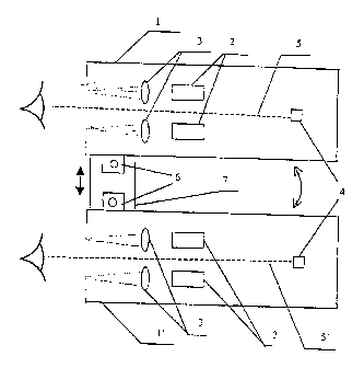
Fig. 1 shows the scheme of one of the variants of the device for laser therapy
in
ophthalmology.
7
CA 02337919 2001-O1-17
Variants of the Invention Embodiment
The device comprises two optical units 1 and 1', in each of which two lasing
emitters 2 (semiconductor losers) are :installed, each of which is equipped
with the
forming optics 3 (in the forth of a lens., installed on the axis of this
emitter) for trans
scleral action by laser radiation upon a corresponding section (at 3 or 9
hours, is not
shown in fig. 1). Besides in each optical unit 1 (1') in its axis a light
diode souxce of light
4 is installed, serving for the formation in the field of vision of a patient
of a light mark
for fixing the sight in a reference direction 5 (5') during the process of a
laser therapy
to session. By that, lasing emitters are installed in an azimuthal plane,
passing through
reference directions 5 and 5', symmetrically with respect to a corresponding
direction 5
(5') and their optical axis (are not shown in fig. 1 ) form an angle of an
order of 0. 1
radian with this direction for providing by this the above action upon said
sections of the
eye. For a corresponding ch<~nge of the angle between reference directions 5
and 5',
optical units 1 and 1' are installed on platforms 6 with the possibility of
turning around
corresponding axes (not shown in fig. 1) in the azimuthal plane and fixation
in the
selected position. In their turn platforms 6 are installed on guide 7 with a
possibility of a
longitudinal displacement along it in the direction transversal to the axis of
optical unit 1)
for coordinating the position of optical uots 1 and 1' with the position of
visual axes of a
2o patient and fixing units in the selected position. Platforms 6, installed
on guide 7, form an
adjustment mechanism of thf; applied for device. The composition of the device
also
includes, similarly to the prototype, a power supply and control unit (not
shown in fig. 1)
connected to the sources of lil;ht 4 and lasing emitters 2 of optical units 1
and 1', and are
equipped with a timer.
The applied for device works in the following way. Before a laser therapy
session,
optical units 1 and 1' are installed into the initial position: at a minimum
angle to each
other (for example, 0.01 radiaw in an absolute value). The same angle is also
formed by
reference directions 5 and 5' in the azimuthal plane, as for this example of
the applied for
device embodiment, sources of light 4 are installed in optical axes of units l
and 1'. A
3o doctor, with a help of a suitable displacement means (for example, a screw
gear)
displaces one or both platforms 6 along guide 7 with respect to each other
until a distance
between optical units l and 1' is not set in accordance to an interpupillary
distance of the
patient which was measured beforehand. Matching of visual images of two
sources 4
observed by the patient into one should be provided in this case. If this does
not occur at
the mutual position of optical units 1 and 1' set or such matching requires
tensions of
accommodation apparatus of the eyes, then the patient, with a help of suitable
rotation
8
CA 02337919 2001-O1-17
means (for example, worm and worm gear) - is not shown in fig. 1) is gradually
changing
the angle between optical units 1 and 1'(or, which is equivalent to it,
between reference
directions 5 and 5') until thc; matching of visual images of two sources of
light 4 is
reached, which testifies to the achievement of an angular interval between
reference
directions 5 and 5', compatible with the value of convergence of patient's
visual axes
while observing with both eyes. After that optical units 1 and 1' are fixed in
the selected
position with a help of a suitable fixing device.
During the whole procedure of the device tuning which was described, light
diode
sources 4 are continuously lit. During this procedure aiming of radiation of
corresponding
lasing emitters 2 to the sections of prelimbic area (at 3 and 9 hours) is
provided. After that
with a help of a power supply and control unit laser emitters 2 are switched
on for the
time, set by the timer. Sources. 4 are transferred into a glittering mode for
the same period
of time for facilitating fixation of patientrs sight in reference directions 5
and 5'. After the
time set passes, power supply and control unit automatically switches lasing
emitters 2 of
both optical units 1 and 1' off, informing the patient about it by
transferring sources 4 into
the mode of continuous lightc,ning and by a sound signal. Upon the end of the
session,
units are installed into the initiial angular position and the device is ready
for the operation
with the next patient. As during the implementation of the described variant
of the
applied for device embodiment known elements the production of which is
mastered by
2o the industry are used, then it is possible to conclude that it satisfies
the conditions of
patentability according to industrial applicability.
At the same time the; above variant of the device implementation cannot be
considered as the restriction o:f the applied for device. It is only an
illustration allowing to
better understand its essence vrhich is most completely described in the
invention claims.
An example of a specific application of the device according to the invention
is
offered.
Patient A., 10 years old. Diagnosis - progressing myopia of a weal degree. The
acuity of vision of both eye wiithout correction is equal to 0.1, with sph -
3.0 diopters it is
0.9. The patient had a course of laser therapy during 10 days (10 sessions)
with a help of a
3o device for laser therapy in its binocular version. The action was applied
to a prelimbic
area of the eye at 3 and 9 hours by infrared laser radiation with a wave
length of 1.3
micron. An aggregate dose of ciliary muscle radiation in each session was
about 0.2 J/cm2
for each eye. As a result of treatment a reserve of relative accommodation was
increased
up to -2.5 Diopters. The acuity of vision of both eyes without correction
became equal to
0.2, with -2.5 Diopters sph it constituted 1Ø An angle between reference
directions of
about 0.09 radian had to be installed for the patient for matching visual
images of the
CA 02337919 2001-O1-17
source of light in two optical units. By that, the treatment session was
conducted without
a visual tension in a comfortable state of the patient.
The example given confirms the possibility of the implementation of the
applied
for invention and of raising the effectiveness of laser therapy of eye
accommodation
apparatus pathologies with its help, decreasing a visual tension and
discomfort of the
patient during it. ',
Industrial applicability
10~ The invention can be used while conducting a stimulation of ciliary muscle
for
raising effectiveness of accommodation apparatus pathologies therapy,
decreasing visual
fatigue and discomfort in the process of conducting it, as well as for
reducing a risk of
undesirable side effects by decreasing a visual tension during keeping a
patient's sight in
such reference directions, which are compatible with the position and
convergence of
visual axes of the patient's eye; during the observation with two eyes.
CA 02337919 2001-O1-17
LITERATURE
1. E.B. Anikina, E.I. Shapiro, N.B Baryshnikov, L.S. Orbachevsky, T.A. Pikus.
Laser
Infrared Therapeutical Device: for Treatment of Disorders of Accommodation
Ability of
the Eye. Thesis of the reports at the 8-th Conference "Laser Optics" and 15-th
International Conference on Coherent and Non-Linear Optics, S-P, 1995.
2. V.E. Avetisov, E.B. Anikina, E.V. Aldimedzhanova. The use of helium-neon
laser in
functional research of the eye; and in pleoptical treatment of amblyopia and
nystagmus.
Methodical recommendations of the Ministry of health of the RSFSR, Moscow
Scientific
1o Research Institute of eye Diseases named after Helmgolts, M., 1990, 14
pages
3. M.G. Vasiliev, E.B. An~ikina, L.S. Orbachevsky. Device for Laser Therapy in
Ophthalmology. RF Patent Nc~. 2092140 of 10.10. 1997 with the priority of
14.10. 1992.
4. E.S. Avetisov. Myopia. Medicine, 1986, pages 19, 61-63.
11