Note: Descriptions are shown in the official language in which they were submitted.
CA 02339861 2001-02-07
WO 00/09002 PCT/AU99/00665
1
SURGICAL VISUAL FEEDBACK AND EYE FIXATION METHOD AND
APPARATUS
Field of the Invention
The present invention relates, in different aspects, to eye fixation and to
the
provision of visual feedback to a surgeon, during the delivery of medical
laser
procedures, particularly in the fields of ophthalmic surgical procedures, such
as
Photorefractive Keratectomy (PRK) and Laser-in-situ Keratomileusis (LASIK), or
any
laser based refractive correction. The invention will be described with
reference to
these applications, though it is to be understood that other applications are
envisaged.
Background Art
Most existing refractive laser delivery systems provide little feedback for
the
operator (typically a surgeon). Usually a crosshair graticule is superimposed
through
the microscope optics to help the operator aim the laser beam correctly onto
the
cornea. A fixation target or light, such as a flashing LED, is used to ensure
that the
patient's eye remains correctly aligned during the surgery. However, this
arrangement does not necessarily provide the best alignment of the eye and the
laser beam, nor does it provide visual feedback for the operator concerning
the
status of the eye or the laser. It may at times be necessary for the operator
to move
his or her attention away from the surgical field to check on instrumentation,
such as
the microkeratome or the laser source. The axis of astigmatism of the
patient's eye is
also likely to be misaligned when the patent is supine and fixating on a point
of light.
Refractive errors are usually assessed when the patient is seated in an
upright
position using structured shapes or symbols, such as letters of the alphabet.
However, refractive surgery is usually performed with the patient reclining in
an
operating chair. It has been found that, when a patient lies recumbent, the
ocular
globe is liable to rotate, altering the position of the axis of astigmatism
between 7°
CA 02339861 2001-02-07
WO 00/09002 PCT/AU99/00665
2
and 16° in 25% of cases (Smith, Talamo, Assil & Petashnick, "Comparison
of
Astigmatic Axis in the Seated and Supine Positions", J. of Refractive &
Corneal
Surgery 10(6), 615 (1994)). This occurs for two reasons: i) the removal of the
reference horizon, and ii) the change from binocular to monocular vision.
Focussing
on a point of light (the flashing LED), instead of the linear horizon, does
not provide a
proper point of horizontal or vertical reference. The globe is therefore
liable to rotate
fractionally, possibly resulting in misalignment of the treatment of the eye's
axis of
astigmatism. The potential end result is under-treatment of the original
astigmatic
error or inducement of astigmatism at another axis.
US Patent 5,549,597 describes a method for determining the axis of
astigmatism of a patient undergoing refractive surgery, so as to provide real-
time
alignment information for the surgical procedure. The patient is required to
focus on
a target such as three sets of three lines of variable line spacing, each set
corresponding to a different visual acuity, and then to focus on the best
resolved set
of lines and rotate the target until the finest line is seen most clearly.
This method of
determining the axis of astigmatism and aligning the surgical laser is not
ideal. The
patient is forced to make subjective comparisons at a highly stressful time.
In
addition, the globe may still rotate after the alignment has been performed,
and prior
to surgery.
An earlier configuration for determining the axis of astigmatism is described
in
US Patent 3,785,723, and involves rotation of an opaque disk having multiple
small
apertures backlit by a light source so as to resemble a set of point light
sources
arranged in a straight line along the diameter of the disk.
US Patent 5,442,412 discloses a patient responsive eye fixation target for use
in ophthalmic procedures in which respective light sources produce a ring of
light and
a dot of light centred on the same optical axis, but respectively closer to
and further
from the eye. In response to detection of a quantifiable amount of eye
movement,
the dot is altered in appearance, eg. by flashing or colour changes, to alert
the
patient that his or her eye is no longer aligned with the dot and ring.
5!00370782.1 CA 02339861 2001-02-07
PCT/AU99/00665
Received 11 October 2000
3
Corresponding to the patient fixation apparatus is the apparatus used by the
surgeon to view and assess the extent of fixation and the alignment of the
laser
beam. The surgeon views this display when looking down the surgical
microscope.
Current technology provides a display including a graticule or crosshair. A He-
Ne
beam is sometimes provided for aiming the surgical beam.
US Patent 4,870,964 provides a head-up display for use with an operating
microscope during phaco-emulsification procedures. This apparatus allows the
operating surgeon to view information about the status of the patient, the eye
and
operating equipment, such as vacuum pressure, without removing their gaze from
the operating field. It does so by projecting light onto the operating field
of the eye
and conditioning the reflections from the cornea so that interpretable images
may be
viewed by the surgeon as they look down the microscope. US Patent 5,135,299
describes a similar operating microscope featuring a head-up display, produced
by
reflecting operational information from the sclera! portion of the eye.
It is an object of the present invention, in at least one aspect, to provide
an
eye fixation method and apparatus that is simple and reliable, and involves
minimal
expectation of the patient. For particular applications, it is further
preferred that the
arrangement reduces the angular rotation of the ocular globe to facilitate
alignment
of an instrument with the axis of astigmatism.
It is an object of another aspect of the present invention to provide a
surgical
visual feedback method and apparatus that provides increased information to
the
surgeon or operator.
Summary of the Invention
According, therefore, to a first aspect of the present invention, there is
provided a method for limiting the rotation of the ocular globe of an eye to
facilitate
alignment of an instrument with the axis of astigmatism of the eye. The method
includes providing fixation target means in the field of view of the eye so
that the eye
may fixate on the target. The fixation target means includes or consists of at
least
one elongate component having a fixed orientation.
(~ 4 v'
w:,'.;_. . .. .. .
5/00370782.1 CA 02339861 2001-02-07
PCT/AU99/00665
Received 11 October 2000
4
Preferably the method includes providing the fixation target means by way of
light emitting means. Preferably, the light emitting means is strobed.
The present invention also provides, in its first aspect, a fixation apparatus
that limits rotation of the ocular globe of an eye, to facilitate alignment of
an
instrument with the axis of astigmatism of the eye. The apparatus includes
fixation
target means for locating in the field of view of the eye so that the eye may
fixate on
the target. The fixation target means includes or consists of at least one
elongate
component having a fixed orientation.
Preferably, said fixation target means includes or consists of at least two
intersecting substantially mutually perpendicular elongate components. The
fixation
target means may consist substantially of a cross, and/or it may include more
than
two elongate components arranged as a grid. The fixation target means
preferably
has a fixed orientation.
The fixation target means may be a light emitting means. Moreover, the or
each elongate component may be defined by the light emitting means.
Preferably, the light emitting means includes a plurality of light emitting
diodes
(LEDs) arranged in a respective linear array to define the or each elongate
component.
Preferably the apparatus includes a printed circuit board (PCB) on which the
LEDs are mounted.
Preferably the apparatus is controllable to strobe the light emitting means.
The apparatus may include a pulsable power supply to strobe the light
emitting means.
In its first aspect, the invention extends to laser surgery apparatus
incorporating patent observable fixation apparatus as described above.
In a second aspect of the present invention there is provided a method for
CA 02339861 2001-02-07
WO 00109002 5 PCT/AU99/00665
supplying visual feedback to an operator during refractive surgery of an eye
of a
patient, including:
1 ) providing fixation target means for the eye to fixate upon;
2) locating the eye for viewing by viewing means while it is fixated
upon said fixation target means;
3) generating an information display of information pertinent to said
surgery and suitable for displaying visually; and
4) transmitting the information display to the viewing means for
viewing by the operator;
whereby the eye and the information display may be viewed
simultaneously by the operator.
Preferably the method includes updating the information display.
Preferably step 3) includes generating the information display with a
controller
means.
Preferably the controller means is a computer.
Preferably the method includes transmitting the information display to a
display means and displaying the information display on the display means.
The display means may be miniature TV or LCD screen or a plurality of LEDs.
Preferably step 1 ) includes the alignment facilitating method according to
the
first aspect of the invention.
Preferably the viewing means includes left and right optics means, and the
target is located between the left and right optics means.
CA 02339861 2001-02-07
WO 00/09002 PCT/AU99/00665
6
Preferably the viewing means is a surgical microscope.
The invention extends to a method of performing refractive surgery on an eye
of a patient, wherein visual feedback is supplied in accordance with the
method of
the second aspect of the invention. The refractive surgery may eg. be PRK or
LASIK,
thermal keratoplasty, intrastromal ablation or any other surgical method that
alters
the refraction of the eye.
The method may be performed with any laser suitable for use in surgery that
involves altering the refractive properties of the eye, e.g. an ultraviolet
ablation laser,
a Holmium laser, or an Erbium laser at 3 microns.
Preferably step 4) includes viewing said information by means of a beam
splitter or plate of glass.
In its second aspect, the invention also provides an apparatus for
supplying visual feedback to an operator during refractive surgery of an eye.
The
apparatus includes fixation target means for the eye to fixate upon, and
viewing
means for viewing the eye while it is fixated upon the fixation target means.
Controller means is provided for generating an information display, and screen
means displays the said information display, for viewing by the viewing means,
whereby the eye and the information display may be viewed simultaneously by
the
operator.
Preferably the apparatus includes display means for displaying the information
display.
The apparatus may be provided in combination with a surgical laser and
thereby comprise laser surgery apparatus.
The laser may be any laser suitable for use in surgery that involves altering
the refractive properties of the eye, such as a ultraviolet ablation laser, a
Holmium
laser, an Erbium laser at 3 microns or any other appropriate laser source.
Preferably the target means is a fixation apparatus according to the first
CA 02339861 2001-02-07
WO 00/09002 7 PCT/AU99/00665
aspect of the invention.
The display means and/or screen means may be viewed by means of a beam
splitter or plate of glass.
Preferably the display means is a miniature TV or LCD screen or a plurality of
LEDs.
Preferably the viewing means is a surgical microscope.
Preferably the controller means is a computer.
Preferably the viewing means includes left and right optics, and the target is
located between the left and right optics.
In the method and apparatus of the second aspect of the, invention, the
information may include an alert signal indicating misalignment of the
patient's eye,
eg. due to straying from fixation upon the fixation target means.
The information may pertain to one or more of: the status of the patient, the
surgery or the equipment, the position of the eye or where an eye-tracker is
aiming
the laser.
The information may include one or more of the following elements of
operational information: type of treatment, number of laser pulses required to
finish,
operation time remaining, patient identification and which eye is being
treated,
keratometry information, refraction information, and/or topographical
information.
The information may include microkeratome status information, such as
suction and blade speed readings.
Brief Description of the Drawings
In order that the invention be more fully understood, preferred embodiments
will now be described, by way of example, with reference to the accompanying
CA 02339861 2001-02-07
WO 00/09002 PCT/AU99/00665
8
drawings, in which:
Figure 1 is a schematic view of an eye fixation apparatus according to a
preferred embodiment of the first aspect of the present invention;
Figure 2 is a fragmentary view illustrating a suitable location for the eye
fixation target; and
Figure 3 is a diagram of the layout of the principal components of an
arrangement for supplying visual feedback to an ophthalmic surgeon during
refractive laser eye surgery procedures, according to a preferred embodiment
of the
second aspect of the present invention, but preferably incorporating the
embodiment
of Figures 1 and 2.
Preferred Embodiments
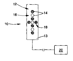
Referring to Figures 1 and 2, there is presented a schematic view of a
patient's eye fixation apparatus 10 according to a preferred embodiment of the
present invention. The apparatus 10 includes a fixation target in the form of
a cross
12 formed by surface mounted light emitting diodes, LEDs 14, arranged in two
linear
arrays to define intersecting elongate components or axes 16, 18 perpendicular
to
one another. The LEDs are fixed to a printed circuit board (PCB) 13, in turn
arranged
on an elongate tipped substrate 15. Substrate 15 is positioned on the front of
the
surgical microscope 28, symmetrically between the adjacent stereo oculars
40,42, so
as to be clearly observable by the patient. Alternatively, cross 12 may be
located
elsewhere within the surgical laser, and projected to optically appear as if
it is placed
between the oculars of the microscope.
Substrate 15 is fixed in position on the microscope so that cross 12 has a
fixed orientation. "Vertical" axis 16 of the cross 12 of LEDs 14 is longer
than
"horizontal" axis 18, by providing several more LEDs 14 in axis 16 than axis
18. By
"vertical" is meant the axis that extends normal to the lines joining the
oculars. The
LEDs 14 may alternatively be positioned to form any other pattern of elongate
or
linear elements, such as a line or a grid.
CA 02339861 2001-02-07
WO 00/09002 PCT/AU99/00665
9
The cruciform arrangement of LEDs i 4 allows the patient to better judge
horizontal and vertical directions, so that the ocular globes) of the patient
does not
rotate and the axis of astigmatism is naturally aligned.
A suitable control circuit 20, of a simple form readily apparent to those
skilled
in the art, is provided for strobing LEDs 14 in a pre-selected, perhaps
adjustable,
sequence. The LEDs may, for example, flash in unison or in a more complex
pattern. LEDs 14 may be of varying colours to facilitate patient concentration
and
gaze control. In use, the patient is required to fixate his or her gaze on the
flashing
cross 12, thereby preventing angular rotation of the ocular globe and
misalignment of
the treatment eye's axis of astigmatism. A preferred flash rate is about 1.5
Hz with a
duty cycle of about 50%. The duty cycle may be adjustable, for example to
allow
more light during LASIK and less light during PRK.
Figure 3 is a diagram of an apparatus 22 for supplying visual feedback to an
ophthalmic surgeon during refractive laser eye surgery procedures being
performed
on an eye 24. This apparatus is an embodiment of the second aspect of the
invention. The apparatus 22 includes a surgical microscope 28 ~ , a fixation
target 26,
which is preferably a flashing cross 12 ~ as in Figures 1 and 2, on microscope
28 ~ , a
head-up display 30 to give the surgeon feedback regarding patient fixation,
the
operating conditions and other pertinent information, and an imaging device in
the
form of a miniaturised TV or LCD screen 32 supplied within the laser delivery
head
(not shown). The head-up display 30 may be produced by projecting lights onto
a
surface, as is known in the art, and may be displayed on screen 32. The
imaging
device may alternatively comprise a combination of light emitting diodes.
The apparatus 22 further includes a controller in the form of computer 34 and
communications link 36 between computer 34 and screen 32. Computer 34
generates the information content (comprising information pertinent to the
operation
being carried out) of the head-up display 30, and transmits this content via
link 36 to
screen 32 to display. This content could include a pulse countdown, operation
time
remaining, an alert signal indicating misalignment of the patient's eye, a
cross 31
indicating where the laser is currently aimed, patient information such as
name or ID,
treatment zone information, topographical information and any other
information that
CA 02339861 2001-02-07
WO 00/09002 PCT/AU99/00665
the surgeon may deem useful.
The apparatus 22 also includes a beamsplitter 38, by which the head-up
display 30 is viewed. The beamsplitter 38 forms a part of the optics of the
laser (not
shown), for relaying this information towards oculars 40 ~ , 42 ~ of the
microscope
5 28 ~ , so that the operator may see the information when he or she looks
down the
microscope 28
Thus, in use, while the patient views (50) a suitable fixation target 26 (such
as
a fixation cross 12 ~ as described above), the surgeon is able to view the
patient's
eye 24 (51 ) and the head-up display 30 (52) through the surgical microscope
28 ~ .