Note: Descriptions are shown in the official language in which they were submitted.
" CA 02340176 2001-02-09
WO 00/09070 PCT/IL99/00440
INHIBITION OF PATHOGENIC PROCESSES
RELATED TO TISSUE TRAUMA
FIELD OF THE INVENTION
The present invention relates to the inhibition of pathogenic processes
associated with tissue trauma by regulating, at the molecular level, the
extracellular
matrix economy. More particularly, the present invention relates to
compositions
containing a quinazolinone derivative which are useful for such regulation,
especially
the quinazolinone Halofuginone, and other molecules which may exert effects
through
the same mechanisms at the molecular level.
In particular, it is now disclosed that these molecules are potent inhibitors
of
nuclear factor xB (NF-oB) transcription, thereby preventing the damaging
cascade of
pathogenic processes that is initiated by trauma, without subverting the
normal repair
mechanisms.
BACKGROUND OF THE INVENTION
Degradation and remodeling of the ECM are essential processes for normal
repair after tissue trauma. However, these mechanisms are also involved in of
a
number of different pathological processes, including the formation of
adhesions,
hepatic fibrosis and cirrhosis, the formation of keloids and hypemophic scars,
and
pulmonary fibrosis. All of these pathophysiological processes are abnormal
responses
to tissue trauma. Yet, each such process represents a different type of
different
fibrosis, involving very different types of tissues, with potentially
different underlying
mechanisms.
The physiological response to tissue trauma is a complex process involving
multiple factors including cell migration and replication, turnover of
extracellular matrix
(ECM) components and changes to the cellular microenvironment. Essentially,
such a
response involves the repair or replacement of damaged tissues. The precise
nature of
such repair or replacement depends upon the tissues involved, although all
such
processes involve certain basic principles. The normal and necessary repair of
any tissue
after any trauma requires the coordination of a wide array of factors by
regulated gene
expression.
The pathophysiological response to the tissue trauma may differ in these
tissues
1
SUBSTITUTE SHEET (RULE 26)
CA 02340176 2001-02-09
WO 00/09070 PCT/1L99/00440
as well, but often results in the formation of adhesions or other types of
abnormal tissues
which do not duplicate the functionality of the original organ tissue, so that
the repair of
tissue trauma does not lead to a complete restoration of organ capacity and
function.
One example of a fibrotic process which results from pathophysiological
S responses to tissue trauma is cardiac fibrosis.
Cardiac fibrosis has a number of causes, which lead to the deposition of
fibrotic tissue. As the deposition of such fibrotic tissue increases, the
ability of the
heart to function decreases, leading to disability and eventually death of the
patient.
The formation of fibrotic tissue in the heart is characterized by the
deposition of
abnormally large amounts of extracellular matrix components, including
collagen, as
well as other matrix proteins. Therefore, the cardiac fibrotic process needs
to be
inhibited in order to prevent damage to the cardiac tissue and hence to the
ability of
the heart to function.
Unfortunately, currently available treatments to inhibit various abnormal
responses to tissue trauma, such as the formation and growth of keloids and
hypernophic scars, cardiac fibrosis and other types of fibrotic disease
processes, are not
completely successful. For example, surgery can be used to reduce the size or
extent of
the lesion, while physical pressure can be used to reduce the size and extent
of keloids
and hypertrophic scars, as well as to prevent their initial formation [D.D.
Datubo-
Brown, Brit. J. Plas. Surg., Vol 43, p. 70-77, 1990]. However, neither
treatment can
prevent the lesion from recurring, and surgery especially carries a risk of
increased
morbidity.
Other forms of treatment include the administration of corticosteroids. For
example, triamcinolone appears to reduce the size of keloids and hypertrophic
scars by
increasing the rate of collagen degradation [Rockwell, W.B. et al., Plastic
and Recon.
Surg., Vol. 84, p. 827-835, 1989]. However, the side effects of such
medications are
potentially dangerous and are not universally successful. Other treatments,
such as
radiation, also showed variable effectiveness and are associated with other
potential side
effects [Rockwell, W.B. et al., Plastic and Recon. Surg., Vol. 84, p. 827-835,
1989].
Thus, clearly improved treatments for these diseases, which are associated
with
pathophysiological fibrotic processes, are required.
As noted above, collagen synthesis and deposition plays an important role in
keloid and hypertrophic scar formation, and in the formation of adhesions, as
well as in
2
SUBSTITUTE SHEET (RULE 26)
CA 02340176 2001-02-09
WO 00/09070 PCT/IL99/00440
the cell hyperproliferation associated with psoriasis and in the many
different forms of
pathological fibrosis such as cardiac fibrosis, pulmonary fibrosis and hepatic
fibrosis.
The synthesis of collagen is also involved in a number of other pathological
conditions,
particularly those associated with primary or secondary fibrosis. The crucial
role of
S collagen in fibrosis has prompted attempts to develop drugs that inhibit the
accumulation of collagen [K.I. Kivirikko, Annals of Medicine, Vol. 25, pp. 113-
126
( 1993)].
However, the deposition of ECM components, such as collagen, is currently
believed to also be important for healing of the wound, as well as for general
I O maintenance of the structure of the tissues. Indeed, the prior art teaches
that the strength
of the healing wound is ultimately dependent upon collagen deposition
[Haukipuro, K.,
et al., Ann. Surg., Vol. 2I3, p. 75-80 , 1991]. Thus, according to the prior
art, collagen
deposition must be present at a sufficient level to give the healing wound
strength and
support, yet not at such a high level to cause the formation of scars.
15 Furthermore, according to the prior art, simply abolishing collagen
synthesis
would be expected to have highly deleterious side effects. Unfortunately,
certain
medicaments which did abolish collagen synthesis, such as general inhibitors
of
collagen formation, were examined for their effect on collagen-dependent
processes
such as tumor growth, and were found to inhibit tumor growth in mice but
proved too
20 toxic for long-term safe administration. Thus, currently available
inhibitors of collagen
synthesis and deposition which were tested for their effects on fibrotic
and/or collagen-
related conditions, were found to be unsuitable for the treatment of
malignancies and
other collagen dependent or related disease conditions, such as the fibrotic
diseases
which were described above.
25 In addition, many other available inhibitors of collagen synthesis and
deposition,
although not examined for their effects on various fibrotic processes, are
generally
undesirable because they lack specificity for the collagen metabolic pathway.
Thus,
many currently available drugs have deleterious side effects.
For example, cytotoxic drugs have been used in an attempt to slow the
30 proliferation of collagen-producing fibroblasts [J.A. Caws, et al., Ann.
Rhem. Dis., Vol.
46, p. 763 ( 1987)], such as colchicine, which slows collagen secretion into
the
extracellular matrix [D. Kershenobich, et al., N. Engl. J. Med., Vol. 318, p.
1709
( i 988)]. Other drugs act as inhibitors of key collagen metabolism enzymes
[K.
3
SUBSTITUTE SHEET (RULE 26)
CA 02340176 2001-02-09
WO 00/09070 PCT/IL99/00440
Karvonen, et al., J. Biol Chem., Vol. 265, p. 8414 (1990); C.J. Cunliffe, et
al., J. Med.
Chem., Vol. 35, p.2652 (1992)]. However, none of these inhibitors have
specific effects
for the metabolism and deposition of specific types of collagen. Also, these
drugs may
interfere with the biosynthesis of other vital collagenous molecules, such as
Clq in the
classical complement pathway, acetylcholine esterase of the neuro-muscular
junction
endplate, conglutinin and pulmonary surfactant apoprotein. Such interference
and lack
of specificity could have potentially serious adverse effects.
Other drugs which can inhibit collagen synthesis, such as nifedipine and
phenytoin, inhibit synthesis of other proteins as well, thereby non-
specifically blocking
the collagen biosynthetic pathway [T. Salo, et al., J. Oral Pathol. Med., Vol.
19, p. 404
(1990)]. Again, the lack of specificity significantly reduces the clinical use
of these
drugs, because the non-specific inhibition of protein synthesis can result in
adverse side-
effects when the drug is administered to the patient.
Indeed, clinically available anti-fibrotic drugs, including the collagen cross-
linking inhibitors such as beta-amino-propionitrile discussed previously, are
also non-
specific. Unfortunately, the lack of specificity of these collagen cross-
linking inhibitors
ultimately results in severe side effects after prolonged use. These side
effects include
lathritic syndrome, as well as disrupted elastogenesis. The latter side effect
is a result of
the disruption of cross-linking of elastin, another fibrous connective tissue
protein. In
addition, the collagen cross-linking inhibitory effect of these drugs is
secondary, so that
collagen must first be overproduced before degradation by collagenase. Thus, a
type-
specific inhibitor of the synthesis of collagen itself is clearly required.
Such a type-specific collagen synthesis inhibitor is disclosed in U.S. Patent
No.
5,449,678 for the treatment of certain fibrotic conditions such as scleroderma
and Graft
Versus Host Disease. Both of these conditions are associated with excessive
collagen
deposition, which can be inhibited by Halofuginone. This specific inhibitor is
a
composition with a pharmaceutically effective amount of a pharmaceutically
active
compound of a formula:
1a
N
N
i
p Rs
4
SUBSTITUTE SHEET (RULE 26)
CA 02340176 2001-02-09
WO 00/09070 PCT/IL99/00440
wherein:
R, is a member of the group consisting of hydrogen, halogen, vitro, benzo,
lower alkyl,
phenyl and lower alkoxy;
RZ is a member of the group consisting of hydroxy, acetoxy and lower alkoxy;
and
R3 is a member of the group consisting of hydrogen and lower alkenoxy-
carbonyl. Of
this group of compounds, Halofuginone has been found to be particularly
effective for
such treatment.
PCT Patent Application No. WO 96/06616 further discloses that these
compounds are able to effectively treat restenosis by preventing the
proliferation of
vascular smooth muscle cells. Restenosis is characterized by smooth muscle
cell
proliferation and extracellular matrix accumulation within the lumen of
affected blood
vessels in response to a vascular injury [Choi et al., Arch. Surg., Vol. 130,
p. 257-261
( 1995)]. One hallmark of such smooth muscle cell proliferation is a
phenotypic
alteration, from the normal contractile phenotype to a synthetic one. Type I
collagen
has been shown to support such a phenotypic alteration, which can be blocked
by
Halofuginone [Choi et al., Arch. Surg., Vol. 130, p. 257-261 (1995); PCT
Patent
Application No. 96/06616]. Thus, Halofuginone can prevent such abnormal
redifferentiation of smooth muscle cells after vascular injury by blocking the
synthesis
of type I collagen. Other in vitro studies show that Halofuginone can also
inhibit the
proliferation of 3T3 fibroblast cells [U.S. Patent No. 5,449,678].
However, the process of restenosis differs from cardiac fibrosis. Furthermore,
cardiac tissue is generally different than the tissue of other organs. In
particular,
cardiac tissue must maintain the ability to function as a single muscle
according to a
wave of electrical activity in order to pump blood effectively. Therefore, the
heart
must maintain a high level of functionality, in contrast to an organ such as
the liver for
example, which can be significantly compromised and still provide the required
level
of function in order to maintain the body. Thus, any amount of cardiac
fibrosis is
detrimental to the functioning of the heart, such that treatments which would
be
suitable for other organs would not be expected to be suitable for treating
and/or
preventing cardiac fibrosis.
Furthermore, the in vitro action of Halofuginone does not always predict its
in
vivo effects. For example, Halofuginone inhibits the synthesis of collagen
type I in
bone chrondrocytes in vitro, as demonstrated in U.S. Patent No. 5,449,678.
However,
S
SUBSTTTUTE SHEET (RULE 26)
CA 02340176 2001-02-09
WO 00/09070 PCT/IL99/00440
chickens treated with Halofuginone were not reported to have an increased rate
of
bone breakage, indicating that the effect is not seen in vivo. Thus, the exact
behavior
of Halofuginone in vivo cannot always be predicted from in vitro studies.
Indeed, the initial discovery of the ability of Halofuginone to successfully
treat
several different disease states was completely serendipitous. Halofuginone
has been
shown to be effective for these disease states by trial-and-error, since the
precise
underlying mechanism of action of Halofuginone was not known. Such a lack of
knowledge, coupled with the inability to completely predict the in vivo
behavior of
Halofuginone from its in vitro effects, has limited the development of new
agents for
these pathophysiological conditions.
The elucidation of the underlying mechanism of the actions) of Halofuginone
would enable new and potentially even more effective treatments to be
developed.
Furthermore, such treatments could be designed to precisely pinpoint the
molecular
targets of Halofuginone and other quinazolinone derivatives, thereby
potentially
1 S reducing the unwanted side effects of treatment. Such treatments could
also regulate
the overall extracellular matrix economy, and thus lead to the amelioration of
many
different pathological conditions associated with disturbances in this
economy.
There is thus a widely recognized unmet medical need for specific effectors
capable of regulating the extracellular matrix economy, whose mechanism of
action
includes targeted intervention at the transcriptional or other molecular
level, such that a
specific effect on the pathological responses to tissue trauma is detemlined
by a precise
intervention at a particular molecular target, which could therefore act as an
inhibitor of
tumor growth, progression and metastasis, which is particularly effective in
vivo,
substantially without adversely affecting other physiological processes, and
which is
able to inhibit angiogenesis and the deposition of collagen, which is able to
selectively
induce apoptosis in tumor cells, and which is able to inhibit a variety of
fibrotic disease
processes, including cardiac fibrosis, in a specific and focused manner, such
that the
treatment does not result in untoward side effects.
SLTryIMARY OF THE INVENTION
It is now disclosed that the ability of the compositions according to the
present
invention to prevent the pathogenic aspects of tissue trauma while preserving
normal
tissue repair mechanisms, is based on the fact that these molecules abrogate
the
6
SUBSTITUTE SHEET (RULE 26)
CA 02340176 2001-02-09
WO 00/090?0 PCT/IL99/00440
cascade of damage initiated by tissue trauma, while maintaining the proper,
healthy
extracellular matrix economy.
According to one aspect of the invention, Halofuginone has been shown to be
effective for the coordination or co-regulation of multiple genes whose
activity is not
known to be co-regulated. Such co-regulation is specific, in that certain
genes which
are known to be linked to the regulated genes in other systems are not co-
regulated by
Halofuginone. Indeed, such specific co-regulation indicates a common,
underlying
mechanism for all of these effects of quinazolinone derivatives such as
Halofuginone.
According to another aspect of the invention, Halofuginone causes a panoply
of effects which were not previously known to be related and which include:
a) decreasing the expression of collagen type I, in particular by inhibiting
the
expression of the collagen aI(I) gene;
b) decreasing the release of the cytokines IL-1 (3 and TNFa, and inhibiting
the
transcription of NF-~cB;
c} inhibiting collagenase type IV production;
d) promotion of activity of cKrox transcription factor;
e) decreasing the expression of the collagen chaperone heat shock protein
HSP47
gene in parallel to the inhibition of the expression of the collagen al(I)
gene; and
f) lack of affect on the expression of TGF[3.
According to the present invention it is now disclosed that the underlying
mechanism of action of Halofuginone and related quinolinones in the inhibition
of
pathogenic responses to tissue trauma involves the regulation of the
extracellular
matrix economy at the molecular level. Such regulation involves at least the
following factors: inhibition of expression of collagen a 1 (I) gene,
inhibition of
transcription of NF-oB and inhibition of collagenase type IV production.
Another
important factor in the efficacy of regulation by Halofuginone is the
promotion of the
activity of the cKrox transcription factor.
NF-xB has become the focus of intense interest, as it has been shown to
become activated in cells through a wide variety of stimulating factors which
are
associated with stress or injury. NF-oB has been shown to be induced through a
large
number of factors, such as IL-1 (3 (interleukin-1 (3) and tumor necrosis
factor a (TNFa)
[Mercurio and Manning; Curr. Op. in Cell Biol., 11:226-232, 1999). Many
inflammatory factors have also been shown to induce NF-xB, such that a wide
variety
7
SUBSTITUTE SHEET (RULE 26)
CA 02340176 2001-02-09
WO 00/09070 PCT/IL99/00440
of induction mechanisms are considered to converge on this particular target.
Thus,
the effect of Halofuginone on the inhibition of NF-~cB expression may indicate
at least
one feature of the mechanism for inducing the effect of Halofuginone on the
inhibition
of fibrotic processes, while regulating and maintaining the physiologically
normal and
S desirable extracellular matrix economy.
The cKrox transcription factor for the expression of the collagen a 1 (I) gent
is a
novel zinc finger-containing transcription factor which binds to the al(I) and
a2(I)
collagen gene promoters, and was shown to repress transcription of the a 1 (I)
procollagen promoter (Widom R L; Culic I; Lee J Y; Korn J H; GENE, (1997 Oct 1
)
198:407-20). As described in further detail in the examples below,
Halofuginone, in
turn, was shown to potentiate the effect of cKrox and thus to potentiate
inhibition of
collagen synthesis.
As described above, additional factors in the ability of Halofuginone to
regulate the extracellular matrix economy may include a decrease in the
release of the
cytokines IL-1 ~3 and TNFa, and a decrease in the expression of the collagen
chaperon
HSP47. Concomitant with all of these actual effects on the extracellular
matrix
economy is a lack of any effect on the expression of TGF~i. Thus, clearly the
regulation of the extracellular matrix economy by Halofuginone, while
maintaining a
healthy extracellular matrix, is specific for a particular underlying
mechanism.
Some of these molecular targets of Halofuginone and other molecules of its
class, such as the inhibition of the tumor marker H19 gene expression, as well
as the
overall regulation of ECM (extracellular matrix) deposition and remodeling,
are likely
to either be secondary targets for the mechanism of action of Halofuginone, or
to only
be indirectly inhibited by Halofuginone. Indeed, these inhibitory effects may
in turn
be related to the potentiation of the cKrox transcriptional factor, or to the
regulation of
the other mechanisms described above.
In any case, all of these mechanisms are related to the regulation of the
"extracetlular matrix economy". Regulation is not merely inhibition of all
processes
related to the extracellular matrix turnover and collagen deposition, since
Halofuginone was previously shown by the inventors to inhibit excessive
collagen
deposition without interfering with basal levels of collagen expression
necessary for
wound repair as shown in US Patent 5,852,024, incorporated by reference as if
fully
set forth herein.
8
SUBSTITUTE SHEET (RULE 26)
CA 02340176 2001-02-09
WO 00/09070 PCT/IL99/00440
The term "extracellular matrix economy" is intended to indicate the
constellation of processes related to the synthesis, deposition and
maintenance of the
extracellular matrix and related tissue structures. The rp oper regulation of
the
extracellular matrix economy leads to the inhibition of the pathological
response to
tissue trauma, such that all of the potential targets for the action of
Halofuginone and
other effectors are able to prevent such pathological responses substantially
without
inhibiting or altering other desirable physiological activity.
Furthermore, these specific effects of Halofuginone, and other compounds
which achieve regulation of the extracellular matrix economy, are useful for
the
treatment of various diseases related to ECM deposition and other aspects of
the ECM
economy, as described in greater detail below. For example and unexpectedly,
it has
been found, as described in the examples below, that Halofuginone can inhibit
the
pathophysiological process of cardiac fibrosis in vivo.
According to one embodiment of the present invention, there is provided a
composition for regulation of the extracellular matrix economy, comprising a
pharmaceutically effective amount of an effector in combination with a
phamaceutically acceptable carrier, wherein regulation of the extracellular
matrix
economy includes inhibition of expression of collagen al(I) gene, together
with
inhibition of transcription of NF-KB and inhibition of collagenase type IV
production.
Preferably, regulation of the extracellular matrix economy includes inhibition
of
expression of collagen a 1 (I) gene and promotion of activity of cKrox
transcription
factor, together with inhibition of transcription of NF-KB and inhibition of
collagenase
type IV production. More preferably, the regulation of the extracellular
matrix
economy includes inhibition of expression of collagen al(I) gene, and
promotion of
activity, of cKrox, together with decreased release of cytokines IL-1 (3 and
TNFa,
inhibition of transcription of NF-fcB and inhibition of collagenase type IV
production,
substantially without affecting expression of TGF-(3.
According to preferred embodiments of the present invention, the regulation of
the extracellular matrix economy includes decreased expression of HSP47 in
parallel
to inhibition of expression of collagen al(I) gene, inhibition of expression
of NF-xB,
inhibition of collagenase type IV production, and decreased release of
cytokines IL-1 (3
and TNFa, substantially without affecting an expression of TGF-~3.
According to another embodiment of the present invention, there is provided a
9
SUBSTITUTE SHEET (RULE 26)
CA 02340176 2001-02-09
WO 00/09070 PCT/IL99/00440
composition for inhibition of at least one pathological process associated
with tissue
trauma, comprising a pharmaceutically effective amount of an effector in
combination
with a pharmaceutically acceptable earner, wherein the effector regulates the
extracellular matrix economy in order to inhibit the at least one pathological
process
associated with tissue trauma, wherein regulation of the extracellular matrix
economy
includes inhibition of expression of collagen al(I} gene, together with
inhibition of
transcription of NF-xB and inhibition of collagenase type IV production.
Preferably, regulation of the extracellular matrix economy includes inhibition
of expression of collagen a 1 (I) gene and promotion of activity of cKrox
transcription
factor, together with inhibition of transcription of NF-xB and inhibition of
collagenase
type IV production. More preferably, the regulation of the extracellular
matrix
economy includes inhibition of expression of collagen a 1 (I) gene, and
promotion of
activity of cKrox, together with inhibition of transcription of NF-~cB,
inhibition of
collagenase type N production and decreasing release of cytokines IL-1(3 and
TNFa,
substantially without affecting expression of TGF-~3. Most preferably, the
effector
decreases an expression of HSP47 in parallel to inhibition of expression of
collagen
a 1 (I) gene, inhibits expression of NF-oB, inhibits collagenase type IV
production and
decreases release of cytokines IL-1 ~ and TNFa, substantially without
affecting
expression of TGF-(3.
Preferably, the at least one pathological process is selected fi-om the group
consisting of cancers, fibrotic conditions including but not limited to
hepatic fibrosis
and cirrhosis, chronic inflammatory disease, pulmonary fibrosis, cardiac
fibrosis, neo-
angiogenesis, formation of adhesions, psoriasis, keloids, hypertrophic scars,
and a
pathological condition which can be ameliorated, reduced or otherwise treated
by an
effector-capable of regulating the extracellular matrix economy.
According to particularly preferred embodiments of the present invention, the
effector is a quinazolinone derivative. More preferably, the quinazolinone
derivative
is a member of a group having a formula:
SUBSTITUTE SHEET (RULE 26)
CA 02340176 2001-02-09
WO 00/09070 PCT/IL99/00440
t O I.
Rr
N
N
I
O Rs
wherein:
Rl is a member of the group consisting of hydrogen, halogen, vitro, benzo,
lower alkyl, phenyl, and lower alkoxy; RZ is a member of the group consisting
of
S hydroxy, acetoxy, and lower alkoxy, and R3 is a member of the group
consisting of
hydrogen and lower alkenoxy; and n is either 1 or 2; and pharmaceutically
acceptable
salts thereof. Most preferably, the compound is Halofuginone and
pharmaceutically
acceptable salts thereof.
According to another preferred embodiment of the present invention, there is
provided a composition for inhibiting cell proliferation enabled by a
deposition of an
extracellular matrix, comprising a pharmaceutically effective amount of a
compound
having a formula:
..
R~r O
N
N
I
p Rs
wherein:
R, is a member of the group consisting of hydrogen, halogen, vitro, benzo,
lower alkyl, phenyl and lower alkoxy; R, is a member of the group consisting
of
hydroxy, acetoxy and lower alkoxy, and R3 is a member of the group consisting
of
hydrogen and lower alkenoxy-carbonyl; n is either 1 or 2; and pharmaceutically
acceptable salts thereof.
According to yet another embodiment of the present invention, there is
provided a composition for treating cardiac fibrosis, comprising a
pharmaceutically
effective amount of a compound in combination with a pharmaceutically
acceptable
carrier, the compound being a member of a group having a formula:
11
SU9ST1TUTE SHEET (RULE 26)
CA 02340176 2001-02-09
WO 00/09070 PCT/IL99/00440
0
N
N
I
p
wherein:
R1 is a member of the group consisting of hydrogen, halogen, vitro, benzo,
lower alkyl, phenyl, and lower alkoxy; R2 is a member of the group consisting
of
hydroxy, acetoxy, and lower alkoxy; R3 is a member of the group consisting of
hydrogen and lower alkenoxy-carbonyl; and n is either 1 or 2; and
pharmaceutically
acceptable salts thereof.
According to still another embodiment of the present invention, there is
provided a method of manufacturing a medicament for treating cardiac fibrosis,
including the step of placing a pharmaceutically effective amount of a
compound in a
pharmaceutically acceptable carrier, the compound being a member of a group
having
a formula:
1p
N
N
I
p R3
wherein:
R1 is a member of the group consisting of hydrogen, halogen, vitro, benzo,
lower
alkyl, phenyl, and lower alkoxy; R2 is a member of the group consisting of
hydroxy,
acetoxy, and lower alkoxy, R3 is a member of the group consisting of hydrogen
and
lower alkenoxy-carbonyl; and n is either 1 or 2. Pharmaceutically acceptable
salts
thereof are also included.
According to yet another embodiment of the present invention, there is
provided a method for the treatment of cardiac fibrosis in a subject,
including the step
12
SUBSTITUTE SHEET (RULE 26)
CA 02340176 2001-02-09
WO 00/09070 PCT/IL99/00440
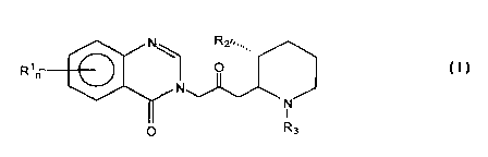
of administering a pharmaceutically effective amount of a compound having a
formula:
. , R2,,,1,
R, r 1 O
N
N
I
O Rs
S
wherein:
R1 is a member of the group consisting of hydrogen, halogen, vitro, benzo,
lower
alkyl, phenyl, and lower alkoxy; R~ is a member of the group consisting of
hydroxy,
acetoxy and lower alkoxy, R3 is a member of the group consisting of hydrogen
and
lower alkenoxy-carbonyl; and n is either 1 or 2. Pharmaceutically acceptable
salts
thereof are also included.
According to other embodiments of the present invention, there is provided a
composition for substantially preventing cardiac fibrosis, comprising a
pharmaceutically effective amount of a compound in combination with a
pharmaceutically acceptable carrier, the compound being a member of a group
having
a formula:
AI
R2~~,,
R, ~ O
r
N
N
I
O R3
wherein:
R1 is a member of the group consisting of hydrogen, halogen, vitro, benzo,
lower alkyl, phenyl, and lower alkoxy; R2 is a member of the group consisting
of
hydroxy, acetoxy, and lower alkoxy; R3 is a member of the group consisting of
hydrogen and lower alkenoxy-carbonyl; and n is either 1 or 2; and
pharmaceutically
acceptable salts thereof.
13
SUBSTITUTE SHEET (RULE 26)
CA 02340176 2001-02-09
WO 00/09070 PCT/IL99/00440
According to further preferred embodiments of the present invention, the
compound is preferably Halofuginone. Hereinafter, the term "HaIofuginone" is
defined as a compound having a formula:
N HOn,,,
Br
O
N
CI N
I
H
O
and pharmaceutically acceptable salts thereof. The composition preferably
includes a
pharmaceutically acceptable carrier for the compound.
Preferably, all of the compounds referred to hereinabove can be either the
compound itself as described by the formula, and/or pharmaceutically
acceptable salts
thereof.
BRIEF DESCRIPTION OF THE DRAWINGS
The invention is herein described, by way of example only, with reference to
the
accompanying drawings, wherein:
FIG. 1 illustrates certain exemplary aspects of the extracellular matrix
economy;
FIG. 2 illustrates the dose-dependent inhibition of type IV collagenase
activity
in T50 bladder carcinoma cell cultures in the presence of Halofuginone;
FIG. 3 illustrates the inhibition of the expression ofthe H19 gene in the
RT112
and 5376 bladder carcinoma cell lines by Halofuginone;
FIG. 4 shows a Northern blot with the effect of Halofuginone on Integrin a~
chain expression;
FIG. 5 shows the effect of Halofuginone on ~3 subunit expression as
determined by RT-PCR;
FIG. 6 illustrates the effect of Halofuginone on ventricular collagen volume
fraction (CVF) in rat heart;
FIG. 7 illustrates the effect of Halofuginone on collagen al(I) gene
expression
in rat heart; and
FIG. 8 illustrates the effect of Halofuginone on TGF-~i expression in rat
heart.
14
SUBSTITUTE SHEET (RULE 26)
CA 02340176 2001-02-09
WO 00109070 PCT/IL99/00440
BRIEF DESCRIPTION OF THE INVENTION
Unexpectedly, it has been found, as described in the examples below, that the
underlying mechanism of action of Halofuginone in the inhibition of all of
these
S pathogenic responses to tissue trauma involves the regulation of the
extracellular
matrix economy at the molecular level.
As disclosed herein the present experimental results also show that
Halofuginone clearly inhibits the expression of NF-~cB (nuclear factor xB),
also in
parallel with the inhibition of the expression of collagen. NF-xB has become
the focus
of intense interest, as it has been shown to become activated in cells through
a wide
variety of stimulating factors which are associated with stress or injury. NF-
oB has
been shown to be induced through a large number of factors, such as IL-1 [3
(interleukin-1 [3) and tumor necrosis factor a (TNFa) [Mercurio and Manning;
Curr.
Op. in Cell Biol., 11:226-232, 1999]. Interestingly, these specific factors
have been
shown in the present experimental results also to be inhibited by
Halofuginone. Many
inflammatory factors have also been shown to induce NF-xB, such that a wide
variety
of induction mechanisms are considered to converge on this particular target.
Thus,
the effect of Halofuginone on the inhibition of NF-oB expression may indicate
at least
one feature of the mechanism for inducing the effect of Halofuginone on the
inhibition
of fibrotic processes, while regulating and maintaining the physiologically
normal and
desirable extracellular matrix economy.
In tum, NF-tcB has been shown to play an important role in the regulation of
the expression of the type VII collagen gene (COL7A1). NF-xB mediates the
effect of
TNF-a on the expression of COL7A1, by binding to a particular portion of the
2~ promoter of this gene which is known as a "TNF-a response element" [Kon et
al.,
Oncogene, 18:1837-1844, 1999]. Interestingly, COL7A1 has two separate elements
in the promoter region: the previously mentioned "TNF-a response element"; and
a
separate response element for TGF-[3. Thus, the apparent effects of
Halofuginone
with regard to TNF-a and NF-oB, but not TGF-~, may ultimately enable
Halofuginone to have the desired inhibitory effects on the pathological
processes of
fibrosis, substantially without detrimentally inhibiting or downregulating the
physiologically normal and desirable extracellular matrix economy.
SUBSTITUTE SHEET (RULE 26)
CA 02340176 2001-02-09
WO 00/09070 PCT/IL99/00440
Another important molecular target of Halofuginone is the cKrox transcription
factor for the expression of the collagen a I (I) gene. cKrox is a novel zinc
finger-
containing transcription factor which binds to the a 1 (I) and a2(I) collagen
gene
promoters, and was shown to repress transcription of the al(I) procollagen
promoter
(Widom R L; Culic I; Lee J Y; Korn J H; GENE, ( 1997 Oct I ) 198:407-20). As
shown
in the examples below, Halofuginone, in tum, was shown to potentiate the
effect of
cKrox and thus to potentiate inhibition of collagen synthesis.
c-Krox was also found to be present at much higher levels in skin than bone,
which may explain why Halofuginone was able to decrease excessive collagen
deposition in skin while not increasing the fragility of bone (Galera P; Musso
M; Ducy
P; Karsenty G; PNAS, (1994) 91: 9372-6).
Other important effects which have been shown to occur through the
administration of Halofuginone include decreasing the expression of the
specific
collagen chaperone heat shock protein HSP47 gene in parallel to the inhibition
of the
I5 expression of the collagen al(I) gene and decreasing the release of the
cytokines IL-
1 [i and TNFa, while inhibiting the expression of NF-~cB; but without
affecting the
expression of TGF~3.
HSP47 has been shown to be a molecular chaperone which is specific for
collagen, and in particular for procollagen [Nagata and Hosokawa; Cell
Structure and
Function; 21:425-430, 1996]. HSP47 is a protein which resides in the
endoplasmic
reticulum (ER), and which binds to newly synthesized procollagen until the
procollagen chain enters the Golgi body. HSP47 may assist in the formation of
proper
collagen protein structures, such as the triple helix structure of collagen.
In further
support of the functional relevance of HSP47 to collagen, previous results
have shown
that the.expression of HSP47 in various cells is closely related to the
expression of
collagen [Nagata and Hosokawa; Cell Structure and Function; 21:425-430, 1996].
In
particular, increased expression of HSP47 was found during the progression of
liver
fibrosis which was induced by the administration of carbon tetrachloride to
rats, also
in parallel to the increased expression of collagen type I.
Interestingly, the present experimental results show that Halofuginone clearly
inhibits the expression of HSP47 in parallel with the expression of collagen.
These
results support the hypothesis that Halofuginone acts at a common, root
mechanism
for a number of different processes related to the pathological synthesis and
16
SUBSTITUTE SHEET (RULE 26)
CA 02340176 2001-02-09
WO 00/09070 PCT/IL99/00440
deposition of collagen and other ECM components within the fibrotic disease
state.
In any case, all of these mechanisms are related to the regulation of the
extracellular matrix economy. Such regulation is not merely inhibition of all
processes
related to the extracellular matrix and collagen deposition, since
Halofuginone was
S previously shown by the inventors to inhibit excessive collagen deposition
associated
with keloids and other abnormal scar formation, yet Halofuginone did not
decrease
wound strength, as shown in U.S. Patent 5,852,024, incorporated by reference
as if
fully set forth herein.
The proper regulation of the extraceilular matrix economy leads to the
inhibition of the pathological response to tissue trauma, such that all of the
potential
targets for the mechanism of action of Halofuginone and other effectors are
able to
prevent such pathological responses substantially without inhibiting or
altering other
desirable physiological activity. Indeed, the term "effector" is used herein
to refer to
substantially any compound or combination thereof which is capable of
regulating the
extracellular matrix economy, thereby specifically inhibiting pathological
processes
related to tissue trauma and mechanistically related processes.
The term "mechanistically related processes" includes those pathological
conditions which share at least one underlying mechanism with abnormal
responses to
tissue trauma. For example, the metastasis of malignant cells is an example of
a
mechanistically related process, because such metastasis depends upon neo-
angiogenesis, which in turn depends upon the deposition of extracellular
matrix
components including collagen, as shown in PCT Application No. WO 98/34613,
filed on February 11, 1998, incorporated by reference as if fully set forth
herein.
Similarly, abnormal responses to tissue trauma such as adhesion formation also
depend upon collagen deposition. Thus, all of these pathological processes can
be
said to be mechanistically related.
Other mechanisms of the "extracellular matrix economy" include, but are not
limited to, the inhibition of angiogenesis, the prevention of ECM deposition,
the
inhibition of collagenase type IV production, the inhibition of integrin
expression, the
induction of apoptosis and the inhibition of H19 gene expression. The overall
structure
of the extracellular matrix economy, and the effect of Halofuginone in
relation to this
economy, is shown in Figure 1. As shown, the specific effects of Halofuginone
on the
extracellular matrix economy include the inhibition or amelioration of
conditions
17
SU8ST1TUTE SHEET (RULE 26)
CA 02340176 2001-02-09
WO 00/09070 PCT/IL99/00440
associated with tumors, fibrosis, including renal fibrosis, scleroderma and
GVHD,
restenosis and skin injury. By mediating certain effects or factors associated
with the
extracellular matrix economy, Halofuginone and other effectors are able to
inhibit
collagen type I synthesis, hence ameliorating pathological conditions
associated with
tissue trauma while permitting normal, desirable physiological processes to
continue.
With regard to specific aspects of these mechanisms, angiogenesis and ECM
deposition have been previously described. Collagenase type IV is a pivotal
metallaprotease enzyme involved in metastasis and cell invasion. Apoptosis is
programmed cell death which, as noted previously, is blocked in malignant
cells, which
are therefore also described as "immortal". The H 19 gene is a tumor-marker
gene
associated with the early stages of bladder carcinoma. More specifically, the
H19 gene
is a developmentally regulated gene whose expression peaks during fetal
development
when tissue differentiation is occurring. Chromosomal abnormalities within the
region
containing H 19 are associated with early stages of malignancies such as
Wilms' tumor,
adrenocortical carcinoma, hepatoblastoma, rhabdomyosarcoma, lung tumors,
trophoblastic tumors and bladder carcinoma [B. Tycko, Am. J. Path., Vol. 144,
p. 431-
439, 1994; de Groot, N. et al., Trophoblast Res., Vol. 8, p. 2285-2302, 1994;
Rachmilewitz, J. et al., Oncogene, Vol. 11, p. 863-870, 1995.
The importance of the extracellular matrix economy is that the mechanism of
action of Halofuginone is able to promote many desirable activities, such as
the
cytostatic inhibition of malignancies, the reduction or prevention of fibrosis
and
adhesions, and other pathological responses to tissue trauma, or
mechanistically related
activities thereof, substantially without causing undesirable side effects
such as
decreasing the strength of healed wounds or altering the remodeling of the
structure of
bone.
DESCRIPTION OF PREFERRED EMBODIMENTS
Unexpectedly, it has been found, as described in the examples below, that the
underlying mechanism of action of Halofuginone in the inhibition of all of
these
pathogenic responses to tissue trauma involves the regulation of the
extracellular
matrix economy at the molecular level. Such regulation includes the following
effects: enhancing the activities of the cKrox transcription factor, which in
turn
represses transcription of the a.l(I) procollagen promoter; decreasing the
expression of
18
SUBSTITUTE SHEET (RULE 26)
CA 02340176 2001-02-09
WO 00/09070 PCT/IL99/00440
the collagen molecular chaperone, HSP47, in parallel to the inhibition of the
expression of the collagen a 1 (I) gene; decreasing the release of the
cytokines IL-1 ~i
and TNFa, while inhibiting transcription of NF-xB; but without affecting the
expression of TGF~3.
Furthermore, Halofuginone is also involved in other aspects of the
extracellular matrix economy, including the inhibition of collagenase type IV
production and the inhibition of H19 gene expression, as well as the overall
regulation
of ECM (extraceilular matrix) deposition and remodeling, the amelioration
and/or
prevention of chronic inflammatory disease, and the inhibition of neo-
angiogenesis.
Other mechanisms of the "extracellular matrix economy" include, but are not
limited
to, the inhibition of angiogenesis, the prevention of ECM deposition, the
inhibition of
collagenase type N production, the inhibition of integrin expression, the
induction of
apoptosis and the inhibition of H19 gene expression. Such specific regulation
of the
extracellular matrix economy has never been demonstrated before, particularly
in vivo.
While the invention will now be described in connection with certain preferred
embodiments in the following figures and examples so that aspects thereof may
be
more fully understood and appreciated, the invention is not intended to be
limited to
these particular embodiments. On the contrary, all alternatives, modifications
and
equivalents are included as within the scope of the invention as defined by
the
appended claims. Thus, the following figures and examples which include
preferred
embodiments will serve to illustrate the practice of this invention, it being
understood
that the particulars shown are by way of example and for purposes of
illustrative
discussion of preferred embodiments of the present invention only, and are
presented
in the cause of providing what is believed to be the most useful and readily
understood
description of formulation procedures as well as of the principles and
conceptual
aspects of the invention.
The present invention may be more readily understood with reference to the
following illustrative examples and figures. It should be noted that although
reference
is made exclusively to Halofuginone, it is believed that the other
quinazolinone
derivatives described and claimed in U.S. Patent 3,320,124, the teachings of
which are
incorporated herein by reference, have similar properties.
19
SUBSTITUTE SHEET (RULE 26)
CA 02340176 2001-02-09
WO 00/09070 PCT/IL99/00440
Example 1
Promotion of cKrox Activity_and
Inhibition of Collagen T~~e I
Gene Expression by Halofu~inone
As noted previously, one of the most important targets for the action of
Halofuginone and other related quinazolinones and effectors is the promotion
of
cKrox activity and the concomitant inhibition of collagen type I gene
expression.
These-two activities were demonstrated with Halofuginone as follows.
First, the ability of Halofuginone to inhibit collagen type I gene expression
was demonstrated as follows. Myometrial and leiomyosarcoma cells were taken
from
the same patient and were plated into 10 cm plates in DMEM supplemented with
10%
FCS. When the cells reached 80% confluence, the medium was replaced by serum
free DMEM plus 0.1% BSA for 48 hours, washed and exposed to increasing
concentrations of Halofuginone in the same medium for about 48 hours at about
37
°C. The cells were then harvested and subjected to RNA extraction and
Northern blot
analysis for collagen type I gene expression. Halofuginone inhibited collagen
type 1
gene expression (products at 5.4 and 4.8 kb) in a dose-dependent manner.
Next, human skin fibroblasts were taken from a subject and were maintained
in primary culture. Control cells were treated with vehicle while Halofuginone-
treated
cells were treated with Halofuginone as previously described. The cells were
then
harvested and subjected to RNA extraction and Northern blot analysis for
collagen
type I gene expression and for cKrox gene expression. Halofuginone promoted
cKrox
gene expression while simultaneously inhibiting collagen type I gene
expression.
Thus, one important molecular target for the mechanism of action of
Halofuginone is
clearly the enhancement of cKrox gene expression and hence cKrox activity,
which in
turn leads to the inhibition of collagen type I gene expression.
Such targeting of the action of Halofuginone is novel, and has not been taught
nor suggested by the background art. However, clearly the elucidation of this
mechanism is important for the development and design of new treatments for
the
pathological processes associated with tissue trauma. Furthermore, such
results
provide a clear mechanistic explanation for the ability of Halofuginone to
inhibit
pathological collagen synthesis, while enabling normal physiological processes
associated with collagen to proceed without unwanted side effects. In
particular,
SUBSTITUTE SHEET (RULE 26)
CA 02340176 2001-02-09
WO 00/09070 PCT/IL99/00440
molecules and chemical compositions which also are able to inhibit
pathological
collagen synthesis, while enabling normal physiological processes associated
with
collagen to proceed without unwanted side effects, are now possible by
targeting the
potentiation of cKrox gene expression andlor activity for therapeutic
intervention.
Example 2
Inhibition of Type IV Colla~enase Production
by Halofuginone in vitro
Another important feature of the regulation of the extracellular matrix
economy is the inhibition of type IV collagenase production by Halofuginone.
Tumor cells secrete enzymes which digest the ECM, enabling the cells to
burrow through neighboring tissue and to invade other tissues. Numerous
studies
have Linked matrix metalloproteases (MMP), especially type IV collagenase, to
the
process of tumor invasion and metastasis. Type IV collagenase appears as two
72 and
92 kDa proteins encoded by a unique mRNA.
As demonstrated in Figure 2, a profound inhibition of the activity of MMP2
(72 kDa type IV collagenase) in TSO bladder carcinoma cell cultures was
exerted in
the presence of 25 ng/ml Halofuginone, while an almost complete inhibition was
obtained at 100 ng/ml Halofuginone. Sub-confluent cell cultures were incubated
for 6
- 24 h in serum-free DMEM. The collagenolytic activity was determined on a
gelatin
impregnated ( 1 mg/ml. Difco, Detroit, MI) SDS-PAGE 8% gel. Briefly, culture
media
samples were separated on the substrate impregnated gels under non reducing
conditions, followed by 30 min incubation in 2.5% Triton X-100 (BDH, England).
The gels were then incubated for 16 h at 37oC in 50 mM Tris, 0.2 M NaCI, 5 mM
CaCl2, 0.02% Brij 35 (weight/volume) at pH 7.5. At the end of incubation
period, the
gels were stained with 0.5% Coomassie 6250 (Bio-Rad Richmond CA) in
methanol/acetic acid/H20 (30:10:60). The intensity of the various bands was
determined on a computerized densitometer (Molecular Dynamics type 300A).
Halofuginone was also found to inhibit cell invasion through matrigel ECM,
using the Boyden chamber invasion assay (data not shown). Such inhibition
supports
the inclusion of type IV collagenase inhibition as part of the extracellular
matrix
economy mechanism, in which Halofuginone inhibits undesirable pathological
processes such as tumor growth, progression and metastasis, as described
previously.
21
SUBSTITUTE SHEET (RULE 26)
CA 02340176 2001-02-09
WO 00/09070 PCT/IL99/00440
Example 3
Inhibition of Tumor-Marker Gene Expression
by Halofu~inone in vitro
Yet another aspect of the extracellular matrix economy is the regulation of
tumor-marker gene expression, and in particular of the inhibition of the
expression
of the H19 gene.
The H19 gene is a developmentally regulated gene whose expression peaks
during fetal development when tissue differentiation is occurring. The H 19
gene is
parenterally imprinted, expressed only by the maternal allele. HI9 is also a
tumor
marker gene, associated with early stages of malignancies such as Wilms'
tumor,
adrenocortical carcinoma, hepatoblastoma, rhabdomyosarcoma, lung tumors,
trophoblastic tumors and bladder carcinoma. The experimental method was as
follows.
I S RT112 and 5376 human bladder carcinoma cell lines were cultured in the
absence and presence of Halofuginone (130 ng/ml, added 24 h or 72 h after
seeding),
and the expression of the H19 gene was evaluated by Northern blot analysis
(Nagler,
A. et al., Arterioscler. Thromb. Yasc. Biol., Vol. I7, p. I94-202, 1997).
Exposure to
Halofuginone resulted in a substantial reduction in the expression of the HI9
gene in
the RT112 and 5376 bladder carcinoma cell lines which were tested, as shown in
Figure 3. Such inhibition also supports the inclusion of the inhibition of H19
gene
expression as part of the extracellular matrix economy mechanism, as described
previously.
Example 4
Inhibition of Integrin Expression
Another important aspect of the effect of Halofuginone and related
quinazolinones on the extracellular matrix economy involves the ability of
these
compounds to inhibit integrin expression, as exemplified by the effect of the
illustrative compound Halofuginone.
Integrins have been shown to function in vivo in vasculogenesis and
angiogenesis. Injection of neutralizing antibody against the X31 subunit
blocked the
formation of an aortic lumen in quail embryos (Drake, C.J. et al., in vivo.
Dev. Dyn.
22
SUBSTfTUTE SHEET (RULE 26)
CA 02340176 2001-02-09
WO 00/09070 PCT/IL99/00440
Vol. 193, p. 83-91, 1992). Cheresh and colleagues have provided evidence that
av(33
is required for blood vessel growth (Brooks, P.C. et al., Science Vo. 264, p.
569-571,
1994). An antibody (LM609) against the av~33 integrin complex inhibited normal
vessel growth and also FGF-2 stimulated or tumor-induced angiogenesis in the
CAM
assay, but did not disrupt preexisting vessels. The mechanism by which anti-
av(33
mAb disrupts angiogenesis appears to involve apoptosis. A single intravascular
injection of a cyclic RGD peptide antagonist of av~33 integrin or of the LM609
monoclonal antibody leads to the rapid regression of human tumors transplanted
into
the CAM. Tumor cells that fail to express the av gene and hence the av~i3
integrin,
lose their adhesion capability and exhibit a significantly reduced
tumorigenicity upon
transplantation into athymic nude mice. Stable transfection of the av cDNA to
these
cells resulted in the full restoration of their tumorigenic potential (Felding-
Habermann, B. et al, ,l. Clin. Invest., Vol. 89, p. 2018-2022, 1992).
Furthermore,
Halofuginone has been found to inhibit angiogenesis and tumor growth.
Therefore, the effect of Halofuginone was investigated on the expression of
av~ (33 and ~i5 integrin subunits in the highly aggressive MDA 435 human
breast
carcinoma cell line. Cells were cultured in the absence (Figure 4, lanes A &
B) or
presence of increasing concentrations (10-400 ng/ml) of Halofuginone for 24 h
(lane
C: 400 ng/ml), 48 h (lanes D-G: 10, 50, 200, and 400 ng/ml, respectively) or
72 h
(Figure 4, lane H: 400 ng/ml}. Total RNA was extracted, subjected to 1.1
formaldehyde-agarose gel electrophoresis, transferred to a nylon membrane and
hybridized with 32P-labeled PCR probe corresponding to av. As demonstrated in
Figure 4, exposure of the breast carcinoma cells for 48 h to 10 and 50 ng/ml
Halofuginone resulted in up regulation of the av mRNA (Figure 4, lanes D & E).
This effect was minimal at higher concentrations (200-400 ng/ml) of
Halofuginone
(Figure 4, lanes F-H). Next, RT-PCR was used to analyze the effect of
Halofuginone
on expression of the (33 and [35 integrin chains by the MDA 435 breast
carcinoma
cells. As shown in Figure 5, Halofuginone inhibited the expression of the mRNA
in a
dose-dependent manner, yielding an almost complete inhibition at 200 ng/ml
(Figure
5, lane 1: control; lanes 2-5: 24 h exposure to 10, S0, 200 and 400 nglml
Halofuginone, respectively). In contrast, there was no effect on expression of
the ~5
23
SUBSTITUTE SHEET (RULE 26)
CA 02340176 2001-02-09
WO 00/09070 PCT/IL99/00440
mRNA. As the ocv(33 integrin complex plays an important role in tumor
angiogenesis,
the anti-angiogenic effect of Halofuginone may be mediated in part by its
inhibition of
the (33 gene expression.
Thus, the inhibition of the expression of integrin genes is clearly another
aspect of the regulation of the extracellular matrix economy by Halofuginone.
Example S
Inhibition of Cardiac Fibrosis
As noted previously, the ability of certain molecules to regulate the
IO extracellular matrix economy by inhibiting abnormal responses to tissue
trauma and
other mechanistically related pathological processes, while maintaining normal
physiological processes, has been exemplified by the effect of the
illustrative
compound Halofuginone. However, none of these results taught or suggested the
suitability of quinazolinone-containing compounds such as Halofuginone as a
treatment for cardiac fibrosis. Such a result is unexpected because cardiac
tissue is
composed of highly differentiated cells which must maintain a high overall
level of
organization in order to function effectively.
Furthermore, myocardial tissue must contract as a single unit in response to
an
electrical signal, which is not a property associated with other, previously
studied
tissues for the treatment of fibrosis and other types of tissue traumas with
Halofuginone. This property is specific to cardiac tissues, and increases the
damaging
effect of fibrosis, since fibrotic tissue cannot contract in this manner.
Second,
damaged myocardial tissue contracts improperly: rather than initiating the
contraction
of the heart at a single focal point, followed by the sweep of the potential
throughout
the heart tissue, arrhythmias can develop in damaged tissues in which many
such focal
points arise, causing improper contractions and eventually death. Third, the
tissue of
the heart must function as a single unit. Other tissues, such as lung and
liver, are
composed of different tissue types and structures which can more or less
function
independently. However, the entire heart must function as a single unit. Thus,
the
preservation restoration of proper myocardial function by Halofuginone, either
before
or after the fibrotic process has begun, cannot be predicted or taught from
the prior art.
Furthermore, Halofuginone has only been shown to be a collagen type I
inhibitor. However, the formation of fibrotic tissue in the heart is
characterized by the
24
SUBSTITUTE SHEET (RULE 26)
CA 02340176 2001-02-09
WO 00/09070 PCT/IL99100440
deposition of abnormally large amounts of extracellular matrix components.
Thus, the
ability of Halofuginone to inhibit collagen type I synthesis and deposition
cannot
predict the ability of Halofuginone to slow, reduce or other ameliorate the
pathogenesis of cardiac fibrosis.
Furthermore, as demonstrated below, Halofuginone is able to prevent cardiac
fibrosis by inhibiting the deposition of type I collagen, without
downregulating or
otherwise altering the synthesis of TGF (3 (transforming growth factor), which
is a
cytokine generally affecting the synthesis and deposition of several ECM
components.
Without wishing to be limited by a single mechanism, Halofuginone may be
exerting
its effect through an influence on type I collagen transcription. Thus, the
effects of
Halofuginone are specific and restricted, yet are able to prevent cardiac
fibrosis.
Although the pathogenesis of cardiac fibrosis is not fully understood, animal
models for the disease have been successfully developed. Cardiac fibrosis has
been
induced in rats by the chronic administration of angiotensin II (Ang II).
Compounds which are intended for the inhibition of cardiac fibrosis must be
tested in an in vivo model, such as the Ang II model described above, for
their ability
to slow or halt the pathological process leading to deposition of fibrotic
tissue. Such
experiments were conducted for the collagen type I synthesis inhibitor
Halofuginone,
as described in greater detail below.
Histological examination of heart samples from control and Ang II
(angiotensin II)-treated rats revealed that Ang II induced specific
morphological
changes in rat heart, including increased collagen fiber content. Halofuginone
substantially inhibited the occurrence of these morphological changes,
resulting in rat
heart of more normal appearance.
The experimental method was as follows. Male Sprague-Dawley rats were
divided into four groups. Two groups were chronically infused with Ang II at
the rate
of 0.150 ng/min by implanted mini-pump. This dosage regimen will induce severe
cardiac fibrosis. The other two groups of rats, control rats, were injected
with saline.
One group of Ang II-treated rats and one control group were daily injected
intraperitoneally with 16 micrograms of Halofuginone. At the end of the
experimental
period, the rats were sacrificed and the heart was removed and weighed.
Heart samples were taken for histological examination. Briefly, the tissue
samples were collected into phosphate-buffered saline (PBS) and fixed
overnight in
SUBSTITUTE SHEET (RULE 26)
CA 02340176 2001-02-09
WO 00/09070 PCT/IL99/00440
O
4% paraformaldehyde in PBS at 4 C. Serial 5 hem sections were prepared after
the
samples had been dehydrated in graded ethanol solutions, cleared in chloroform
and
embedded in Paraplast. Differential staining of collagenous and non-
collagenous
proteins was performed with 0.1 % Sirius red and 0.1 % fast green as a counter-
stain in
picric acid. This procedure stains collagen red [Gascon-Barre, M., et al., ,l.
Histochem. Cytochem., 37:377-381, 1989].
Heart samples were then hybridized with a probe for rat collagen a 1 (I)
expression, or with a probe for TGF [i l expression. For hybridization with
one of the
genetic probes, the sections were deparafinized in xylene, rehydrated through
a graded
series of ethanol solutions, rinsed in distilled water for 5 minutes and then
incubated
in 2X SSC at 70 °C for 30 minutes. The sections were then rinsed in
distilled water
and treated with pronase, 0.125 mg/ml in 50 mM Tris-HCI, 5 mM EDTA, pH 7.5,
for
10 minutes. After digestion, the slides were rinsed with distilled water, post-
fixed in
10% formalin in PBS and blocked in 0.2% glycine. After blocking, the slides
were
rinsed in distilled water, rapidly dehydrated through graded ethanol solutions
and air-
dried for several hours. Before hybridization, the I 600 by rat collagen a I
(I) insert
was cut out from the original plasmid, pUC 18, and inserted into the pSafyre
plasmid.
The sections were then hybridized with this probe after digoxigenin-labeling
[M.
Pines et al., Matrix Biology, 14:765-71, 1996]. Similarly, other slides were
hybridized with a probe for TGF-[i I .
Figure 6 shows the result of collagen volume quantitation of rat heart after
videodensitometry. Sections of rat liver tissue were stained with Sirius red
to
demonstrate collagen content of the tissue. A low volume of collagen was
observed in
control rats (CON) and rats which received Halofuginone alone (HAL). A high
volume of collagen was observed in rats which received Ang II (A II) alone,
but was
markedly reduced in rats given both Ang II and Halofuginone (A II + HAL),
indicating the ability of Halofuginone to substantially inhibit the
pathophysiological
process of fibrosis induced by Ang II. Indeed, the ventricular collagen volume
fraction (CVF) was increased by threefold (p<0.05) in rats treated with Ang
II,
compared to rats treated with either Halofuginone alone or Halofuginone plus
Ang II.
In addition, the ventricular CVF for control rats was identical to that for
rats treated
with Halofuginone alone. Indeed, over a two week period, Halofuginone did not
alter
the ventricular CVF when administered alone, without Ang II. Thus,
Halofuginone
26
SUBSTITUTE SHEET (RULE 26)
CA 02340176 2001-02-09
WO 00109070 PCT/IL99/00440
had a strong ability to prevent the increase in ventricular CVF which is
induced by
Ang II, without having effect alone on ventricular CVF.
Figure 7 shows the results of densitometery measurements after in situ
hybridization of a section of rat heart tissue with rat collagen a 1 (I)
probe. A low
S expression of collagen al(I) gene is seen in heart tissue of rats given
Halofuginone
alone (HALO). A marked increase in the expression of collagen a 1 (I) gene was
seen
in the liver of rats given Ang II alone (Ang II). Rats given both Halofuginone
and Ang
II show a marked reduction in the expression of collagen al(I) gene (Ang II +
HALO), as compared to rats given Ang II alone. Although this dose of
Halofuginone
substantially reduced the increase in rat collagen al(I) gene expression
caused by Ang
II, it did not completely inhibit such expression. However, the substantially
reduced
rat collagen al(I) gene expression indicates that Halofuginone is effective
against the
pathological induction of expression by Ang II. Thus, the five fold rise in
type I
collagen mRNA level (p<0.05), induced by Ang II, was attenuated by
Halofuginone.
Figure 8 shows that although Ang II caused an increase in the expression of
mRNA for TGF (3 (p<0.05), this enhancement was not affected by the
administration
of Halofuginone. In particular, the level of TGF (3 expression was
significantly and
equally higher in rats given either Ang II (AngII) or Ang II plus Halofuginone
(Ang II
+ HALO), than in rats given Halofuginone alone (HALO). Thus, Halofuginone does
not appear to inhibit the processes of extracellular matrix synthesis and
deposition
which are controlled by TGF ~3.
Thus, Halofuginone is able to prevent cardiac fibrosis by inhibiting the
deposition of type I collagen, without altering the synthesis of TGF ~3, which
is a
cytokine affecting the synthesis and deposition of several ECM components. The
effects of Halofuginone appear to be specific to a particular aspect of the
pathway for
extracellular matrix synthesis, which is neither taught nor suggested by the
background art. Therefore, the specificity of Halofuginone appears to be
directed to a
particular mechanism of collagen synthesis and deposition, a mechanism which
does
not appear to be controlled by TGF ~3.
Overall, Halofuginone was able to prevent the appearance of the effects of Ang
II-induced fibrosis on all levels, including marked reduction of gross and
fine
morphological changes caused by Ang II-induced fibrosis. Clearly, the effects
of
Halofuginone are both potent and specific for the prevention of the
morphological
27
SUBSTtTUTE SHEET (RULE 26)
CA 02340176 2001-02-09
WO 00/09070 PCT/IL99100440
changes produced during the pathological process of cardiac fibrosis.
Example 6
Co-regulation of Multiple Genes
with Halofu inone
As described in greater detail below, unexpectedly Halofuginone has been
shown to be effective for the co-regulation of multiple genes whose activity
is not
lrnowrr to be co-regulated. Such co-regulation is specific, in that certain
genes which
are known to be potentially linked to the regulated genes in other systems are
not co-
regulated by Halofuginone. Furthermore, such specific co-regulation clearly
indicates
a common, underlying mechanism for all of these effects of quinazolinone
derivatives
such as Halofuginone.
In particular, experimental results show that Halofuginone causes a number of
effects which were not previously known to be necessarily related and which
include
the decrease in the expression of collagen type l, in particular by inhibiting
the
expression of the collagen ai(I) gene; inhibiting collagenase type IV
production;
inhibiting H 19 gene expression; decreasing the expression of the HSP47 gene
in
parallel to the inhibition of the expression of the collagen ocl(I) gene;
decreasing the
release of the cytokines IL-1 ~3 and TNFa,, while inhibiting the expression of
NF-tcB;
but without affecting the expression of TGF~i.
As previously described in greater detail, Halofuginone appears to exert a
number of related inhibitory effects with regard to the expression of these
genes. For
example, Halofuginone has been shown to downregulate the expression of HSP47,
a
molecular chaperone which is specific for collagen, in parallel with the
expression of
type I collagen. On the other hand, increased expression of HSP47 was found
during
the progression of liver fibrosis which was induced by the administration of
carbon
tetrachloride to rats, also in parallel to the increased expression of
collagen type I,
such that the parallel inhibition of both HSP47 and collagen type I by
Halofuginone
may indicate one aspect of the mechanism according to which Halofuginone
affects a
number of different processes related to the pathological synthesis and
deposition of
collagen and other ECM components within the fibrotic disease state.
Furthermore, Halofuginone was clearly shown below to inhibit the expression
of NF-oB (nuclear factor oB), also in parallel with the inhibition of the
expression of
28
SUBSTITUTE SHEET (RULE 26)
CA 02340176 2001-02-09
WO 00/09070 PCT/IL99/00440
HSP47 and with the expression of collagen. NF-xB was shown to be induced
through
a large number of factors, such as IL-1 ~3 (interleukin-1 [i} and tumor
necrosis factor a
(T'NFa) [Mercurio and Manning; Curr. Op. in Cell Biol., 11:226-232, 1999],
which
were among the specific factors which were also be inhibited by Halofuginone
in the
present experiments.
Thus, the apparent effects of Halofuginone with regard to TNF-a and NF-~cB,
but not TGF-~3, may ultimately enable Halofuginone to have the desired
inhibitory
effects on the pathological processes of fibrosis, substantially without
detrimentally
inhibiting or downregulating the physiologically normal and desirable
extracellular
matrix economy.
Inhibition of Biochemical Processes
The ability of Halofuginone to inhibit various biochemical processes according
to a set of assays for determining the percent inhibition of specific binding
or activity
of particular cellular proteins is described in greater detail in Table 1
below. The
assays were performed by MDS Panlabs Inc. (Bothell, Wisconsin, USA). These
assays included: the inhibition of release of the cytokines IL-1 (3 and TNF-a;
and the
inhibition of the transcription of NF-KB. The results were as follows:
Table I
Primary Assay ICS
cytokine IL-I ~3 release 31.7 nM
cytokine TNF-a release 6.8 nM
NF-~cB transcription 25.0 nM
Thus, these results clearly show the constellation of inhibitory effects by
Halofuginone as previously described. Furthermore, these results show a strong
and
significant effect by Halofuginone for these specific functions, while other
assayed
functions remained substantially unaffected by Halofuginone (data not shown).
Investigation of Changes in Gene Expression with Halofu~none
As described in greater detail below, the effect of Halofuginone on the
expression of multiple genes was investigated by using the Atlas cDNA
Expression
29
SUBSTITUTE SHEET (RUt~ 26)
CA 02340176 2001-02-09
WO 00/09070 PCTIIL99/00440
Arrays (Clontech Ltd.), which enable a large number of genes to be
investigated
simultaneously. The results are shown in Table 2 (down-regulation of genes
after
treatment with Halofuginone) for Atlas functional arrays; and in Tables 3 (up-
regulation of genes after treatment with Halofuginone) and 4 (down-regulation
of
genes after treatment with Halofuginone) for Atlas human interaction arrays.
The
experimental method was as follows.
The Atlas Human cDNA Expression Array, cat. No. 7740-l, was used. The
Atlas cDNA Expression Array includes 588 human cDNAs spotted in duplicates on
a
positive charged nylon membrane, nine housekeeping control cDNAs for
normalizing
mRNA abundance, along with plasmid and bacteriophage DNAs which are included
as negative controls to confirm hybridization specificity. The cDNAs are
represent
and arrayed into several different functional classes, including transcription
factors,
cytokines, oncogenes, and tumor suppressor genes. The cDNAs immobilized on
each
Atlas Array have been specially prepared to minimize the problem of
nonspecific
1 S hybridization. Each cDNA fragment is 200 to 600 by and has been amplified
from a
region of the transcript that lacks the poly-A tail, repetitive elements, or
other highly
homologous sequences. A transparent Orientation Grid was also used to allow
identification of cDNAs corresponding to positive hybridization signals.
First, 1 pg of each RNA population was reverse-transcribed by using the
reagents provided and [a-32P]dATP. The radioactively labeled, complex cDNA
probes were separately hybridized overnight to the Atlas Arrays using
ExpressHyb
Hybridization Solution, which was also provided. After a high-stringency wash,
the
hybridization pattern was analyzed by autoradiography and/or quantified by
phosphorimaging, as described in greater detail below. The relative expression
levels
of a given cDNA in two different RNA sources was assessed by comparing the
signal
obtained with a probe from one RNA source to that obtained with a probe from
another source.
Total RNA was prepared from primary human fibroblasts. Cells were grown in
15 mm culture dishes in the presence of 10% FCS in DMEM (Dulbecco's Modified
Eagle Medium) culture media. At about ?0-90% confluency, the cells were
treated
with 10 g M Halofuginone for lhr. Both treated and untreated cells, were
subjected to
total RNA preparation using SV Total RNA Isolation System (Promega, cat. No.
23100) or Tri Reagent (Molecular Research Center, Inc., cat No. TR-118).
SUBSTITUTE SHEET (RULE 26)
CA 02340176 2001-02-09
WO 00/09070 PCT/IL99/00440
According to the protocol of Promega, 2 ml lysis buffer was added to 1 S mm
plate, the lysed cells were collected using a 200-1000 microliter tip and
transferred to
the next plate until the cells in the whole 5 plates were lysed and collected.
The lysate
was transferred to microcentrifuge tubes, 175 p.l of lysate per tube, total of
10 tubes
per each 5 plates. 350p1 of SV RNA Dilution Buffer was added to each 175 pl of
cell
lysate and the solution was mixed by inverting the tube 3-4 times. Following
incubation at 70 °C for 3 min, the lysates were centrifuged at I 1,000g
for 10 min at
R.T.
For each sample, a Spin Column Assembly was prepared. Each Spin Column
Assembly consists of a Spin Basket and a Collection Tube. The caps on the Spin
Baskets were removed and the Collection Tubes were labeled and placed in a
microcentrifuge tube rack. The cleared lysate was transferred to fresh
microcentrifuge
tubes, without disturbing the debris. 200 pl of 9~°ro ethanol was added
to each lysate
tube and mixed by pipetting 3-4 times. The mixture was transferred to the Spin
Column Assembly and centrifuged at 11,OOOg for 1 min at R.T.
The Spin Basket was taken from the Spin Column Assembly and the liquid in
the Collection Tube was discarded. The Spin Basket was returned to the
collection
Tube and 6001 of SV RNA Wash solution was added to the Spin Column Assembly
and the columns were centrifuged at 11,OOOg for 1 min. The Collection Tube was
emptied as before and set in a rack. For each isolation to be performed, DNase
incubation mix was prepared by combining 40 pl Yellow Core Buffer, Spl 0.90M
MnCl2 and Spl of DNase I enzyme per sample in a sterile tube (in this order).
The
incubation mix was mixed gently by pipetting (not vortexing) and kept on ice.
SOpI of
this freshly prepared DNase incubation mix was applied directly to the
membrane
inside the Spin Basket, being sure that the solution was in contact with and
thoroughly
covering the membrane. Following incubation for 15 min at R.T., 200p1 of SV
DNase
Stop Solution was added to the Spin Basket and centrifuged at 11,OOOg for 1
min. 600
pl of SV RNA Wash Solution was added followed by centrifugation at 11,OOOg for
1
min. The Collection Tube was emptied, 250p1 of SV RNA Wash Solution was added
and centrifuged at high 11,OOOg for two min.
For each sample, one Elution Tube was prepared; the Spin Basket was
transferred from the Collection Tube to the Elution Tube, and 100 pl Nuclease-
Free
Water was added to the membrane, while completely covering the surface of the
31
SUBSTITUTE SHEET (RULE 26)
CA 02340176 2001-02-09
WO 00/09070 PCT/IL99/00440
surface of the membrane with the water. The Spin Basket Assemblies were placed
in
the centrifuge with the lids of the Elution Tubes facing out and centrifuged
at 11,OOOg
for one min. The Spin Basket was removed and discarded.
According to Tri Reagent Procedure, cells were lysed directly in the culture
dishes. 1 ml of Tri Reagent was added to each plate and the cell lysate was
passed
several times through a pipette. Each ~ plates (treated and untreated) were
collected
together. The homogenates were stored at R.T. for S min to permit the complete
dissociation of nucleoprotein complexes. The homogenates were supplemented
with
0.2 ml chloroform per each 1 ml of Tri Reagent and the samples were shaken
vigorously for 15 seconds. The resulting mixture was incubated at R.T. for 15
min and
centrifuged at 12,OOOg for 15 min at 40 °C. Following centrifugation,
the mixture was
separated into a lower red, phenol-chloroform phase, interphase, and the
colorless
upper aqueous phase containing the RNA. The aqueous phase was transferred to a
fresh tube and RNA was precipitated by introducing of 0.5 ml of isopropanol
per each
lml of Tri Reagent used for the initial step.
Samples were incubated at R.T. for 10 min and centrifuged at 12,OOOg for 8
min at 40 °C. The supernatant was discarded and RNA pellet was washed
with 75%
ethanol (at least 1 ml per initially used Tri Reagent) by vortexing and
subsequent
centrifugation at 7,SOOg for 5 min at 40 °C. The resulted RNA was air
dried and
resuspended in water.
The yield of total RNA obtained was determined spectrophotometrically at 260
nm, where I absorbance unit (A260) equals 40p.g of single-stranded RNA/ml. The
integrity of the purified RNA was determined also by agarose gel
electrophoresis in
which the ratio of 28S to 18S eucaryotic ribosomal RNAs should be
approximately
2: I by ethidium bromide staining, indicating that no gross degradation of RNA
has
occurred.
Contamination by genomic DNA is particularly troublesome. Therefore, all
RNA samples were treated with RNase-free DNase I prior to being used as a
probe.
The following reagents were combined for each sample:
500 pl Total RNA (0.5 fig)
100 pl lOx DNase I Buffer (400mM Tris-HCl pH 7.5; 100mM NaCI; 60mM
MgClz}
50 pl DNase I (RQ1 RNase-Free DNase, Promega, cat No. M6101;
32
SUBSTITUTE SHEET (RULE 26)
CA 02340176 2001-02-09
WO 00/09070 PCT/IL99/00440
undiluted; 1 unit/pl)
395 pl Deionized H20
The solution was mixed well by pipetting up and down several times. The
reacting mixtures were incubated at 37 °C for 1 hr in a water bath
followed by
addition of 100 pl of lOX Termination Mix (O.1M EDTA [pH 8.OJ; 1 ng/ml
glycogen
[type IX from bovine liver, Sigma, Catalog No. 60885]) and mixing by pipetting
up
and down.
Each reaction was split into two 1.5-ml microcentrifuge tubes (550 ul per
tube)
and SSO pl ofphenol:chloroform:isoamylalcohol (25:24:1) was added to each
tube,
the mixtures were vortexed thoroughly and centrifuged in a microcentrifuge at
14,000
rpm for 10 min to separate phases. The top aqueous layer was carefully
transferred to
a fresh 1.5-ml microcentrifuge tube while the interface and lower phase were
discarded. After repeating this extraction on the obtained aqueous phase, 550
~I of a
mixture of chloroform:isoamylalcohol (24:1 ) was added to the final obtained
aqueous
layer, followed by vigorous vortexing. The tubes were centrifuged at 11,0008
for 10
min to separate phases and the resulting top aqueous layer was removed and
transferred to a 2.0-ml microcentrifuge tube. 1/5 volume (100111) of 7.5 M
NH40Ac
and 2.5 volumes (1.5 ml) of 96% ethanol were added, the mixtures were vortexed
thoroughly and centrifuged at 14,000 rpm for 20 min. The supernatant was
remcved
carefully and the pellet was gently overlaid with 100p1 of 80% ethanol and
centrifuged at 14,000 rpm for 10 min. After removing the supernatant, the
pellet was
dried for about 10 min to evaporate residual ethanol. The pellet was dissolved
in 250
p,l of deionized H20, the two identical tubes were combined and the RNA was
subjected to oligo (dT) purification of poly A+ R.NA.
Total RNA was subjected to poly A+ RNA isolation using Promega's
PolyATract mRNA Isolation Systems III (cat No. Z5300). S00 pl volume of total
RNA was incubated in a water bath at 65 °C for 10 min. 3 pl of the
Biotinylated-
Oligo (dT) Probe and 13 pl of 20x SSC were added to the RNA, mixed gently and
incubated at R.T. until completely cooled (about 10 min). Streptavidin-
Paramagnetic
Particles (SA-PMPs) (tube per RNA sample) were resuspended by gently flicking
the
bottom of the tube until they were completely dispersed and then they were
captured
by placing the tube in the Magnetic Stand until the SA-PMPs have been
collected at
the side of the tube (approximately 30 seconds). The supernatant was removed
and the
33
SUBSTITUTE SHEET (RULE 26)
CA 02340176 2001-02-09
WO 00/09070 PCT/IL99/00440
SA-PMPs were washed three times with O.Sx SSC (0.3 ml per wash), each time
they
were captured using the Magnetic Stand and then the supernatant was removed
carefully. The washed SA-PMPs were resuspended in 0.1 ml of O.Sx SSC.
The entire contents of the annealing reaction was added to the tube containing
the washed SA-PMPs, the mixture was incubated at R.T. for 10 min (the mixture
was
gently mixed by inversion the tube every 1-2 min) and the SA-PMPs were
captured
using the Magnetic Stand and the supernatant was carefully removed without
disturbing the SA-PMP pellet. The particles were washed four times with O.lx
SSC
(0.3 ml per wash) by gently flicking the bottom of the tube until all of the
particles
were resuspended. After the final wash attention was paid to remove as much of
the
aqueous phase as possible without disturbing the SA-PMP particles. To elute
the
mRNA, the final SA-PMP pellet was resuspended in 0.1 ml of the RNase-Free
Water
and the particles were gently resuspended by gently flicking the tube. The SA-
PMPs
were magnetically captured and the eluted mRNA aqueous phase was transferred
to a
fresh tube.
The elution procedure was repeated by resuspending the SA-PMP pellet in
0.15 ml of RNase-Free Water, capturing the particles and pooling the eluted
mRNA
with the mRNA eluted in the previous step. In order to remove any particles,
the
eluted mRNA was centrifuged at 10,000g for 10 min and the mRNA was carefully
transferred to a fresh tube.
The concentration and purity of the eluted mRNA was determined by
spectrophotometry as described above.
In order to prepare cDNA probes, cDNA was synthesized from poly A+ RNA.
1 ug of Poly A+ RNA was subjected to a cDNA synthesis using Clontech Atlas
cDNA Expression Array ingredients. A Master Mix for all labeling reactions was
prepared:
Sx Reaction Buffer 2.0 pl
lOx dNTP Mix (for dATP label) 1.0 lcl
[u-32P]dATP (3,000 Ci/mmol, 10 mCi/ml; Amersham #PB10204) 3.5 pl
DTT (100 mM) 0.5 ~l
MMLV Reverse Transcriptase (50 units/pl) 1.0 p.l
For each reaction 8.0 pl of the Master Mix was added. The PCR thermal
cycler (T3 Themocycler, Biometra) was preheated to 70 °C. For each
experimental
34
SUBSTITUTE SHEET (RULE 26)
CA 02340176 2001-02-09
WO 00/09070 PCT/IL99/00440
poly A+ RNA and the Control Poly A+ RNA, the following ingredients were
combined in a 0.2 ml PCR tube (Tamar, cat No. 43 I M):
Poly A+ RNA sample lpg (2pl)
lOx CDS Primer Mix 1 pl
The tubes were mixed by vortexing and spun briefly in a microcentrifuge. The
tubes were incubated in the preheated PCR thermal cycler at 70 °C for 2
min followed
by incubation for 2 min at 50 °C. 8 pl of Master Mix was added to each
reaction tube
(paying attention not to remove the RNA samples from the thermal cycler for
longer
than is necessary to add the Master Mix), the contents of the tubes were mixed
gently
by pipetting and immediately returned to the thermal cycler. The tubes were
incubated
in the PCR thermal cycler at 50 °C for 25 min and the reaction was
stopped by adding
1 pl of l Ox Termination Mix (0.1 M EDTA pH 8.0; 1.0 mg/ml Glycogen [type IX
from bovine liver; Sigma]).
To purify the 32P-labeled cDNA from unincorporated 32P -labeled nucleotides
and small (<O.lkb) cDNA fragments, each reaction tube was subjected to the
following procedure: for each reaction, one CHROMA SPIN-200 DEPC-H20 column
was removed to completely resuspend the gel matrix. (In case of air bubbles in
the
column matrix the column was inverted again). The bottom cap was removed from
the
column followed by removing the top cap slowly. The column was placed into a I
.S-
ml microcentrifuge tube refrigerator and allowed to warm up at room
temperature for
about 1 hr. The column was inverted to let the fluid to drain through the
column until
the surface of the gel beads in the column matrix could be seen. The top of
the column
matrix should be I.0-ml. The collected fluid was discarded and the sample was
applied carefully and slowly to the center of the gel bed's flat surface and
the sample
was allowed to be fully absorbed into the resin bed before proceeding to the
next step.
40 pl of deionized HZO was applied to the column and was allowed to
completely drain out of the column and discarded. 250 pl of deionized H20 was
applied to the column and allowed to completely drain out of the column until
no
liquid remained above the gel bed, and discarded.
Four fractions were collected as follows. The column was transfer to a clean
1.5-ml microcentrifuge tube; I00 ul of deionized H20 was added to the column
and
allowed to completely drain out of the column. The procedure was repeated four
times. The incorporation of 32P into the probe was checked using the Cherenkov
SUBSTITUTE SHEET (RULE 26)
CA 02340176 2001-02-09
WO 00!09070 PCT/IL99/00440
method for scintillation counting by counting the entire sample on the tritium
channel
without adding scintillation cocktail to the tube.
The hybridization procedure was performed as follows, by first performing a
test hybridization to a blank membrane. Before hybridizing the prepared 32P -
labeled
cDNA probes to the Atlas Array, the quality of each experimental cDNA probe
was
checked by hybridizing it to the control (blank) nylon membrane supplied. This
allowed estimation of the level of nonspecific background resulting from
impurities in
the R~-A samples.
The nylon membrane was stored at -20 °C and was kept moistened
throughout
the procedure. 15 ml of ExpressHyb solution was prewarmed at 68 °C and
1.5 mg of
sheared salmon testes DNA ( l Omg/ml; Sigma #D-7656) was heated at 95-100
°C for
S min and then chilled quickly on ice; The heat-denatured sheared salmon
testes DNA
was mixed with prewarmed ExpressHyb and kept at 68 °C until use.
The Atlas Array was wet with deionized HzO, the excess was shaken off and
the Atlas Array was placed into a 10 ml hybridization solution prepared
previously.
Prehybridization was performed for 30 min with continuous agitation at 68
°C.
The entire pool of labeled cDNA probe, (about 200 p.l, 2 and 4-10 x 106 cpm,
for blank and Atlas Array, respectively) was mixed with 1/1 Oth of the
reaction total
volume of 1 Ox denaturing solution ( 1 M NaOH, 10 mM EDTA) and incubated at 68
°C for 20 min. Then, 5 111 (1 pg/pl) of Cot-1 DNA and an equal volume
(about 225 pl)
of 2x neutralizing solution (1 M NaH~P04 [pH 7.0]) was added and incubation
continued at 68 °C for 10 min.
The radioactive mixture was added to the remaining 5 ml of ExpressHyb
solution; the prehybridization solution was poured out and the hybridization
solution
was introduced. Hybridization took place overnight with continuous agitation
at 68
°C.
Solutions 1 (2x SSC; 1% SDS) and 2 (O.lx SSC; 0.5% SDS) were prewarmed
at 68 °C. The hybridization solution was carefully removed and the
Atlas Array was
washed with 200 ml of solution 1 for 30 min with continuous agitation at 68
°C. This
step was repeated four times. Two additional 30-min washes were performed with
solution 2 as well.
The Atlas Array was removed from the container and excess solution was
shaken off without allowing the membrane to dry. Immediately, the membrane was
36
SUBSTITUTE SHEET (RULE 26)
CA 02340176 2001-02-09
WO 00/09070 PCT/IL99/00440
wrapped in plastic wrap and the Atlas Array was exposed to x-ray film at -70
°C with
an intensifying screen for 3 days.
Following development of the film, the cDNA Probes were stripped from the
Atlas Array and the membrane was reused. 0.5% SDS solution was heated to boil,
the
plastic wrap was removed from the Atlas Array and the membrane was immediately
placed into the boiling solution for 10 min. The solution was removed from
heat and
allowed to cool for 10 min. The efficiency of the stripping was checked by
exposure
to x-ray film, and the stripping procedure was repeated, until radioactivity
was no
longer detected.
The genes on the Atlas Arrays were grouped in various functional classes:
A. Oncogenes; Tumor Suppressor genes; Cell Cycle Control Proteins.
B. Stress Response proteins; Ion Channels and Transport proteins;
Intracellular Signal Transduction Modulators and Effectors.
C. Apoptosis genes; DNA synthesis , Repair and Recombination genes.
I S D. Transcription Factors, General DNA-binding proteins.
E. Receptors: Growth factors and Chemokines, Interleukins and Interferons,
Hormones, Neurotransmitters; Cell-surface Antigens; Cell Adhesion proteins.
F. Cell-cell Communication proteins: Growth Factors, Cytokines and
Chemokines; Interleukins and Interferons; Hormones.
G. Housekeeping Genes
Negative Controls: G2-4, 9-1 1, 16-18.
Blank Spots: G 1, 8, 15.
Both negative controls and blank spots behaved as they should upon
hybridization. Following 3 days exposure, the two identical Atlas Arrays,
hybridized
to the two different probes, were compared, and the genes that showed
different
expression intensities between the two were chosen for further investigation.
37
SUBSTITUTE SHEET (RULE 26)
CA 02340176 2001-02-09
WO 00/09070 PCT/IL99/00440
Table 2~ Down-regulation of genes after treatment with Halofu~inone
Gene Effect of Halofuuinone
RAS-related Protein RAB-SA --
Apopain (cystein protease CPP32 isoform -
alpha)
Apopain (cystein protease Mch2 isoform --
beta)
MUTL Protein Homolog --
UV Excision Repair Protein RAD23 --
Nuclear Factor NF90 ---
Homeobox Protein HOX-D3 --
Glyceraldehyde 3-Phosphate -----
Dehydrogenase (G3PDH)
The reeulation of human cell interaction ~enesafter. treatment with
HalofuQinone
The same protocol was used as previously. Primary human fibroblasts were
treated for lhr with 10-8 M Halofuginone. The Halofuginone was previously
tested at
the level of RNA and collagen a 1 (I) mRNA was found to be reduced upon
treatment
with Halofuginone. cDNA probes were prepared from either mRNA from cells
treated
or not treated with Halofuginone and hybridized against Clontech's Atlas cDNA
Expression Arrays: Atlas Human Cell Interaction Array, cat No. 7746-1. Arrays
include 265 genes known to be involved in various types of cell interactions.
Following hybridization, the two membranes were compared, and genes that
showed
different expression intensities between the two were chosen for further
investigation.
38
SUBSTITUTE SHEET (RULE 26)
CA 02340176 2001-02-09
WO 00/09070 PCT/IL99/00440
Table 3: Un-regulation of human cell interaction genes after treatment with
HalofuEinone
Gene Effect of Halofu~inone
Laminin receptor +++
IGFBP6 +
Integrin alpha 3 +
TIMP-1 ++
TIMP=2 +-+-
CD59 ++
Nucleoside Diphosphate ++
Kinase B
Transforming protein RHOA ++
Table 4: Down-regulation of human cell interaction genes after treatment with
Halofuginone
Gene Effect of Halofu inone
G3 PDH -----
23 kDA highly basic protein --
CD9 --
Wnt-13 --
Caveolin-1 --
rhoG ---
From these experimental results, Halofuginone clearly alters the regulation of
a number of different genes, thereby affecting the extracellular matrix
economy with
regard to a large number of different gene products. However, many of these
effects
on different genes are presumably secondary to the main mechanism of action of
Halofuginone and related molecules of this class, which includes inhibiting
the
expression of the collagen al(I) gene; inhibiting collagenase type IV
production;
inhibiting H19 gene expression; decreasing the expression of the HSP47 gene in
parallel to the inhibition of the expression of the collagen al(I) gene;
decreasing the
release of the cytokines IL-1 ~3 and TNFa, while inhibiting the transcription
of NF-mB;
but without affecting the expression of TGF~3.
Thus, the apparent effects of Halofuginone with regard to TNF-a and NF-oB,
39
SUBSTITUTE SHEET (RULE 26)
CA 02340176 2001-02-09
WO 00/09070 PCT/IL99/00440
but not TGF-/3, may ultimately enable Halofuginone to have the desired
inhibitory
effects on the pathological processes of fibrosis, substantially without
detrimentally
inhibiting or downregulating the physiologically normal and desirable
extracellular
matrix economy.
Example 7
Effectors for Re ulatin~ the
Extracellular Matrix Economy
As clearly demonstrated above, the extracellular matrix economy can be
regulated by Halofuginone and other effectors at a number of intervention
points.
These intervention points enable pathological processes associated with tissue
trauma,
and other mechanistically related processes, to be selectively inhibited while
maintaining substantially normal activity of physiologically desirable
processes. As
noted previously, the term "mechanistically related processes" refers to those
pathological conditions which share one or more underlying mechanisms with the
abnormal responses to tissue trauma. An example of such a mechanistically
related
process is the growth and metastasis of malignant cancer cells.
The underlying mechanism of action of Halofuginone in the inhibition of all of
these pathological processes involves the inhibition of collagen type I
synthesis at the
molecular level, in particular by promoting cKrox activity and hence
inhibiting the
expression of collagen a 1 (I) gene expression, as well as by decreasing the
expression
of the HSP47 gene in parallel to the inhibition of the expression of the
collagen a 1 (I)
gene; decreasing the release of the cytokines IL-I~3 and TNFa, while
inhibiting the
transcription of NF-xB; but without affecting the expression of TGF(3.. The
term
"promotion of cKrox activity" includes, but is not limited to, the
upregulation of
cKrox gene expression and the enhancement of the activity of the cKrox protein
or
proteins.
Other potential molecular targets for Halofuginone include the inhibition of
collagenase type N production and the inhibition of H19 gene expression, as
well as the
overall regulation of ECM (extracellular matrix) deposition and remodeling,
and the
inhibition of neo-angiogenesis. Other mechanisms of the "extracellular matrix
economy" include, but are not limited to, the induction of apoptosis and the
inhibition
SUBSTITUTE SHEET (RULE 26)
CA 02340176 2001-02-09
WO 00/09070 PCT/IL99/00440
of H19 gene expression. Clearly, the elucidation of these mechanisms and of
the overall
selective regulation of the extracellular matrix economy demonstrates novel
and non-
obvious methods for therapeutic intervention which have not been taught nor
suggested
by the background art.
S Therefore, the present invention is also contemplated to include methods and
compositions for the regulation of the extracellular matrix economy, and in
particular for
the promotion of cKrox activity and for the inhibition of the HSP47 gene in
parallel to
the inl'iibition of the expression of the collagen al(I) gene; the decrease in
the release
of the cytokines IL-1 ~3 and TNFa, with the inhibition of the transcription of
NF-KB;
but substantially without affecting the expression of TGF(3.
These methods and compositions include the administration of an effector to a
subject. The term "effector" as used herein refers to substantially any
chemical
compound or combination thereof which is able to regulate the extracelIular
matrix
economy as described above. Preferably, the effector is able to promote cKrox
activity.
More preferably, such promotion is caused by enhancing the expression of the
cKrox
gene. Alternatively and preferably, the effector is able to inhibit the HSP47
gene in
parallel to the inhibition of the expression of the collagen al(I) gene;
decrease the
release of the cytokines IL-1~3 and TNFa, as well as inhibiting the
transcription of
NF-oB; yet is able to exert these effects substantially without affecting the
expression
of TGF(3. More preferably, the ef~ector is capable of inducing this
constellation of
effects as a group.
In preferred embodiments of compositions of the present invention, these
compositions include a quinazolinone derivative as the effector. In further
preferred
embodiments of the present invention, these compositions include Halofuginone
as the
effector.
The effector of the present invention can be administered to a subject in a
number of ways, which are well known in the art. Hereinafter, the term
"subject" refers
to the human or lower animal to whom the effector was administered. For
example,
administration may be done topically (including opthalmically, vaginally,
rectally,
intranasally), orally, or parenterally, for example by intravenous drip or
intraperitoneal,
subcutaneous, or intramuscular injection.
Formulations for topical administration may include but are not limited to
lotions, ointments, gels, creams, suppositories, drops, liquids, sprays and
powders.
41
SUBSTITUTE SHEET (RULE 26)
CA 02340176 2001-02-09
WO 00/09070 PCT/IL99/00440
Conventional pharmaceutical carriers, aqueous, powder or oily bases,
thickeners and the
like may be necessary or desirable.
Compositions for oral administration include powders or granules, suspensions
or solutions in water or non-aqueous media, sachets, capsules or tablets.
Thickeners,
diluents, flavorings, dispersing aids, emulsifiers or binders may be
desirable.
Formulations for parenteral administration may include but are not limited to
sterile aqueous solutions which may also contain buffers, diluents and other
suitable
additives.
Dosing is dependent on the severity of the symptoms and on the responsiveness
of the subject to the effector. Persons of ordinary skill in the art can
easily determine
optimum dosages, dosing methodologies and repetition rates.
The following example is an illustration only of a method of regulating the
extracellular economy with an effector such as Halofuginone in order to treat
a
pathological condition associated with tissue trauma or a mechanistically
related
1 ~ condition, and is not intended to be limiting.
The method includes the step of administering the effector, in a
pharmaceutically acceptable carrier as described above, to a subject to be
treated. The
effector is administered according to an effective dosing methodology,
preferably
until a predefined endpoint is reached, such as a reduction or amelioration of
the
pathological condition in the subject.
Examples of conditions for which such a treatment would be effective include,
but are not limited to, various types of cancers, fibrotic conditions
including but not
limited to hepatic fibrosis and cirrhosis, pulmonary fibrosis, cardiac
fibrosis, neo-
angiogenesis, formation of adhesions, psoriasis, keloids, hypertrophic scars,
and any
other such pathological condition which can be ameliorated, reduced or
otherwise
treated by an effector capable of regulating the extracellular matrix economy.
Example 8
Method of Treatment of Cardiac Fibrosis
As noted above, Halofuginone has been shown to be an effective inhibitor of
cardiac fibrosis. The following example is an illustration only of a method of
treating
cardiac fibrosis with Halofuginone, and is not intended to be limiting.
The method includes the step of administering Halofuginone, in a
42
SUBSTITUTE SHEET (RUlE 26)
CA 02340176 2001-02-09
WO 00/09070 PCT/IL99/00440
pharmaceutically acceptable carver as described in Example 7 above, to a
subject to
be treated. Halofuginone is administered according to an effective dosing
methodology, preferably until a predefined endpoint is reached, such as the
absence of
further progression of cardiac fibrosis in the subject, the inhibition of
cardiac fibrosis
or the prevention of the formation of cardiac fibrosis.
While the invention has been described with respect to a limited number of
embodiments, it will be appreciated that many variations, modifications and
other
applications of the invention may be made.
43
SUBSTITUTE SHEET (RULE 26)