Language selection

Search

Patent 2340246 Summary

Third-party information liability

Some of the information on this Web page has been provided by external sources. The Government of Canada is not responsible for the accuracy, reliability or currency of the information supplied by external sources. Users wishing to rely upon this information should consult directly with the source of the information. Content provided by external sources is not subject to official languages, privacy and accessibility requirements.

Claims and Abstract availability

Any discrepancies in the text and image of the Claims and Abstract are due to differing posting times. Text of the Claims and Abstract are posted:

  • At the time the application is open to public inspection;
  • At the time of issue of the patent (grant).
(12) Patent: (11) CA 2340246
(54) English Title: ADAPTIVE CANCELLATION OF RING-DOWN ARTIFACT IN IVUS IMAGING
(54) French Title: SUPPRESSION ADAPTATIVE DU PHENOMENE PARASITE DU TYPE <= RESONANCE AMORTIE >= LORS D'UNE IMAGERIE ULTRASONORE INTRAVASCULAIRE
Status: Expired and beyond the Period of Reversal
Bibliographic Data
(51) International Patent Classification (IPC):
  • A61B 8/00 (2006.01)
  • A61B 8/12 (2006.01)
  • G01S 7/52 (2006.01)
  • G01S 7/524 (2006.01)
(72) Inventors :
  • GRUNWALD, SORIN (United States of America)
  • TEO, TAT-JIN (United States of America)
(73) Owners :
  • BOSTON SCIENTIFIC LIMITED
(71) Applicants :
  • BOSTON SCIENTIFIC LIMITED (Bermuda)
(74) Agent: SMART & BIGGAR LP
(74) Associate agent:
(45) Issued: 2011-04-05
(86) PCT Filing Date: 1999-09-13
(87) Open to Public Inspection: 2000-04-13
Examination requested: 2004-07-26
Availability of licence: N/A
Dedicated to the Public: N/A
(25) Language of filing: English

Patent Cooperation Treaty (PCT): Yes
(86) PCT Filing Number: PCT/IB1999/001542
(87) International Publication Number: WO 2000019904
(85) National Entry: 2001-02-12

(30) Application Priority Data:
Application No. Country/Territory Date
09/165,807 (United States of America) 1998-10-02

Abstracts

English Abstract


A method and apparatus for reducing or eliminating ring-down artifact in a
signal from an intravascular ultrasonic (IVUS) imaging system. The method
comprises the steps of emitting an ultrasonic signal, collecting a return
signal which includes an artifact component and a blood component, identifying
a transition region in the return signal (D), determining, for example by FFT
analysis, a ring-down artifact pattern in the transition region (E) and
filtering at least some of the artifact component from the return signal (H).
The method may further comprise the step of initially enhancing the ring down
artifact (C) so that it may be more easily characterised.


French Abstract

Cette invention a trait à un procédé et à l'appareil correspondant permettant de réduire ou d'éliminer un phénomène parasite du type <= résonance amortie >= dans un système d'imagerie ultrasonore intravasculaire (IVUS). Le procédé consiste à émettre un signal ultrasonore, à recueillir un signal de retour comportant une composante de phénomène parasite et une composante du sang, à identifier une région de transition dans le signal de retour (D), à déterminer, par exemple au moyen d'une analyse par transformation de Fourier rapide, une configuration du phénomène parasite du type <= résonance amortie >= dans la région de transition (E) et à filtrer au moins une partie de la composante du phénomène parasite dans le signal de retour (H). Ce procédé peut également comprendre une opération consistant à renforcer dès le début le phénomène parasite du type <= résonance amortie >= (C) de manière à pouvoir le caractériser plus aisément.

Claims

Note: Claims are shown in the official language in which they were submitted.


9
CLAIMS
1. A method for filtering an in-vivo ultrasonic signal, comprising:
emitting an ultrasonic signal;
collecting a return signal which includes at least an artifact component
and a blood component;
identifying a transition region in the collected return signal, wherein the
transition region includes the artifact component and the artifact component
combined with the blood component;
determining a ring-down pattern in the transition region based at least in
part on the artifact component; and
filtering at least some of the artifact component from the collected return
signal based on the ring-down pattern.
2. The method of claim 1, wherein the identifying step further comprises
examining amplitude patterns in the collected return signal.
3. The method of claim 2, further comprising examining spectral patterns in
the collected return signal after examining the amplitude patterns.
4. The method of claim 2, wherein the examined patterns in the collected
return signal include a low frequency, high amplitude pattern indicative of
ring-
down artifact, and a high frequency, low amplitude pattern indicative of
blood.
5. The method of claim 1, further comprising enhancing the artifact
component so the artifact component is readily identified.
6. The method of claim 1, further comprising repeating the emitting and
collecting steps at different locations to obtain multiple scans, and
convolving
sequential ones of the scans to dynamically enhance a pattern of ringdown
artifacts as an accumulated ring-down pattern.
7. The method of claim 1, further comprising:
storing the ring-down pattern for use in analyzing subsequent scans; and
using the stored ring-down pattern for said filtering where a ring-down-to-
blood

10
transition is not found in a subsequent scan.
8. The method of claim 1, wherein the step of determining the ring-down
pattern comprises obtaining a Fourier transform of the transition region and a
blood region of the collected return signal and subtracting the transformed
blood
region from the transformed transition region.
9. A method for suppressing ring-down artifact in an in-vivo ultrasonic
imaging system, the method comprising:
exposing a target region to ultrasonic energy;
identifying a transition region between ring-down artifact and blood by
examining amplitude patterns;
determining, from said examining, a ring-down pattern in the transition
region; and
selectively filtering artifact information based on the ring-down pattern.
10. The method according to claim 9, further comprising examining spectral
patterns to assist in identifying the transition region.
11. The method according to claim 9, further including enhancing ring-down
artifact so the ring-down artifact is readily characterized.
12. The method according to claim 9, further including:
convolving sequential scans to dynamically enhance a pattern of ring-
down artifacts as an accumulated ring-down pattern.
13. The method according to claim 9, wherein a low frequency, high
amplitude segment indicates ring-down artifact, and a high frequency, low
amplitude segment indicates blood.
14. The method of claim 9, further including:
storing the ring-down pattern for use in analyzing subsequent scans; and
using the ring-down pattern for said filtering where a ring-down-to-blood
transition is not found in a subsequent scan.

11
15. The method according to claim 10, wherein said ring-down characterizing
step includes obtaining a Fourier transform to yield said spectral patterns.
16. An apparatus for suppressing ring-down artifact in an in-vivo ultrasonic
imaging system comprising:
exciter means for exposing a target region to ultrasonic energy;
a receiver for receiving a return signal resulting from the ultrasonic
energy;
means for identifying a transition region in the return signal , wherein the
transition region includes an artifact component, said identifying means
including
means for examining amplitude patterns;
means for determining, from said examining, a ringdown pattern in the
transition region; and
means for selectively filtering artifact information based on the ring-down
pattern.
17. The apparatus according to claim 16, further including exciter means for
enhancing ring-down artifact so the ring-down artifact is readily
characterized.
18. The apparatus according to claim 16, further including:
means for convolving sequential scans to dynamically enhance a pattern
of ring-down artifacts as an accumulated ring-down pattern;
means for storing the accumulated ring-down pattern for use in analyzing
subsequent scans; and
means for using the accumulated ring-down pattern for said filtering
where a ring-down-to-blood transition is not found.
19. The apparatus according to claim 16, further comprising means for
assisting in the identification of the transition region by examining spectral
patterns.
20. The apparatus according to claim 19, wherein said ring-down determining
means includes means for obtaining a Fourier transform to yield said spectral
patterns.

12
21. An ultrasonic imaging system comprising:
a processor;
a memory to store ultrasonic imaging data, including a return signal which
includes at least an artifact component and a blood component;
a display screen coupled to the processor to display the imaging data;
code providing instructions to be implemented by the processor to identify
a transition region in the return signal, wherein the transition region
includes the
artifact component and the artifact component combined with the blood
component;
code providing instructions to be implemented by the processor to
determine a ring-down pattern in the transition region based at least in part
on
the artifact component; and
code providing instructions to be implemented by the processor to filter at
least some of the artifact component from the collected return signal based on
the ringdown pattern.
22. A system as in claim 21, further comprising a catheter having an
ultrasonic element to produce an ultrasonic signal and to collect the return
signal.
23. Use of an ultrasonic imaging system for filtering an in-vivo ultrasonic
signal, wherein the imaging system is capable of:
emitting an ultrasonic signal;
collecting a return signal which includes at least an artifact component
and a blood component;
identifying a transition region in the collected return signal, wherein the
transition-region includes the artifact component and the artifact component
combined with the blood component;
determining a ring-down pattern in the transition region based at least in
part on the artifact component; and
filtering at least some of the artifact component from the collected return
signal based on the ring-down pattern.
24. The use of the claim 23, wherein identifying the transition region further

13
comprises examining amplitude patterns in the collected return signal.
25. The use of claim 24, wherein the imaging system is further capable of
examining spectral patterns in the collected return signal after examining the
amplitude patterns.
26. The use method of claim 24, wherein the examined patterns in the
collected return signal include a low frequency, high amplitude pattern
indicative
of ring-down artifact, and a high frequency, low amplitude pattern indicative
of
blood.
27. The use of claim 23, wherein the imaging system is capable of enhancing
the artifact component so the artifact component is readily identified.
28. The use of claim 23, wherein the return signals come from different
locations to provide multiple scans, and the imaging system is capable of
convolving sequential ones of the scans to dynamically enhance a pattern of
ringdown artifacts as an accumulated ring-down pattern.
29. The use of claim 23, wherein the imaging system is capable of:
storing the ring-down pattern for use in analyzing subsequent scans; and
using the stored ring-down pattern for said filtering where a ring-down-to-
blood
transition is not found in a subsequent scan.
30. The use of claim 23, wherein determining the ring-down pattern
comprises obtaining a Fourier transform of the transition region and a blood
region of the collected return signal and subtracting the transformed blood
region from the transformed transition region.
31. Use of an ultrasonic imaging system for suppressing ring-down artifact in
in-vivo ultrasonic imaging, the ultrasonic imaging system being capable of:
exposing a target region to ultrasonic energy;
identifying a transition region between ring-down artifact and blood by
examining amplitude patterns;

14
determining, from said examining, a ring-down pattern in the transition
region; and
selectively filtering artifact information based on the ring-down pattern.
32. The use of claim 31, the ultrasonic imaging system being capable of
examining spectral patterns to assist in identifying the transition region.
33. The use of claim 31, the ultrasonic imaging system being capable of
enhancing ring-down artifact so the ring-down artifact is readily
characterized.
34. The use of claim 31, the ultrasonic imaging system being capable of
convolving sequential scans to dynamically enhance a pattern of ring-
down artifacts as an accumulated ring-down pattern.
35. The use of claim 31, wherein a low frequency, high amplitude segment
indicates ring-down artifact, and a high frequency, low amplitude segment
indicates blood.
36. The use of claim 31, the ultrasonic imaging system being capable of:
storing the ring-down pattern for use in analyzing subsequent scans; and
using the ring-down pattern for said filtering where a ring-down-to-blood
transition is not found in a subsequent scan.
37. The use of claim 31, wherein the ring-down characterizing includes
obtaining a Fourier transform to yield said spectral patterns.

Description

Note: Descriptions are shown in the official language in which they were submitted.


CA 02340246 2007-09-06
ADAPTIVE CANCELLATION OF RING-DOWN ARTIFACT IN IVUS
IMAGING
BACKGROUND OF THE INVENTION
This invention relates to ultrasonic imaging and
more particularly to suppression of spurious artifact signals
at-ranges close to an excitation source, herein known as ring-
down artifact.
Ring-down artifact is caused by transients
associated with an exciter which cause interference with
informational signals reflected from sources close to the
exciter (echo signals) . In close-in imaging, such as in
intravascular structures, undesired ring-down artifact can
impede accurate imaging.
One known mechanism for eliminating ring-down
artifact is to gate on the echo signal so that all artifacts
are eliminated in the close-in region where ring-down is
expected to occur. However, useful echo signals are also
eliminated by gating.
Another method described in U.S. Pat. No. 5,601,082
is to generate a reference scan to develop a long-term average
and use the reference scan to subtract on all but useful echo
signals. However, subtraction of a reference scan may also
remove useful echoes having a time constant of the same order
of magnitude as the averaged reference scan. Thus subtraction
based on a simple reference scan is inadequate to analyze a
full range of signal types. What is needed is a more accurate
technique for identifying ring-down artifact so it can be
separated from legitimate signals.
SUMMARY OF THE INVENTION
According to the invention, in an ultrasonic in-vivo
imaging system, ring-down artifact is reduced or eliminated by
dynamically enhancing the ring-down over a plurality of scans,

CA 02340246 2001-02-12
WO 00/19904 PCT/IB99/01542
2
and then determining the ring-down range by keying on a ring-
down-to-blood transition characterized by a rapid change from
high amplitude to low amplitude echoes. A ring-down pattern
is computed for a single or several A-scans within the ring-
down range, using for example an FFT analysis, and then
selectively filtering subsequent images using the recently
computed ring-down pattern.
In one exemplary embodiment, the invention provides
a method for filtering an in-vivo ultrasonic signal.
According to the method, an ultrasonic signal is emitted and a
return signal is collected which includes at least an artifact
component and a blood component. A transition region in the
collected return signal is then identified, with the
transition region having the artifact component and the
artifact component combined with the blood component. A ring-
down pattern in the transition region is then determined based
at least in part on the artifact component. Once the ring-
down pattern is identified, at least some of (and preferably
substantially all of) the artifact component is filtered from
the collected return signal based on the ring-down pattern.
The transition region is preferably identified by
examining amplitude patterns in the collected return signal.
For example, the signal may be analyzed to determine a rapid
change from high amplitude to low amplitude. In many cases,
the return signal will include a low frequency, high amplitude
pattern which is indicative of the ring-down artifact, and a
high frequency, low amplitude pattern which is indicative of
blood. The point at which such a change is detected is
referred to as a transition point and divides the signal into
the transition region and a target or blood region.
Optionally, spectral patterns in the collected
return signal may also be examined. Use of the spectral
patterns can assist in identifying the transition region after
the transition point has been identified or approximated.
Conveniently, a catheter is introduced into a body
lumen and an ultrasonic source is excited within the catheter
to emit the ultrasonic signal. In another aspect, the
artifact component is enhanced so that the artifact component
SUBSTITUTE SHEET (RULE 26)

CA 02340246 2001-02-12
WO 00/19904 PCT/IB99/01542
3
is readily identified. This may be done mechanically by
repositioning the ultrasonic source. Enhancement may also
occur electronically or by software. For example, the
emitting and collecting steps may be repeated at different
locations to obtain multiple scans. These scans are then
convolved to dynamically enhance a pattern of ring-down
artifacts as an accumulated ring-down pattern.
In another aspect, the ring-down pattern is stored
for use in analyzing subsequent scans. The stored ring-down
pattern for is then used for filtering where a ring-down-to-
blood transition is not found in a subsequent scan. In still
another aspect, the step of determining the ring-down pattern
comprises obtaining a Fourier transform of the transition
region and the blood region of the collected return signal and
subtracting the transformed blood region from the transformed
transition region.
This invention will be better understood by
reference to the following detailed description in connection
with the accompanying drawings.
BRIEF DESCRIPTION OF THE DRAWINGS
Fig. 1 is a block diagram illustrating a device
operative according to the invention for identifying the ring-
down region.
Fig. 2 is a graph illustrating a scan having a ring-
down artifact region a target region and a transition region
between the artifact region and the target region.
Fig. 3 is the graph of Fig. 2 showing a ring-down
pattern in the transition region.
Fig. 4 is the graph of Fig. 2 with the ring-down
pattern filtered out.
Fig. 5 is a graph illustrating another scan
generated with the ultrasonic source being adjacent tissue.
Fig. 6 is the graph of Fig. 5 having the ring-down
pattern of Fig. 3 being filtered out.
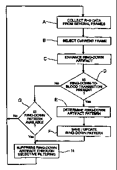
Fig. 7 is a flow chart of the steps according to the
inventive method.
SUBSTITUTE SHEET (RULE 26)

CA 02340246 2001-02-12
WO 00/19904 PCT/IB99/01542
4
DESCRIPTION OF SPECIFIC EMBODIMENTS
The invention provides exemplary systems and methods
for suppressing spurious artifact signals at ranges close to
an excitation source. Although useful with essentially any
type of ultrasonic system, the invention will find its
greatest use with ultrasonic imaging elements which are
disposed within catheters, and particularly, imaging catheters
employed to produce images of the vascular anatomy. As is
known in the art, such catheters include an imaging element
that is held within a housing. As the imaging element is
excited, transients reflected from the housing interfere with
the signals reflected from objects within the anatomy, such as
blood, vessel walls, and the like. The invention is able to
substantially reduce or eliminate the ring-down artifact
caused by such transient signals.
Referring to Fig. 1, there is shown basic elements
of a simple intravascular ultrasonic (IVUS) imaging system 10
providing imaging of the interior 12 of a vascular subject 14,
as shown in an enlarged cross-section. A catheter 16 contains
electrical conduits 18 that communicate between a transducer
20 and a console 22 housing an exciter source 24, a receiver
26 a signal processor 28 with associated controls, the output
of which is provided to an output device 30, such as a
television monitor or a computer display or a combination
thereof. The exciter source 24 generates ultrasonic excitation
signals 32 of a finite duration that are applied to the
transducer 20, which in turn directs those excitation signals
32 in a generally-defined directional beam. Ultrasonic
artifact signal 34 is reflected from the interior of the space
under observation to be intercepted by the transducer 20,
inducing an electrical report which is recovered by the
receiver 26 in the console 22. The electrical signals
recovered are analyzed by a signal processor 28 operative
according to the invention to present an output to the output
device 30 which is preferably a reconstructed two-dimensional
image of the target cross-section displayed in near real-time.
An exemplary medical imaging system that may be used to
implement the techniques of the invention is a Galaxy medical
SUBSTITUTE SHEET (RULE 26)

CA 02340246 2001-02-12
WO 00/19904 PCT/IB99/01542
S
imaging system, commercially available from Boston Scientific
Corporation.
The transducer 20 may be an array disposed around
the skin of the catheter 16, or a single transducer or
transducer set which might rotate around the skin of the
catheter. As is known in the art, the signal which is emitted
from the transducer is referred to as an A-scan. The detected
signal along any axis can be reconstructed as the sum of the
echo and ring-down artifact which is an amplitude as a
function of time.
Fig. 2 is a graph of a trace 40, in this case a
convoluted A-scan, and includes both ring-down artifact and
echo signal. Such a scan is typical of a scan produced when
the transducer is separated from a target region (such as
plaque) by blood. The segment I of trace 40 represents pure
ring-down. The segment R of trace 40 represents the portion
of the overlap of contribution of echo and ring-down, that is,
the region where echo begins before the transducer 20 settles.
The combination of segments I and R are referred to as the
transition region. The segment T is the pure echo without
ring-down of the target, which in this case is blood.
According to the invention the ring-down contribution or
pattern is determined as shown in Fig. 3, then its
contribution is subtracted from the composite echo signal in
order to yield a more accurate image of the target area as
shown in Fig. 4.
The ring-down artifact may be characterized in the
time domain and/or the time domain across consecutive scans:
several sequential scans are convolved or otherwise averaged
together to determine the nature of any repetitive artifacts
while canceling any short-term artifacts. The resultant
convoluted ring-down pattern (see Fig. 3) is subtracted from
the current scan report to yield a scan report 42 with ring-
down effectively eliminated, as shown in Fig. 4.
The computation of the ring-down pattern is made for
scans for which the following assumption holds: along the A-
scan axis, tissue does not intervene between the transducer
and the blood region nearest the transducer, such as for
SUBSTITUTE SHEET (RULE 26)

CA 02340246 2001-02-12
WO 00/19904 PCT/IB99/01542
6
example, in Fig. 2. Within that range, it is assumed that only
ring-down and blood echoes are present. The typical transition
from ring-down to blood echo can be identified by the
distinction between signals produced by ring-down and produced
by blood. As shown in Fig. 2, the signals produced by ring-
down are high amplitude oscillations of relatively low
frequency. The signals produced by blood are of low amplitude
and high frequency. The ring-down contribution is represented
by the crossed hatched area in Fig. 2.
In a system where the target is located abutting the
excitation source, the ring-down signal may detrimentally
overload the finite, lower-amplitude echo from the target
region. With such scans, the previous assumption does not
hold because tissue exists near the transducer. As such, a
previously computed and stored ring-down pattern (such as the
pattern of Fig. 3) is used for selective filtering.
Fig. 5 is a graph of a trace 44 where the transducer
is adjacent tissue. To filter the ring-down artifact, the
pattern of Fig. 3, which was previously computed, is used for
selective filtering. The result is illustrated in Fig. 6
which includes only the signal of the target.
Referring to Fig. 7, there is shown a flow chart of
a signal processing technique according to the invention.
First, the data on a plurality of frames of R-9 data is
collected (Step A), and the latest frame is preferably
selected as the current frame for processing (Step B).
Optionally, the ring-down artifact may be enhanced so it can
be more easily characterized (Step C). This may be done
mechanically by repositioning the transducer to zero tilt,
whereas a slight tilt is normally preferred to suppress such
artifact. Enhancement may also be done electronically or by
software by the process of convolving several sequential A-
scans.
Once the A-scan has been recovered for a frame, the
A-scan is inspected to determine the presence of the
transition region of ring-down to blood (Step D). This can be
an iterative process of examining the time domain signal
searching for the boundary between rapid high-amplitude
SUBSTITUTE SHEET (RULE 26)

CA 02340246 2001-02-12
WO 00/19904 PCT/IB99/01542
7
transitions and low-amplitude transitions. The transition
between the transition region and the blood region is referred
to as a transition point, such as point 48 on Fig. 2.
In some cases, such an amplitude analysis may serve
only as a first approximation of the transition point. If so,
a second processes may be employed to further define the
transition point. For example, the estimated target point may
be varied and a fast Fourier transform may be performed on the
target region T and on the transition region I and R (see Fig.
2) to covert the time-domain data to frequency-domain data for
each variation. This process may be repeated until consistent
results are obtained.
Having found the transition point (and thus the
transition region), the ring-down artifact pattern in the
transition region is computed (Step E). This may be done
dynamically by computing the ring-down pattern for one A-scan
within the ring-down range. A straightforward fast Fourier
transform (FFT) of the transition region and the target region
may be used for frequency domain analysis. Such an FFT
computation can be performed periodically during real-time
imaging for each individual A-scan following other filtering
processes, such as blood speckle reduction. Once the FFT
values are obtained for the transition region and the target
region, a weighted subtraction is performed to selectively
filter out the ring down pattern (such as is shown in Fig. 3).
The ring down pattern is preferably saved, and the filtered
data is converted back to the time domain to produce the
signal shown in Fig. 4.
The ring-down pattern is preferably saved and/or
updated for use in the cases where a ring-down-to-blood
transition is lacking, e.g., where there is tissue residing
next to the transducer (Step F) as shown, for example, in Fig.
5. If there is no ring-down-to-blood transition present, the
system checks to see if there is already a ring-down pattern
available or previously stored (Step G). If not, the process
begins again (Step A) until a pattern emerges, e.g., after a
ring-down-to-blood transition is found. Finally, in A-scans
having clear ring-down-to-blood transition regions, ring-down
SUBSTITUTE SHEET (RULE 26)

CA 02340246 2001-02-12
WO 00/19904 PCT/IB99/01542
8
artifact is suppressed by a selective filtering, i.e., by
subtraction of the ring-down contribution from the signal, to
yield a filtered image (Step H). As previously noted, under
conditions where a clear transition is lacking, ring-down
contribution can be subtracted by using the last known ring-
down pattern.
The invention has now been explained with respect to
specific embodiments. Other embodiments will be apparent to
those of ordinary skill in the art. It is therefor not
intended that the invention be limited, except as indicated by
the appended claims.
SUBSTITUTE SHEET (RULE 26)
Representative Drawing
A single figure which represents the drawing illustrating the invention.
Administrative Status

2024-08-01:As part of the Next Generation Patents (NGP) transition, the Canadian Patents Database (CPD) now contains a more detailed Event History, which replicates the Event Log of our new back-office solution.

Please note that "Inactive:" events refers to events no longer in use in our new back-office solution.

For a clearer understanding of the status of the application/patent presented on this page, the site Disclaimer , as well as the definitions for Patent , Event History , Maintenance Fee  and Payment History  should be consulted.

Event History

Description Date
Time Limit for Reversal Expired 2013-09-13
Letter Sent 2012-09-13
Grant by Issuance 2011-04-05
Inactive: Cover page published 2011-04-04
Inactive: Final fee received 2011-01-19
Pre-grant 2011-01-19
Notice of Allowance is Issued 2010-10-07
Letter Sent 2010-10-07
Notice of Allowance is Issued 2010-10-07
Inactive: Approved for allowance (AFA) 2010-10-05
Amendment Received - Voluntary Amendment 2009-10-08
Amendment Received - Voluntary Amendment 2008-12-18
Inactive: S.30(2) Rules - Examiner requisition 2008-06-30
Amendment Received - Voluntary Amendment 2007-09-06
Inactive: S.30(2) Rules - Examiner requisition 2007-03-06
Inactive: IPC from MCD 2006-03-12
Amendment Received - Voluntary Amendment 2004-09-16
Letter Sent 2004-08-16
All Requirements for Examination Determined Compliant 2004-07-26
Request for Examination Requirements Determined Compliant 2004-07-26
Request for Examination Received 2004-07-26
Inactive: Cover page published 2001-05-15
Inactive: First IPC assigned 2001-05-08
Letter Sent 2001-04-17
Inactive: Notice - National entry - No RFE 2001-04-17
Application Received - PCT 2001-04-09
Application Published (Open to Public Inspection) 2000-04-13

Abandonment History

There is no abandonment history.

Maintenance Fee

The last payment was received on 2010-06-23

Note : If the full payment has not been received on or before the date indicated, a further fee may be required which may be one of the following

  • the reinstatement fee;
  • the late payment fee; or
  • additional fee to reverse deemed expiry.

Please refer to the CIPO Patent Fees web page to see all current fee amounts.

Owners on Record

Note: Records showing the ownership history in alphabetical order.

Current Owners on Record
BOSTON SCIENTIFIC LIMITED
Past Owners on Record
SORIN GRUNWALD
TAT-JIN TEO
Past Owners that do not appear in the "Owners on Record" listing will appear in other documentation within the application.
Documents

To view selected files, please enter reCAPTCHA code :


To view images, click a link in the Document Description column. To download the documents, select one or more checkboxes in the first column and then click the "Download Selected in PDF format (Zip Archive)" or the "Download Selected as Single PDF" button.

List of published and non-published patent-specific documents on the CPD .

If you have any difficulty accessing content, you can call the Client Service Centre at 1-866-997-1936 or send them an e-mail at CIPO Client Service Centre.


Document
Description 
Date
(yyyy-mm-dd) 
Number of pages   Size of Image (KB) 
Representative drawing 2001-05-15 1 8
Cover Page 2001-05-15 1 36
Description 2001-02-12 8 420
Abstract 2001-02-12 1 51
Claims 2001-02-12 5 184
Drawings 2001-02-12 4 51
Description 2007-09-06 8 415
Claims 2007-09-06 6 224
Claims 2008-12-18 6 207
Representative drawing 2011-03-03 1 11
Cover Page 2011-03-03 2 48
Reminder of maintenance fee due 2001-05-15 1 111
Notice of National Entry 2001-04-17 1 193
Courtesy - Certificate of registration (related document(s)) 2001-04-17 1 113
Reminder - Request for Examination 2004-05-17 1 116
Acknowledgement of Request for Examination 2004-08-16 1 177
Commissioner's Notice - Application Found Allowable 2010-10-07 1 163
Maintenance Fee Notice 2012-10-25 1 171
PCT 2001-02-12 10 405
Fees 2003-06-20 1 30
Fees 2001-08-21 1 33
Fees 2002-06-19 1 33
Fees 2004-06-28 1 35
Fees 2005-06-20 1 28
Fees 2006-06-19 1 29
Fees 2007-06-20 1 29
Fees 2008-06-19 1 36
Fees 2009-06-19 1 33
Fees 2010-06-23 1 35
Correspondence 2011-01-19 1 36