Note: Descriptions are shown in the official language in which they were submitted.
CA 02342118 2001-03-09
WO 00/14267 PCT/GB99/02997
BIOSENSOR
The present invention relates to a eukaryotic biosensor for
the detection of xenobiotics and bioactive compounds. It
will be appreciated that xenobiotics are not necessarily
bioactive.
The problems of environmental contamination with toxic
chemicals are becoming increasingly apparent. Such pollution
has fuelled the need to develop novel, rapid and inexpensive
methods for toxin detection in the environment.
Assay systems such as High-Performance Liquid Chromatography
accurately predict quantities of chemical components but will
not indicate toxicity or bioavailability (potential of
compound to react with cellular components).
Prokaryotic biosensors, for example MICROTOX~, were created to
indicate the toxicity and bioavailability of chemical
components. Prokaryotic biosensors are unicellular living
organisms that provide information about in vivo toxicity
rapidly and reliably. They can detect a wide range of
pollutants within a certain narrow pH range, whilst
simultaneously assessing bioavailability in environmental
samples. However, the MICROTOX~ biosensors are generally not
sensitive to eukaryotic-specific molecules, such as DIURON~
3-(3,4-dichloropheny:L)-1,1-dimethylurea, and they only
operate within a narrow pH range.
Existing assays for quantifying in vivo toxicity of chemicals
for eukaryotic cells exploit whole animal models or tissue
CA 02342118 2001-03-09
WO 00/14267 PCT/GB99/025297
- 2 -
culture, which is both time consuming and expensive. The
unicellular yeast. Saccharomyces cerevisiae has previously
been shown to function as a biosensor using BOD (Biochemical
Oxygen Demand) respirometry. However, this BOD assay system
is also time consuming and expensive.
The usual method for selecting plasmids or episomes which
have undergone bioengineering is that during the genetic
engineering procedure antibiotic resistance genes are
transferred in the vector with the desired gene. Therefore
when the transfer is successful the plasmid and therefore the
host cell is resistant to antibiotics. The bioengineered
cells can then be selected~by growing them in the presence of
antibiotics. However, antibiotic resistence is becoming more
and more of a problem. There are now 'Superbugs' which
cannot be treated by antibiotics and many other bacteria are
resistant to all but one antibiotic. Now there is a
concerted effort an the part of scientists to reduce the
number of antibiotic resistence genes that they transfer from
one microorganism to another.
One object of the present invention is to develop a cheap and
quick assay for quantifying the in vivo toxicity of chemical
components for eukaryotic cells which will operate over a
wide pH range and function in the presence of an organic
solvent.
Another object of the present invention is to develop a cheap
and quick assay for quantifying the in vivo toxicity of
chemical components for eukaryotic cells through chromosomal
integration of the luciferase gene without the transfer of
CA 02342118 2001-03-09
13-10-2700 ~~ GB 009902997
13. 10. 2000
6'8
antibiotic resistance.
According to a first aspect of the invention there is
provided a method for evaluating a biological effect of a
substance, the method comprising the following steps:
S a)preparing a eukaryotic biosensor engineered with a gene
which constitutively expresses a light emitting protein;
b)sampling the substance;
c)subjecting the sampled substance at any pH between pHl and
pHl2 to an assay in the presence of the biosensor; and
d)monitoring any changes in light output.
It should be noted that the term "biological effects"
include all toxicity testing and the steps in the above
identified method can be carried out in any order. In
addition, a substance can be taken to be, inter alia, a
:L5 liquid, such as water,. a solid or suspension or colloid or
sediment or sludge.
Typically biosensora have been produced from prokaryotic
cells which can only operate in a narrow pH range. The
inventive eukaryotic biosensor can function at any pH between
:?0 pHl and pHl2 which makes it more useful for environmental
samples and commercially viable. The biosensor is cheap and
easy to produce so i.t can be used in mass and routine
screening of water supplies. The pH tolerance of the yeast
biosensor for example will enable toxicity assessment of
?S industrial waste and toxicity in extreme environmental
samples such as acid mining waste.
In a preferred embodiment the eukaryotic biosensor of the
invention is derived from the Saccharomyces genus and
_f0 preferably from Saccharomyces cerevisiae. S. cerevisiae is
an ideal cell to function as a biosensor because it can
~4MENDED SHEET
CA 02342118 2001-03-09
WO 00/14267 PCT/GB99/02997
- 4 -
tolerate an external pH within the range pH1 and pHl2 and it
is permeable to many xenobiotics and bioactive compounds. It
also senses the toxic effect of the contaminated liquids on
eukaryotic cells and therefore more accurately indicates the
5 liquid samples possible toxicity to higher order organisms,
and particularly to mammals.
Conveniently the :light emitting protein is a luciferase.
10 In a preferred embodiment the luciferase is either a
bacterial luciferase or a eukaryotic luciferase. Preferably
the bacterial luciferase is from Vibrio harveyi and the
eukaryotic luciferase is a firefly luciferase from Photinus
pyralis. Both the luciferases require an exogenous addition
15 of the substrate n-decyl aldehyde and luciferin,
respectively. The luciferin is an amphipathic molecule that
has a carboxyl group charged at physiological pH which
prevents its free passage across cell membranes. This
problem is overcome by acidifying the sample containing the
20 cells and the bi.osensor after exposure to the potentially
toxic sample. It is therefore important that these cells can
remain metabolically active at an acidic pH. Both these
luciferase genes produce light emitting proteins that require
energy from the eukaryotic cell to produce light and
25 therefore the level of light output is dependant on the
health of the cell. Thus if the cell viability is challenged
by components of 'the sample, for example due to the presence
of a toxin, the level of light will fall and the resultant
toxic effect of the sample can be noted.
The substance may be contaminated with a xenobiot:ic compound
CA 02342118 2001-03-09
WO 00/14267 PCT/GB99/02997
- 5 -
or a bioactive compound. For example, a xenobiotic compound
may be selected from copper, 3,5-dichlorophenol,
2,4-dichlorophenol, MECOPROP~ (+/-)-2-(4-chloro-0-tolyloxy)
propionic acid, DIURON~, paralytic shell fish toxins, benzo
5 (a) pyrene and MCPA. Copper, 3,5-dicholorophenol and 2,4-
dichlorophenol are compounds found in industrial waste,
which can find their way into rivers and lakes through
accidental or deliberate dumping. They can also leach out of
the soil around :industrial waste plants. They are toxic to
10 river dwelling organisms and those higher up the food chain.
Accordingly, their levels in river water must be carefully
monitored. MECOPROP~ and DIURON~ are herbicides which are
used liberally by farmers: They leach out of the soil into
rivers where once again they and their biologically active
15 derivatives are toxic to the river dwelling organisms and
those higher up the food chain.
Organic solvents such as ethanol, methanol, acetone and DMSO
are also harmful to organisms. The biosensor herein
20 described is stable in an environment which contains such
organic solvents and it can therefore be used to identify
substances which are being tested for toxicity to higher
organisms, in the presence of these solvents. It will be
appreciated that the presence of these solvents thus does not
25 detract from the assay of the substances.
In a second aspect of the invention there is provided a
biosensor comprising a bioengineered organism from the
Saccharomyces genus expressing a light emitting protein gene
30 wherein the level of light emitted by the organisms is
dependent on the environmental conditions surrounding the
CA 02342118 2001-03-09
WO 00/14267 PCT/GB99/02997
- 6 -
organism. A gene conferring antibiotic resistance is not
necessarily required for biosensor selection. The light
emitting protein gene may be present on a plasmid which has
been transferred into Saccharomyces species.
In a third aspect of the invention there is provided a
biosensor compri:;ing a eukaryotic bioengineered organism with
a chromosomally integrated gene fragment expressing a light
emitting protein wherein the level of light emitted by the
10 organisms is dependent on the environmental conditions
surrounding the organism. Preferably the Eukaryotic
biosensor is adapted for cell division during assay.
A specific embodiment of this invention is a eukaryotic
biosensor S. cerevisiae LUCK deposited at the National
Collection of Industrial and Marine Bacteria at Aberdeen
University, 23 St Machar Drive, Aberdeen, UK on the 28 August
1998 under the number NCIMB 40969.
20 This invention will now be described, by illustration only,
with reference to the following example and the accompanying
figures.
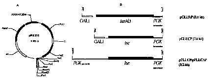
Figure la shows a diagram of a centromeric shuttle vector
pRS316 which is used to produce a biosensor plasmid.
Figure lb shows a diagram of luciferase expression cassette
which have been inserted into relevant restriction sites of
the vector pRS316.
Figure 2a shows the genetic code of primers used in PCR for
CA 02342118 2001-03-09
WO 00/I4267 PCT/GB99/02997
the construction of the biosensor plasmid of Figure lb.
Figure 2b shows the genetic code of primers used in a PCR for
the construction of the integrative luciferase cassette.
Figure 3 is a graphical representation of the luminescence of
S. cerevisiae pPLUC~P after transformation with the biosensor
plasmid. The mean RLU (Relative Light Units) (---c3--)and the
mean OD 600 nm (Optical Density) (-~--) are shown.
Figure 4 is a graphical representation of the effect of pH on
the luminescence of the S. cerevisiae pPLUC~ P biosensor. The
graph shows the mean luminescence.
Figure 5 is a graphical representation of the luminescence of
the S, cerevisiae pPLUC D P biosensor 10 minutes after exposure
to a range of copper concentrations.
Figure 6 is a graphical representation of the luminescence of
the S. cerevisiae pPLUC O P biosensor 10 minutes after exposure
to a range of 3,5-dichlorophenol concentrations.
Figure 7 is a graphical representation of the luminescence of
the S. cerevisiae pPLUCOP biosensor (--~--) compared with the
luminescence of a E. coli biosensor (- --o---) :LO minutes
after exposure to a range of MECOPROP~ concentrations.
Figure 8 is a graphical representation of the luminescence of
the S. cerevisiae ;pPLUC O P biosensor (~ ) compared with the
luminescence of the E. coli biosensor (---0---)10 minutes
after exposure to a range of DIURON~ concentrations.
CA 02342118 2001-03-09
WO 00/14267 PCT/GB99/02997
- g _
Figure 9 is a graphical representation of the effect on the
luminescence of the S. cerevisiae pPLUC~P when placed in a
range of concentrations; ethanol(-1-), methanol (---~---),
acetone ( ~-) and DMSO (- ~-) .
Figures 10a is a graphical representation of the effect of
varying the amount of time the S. cerevisiae pPLUC~ P
biosensor was exposed to varying concentrations of copper.
The amount of exposure was either 5 rains ( ~ ), 10 rains
(--w'-) or 15 rnins (- ~ - ) .
Figure lOb is a graphical representation of the effect of
varying the amount of 'time the S. cerevisiae pPLUC~ P
biosensor was exposed to varying concentrations of copper.
15 The amount of exposure was either 10 rains (--~--), 30 rains
( ~ -) , 60 rains ( ~ -) , 120 rains (---~--- ) or
180 rains (- X --) .
Figure 11 is a graphical representation of the stability of
the S. cerevisiae LUCK biosensor. The amount of exposure to
copper was 10 mires. The sensor cells were either used for
the assay after 10 rains. (-~--) or 48 hours (---~---).
Figure 12 shows t:he rpsl6A gene disruption cassette used in
Luck strain constructs containing the modified luciferase
reporter gene witAz PGK terminator and urs3 selective marker.
Region A is the region which is homologous with the rpsl6A
gene; Region B is the luciferase gene; Region C is the PGKtem,%
Region D is the common region of homology; Region E is the
30 URA3 gene.
CA 02342118 2001-03-09
13-10-x()00 GB 009902997
- 9 -
Figure 13 shows graphically the effect of ethidium bromide on
non-dividing S. cerevi.siae cells; and
Figure 14 shows graphically the effect of ethidium bromide on
dividing S. cerevisiae cells.
Example 1
Production of a S. cerevisiae biosensor
A pBLUESCRIPT~ based yeast centromeric plasmid pRS316 (Figure
la) with a GAZ,1 promoter was used as the vector for a
luciferase gene. A PGK terminator region was added to the
luciferase gene by amplification with primers, designed to
introduce restriction enzyme sites to place the terminator at
the 3' end of the polylinker using PCR. A SacII site
included in the primer S5R (Figure 2a) at the 5'end and a
SacI site included in the primer S3R (Figure 2a) at the 3'
end of this region allowed directional cloning of the PGK
terminator.
Four different luciferase expression cassettes were produced
and placed into the vector to produce four different
biosensors; one using' a bacterial luciferase from Vibrio
harveyi (pGLUXP) and three using a eukaryotic luciferase from
Photinus pyralis which is a firefly luciferase (pGLUCP and
pPLUCOP) (Figure lb) .
The pGLUXP vector was constructed by the addition of a 2.16
kb fused luxAB gene (see Boylan M, et al., Journal of
Biological Chemistry, 264 pp 1915 to 1918, 1989) that had
AMENDED SHEET
CA 02342118 2001-03-09
WO 00/14267 PCT/GB99/02997
- 10 -
been amplified as above to include directional cloning sites
into the vector pRS316. A XbaI site introduced by X5R
(Figure 2a) to the 5' end of the gene and a NotI site
introduced by N3R (Figure 2a) at the 3' end of the luxAB
fusion ensures that the gene would be inserted in the correct
orientation with respect to the promoter and terminator
regions.
The pGLUCP vector was constructed by the addition of a 1.65
kb eukaryotic luciferase gene, luc, into the polylinker
between the GAL1 promoter and PGK terminator. Again PCR
mutagenesis was undertaken to add a BamHI site 5' to the gene
using the primer BSRL (Figure 2a) and a NotI site 3' of the
luciferase using the primer N3RL. The template used for this
PCR reaction was the pGL2 vector from Promega.
The pPLUCP vector was constructed by replacing the GALL
promoter in the polylinker with a PGKl promoter. The
luciferase gene, luc, in pPLUCP was modified tc include an
optimised 5' leader sequence using the primer 5LEADL (Figure
2a). The PCR reaction for amplifying the luciferase gene
with SLEADL included the primer N3RL for the inclusion of a
NotI site 3' to the luciferase gene.
The pPLUC~P vector was created by inserting luc, amplified
using the primers SLEADL and N3RD (Figure 2a) which removes
the 9 by carboxyl terminal peroxisome targeting sequence of
the luciferase gene, into the vector pRS316. The 1.65 kb
luck produced from the PCR reaction is inserted into the
vector in the same restriction sites used for pPLUCP.
CA 02342118 2001-03-09
WO 00/142b7 PCT/GB99/02Q97
- 11 -
Each vector was then inserted in to the S. cerevisiae using
standard transforrnation procedures.
Bioluminescence was monitored using Bio-Orbit 1251
luminometer connected to a Multiuse software package. The
units of luminescence were expressed as RLU (relative light
units ) which equate to 10 mVs-lml'I .
In vivo luciferase activity assays using S. cerevisiae
require acidification to allow the substrate for the light
reaction (luciferin) to freely enter intact cells. Therefore,
a two-step assay procedure was designed to incorporate
separate toxicity analysis without pH adjustment and
subsequent acidification for luminescence quantification. In
preparation for the assay, S. cerevisiae cells were harvested
at peak luminescenca_ (around 3 to 4x10$ cells/ml), centrifuged
at 7008 (3000 rpm) and wasted twice in 5 mM KCl; keeping the
original volume constant for the first wash, but halved for
second wash (allowing for modified dilution in assay). The
first step (exposure), 50 ~1 of washed S. cerevisiae cells
are added to a 450 ,ul sample (pH generally not important) .
In an alternative procedure the S. cerevisiae can also be
resuspended in water rather than in 5 mM Kcl. The second
step (acidification), 500 ~cl citrate phosphate buffer
(pH2.5), also containing the luciferin (0.1 mM final
concentration in 1 ml sample), are added to make up 1 ml
total volume for subsequent luminescence quantification. The
exposure time is generally 10 minutes, but this can be
lengthened or shortened depending on assay requirements (e. g.
Figures; 10a, lOb and 11). The assay allows for the full pH
durability of S. cerevisiae to be exploited as prior pH
CA 02342118 2001-03-09
WO 00/14267 PCT/GB99/02997
- 12 -
adjustment of samples is not required. Luminescence of the
sample containing cuvettes are compared to a blank containing
diluent(s) used in the assay. This blank defines 1000
luminescence.
For bioluminescence monitoring during growth using S.
cerevisiae with vector pGLUXP, 5 ,ul of 100 n-decyl aldehyde
was added to the culture in each 1 ml luminometer cuvette.
In assays using S. cerevisiae with each of the vectors
pGLUCP, pPLUCP and pPLUC~P, 5 ,ul of 20 mM of luciferin
dissolved in Millit~ deionised H20 was added to the culture in
each 1 ml luminometer cuvette. If caged luciferi.n was used
1 ,ul of 200 mM DMNPE "caged" luciferin was used instead of
the 5 /.cl free luc:i:ferin. All assays were performed at 25°C.
As can be seen from Figure 11 S. cerevisiae pPLUC O P biosensor
luminesces for more than 48 hours.
In order to assess the pH tolerance of the biosensor,
deionised water had its pH adjusted with HC1 and NaOH to a
point in the range of pHl to pHl2. The S. cerevi.siae cells
were harvested at peak luminescence, centrifuged at 700 g and
washed twice in 10 mM KC1; keeping the original volume
constant. pH adjusted deionised water (450 ul) was added to
each cuvette. The volume was made up to 500 ml using 3 to
4x10' cells. Bioluminescence was then monitored following a
10 minute exposure to the sample, and the subsequent addition
of citrate/phosphate buffer (pH 2.5) containing O.lmM
luciferin.
Stock concentrations of the toxicants (copper, 3,5-
CA 02342118 2001-03-09
WO 00/14267 PCT/GB99/02997
- 13 -
dichlorophenol, MECOPROP~ and DIURON~) were made i.n deionised
water and their pH adjusted to 5.5 using HC1 and NaOH.
Dilutions were then made of the stock solutions using
deionised water adjusted to pH5.5, as above. The cells were
harvested at peak luminescence, centrifuged at 700 g and
washed twice in 5 mM KCI; keeping the original volume
constant. The toxicant (450 ,ul)was added to each cuvette.
The final volume was then made up to 500 ,ul using 3 to 4x 10'
cells. After 10 min, 500 ~cl of citrate phosphate buffer (pH
2 . 5 ) containing 5 /,cl of 20 mM luciferin was added to each
cuvette and the bioluminescence was monitored.
The results shown in Figures 5 and 6 show that the
transformed S. cerevisiae pPLUC~P biosensor can detect both
inorganic (copper) and organic (3,5-dichlorophenol) toxic
substances and as shown in Figures 10a, lOb the S. cerevisiae
pPLUC~P biosensor can serve as an acute (up to 15 minutes
exposure to sample) and as a chronic (up to 1.80 minutes
exposure to the sample) biosensor. Figure 11 shows that the
biosensor is still actively detecting copper after 48 hours
growth.
A direct comparison of light output from the P. pyralis and
fused V. harveyi luciferases was undertaken through placing
the luciferase genes in the same GAL1/PGK1 expression system.
Expression of the fused bacterial luciferase resulted in a
low light output per millilitre of culture. Expression of
the eukaryotic luciferase produced far higher light output
(around 100 fold) and as a result was selected for use in the
final biosensor construct. The PGK1 promoter was chosen as
it has efficacious properties, such as high levels of
CA 02342118 2001-03-09
WO 00/14267 PCT/GB99/02Q97
- 14 -
expression and no requirement for medium change when inducing
expression. It was also found that light output from the
PGKl containing constructs was araund 10 to 20 fold higher
than GAL1 containing constructs.
Final modifications of the biosensor construct were made to
the luciferase gene itself when 9 by from the carboxyl
terminal was removed using PCR mutagenesis. This adjustment
removed a peroxisome targeting sequence which prevents
targeting of the luciferase to the peroxisome. This strain,
pPLUC~P, was further characterized and applied i.n toxicity
analysis.
Growth of S. cerevis.iae containing pPLUC D P was monitored and
the results are shown in Figure 3. Following inoculation
light output increased with cell numbers and reached a peak
output after 27 hours, as cells entered stationary phase.
Cells were harvested at this point, when OD6oo":~ was around
3.65. It was observed that the luminescence after this time
declines slowly, which is dissimilar to the situation for
bacteria. Bacterial biosensors constitutively expressing
luciferase lose luminescence rapidly as the cells enter
stationary phase, whereas the S. cerevisiae biosensor
maintains bioluminescence into the stationary phase and still
functions as a biosensor (Figure 11).
Assays were carried out to discover if there were differences
between sensing capabilities of the new S. cerevisiae
biosensor and an existing E. coli biosensor. Results are
displayed graphically for MECOPROP~ and DIURON~ in Figures 7
and 8 respectively. The dose response curves obtained for
CA 02342118 2001-03-09
WO 00/14267 PCT/GB99/02997
- 15 -
the herbicides MECOPROP~ and DIURON~ indicate that the S.
cerevisiae is more sensitive in toxicity assays for these
compounds. MECOPROP~ was detected at levels far lower than
E. coli could sense (twice as sensitive). In contrast to the
5 S. cerevisiae pPLUC O P biosensor DIURON~ toxicity was not
observed at all when assaying toxicity using the E, coli
biosensor at the concentrations tested.
In order to assess the solvent tolerance of the biosensor;
methanol, ethanol, acetone and DMSO dilutions were prepared
for analysis. Dilutions ranging from 1°s to 50°s were made. The
S, cerevisiae cells were harvested at peak luminescence,
centrifuged at 700 g and 'washed twice in lOmM KC1; keeping
the original volume constant for the first wash, but halved
15 for second wash, 450 ~cl of the solvent dilutions was added to
the respective cuvettes. 50 ~cl of S. cerevisiae cells were
then added to each cuvette. Simultaneous acidification and
substrate addition was carried out after a 10 minute
exposure. Luminescence was then quantified. Results shown in
20 Figure 9.
Example 2
Production of a S. cerevisiae biosensor with no antibiotic
25 resistance gene.
Luciferase and ura3 genes from the vector pPLUC~P were
separated from the plasmid using PCR. The ST1K1 primer (see
Figure 2b) was designed so that the ura3 gene would be
30 modified to allow the joining of the luciferase gene in a 5'
position and the ura3 gene in a 3' position to form the
CA 02342118 2001-03-09
WO 00/14267 PCT/GB99/02Q97
- 16 -
integrating expression cassette (see Figure 12) in a
subsequent PCR (discussed below). The primers were also
designed so that the whole cassette should be targeted to a
duplicated constitutively expressed ribosomal gene rpsl6a in
5 the S. cerevis.iae genome. The primers rL UC and S3R (see
Figure 2a and b) were used to amplify a promoterless
luciferase gene including the PGK terminator region and
the addition of a 50 by homology region to rpsl6a promoter
for integration included in rL UC primer. The primers rURA and
10 ST1K1 (see Figure 2b) were used to amplify the ura3 gene
including all of its control regions (pramoter and
terminator) and the additional regions for genomic
integration included in rURA and homology to the previously
produced luciferase amplification product included in ST1K1.
15 The common homology allows an ovelap of 20 by between the 3'
end of the rLUC:/S3R and 5' end of the ST1K1/rURA PCR
products . These two products were then added together ire a
final PCR reaction to allow fusion of the luciferase and ura3
genes though this homology and amplification of the resultant
20 3.5 kb product. The primers mrL and mrU (see Figure 3b) are
homologous to secaions of the rpsl6a regions introduced by
the rLUC and rURA primers respectively. Therefore, allowing
amplification of the 3.5 kb product with the ribosomal
flanking regions.
The fragment was then purified (gel electrophoresis) and
concentrated for transformation of S. cerevisiae (Gietz R.D.;
Schiosh, R.H.; Willems. A.R. and Woods, R.A. 1995, Studies
on the transformation of intact yeast cells by the LiAc/S-
30 DNA/PEG procedure:-Yeast 11, 355-60). Very few colonies are
obtained, e.g. 100 to 1000 fold less than can be expected if
CA 02342118 2001-03-09
13-10 ~C~00 GB 009902997
- 17 -
transforming with ~ plasmid, as you are relying on a
spontaneous event.
The S. cerevisiae biosensors of the Examples 1 and 2 have all
the advantages of the prokaryotic systems, including rapid
and cheap quantificat=:on of toxicity. Additionally, their
wide range pH tolerance allows toxicity and bioavailability
analysis to be carried out from at least pH 1 through pH 12.
The loco construct in Example 2 does not contain antibiotic
resistance genes and the organism itself is not a known
pathogen, therefore having minimal disadvantages for field
use. Most importantly, S. cerevisiae is a eukaryotic
organism that is sensing different xenobiotics at different
concentrations compared to bacterial biosensors. This S.
cerevisiae biosensor may not just revolutionise environmental
monitoring as the pharmaceutical industry would be also
benefit by this system as it is able to provide rapid and
cheap preliminary screens for in vivo toxicity to a
eukaryotic cell.
The biosensors are stable over a prolonged period (at least
48 hours; so that as a biosensor reagent it is a particularly
suited for on-line applications.
Example 3
Dividing Cell Assays
The following procedure was designed to monitor the effect of
increasing the exposure time up to 9 hours and to determine
AMENDED SHEET
CA 02342118 2001-03-09
WO 00/14267 PCT/GB99/02997
- 18 -
the different responses of dividing and non-dividing
biosensor cells t:o the presence of DNA-damaging agents. For
assay preparation, the diluent used was ethanol at 4.5°s final
concentration. All ethidium bromide dilution standards were
5 prepared in glassware. Cells for the dividing assays were
only pelleted once at 700 x G for 1 minute before
resuspension in 10 x SC (-ura) medium (Strathern, J.N. (1994)
Ty Insertional mutagenesis. In: Johnston, J.R. [Ed]
Molecular genetics of yeast; a practical approach, pp 118
10 [Oxford University press]) for the dividing cultures (to give
a 1 x SC (-ura) final concentration. Cells for the non-
dividing assay were harvested at 700 x G for 1 minute and
washed twice in deionised water. The different cell
resuspensions for dividing and non-dividing protocols were
15 assayed with duplicated standards for toxicity analysis.
Assays were performed in 96 well plates adding 10 ,ul cells
(ar_ound 4 x 10⁶ cells) to 90 ~cl standard. The black 96-well
plates were covered with a plate sealer and incubated at 30°C
before and between readings. The exposure times were 3 , 5
20 h, 7 h, and 9 h. Additional of 100 ,ul citrate phosphate
buffer (pH 2.5), containing 0.2 mM luciferin, was performed
at each time point before luminescence quantification in a
Lucy Anthos 1 luminometer using the Stingray (v2.Ob31)
software package. Following luminescence quantification
25 100 ~ci was removed from the blank well in the dividing assays
for OD6oo measurements and cell counts. This was performed at
each time point to ensure cell division was or was not
occurring inn the dividing and non-dividing assays
respectively.
The results of the dividing and non-dividing assays are shown
CA 02342118 2001-03-09
WO 00/14267 PCT/GB99/02297
- 19 -
in Figures 13 and 14 respectively .
Thus in Figure 13, the effect of ethidium bromide on dividing
cells was measured by recording luciferase activity from
cells in wells containing ethidium bromide dilutions prepared
in 4.5°s ethanol. The cells were capable of division as they
were resuspended in a 1 x SC (-ura) final concentration. A
clear decrease in cell division with increasing ethidium
bromide concentration with time was observed (3 h (~), 5 h
(~), 7 h (o), 9 h (~)). These results were obtained with the
Luco S. cerevisiae strain in 96-well plates using the Lucy
Anthos 1 luminometer. The experiment was carried out in
triplicate at 25"C and the error bars represent standard
errors of the mean triplicate value.
In Figure 14, the effect of ethidium bromide on non-dividing
cells was measured by recording the light output from cells
in wells containing ethidium bromide dilutions prepared in
4.5~ ethanol. The cells were incapable of division as they
were resuspended in pure ddHzO . The response of the cells to
ethidium bromide does not change with increased exposure time
(3 h (~), 5 h (~), 7 h (a), 9 h (~)). There was no clear
toxic effect in these non-dividing cells, compared to the
situation with dividing cells illustrated in Figure 13.
These results were obtained with the Luck S. cerevisiae
strain in 96-well plates using the Lucy Anthos 1 luminometer.
The experiment was carried out in triplicate at 25°C and the
error bars represent standard errors of the mean triplicate
value.