Note: Descriptions are shown in the official language in which they were submitted.
CA 02344586 2001-03-19
WO 00/19918 PCTIUS99/23209
METHOD FOR DIAGNOSING AND IMPROVING VISION
Field of the Invention
The present invention relates to a method and system
for diagnosing improving the vision of an eye.
Background of the Invention
Most common defects in human vision are caused by the
inability of the eye to focus properly. For example,
nearsightedness can be attributed to an eye which focuses
forward of the retiria instead of on it, farsightedness can be
attributed to an eye which focuses beyond the retina, and
astigmatism can be attributed to an eye which cannot produce
a sharp focus, instead producing an area of blurriness.
Ophthalmologists model the cornea as a portion of an ellipsoid
defined by orthogonal major and minor axes. Current surgical
procedures for correcting visual acuity are typically directed
at increasing or decreasing the surface curvature of the
cornea, while making its shape more spherical.
In conjunction with modern corneal procedures, such
as corneal ablation surgery, and for clinical applications,
high resolution cameras are used to obtain a digitized array
of discrete data poiilts on the corneal surface. One system and
camera useful for mapping the cornea is the PAR. Corneal
Topography System (PAR CTS) available from PAR Vision Systems.
The PAR CTS maps the corneal surface topology in two-
dimensional Cartesian space, i.e., along x- and y- coordinates,
and locates the "line-of-sight", which is then used by the
practitioner to plari the surgical procedure. The "line-of-
CA 02344586 2006-11-15
2
sight" is a straight line segment from a fixation point to the center of the
entrance pupil. As
described more fully in Mandell, "Locating the Corneal Sighting Center From
Videokeratography,"J. Refractive Surgery, V. 11, pp. 253-259 (July/August
1995), a light ray
which is directed toward a point on the entrance pupil from a point of
fixation will be refracted
by the cornea and aqueous and pass through a corresponding point on the real
pupil to
eventually reach the retina.
The point on the cornea at which the line-of-sight intersects the corneal
surface
is the"optical center"or "sighting center"of the cornea. It is the primary
reference point for
refractive surgery in that it usually represents the center of the area to be
ablated in
photorefractive keratectomy. The line-of-sight has conventionally been
programmed into a
laser control system to govern corneal ablation surgery. However, some
surgeons prefer to
use the pupillary axis as a reference line. Experienced practitioners have
employed various
techniques for locating the sighting center. In one technique, the angle
lambda is used to
calculate the position of the sighting center relative to the pupillary
("optic") axis. See Mandell,
supra, which includes a detailed discussion of the angles kappa and lambda.
In current corneal ablation procedures, a portion of the corneal surface is
ablated. The gathered elevational data is used to direct an ablation device
such as a laser
so that the corneal surface can be selectively ablated to more closely
approximate a spherical
surface of appropriate radius about the line-of-sight, within the ablation
zone. The use of the
line-of-sight as a reference line for the procedures may reduce myopia or
otherwise correct
a pre-surgical dysfunction. However, a more irregularly shaped cornea may
result, which may
exacerbate existing astigmatism or introduce astigmatism in the treated eye.
This will
complicate any subsequent vision correction measures that need be taken. Also,
any
substantial surface irregularities which are produced can cause development
CA 02344586 2001-03-19
WO 00/19918 3 PCT/US99/23209
of scar tissue or the local accumulation of tear deposits,
either of which can adversely affect vision.
Implicit in the use of the-line-of sight or the
pupillary axis as a reference axis for surgical procedures is
the assumption that the cornea is symmetric about an axis
extending along a radius of the eye. The cornea, however, is
an "asymmetrically aspheric" surface. "Aspheric" means that
the radius of curvature along any corneal "meridian" is not
a constant (a "meridian" could be thought of as the curve
formed by the intersection of the corneal surface and a plane
containing the pupillary axis) . Indeed, the corneal curvature
tends to flatten progressively from the geometric center to the
periphery. "Asymmetric" means that the corneal meridians do
not exhibit symmetry about their centers. The degree to which
the cornea is aspheric and/or asymmetrical varies from patient
to patient and from eye to eye, within the same person.
Clinical measurements performed with the PAR CTS, as
analyzed in accordance with the method disclosed in U.S. Patent
No. 5,807,381 assigned to the assignee of the present patent
application, reveal that the cornea exhibits a tilt, typically
a forward and downward tilt, relative to the eye. This tilt
may be as great as 6 and, on the average, is between 1 and
3 . Hence, a corneal ablation procedure which utilizes the
line-of-sight or pupillary axis as a reference axis tends to
over-ablate some portions of the cornea and underablate other
portions of the cornea. At the same time, it changes the
geometric relationshi.p between the cornea and the remainder of
the eye. Thus, any ablation procedure which does not take
into account the ti1t: of the cornea is not likely to achieve
the desired shaping of the cornea and may therefore be
unpredictable in its effect.
Analysis of clinical measurements in accordance with
the method of Patent No. 5,807,381 also reveals that the point
on the surface of the cornea which is most distant from the
reference plane of the PAR CTS (hereafter referred to as the
HIGH point) is a far more effective reference point for corneal
ablation than the center of the cornea. Specifically, as
demonstrated in Patent No. 5,807,381 laser ablation about an
CA 02344586 2006-11-15
4
axis passing through the HIGH point produces a much more regularly shaped
cornea and
removes substantially less corneal material than the same operation performed
about an axis
close to the center of the eye, such as the pupillary axis.
Although incorporating corneal tilt and utilizing the HIGH point leads to
improved
and more consistent results with corneal ablation surgery, there is still an
excessively high
degree of unpredictability. For example, recent analyses of clinical
measurements have
revealed that the post-operative cornea begins to change shape a short time
after corneal
ablation surgery. Thus, a nearly perfectly spherical postoperative cornea,
will, over time,
return to an aspheric, asymmetric shape.
The use of a collagen gel has been proposed as a vehicle to facilitate
smoothing
of the corneal undulations. See Ophthalmology Times,"Slick Start, Clear
Finish,"1995, pp.
1 and 24 (June 19-25,1995) and Review of Ophthalmology,"News & Trends:
Researchers
Unveil New Ablatable Mask,"pp. 12-13 (June 1995). A Type 1 collagen is molded
between
a contact lens and the anterior surface of the cornea to form a gel mask. The
surgeon can
adjust the curvature of the postoperative cornea by selecting a flatter or
steeper lens, as
desired. Reportedly, the gel mask does not shift when hit by laser pulses.
Therefore, instead
of selective ablation of predetermined locations of the cornea, the masked
cornea can be
ablated to a uniform depth, thereby conforming the surface contour of the
cornea to the lens.
A smooth post-operative cornea results, and refractive power correction can be
achieved.
However, because the ablation operation is centered on the optical center of
the cornea or
the center of the pupil and does not allow for corneal tilt, the postoperative
eye may exhibit
an irregular shape or more corneal material may be removed than is necessary.
What is needed in the art and has heretofore not been provided is a method of
correcting vision that avoids one or more of these problems, that can produce
predictable
results,
CA 02344586 2001-03-19
WO 00/19918 5 PCTIUS99/23209
and that provides corrected vision with respect to the
particular topology of the patient's eye on which the
correction is being performed.
It is therefore one object of the present invention
to provide a method for improving the vision of an eye.
It is an additional object of the present invention
to provide an improved surgical method for a corneal ablation
procedure.
It is also an object of the present invention to
provide a method and apparatus for diagnosing and analyzing a
pre-surgical eye for the purpose of predicting the post-
operative condition of the eye and planning more effective
surgery.
The present: inventors believe that corneal ablation
surgery has had limited success and predictability, because of
a parochial approach. The conventional wisdom has been to
concentrate on the shape of the cornea, with the expectation
that a smooth, spherical cornea will optimize vision. However,
the human eye is a complex system which includes numerous
optical components besides the anterior surface of the cornea
(for example, the posterior corneal surface, the lens and the
aqueous), all of which affect vision. Also, the mechanical
environment of the eye cannot be ignored. For example, recent
analyses of clinical measurements reveal that the eyelids exert
substantial pressure on the cornea, causing it to flatten near
its upper margin and to form a depression near its lower
margin. It is believed that the mechanical environment of the
eye accounts, in large part, for its shape. This also explains
why a perfectly spherical post-operative cornea would return
to an aspherical, asymmetric shape.
In accordance with the present invention, corneal
ablation procedures of the eye are performed in a manner which
does not interfere with the natural shape of the cornea or its
orientation relative to the remainder of the eye, but which
changes its surface curvature appropriately to achieve the
required correction of vision. Three preferred embodiments are
described, which model the cornea to different degrees of
accuracy. Once the model of the cornea is obtained, surface
CA 02344586 2001-03-19
WO 00/19918 6 PCTIUS99/23209
curvature is modified to achieve the degree of correction in
refraction that is necessary, as determined by an eye test of
the patient. The modified model of the cornea is then utilized
to control the removal of material from the surface of the
cornea in a corneal ablation operation.
In a first embodiment, the cornea is modeled as an
ellipsoid having major and minor axes which are perpendicular
to each other. These are the axes that are revealed by
conventional eye test:s as being appropriate for correction of
refraction. On a mociel of the cornea generated in accordance
with disclosure of
Patent No. 5,807,381 perpendicular planes are constructed which
contain the local or tilted Z axis and are rotated about that
axis to the angle specified by the eye test. The intersection
of each of these planes with the surface model produces an
arcuate curve. Each of these curves is then estimated by a
circular arc which estimates the patient's current radius of
curvature at each axis. A modified arc is then determined
which achieves the required diopter correction at each axis.
A model of the post-operative cornea is then created by
performing a smooth interpolation from one of the arcs to the
other. In this model, the corneal surface is represented as
the surface of an ellipsoid which has the corrected radii of
curvature at the two orthogonal axes specified by the eye test.
In an second embodiment, the cornea is mocieled in
such a manner as to preserve its asymmetry. To achieve this,
a large number of annularly spaced meridians are generated on
the surface model of the cornea. The distance along each
meridian is measured from the HIGH point to the perimeter of
the working area of the cornea, and the curves with the
greatest and least average radius of curvature are each
estimated by a circular arc. The complementary curves
corresponding to the two initial curves (i.e. those extending
from the HIGH point diametrically opposite to the corresponding
curve are then also estimated by circular arcs. Each of the
four arcs is then adjusted for curvature to achieve the desired
degree of visual correction at each arc. The model of the
post-operative cornea is then generated by angularly
CA 02344586 2001-03-19
WO 00/19918 7 PCT/US99/23209
interpolating between pairs of the four arcs mentioned above
and providing smoothing between two partial surfaces at each
of the four initial arcs.
A third embodiment of the invention comes closest to
preserving the initial shape of the cornea. Initially, a large
number of angularly spaced meridians, for example 72, are
generated on the surface model. The curves defining the
meridians, which extend from the HIGH point to the periphery
of the working region of the cornea are each estimated by a
circular arc. Each of these arcs is then corrected in
curvature to achieve the required diopter correction at the
respective arc. The post-operative corneal surface is then
estimated by generating a best-fit surface corresponding to all
of the corrected arcs.
Brief Description of the Drawings
The foregoing brief description, as well as other
objects, features and advantages of the present invention will
be understood more completely from the following detailed
description of presently preferred embodiments, with reference
being had to the accompanying drawings in which:
Figure 1 is a block diagram illustrating a method for
achieving laser ablation of the cornea in accordance with the
present invention;
Figure 2 is a schematic diagram illustrating a plan
view of a point cloud as obtained with a corneal image capture
system;
Figure 3 is a schematic plan view similar to Fig. 2
illustrating a plurality of splines and how they are connected
through the data points of the point cloud;
Figure 4 is a perspective view of a cornea matching
surface illustrating how characterizing curves are constructed;
Figure 5 is a plan view in the tilted plane
illustrating how the cornea matching surface is modified to
provide vision correction in accordance with a first
embodiment;
Figure 6 is a plan view in the tilted plane
illustrating how the cornea matching surface is modified in
CA 02344586 2001-03-19
WO 00/19918 8 PCT/US99/23209
order to achieve vision correction in accordance with a second
embodiment;
Figure 7 is a functional block diagram illustrating
how corneal shaping is achieved when using a moldable mask and
uniform corneal ablat:ion;
Figure 8 is a sectional side view illustrating the
application of a coritact lens to form a moldable mask when
performing uniform corneal ablation;
Figure 9 is a plan view of a contact lens usable to
form the moldable mask for uniform corneal ablation, which
contact lens is manually positioned;
Figure 10 is a plan view of a contact lens similar
to the lens of Fig. 9, except that the lens is constructed to
position itself automatically upon being applied to the eye;
Figure 11 is a side view, with parts in section,
illustrating applanat.ion of the cornea during Lasik surgery;
Figure 12 is a side view illustrating the cornea
after creation of a corneal flap, but prior to laser ablation
during Lasik surgery;
Figure 13A is a side view illustrating an improvement
to a conventional microkeratome in accordance with the present
invention;
Figure 13B is a left side view with respect to Figure
13A; and
Figure 13C is a plan view of one of the rings forming
part of assembly 53 in Figures 13A and 13B.
Detailed Description of the Preferred Embodiments
A process for achieving laser ablation of the cornea
in accordance the present invention is illustrated in block
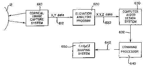
diagram form in Figure 1. The process makes use of a Corneal
Image Capture System 610, an Elevation Analysis Program 620,
a Computer Aided Design System 630, a Command Processor 640 and
a Cornea Shaping System 650. The Corneal Image Capture System
610, in conjunction with the Elevation Analysis Program 620,
generates a three dimensional topographic map of the cornea of
the patient. The Computer Aided Design System 630 is used as
an aid in editing or modifying the corneal topographic data,
CA 02344586 2001-03-19
WO 00/19918 9 PCT/US99/23209
to create a surface model, and data relating to the model is
sent to a Cornea Shaping System 650 via the Command Processor
640. The Command Processor 640 uses the topographic data
describing the surface of the cornea to be shaped from the
Computer Aided Desicin System 630 to generate a sequence of
commands/control signals required by the Cornea Shaping System
650. The Cornea Shaping System 650 accepts, from the Command
Processor 640, a sequence of commands that describe the three
dimensional movements of the Cornea Shaping System (any
coordinate system may be used; e.g., cartesian, radial or
spherical coordinates) to shape the cornea.
The Corneal Image Capturing System 610 and the
Elevation Analysis Program 620 are preferably components of the
PAR Corneal Topography System ("the PAR System"), which is
available from PAR Vision Systems. The Elevation Analysis
Program 620 is a software program executed by a processor, for
example an IBMT"' compatible PC. Program 620 generates a third
dimension element (a Z coordinate representing distance away
from a reference plane inside the eye) for each of a plurality
of sample points on the surface of the cornea measured by
system 610. Each point is defined by its X-Y coordinates as
mapped into the reference plane, and its Z coordinate is
determined from brightness of the point. One method of
calculating the elevation of each point, i.e., the Z
coordinate, is by comparing the X-Y and brightness values
measured from the patient's cornea 14 with the coordinates and
brightness of some reference surface with known elevation,
e.g., a sphere of a known radius. The reference values can be
pre-stored.
The final output of the Elevation Analysis Program
620 is the X-Y-Z coordinates for a multiplicity of sample
points, known as a point cloud, on the surface of the cornea
14. It will be apparent to those skilled in the art that any
method can be used that can generate X, Y, Z corneal data
providing both location and elevation information for points
on the corneal surface with the required accuracy. In the
preferred embodiment about 1500 points are spaced in a grid
CA 02344586 2006-11-15
pattern, as viewed in the X-Y plane, so the projections of the points into the
X-Y plane are
about 200 microns apart.
The X-Y-Z data output from the Elevation Analysis Program 620 can be
5 formatted in any number of well-known machine-specific formats. In the
preferred
embodiment, the data are formatted in Data Exchange File (DXF) format, an
industry
standard format which is typically used for the interapplication transfer of
data. A DXF file is
an ASCII data file, which can be read by most computer aided design systems.
10 Referring now to Figures 2 and 3, a point cloud 100 is depicted as it would
appear when viewing the reference plane along the Z-axis (i. e., as projected
into the X-Y
plane). Each point corresponds to a particular location on the patient's
cornea. The data are
usually generated from an approximately 10mm x 10mm bounded area of the
cornea, the
working area. Thus, there may be as many as 50 rows of data points. A surface
108 (see Fig.
4) that models or matches the topography of the surface of the patient's
cornea is generated
by the computer aided design system 630 from the data points generated by the
Elevation
Analysis Program. In a preferred embodiment, Computer Aided Design System 630
is the
Anvil 5000TM program which is available from Manufacturing Consulting Services
of
Scottsdale, Arizona.
Cornea matching surface 108 is preferably produced by first generating a
plurality of splines 102, each defined by a plurality of the data points of
the point cloud 100.
The generation of a spline that intersects a plurality of data points (i. e.,
knot points) is, per
se, known to those skilled in the art and can be accomplished by the Anvil
5000TM program
once the input data have been entered. For more information regarding the
generation of a
surface model, see U. S. Patent No. 5,807,381. In a preferred embodiment, the
known
nonrational uniform B-spline formula is used to generate the splines, but they
could be
generated by other well-known mathematical formulas for splines, such as the
cubic spline
formula or the rational uniform B-spline formula. As illustrated in Figure 3,
in a preferred
embodiment, each of the
CA 02344586 2001-03-19
WO 00/19918 11 PCT/US99/23209
splines 102 lies in a plane that is parallel to the X and Z
axes and includes a row of points from the cloud 100 in Fig.
3.
Surface 108,, which matches the corneal surface of the
scanned eye, is then generated from splines 102. There are a
number of well-known mathematical formulas that may be used to
generate a surface from a plurality of splines 102. In the
preferred embodiment, the well known nurb surface equation is
used to generate a corneal surface from splines 102. In the
embodiment, because the scanned area of the eye is
approximately 10mm x 10mm, approximately 50 splines 102 are
created. As illustrated in Figure 3, a skin surface segment
104 is created for a small number (e.g., five) of the adjacent
splines. Adjacent skin surface segments 104 share a common
border spline. Thus, about ten skin surface segments are
generated from the point cloud and are then merged together by
the Anvil 5000T"' program in a manner known to those skilled in
the art, to produce one composite surface 108.
Neither the original data points, nor the knot points
of splines 102 necessarily lie on surface 108, owing to the
mathematical generation of the surface when using the nurb
surface equation formula. However, the surface 108 estimates
those points within a. predefined tolerance.
The HIGH point on the generated corneal matching
surface 108 (i.e., the point having the greatest Z value) is
determined. A cylinder 106 of a predetermined diameter, is
then projected onto the corneal matching surface 108 along an
axis which is parallel to the Z-axis and passes through the
HIGH point. Cylinder 106 preferably has a diameter of 4mm -
7mm, typically 6mm, and the closed contour formed by the
intersection of cylirider 106 with surface 108 projects as a
circle 106' in the X--Y plane. On the matching surface 108,
this contour defines the outer margin 26 of the working area
of the cornea. The cornea is the most symmetric and spherical
about the HIGH point and, therefore, provides the best optics
at this point.
The outer margin 26 must fit within the point cloud,
so that the surfaces of the cornea can be formed based on the
CA 02344586 2001-03-19
WO 00/19918 12 PCTIUS99/23209
measured corneal data. The computer aided design system 630
can then illustrate a default circle 106' (in the X-Y plane)
with respect to the point cloud, for example on a monitor
screen, so that the operator can be assured that circle 106'
falls within the poiiit cloud. Additionally, system 630 can be
set up to determine if circle 106' falls within point cloud 100
and, if it does not fall completely within point cloud 100, to
alert the user to mariipulate the circle (i.e., move the center
point and/or change the radius of the circle) so that circle
106' lies within the corneal data point cloud 100. In a worst
case scenario, the eye should be rescanned if insufficient data
is available from the scanned eye to ensure that the cornea
will fit properly on the patient's cornea. Alternatively, the
area of the point cloud can be made larger.
It is to be understood that circle 106' is only a
circle when viewed in the X-Y plane (i.e., looking along the
Z-axis). Actually, the periphery 26 is approximately
elliptical and lies .Ln a plane which is tilted relative to the
reference plane. A line perpendicular to this tilted plane
which passes through the HIGH point will be referred to as the
"local Z-axis" and the tilt of the tilted plane relative to the
reference plane will be considered the tilt angle of the
working area of the cornea.
The cornea is about 600,um thick. In most corneal
ablation procedures, less than l00 m depth of cornea is
ablated, because there is virtually no risk of scarring with
the type of lasers that are typically used. Beyond the 100 m
depth, the risk of scarring increases. For example, 1204m
depth ablation is known to cause scarring. However, there
exists the possibilit:y that the risk of scarring for deeper
ablations may be reduced by drug therapy prior to or
contemporaneous with the laser treatment. The magnitude of the
corneal undulations is typically about fifteen to twenty
microns from the crest of a hill to the trough of a valley and
may be as great as about thirty microns.
The proposed use of a collagen gel, for example A
Type 1 collagen, to mold a smooth spherical surface on the
cornea using a tempo:rary mask allows the cornea to be ablated
CA 02344586 2001-03-19
WO 00/19918 13 PCT/US99/23209
uniformly to the spherical shape defined by the mask. However,
because conventional lenses do not seat themselves predictably
about a particular point on the eye, the ablation procedure
relying on them will result in not maintaining corneal tilt or
proper orientation, because the art has not recognized the need
to orient the lens sc> as to retain corneal tilt, to locate the
optical center of the eye at the HIGH point of the cornea, and
to maintain proper rotational orientation.
The surgical procedures performed in accordance with
the present invention will seek to correct the patient's vision
in accordance with the required corrections established in a
"refraction test." 'When this test is performed, the patient
sits in chair which is fitted with a special device called a
"phoropter", through which the patient looks at an eye chart
approximately 20 feet away. As the patient looks into the
phoropter, the doctor manipulates lenses of different strengths
into view and, each time, asks the patient whether the chart
appears more or less clear with the particular lenses in place.
In practice, the doctor is able to vary the power or diopter
correction about two orthogonal axes, as well as the degree of
rotation of those axes about a Z-axis along the line-of-sight.
The doctor continues to modify these three parameters until he
achieves the optimum vision. The results of the refraction
test are usually given in the form "a, b, c " , where "a" is the
diopter correction at the first axis, "b" is the additional
diopter correction required at the second, orthogonal axis, and
"c " is the angle of rotation of the first axis relative to the
horizontal. This forrn of information is given for each eye and
is immediately usef=ul in grinding a pair of lenses for
eyeglasses.
For the purposes of the present invention, it is
preferred to perform a modified form of refraction test. For
this modified form of refraction test, the eye doctor adjusts
the phoropter at a series of equally spaced angles, say every
15 from the horizontal, and obtains the optimum refraction at
each angle. Typically, the more angles that are measured, the
better the results. However, since the refraction measurements
can be time consuming, 15 increments, which results in the
CA 02344586 2001-03-19
WO 00/19918 14 PCT/US99/23209
total of 12 readings seems to be a reasonable number. The
manner of using the modified refraction test will be described
in detail below.
There will. now be described a technique for
generating characterizing curves on surface 108, which will be
useful below. A plane 110 is constructed which contains the
local Z-axis (See Fig. 4) The intersection between plane 110
and surface 108 def'ines a first characterizing curve 112.
Plane 110 is then rotated about the local Z-axis, for example
by a 5 increment counterclockwise, as represented by :Line 114,
where its intersection with surface 108 defines a second
characterizing curve 116, which is illustrated as a dashed line
in Fig. 4. This process continues at fixed rotational
increments about the local Z-axis, for example every 5 , until
plane 110 has swept 360 , to produce a complete set of
characterizing curves, in this case seventy-two (360" = 5 ).
In accordance with a first embodiment of the present
invention, corneal ablation surgery is performed so as to
effect the vision corrections specified in a conventional
refraction test. This procedure requires the generation of two
characterizing curves as described above. The first
characterizing curve is obtained by constructing a plane which
contains the local Z-axis and forms an angle of c with the
X axis, that is, the rotational angle obtained in the
conventional refraction test. The first characterizing curve
is formed by the intersection of this plane with the surface
108. The second. characterizing curve is obtained by
constructing a plane which contains the local Z-axis and is
perpendicular to the first plane. The intersection of the
second plane with the surface 108 defines the second
characterizing curve.
Figure 5 is a plan view in the tilted plane of
contour 106' illustrating the derivation of these two
characterizing curves. The contour 106' is the periphery of
the working area of the cornea as it appears in the tilted
plane. Plane 20 contains the local Z-axis and therefore the
HIGH point H and is also perpendicular to the plane of the
contour 106' (the ti:Lted plane). Plane 20 forms angle of c
CA 02344586 2001-03-19
WO 00/19918 15 PCT/US99/23209
with the X-axis in the tilted plane. The intersection of plane
20 and surface 108 defines a characterizing curve 22 which
touches the contour :106' at two points and passes through the
HIGH point H. P:1ane 25 is constructed so as to be
perpendicular to plane 20 and to contain the local Z-axis.
Plane 25 therefore also contains the HIGH point and is
perpendicular to the plane of contour 106'. The intersection
of plane 25 and surface 108 defines a second characterizing
curve 26, which touches the contour 106' at two points and
passes through the H:IGH point H.
Each of the characterizing curves may be estimated
by a best-fit spherical arc. One manner of doing this is
simply to select a c_Lrcular arc which passes through the three
known points for each curve (i.e. the points at which it
touches the contour :106 and the HIGH point. With the radius
of curvature of each characterizing curve determined, the Zeiss
lens formula provides a diopter value for each characterizing
curve. The diopter value "a" is then added to the diopter
value for curve 22 and the diopter value "a+b" is added to the
diopter value for characterizing curve 25. Those skilled in
the art will appreciate that the values a and b may be positive
or negative. With the corrected diopter va:lues for
curves 22 and 26 determined, the Zeiss lens formula now
provides the correcte:d average radii of curvature for the two
curves. The two curves are then replaced by circular arcs
having those radii of curvature. A corrected surface model
108' for the cornea is then generated within the bounded area
106' by producing a curve driven surface which interpolates
from the circular arc for curve 22 to the circular arc for
curve 26, while driving along contour 106'. The generation of
curve driven surfaces is a feature available in most CAD/CAM
programs. In effect, a surface of rotation is produced which
is bounded by contour 106 and is made up of a continuum of
circular arcs centered about the HIGH point H and ranging from
the arc for curve 22 to the arc for curve 26.
From the above description, it will be appreciated
that the corrected corneal surface 108' will conform precisely
to the specifications of the refraction test in the two planes
CA 02344586 2001-03-19
WO 00/19918 16 PCT/US99/23209
20 and 25 and will vary gradually therebetween. Since all
operations were done about the HIGH point and with respect to
the local Z-axis, the tilt of the cornea relative to the eye
is maintained, as is its general geometry. Only the small area
within the contour 106' has been changed in shape in order to
achieve the required degree of correction.
In accordarice with a second embodiment, the area of
the surface 108 bounded by the contour 106' is modified in
shape in a manner to retain the asymmetry originally present
in the cornea. In the manner described above, a multiplicity
of characterizing curves (meridians), preferably 72, is
obtained about the HIGH point within the contour 106'. The
average radius of curvature of each characterizing curve is
determined, and the curves with the greatest and smallest radii
of curvature (curves 30 and 32, respectively in Fig. 6) are
found. The extensions of curves 30 and 32 towards the opposite
margins of contour 106' are then created, to define the curves
30' and 32', respectively. Figure 6 shows the projection of
the contour 106' and the curves 30, 30', 32 and 32' into the
tilted plane of contour 106'. In each instance, instead of
using the average radius of curvature for a curve, the curve
may be estimated by a circular arc which passes through the
HIGH point, the intersection of the curve with the contour
106', and that point which in Fig. 6 is halfway between those
two points (for example, point 34 in contour 30).
Having a radius of curvature for each of the four
curves, it is now possible to generate corrected average radii
of curvature. In order to do so, use is made of the results
of the modified refraction test described above. In each
instance, a curve will receive the diopter correction of the
arc measurement which is closest to it in the modified
refraction test. Each of the arcs 30, 30', 32 and 32' is then
replaced by a circular arc having the corrected average radius
of curvature, and four curve driven quadrant surfaces are
generated. For example, an upper right-hand quadrant surface
is generated by driving the circular arc for curve 32 into the
circular arc for curve 30 along the contour 106' as a drive
rail. This produces a curve driven quadrant surface having a
CA 02344586 2001-03-19
WO 00/19918 17 PCT/US99/23209
perimeter of the contour 106' which merges smoothly in shape
from the arc for curve 32 to the arc for curve 34. The three
additional quadrant surfaces are similarly generated, and the
interfaces between the surfaces are smoothed, to produce the
finished, corrected surface model 108' bounded by the contour
106'.
It should be appreciated that the preceding
construction of model 108' can be undertaken even if the only
test results available are those from a conventional refraction
test. The required correction at each of arcs 30, 301, 32 and
32' would then be determined by interpolation. For example,
suppose arc 30 extended 20 beyond c (the refraction test
angle), the interpolated diopter correction for arc 30, d30
could be computed as::
d3 o= a + 20 b.
The remaining diopter corrections could be determined
similarly by interpolation. The corrected surface model 108'
would then be generated the same manner as described above
relative to Fig. 6.
The surface model 108' achieves the required
correction along four different arcs, while conforming more
closely to the original shape of the cornea than the model of
Figs. 5. Specifically, it has retained the original asymmetry
of the cornea.
In accordance with a third embodiment, the required
correction of vision is achieved by modifying the curvature of
the cornea while retaining its overall original shape. For
this embodiment, it would be preferred to perform the modified
refraction test at a niultiplicity of angles and to generate the
characterizing curves at the same multiplicity of angles.
However, the procedui=e can be performed using the results of
a conventional refraction, as will be explained below.
Preferably, the characterizing curves and refraction
measurements are taken at every 5 , so that there will be a
total of 72 characterizing curves. As was the case with the
second embodiment, the average radius of curvature of each
CA 02344586 2001-03-19
WO 00/19918 18 PCT/US99/23209
curve is determined, and the required diopter correction is
applied to each curve, to obtain a corrected average radius of
curvature. Each char=acterizing curve is then replaced by a
circular arc having the corrected average radius of curvature,
and the corrected surface model is generated by interpolating
between all of the corrected circular arcs. Smoothing is then
applied to produce the corrected surface model within the
bounding contour 106'. This surface model will riot only
include the requireci diopter correction, but will closely
approximate the original shape of the cornea as well.
The present: procedure can be performed even if the
only available test results for vision correction are a
conventional refraction test. As was done for the second
embodiment, the diopter correction at each of the 72 arcs can
be computed by interpolating between the conventional
refraction test measurement a and b. The procedure then
continues as already described.
Once the desired corrected surface model 108' within
the contour 106' is obtained, Computer Aided Design system will
provide information to Command Processor 640 which will permit
it to generate appropriate control signals for operating the
Cornea Shaping System 650. Preferably, system 670 produces
information which represents the differences between models 108
and 108', so that the appropriate material may be removed from
the cornea. Typically, when selective corneal ablation is
being performed, system 650 will include a station in which the
patient's head and eyes are held in a fixed manner, and a high
precision laser is maintained in close registry with the cornea
so as to achieve precise movement and controlled degrees of
ablation. Preferably, the laser is a spot laser which is moved
to precise locations under control of Command Processor 640 and
is then precisely controlled to apply the required degree of
ablation at each location.
The components utilized to achieve the process
depicted in Fig. 1 can prove costly and not within the budget
of the average doctor's office. It is therefore contemplated
that corneal shaping could, alternately, be performed by a
process of uniform ablation utilizing a smoothing mask. As
CA 02344586 2006-11-15
19
will be explained below, the mask is shaped by the posterior surface of a
contact lens which
has been formed to conform to the corrected matching surface 108'. Uniform
ablation with
an inexpensive laser to the maximum thickness of the mask will then result in
appropriate
shaping of the working area of the cornea. Moreover, this process is performed
with an
inexpensive wide beam laser and can be done relatively slowly so as to
eliminate the need
for extreme precision.
It should also be appreciated that when uniform ablation is performed, the
only
steps performed by the doctor preliminary to ablation would be the eye test.
The patient would
then be sent to a laboratory which would have all of the equipment illustrated
in Fig. 1. The
laboratorywould generate the precision contact lenses for molding the mask and
furnish them
to the doctor. The uniform corneal ablation could then take place in the
doctor's office.
From the preceding description, itwill be appreciated that, in the case of
uniform
corneal ablation, the block diagram of Fig. 1 is modified as illustrated in
Fig. 7. That is, the
elements of Fig. 7 represent the contents of block 650 (Cornea Shaping
System). Block 650'is
a Lens Shaping System. Systems for making custom contact lenses are well
known. In this
case, the contact lens could be provided with appropriate markings to guide
the doctor in
orienting the lens. Alternatively, the lens could be made with a custom
peripheral skirt portion
to assure that it will orient itself on the patient's cornea in a
predetermined position and
orientation. Lenses of this type and their method of manufacture are disclosed
in U. S.
Patent No. 5,502,518 issued March 26,1996.
Once a corrected corneal surface model 108' has been generated, a contact
lens 72 (see Fig. 8) having a posterior surface 76 shaped to conform to the
corrected corneal
surface 108'can be made. Uniform ablation can be performed by depositing a
moldable mask
70 onto the cornea 18 (block 660 in Fig. 7), and placing the posterior surface
76 of lens 72
over the moldable mask 70 with correct rotational orientation and so that the
optical center
74 of the lens 72 aligns with the
CA 02344586 2001-03-19
WO 00/19918 20 PCT/US99/23209
HIGH point H. The moldable mask 70 is then molded to the shape
of the posterior surface 76 of the lens as the lens is pressed
into it (block 670 in Fig. 7).
A presently preferred material for the mask 70 is A
Type 1 collagen. The collagen mask is heated to a temperature
of about 42 C to 45 C so that it assumes a syrup-like
viscosity. The heated collagen is deposited as a film on the
cornea where it immediately begins to cool to body temperature
(37 C) at which temperature it assumes a gel-like consistency.
Prior to cooling, the lens .72 is positioned on the collagen
film as explained above and illustrated in Fig. 7. Once the
collagen gel has coo:led and set, the positioned lens 72 will
have molded the collagen into a surface having the desired
corrected shape of the cornea. The lens 72 can then be
discarded.
The cornea plus collagen gel have a smooth,
undulation free surface. Uniform ablation of the masked
anterior surface of the cornea (block 680 in Fig. 7) can then
proceed by ablating the masked cornea to a depth sufficient to
remove all of the gel, in a manner known to those skilled in
this art. Because the collagen and cornea ablate at the same
rate (they are virtually identical materials, hence the
preference for this material), uniform ablation will result in
a smooth corneal surface of the desired shape.
For reasons already explained, the collagen mask is
preferably formed with a width of 6 mm, and a lmm lip including
a transition region. This transition region may be formed in
a separate step, or the posterior surface of the contact lens
may be ground so as to have a properly shaped transition lip.
Figure 9 illustrates one form of contact lens 40
useful in forming the collagen mask when performing uniform
ablation. The lens is formed as described above by
conventional lens manufacturing techniques, such as molding or
shaping on a lathe. When the original corrected model 108' of
the cornea is produced, the operator also defines a point on
the cornea which corresponds to the center of the pupil. When
the lens is manufactured, a visible index marking 42 is placed
at the location on the anterior surface of the lens which
CA 02344586 2001-03-19
WO 00/19918 21 PCT/US99/23209
should cover the center of the pupil. Similarly, during
manufacture of the lens, a visible index marking 44 is placed
at the bottom edge of lens 40 or, alternatively, at oile of the
apexes at the corner= of the eye, or any other predefined
orientation. In positioning lens 40 over the collagen on the
patient's eye, the doctor locates index 42 over the center of
the pupil and assures that index 44 is pointing downward (or
any other predefined orientation). Lens 40 will then be
positioned properly over the HIGH point of the cornea with the
proper rotational orientation. Pressing the lens into the
collagen will then shape it appropriately.
Figure 10 illustrates a contact lens 40' which may
be used to shape the collagen mask, in the event that more
precision in orientation is required than can be obtained
manually. The lens includes a central portion 41, the
posterior surface of which is constructed to achieved the
desired shaping of the collagen mask. Surrounding the central
portion in the posterior surface of the lens is a channel 43.
The lens may be formed with a ridge, to allow the channel 43
to be deeper. At spaced locations along the channel 43, there
are provided openings 45, which extend through the lens. Four
spaced openings are illustrated, but it will be appreciated
that a larger or smaller number may be provided. Outward of the
channel 43, lens 40' includes a peripheral skirt 46, the
posterior surface of which is designed to conform closely to
the shape of the surface of the cornea outside the working
area. As explained above, the construction of the skirt is
intended to make the lens 40' position itself automatically on
the cornea in a precietermined position and orientation.
The lens is applied immediately after the collagen
material is placed on the cornea. The skirt will assure
automatic alignment of the lens and, as the center portion 41
is pressed down, the thinned out material of the channel 43
allows a certain amount of rearward movement of the central
portion relative to the skirt. When the posterior surface of
portion 41 of the leiis comes into contact with the collagen,
it will force it to spread out and flow into the channel 43,
then out of the channel through the openings 45. Excess
CA 02344586 2001-03-19
WO 00/19918 22 PCT/US99/23209
material exiting from the openings 45 may be wiped away
immediately. When the central portion 41 is fully depressed,
the collagen materia.l. has been appropriately shaped for the
ablation process. The lens 40' may then be removed and
discarded.
Another form of selective laser ablation surgery
which is commonly used for corneas requiring removal of a
relatively large amount of material is known as laser assisted
interstromal kerotop:Lasty (hereafter referred to as "Lasik".)
The method for performing conventional Lasik surgery is
illustrated schematically in Fig. 11. It is performed with the
aid of an instrument called a microkeratome, which includes a
vacuum cylinder 50 which is positioned over the cornea 18. In
practice, a fitting is provided (not shown) which is centered
over the pupillary axis and the cylinder is attached to the
fitting. Typically, the microkeratome is positioned over the
cornea with its axis aligned with the pupillary axis. A strong
vacuum is then applied to the cylinder 50 which draws the
cornea into the cylinder and simultaneously causes it to
flatten or "applanate". Following applanation, a blade 52 is
passed beneath the applanated portion 18a of the cornea and
parallel to it. Preferably, a cut is made in the cornea
approximately 180 m thick. The cut stops short of the remote
end 18b of the applariated portion 18a, leaving an attached,
thin flap of corneal material 18c (see Fig. 12). Air is then
admitted into the cylinder 50, and the microkeratome is
removed, allowing the cornea to return to its normal shape.
The flap 18c is then folded back and corneal ablation, surgery
is performed on the underlying, exposed surface of the cornea.
The reason for performing this form of surgery is that the
corneal surface under the flap 18c is less likely to form scar
tissue. Upon completion of corneal ablation, the flap c is
carefully folded back down over the underlying surface of the
cornea and, upon healing will form a integral, reshaped cornea.
The theory behind Lasik surgery is that the flap 18c
is of uniformed thickness. Therefore, the underlying cornea
may be formed to the desired shape and will retain that shape
when the flap 18c is replaced. However, since Lasik surgery
CA 02344586 2001-03-19
WO 00/19918 23 PCT/US99/23209
is performed by taking a slice perpendicular to the pupillary
axis, it does not take account of corneal tilt.
As a result, the flap 18c exhibits a substantial
amount of variation in thickness. Thus, when flap 18c is
replaced over a cornea ablated in the conventional manner, it
actually changes the shape of the cornea. The results obtained
with traditional Lasik surgery are therefore far from
predictable.
It is should be noted that, since, in accordance with
the present inventior.t, selective ablation surgery is performed
by operating over the working area of the cornea in order to
change curvature at every location, and not to produce a
particular resulting shape, having a flap 18c with an irregular
thickness will not affect the outcome of the surgery. That is,
since each point on the area underlying the flap 18c is ablated
to achieve the desired correction or change and not a desired
overall shape, when the flap 18c is replaced, the desired
changes will, in fact, be achieved in the overall cornea.
However, having a flap 18c with uneven thickness is
still undesirable, in that certain areas may be excessively
thin. Accordingly, in accordance with the present invention,
certain modifications are made to the microkeratome cylinder
to cause the flap 18c to be cut so that it takes account of the
tilt of the cornea. Referring to Figs. 13A and 13B, there are
shown two schematic diagrams of a microkeratome cylinder
including the modification proposed by the applicants. It
should be kept in mind that Fig. 13B shows the same apparatus
as Fig. 13A, but as seen when looking from the left in Fig.
13A. The present improvement constitutes the addition of a
shim apparatus 53 at the bottom of the cylinder 50. In
actuality, the apparatus 53 would be mounted between the bottom
of the cylinder and the fitting that holds it to the cornea.
As can be seen, the shim apparatus preferably comprises two
rings, 52, 56 which are tapered in thickness. The ring 54 is
designed to be secured to the bottom of cylinder 50, as by
complimentary screw threads or a bayonet connection, both of
which are well-known to those skilled in the art. Similarly,
ring 56 is designed to be secured at the bottom of ring 54, as
CA 02344586 2001-03-19
WO 00/19918 24 PCTIUS99/23209
by complementary screw threads or bayonet connection. As can
be seen in Figs. 13A and 13B, the rings 54, 56 are preferably
constructed so that their tapers form solid angles which are
rotationally perpendicular to each other about the local Z-
axis. That is, one provides the solid angle or tilt relative
to the X-axis, whereas the other provides the solid angle or
tilt relative to the Z-axis.
When using the shim apparatus 53, the doctor would
be aware of the corneal tilt of each eye relative to the X- and
Y-axes, based upon the results of the corneal model 108. He
would then select a ring 54 to give him the appropriate X tilt
and a ring 56 to give him the Y tilt and mount them at the
bottom the cylinder E.O. When fully mounted, the rings provide
a lower lip 58 below the cylinder 50 which is tilted relative
to the bottom of the cylinder in the same manner that the
cornea is tilted. When the doctor subsequently applies the
cylinder 50 to the cornea, the shimming device 52 causes the
entire cylinder to be tilted in conformity with the corneal
tilt. The knife 52 then performs its cut with the same tilt,
avoiding substantial irregularities in thickness of the flap
18c.
Although preferred embodiments of the invention have
been disclosed for illustrative purposes, those skilled in the
art will appreciate that many additions, modifications and
substitutions are possible, without departing from the scope
and spirit of the invention.