Note: Descriptions are shown in the official language in which they were submitted.
CA 02345322 2001-03-26
WO 01/06932 PCT/IT00/003I4
1
DEVICE FOR MONITORING FERTILITY IN WOMEN BY OBSERVING PHYSICAL CHANGES IN BODY
FLUIDS
The present invention relates to the sector of devices used to obtain
and/or improve knowledge of the times of greatest or least "fertility" in
women during the monthly menstrual cycle and to identify with a good
measure of probability the day when ovulation occurs, for the purposes
of contributing to knowledge of the most or least appropriate periods
for conception.
More particularly, the present invention relates to a device which
proves useful and effective in detecting, by means of a totally natural
method and without the use of either chemicals or "invasive"
procedures, the fertile days and the time of ovulation in women with a
very high degree of approximation (such as to allow various
applications both in the field of physiological events and in that of
medical intervention).
The invention relates essentially to a detection system for assessing
information closely related to changes of a physical type in a number of
body fluids which can be collected without invasive measures, such as
saliva and other fluids which will be specified here below.
In the light of the present state of our technical and scientific
knowledge, it is known that, in the course of the menstrual cycle,
important, essentially hormone-based physiological transformations
take place in the woman for the purposes of optimising the conditions
for possible conception. Since this "natural" program has a very strong
functional purpose, it comes about that various biological variables
related to it, despite the "biological variability" which is always
present, take on a character and values which tend to be deterministic
and leave little room for chance.
These variables are numerous and different in nature, such as
hormone levels, body temperatures, density and viscosity of certain
CA 02345322 2001-03-26
WO 01/06932 PCT/IT00/00314
2
fluids, i.a. As the days of the cycle leading up to the time of ovulation
and then following ovulation pass, these variables change in value and
thus reflect actual physical changes in a number of elements of the
body.
It is also well known that the woman's fertile period occurs only once in
the course of each menstrual cycle. The ovum matures around mid-
cycle, roughly 14 days after the start of the last menstruation.
The fertility period, i.e. the days when the ovum can be fertilised,
covers a maximum of 5-6 days (with a greater chance of fertilisation on
the 2-3 central days of the period).
Identification of this short period of "maximum fertility" is not easy,
unless sophisticated, expensive and sometimes also "invasive" methods
are used. The various traditional methods based on calculations and
subjective observations are very imprecise and not always easy to use.
All this often leads to practical consequences of substantial distress in
couples that desire to conceive a child or who would like to implement
proper family planning on the basis of wholly natural methods. In
recent years, then, substantial efforts have been made in an attempt to
develop reliable, easy-to-use predictive tests, based on changes in the
above-mentioned biological variables which mark the various phases of
the cycle in the woman.
One variable often used for this purpose is basal temperature, which,
as is known, tends to rise at the time of ovulation. The use of this
variable, which can easily be measured with special ad-hoc
thermometers, yields information which is sometimes not particularly
accurate and is often influenced by other factors.
Another variable considered is the viscosity (either subjectively
assessed or measured using an instrument called a viscosimeter) of the
uterine cervical mucus, which is not always easy to assess.
CA 02345322 2001-03-26
WO 01/06932 PCT/IT00/00314
3
All these variables, moreover, require evaluation not only of the
"present" value, but also of the variations compared to the last few
days. Their reliability in practical use has therefore often proved fairly
poor.
One very reliable variable which is less influenced by other factors is
the luteinising hormone (LH) level in the female body. It can be
measured precisely with sophisticated laboratory equipment and, more
recently, with the introduction of special kits on the market, it can also
be measured at home; these kits are quite expensive and, for reliable
conclusions regarding fertility, again require comparison with results
obtained on a number of consecutive days.
Lastly, we should recall that comparative tests performed by
authoritative investigators have shown and confirmed that, during the
menstrual cycle, the woman's saliva (or other fluids such as cervical
mucus) undergoes structural changes as a result of the oestrogen levels
circulating in the body; as a result, over a period ranging from 2-3 days
before ovulation (oestrogen peak) to 2-3 days after ovulation, a physical
phenomenon of microscopic "crystallisation" of saliva occurs, which, in
turn, can be recognised and, if properly interpreted, used to
understand which phase of the cycle the woman is in from the fertility
point of view.
The above-mentioned observations are summarised in the following
specification which also allows comparison with the information that
the woman can obtain using the various "natural" methods outlined
above.
In the light of the present state of our technical and scientific
knowledge, it can be stated that this latter effect of crystallisation of
saliva, known as the "fern effect", in that the crystals present the
appearance of the fronds of a fern, has been used in laboratories and in
specialist medical studies in order to "see" the crystalline structure
indicating a pre- or post-ovulation condition under the microscope, thus
CA 02345322 2001-03-26
WO 01/06932 PC1'/IT00/00314
4
allowing conclusions to be drawn as to the woman's fertility status.
Small microscopes for personal use have also been produced for said
purpose.
The above-mentioned approach also affords advantages particularly
when used in conjunction with other natural methods, but it also
presents a number of drawbacks related mainly to the need to perform
calculations and take account of the results of previous days, as well as
a certain amount of objective difficulty in collecting samples of saliva
(which prove hard to compare) in a simple, standardised manner over
time.
The object of the present invention is a device suitable for detecting
changes in the "state" of fluids, such as saliva, in response to a rapid
increase in oestrogen levels in the body and other changes closely
linked to the approach and occurrence of the physiological phenomenon
of ovulation (which, as already mentioned, is a phenomenon with an
intense deterministic component, that strongly influences the changes
observed).
More particularly, the object of the present invention is a kit as
described in the preamble of claim 1 attached hereto, characterised in
the characterising clause of the same claim.
The present invention makes it possible to overcome the various
limitations of the above-mentioned systems (difficulty in collecting
standardised samples of saliva or other fluids; poor sensitivity related
to visual observation of saliva placed on surfaces with undefined limits,
such as slides, lenses or the like; the need to save the results of
preceding days with the difficulty of detecting the onset of changes
which are not particularly marked as compared to previous findings).
In fact, the object of the present invention consists in a kit made up of:
- a device for collecting and storing samples consisting of a set of flat
plate-shaped supports (hereinafter called "petals") made of special
CA 02345322 2001-03-26
WO 01/06932 PCT/IT00/00314
material, as specified here below, with an entirely original design
which enables the fluid samples (saliva or other fluids) to be collected
in a homogenous, standardised manner by implementing a kind of
automatic mechanism as will be explained later in this description.
Said set of petals makes it possible to obtain: greater reliability of
results due to the standardised collection of fluid in constant amounts;
greater sensitivity due both to the quality of the sample and to the way
the petal is constructed, with the possibility of easy comparison with
the results of groups of subsequent days with immediate detection of
any changes and with the further possibility of saving indefinitely the
effective results ("values" of the variables used, with the consequent
possibility of comparing them over time with later cycles, checks,
interpolations and extrapolations);
- a petal readout device consisting in a viewer of appropriate shape,
as described here below, in which the petals can be inserted for the
purposes of the optical or electrical or mixed optical-electrical detection
of the crystallisation of saliva or other fluids. The mixed system may
substantially enhance the sensitivity of the device with only a slight
increase in cost, inasmuch as the electrical component can be realised
at only limited extra expense.
A preferred embodiment of the kit according to the invention will be
described in greater detail here below, making reference to the
attached drawings which represent:
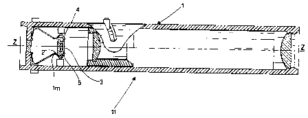
- in Figure 1, the longitudinal section of a viewer according to the
invention in which a flat plate-shaped support (or "petal") is inserted
for physiological fluid samples;
- in Figure 2, ~ the plan view of one of said petals according to the
invention;
- in Figure 3, the cross sectional view of the same petal;
- in Figure 4, a sequence of images obtained optically with the viewer
in Figure 1, showing an example of successive changes in the image
over a period concluding with ovulation; and
- in Figure 5, a graph showing the trend of the ovulation phase
during the menstrual cycle of a woman.
CA 02345322 2001-03-26
WO 01/06932 PCT/IT00/00314
6
As shown in Figure 1, the viewer 1 equipped with flat-convex lenses
and other features which are well known in the field of optical
instruments presents a compartment 2, perpendicular to the Z-Z
optical axis of viewer 1, suitable for housing a flat plate-shaped support
(see also Figures 2 and 3) carrying a sample of physiological fluid F.
This flat plate-shaped support, or petal 3, is obtained from special
high-transparency synthetic material (preferably high-transparency
polystyrene) and presents a shallow basin or trap 3p with a slightly
convex bottom entirely surrounded by a raised rim 3r. As a result of
the surface tensions exerted by the raised rim, a sample of
physiological fluid F deposited in said shallow basin necessarily takes
on a fixed conformation depending on the geometry of the system, with
a flat part of predetermined thickness positioned at the centre of the
visual field of viewer 1.
A spring clip lm holds petal 3 in a fixed position after its insertion in
compartment 2, while two projecting sidepieces, in the form of the two
fins 3t, act as end-stops or locking elements in contact with the outer
edges of said compartment 2.
This enables petal 3 to be positioned consistently in the same position
in relation to the viewer in which it is inserted, which is a very
important feature for reliable standardisation of the method in terms
of readouts of results and their comparison.
The system produced presents undoubted advantages compared to
previous systems and allows any woman to carry out the simple,
inexpensive and continuous monitoring of her fertility status with very
precise identification of the time of ovulation.
The multi-petal system (a complete set contains 32 petals with a
container, not illustrated here, for their collection) allows sample
collection and thus the monitoring of the history of an entire cycle (or
even of several cycles), enabling the woman to trace the changes in the
CA 02345322 2001-03-26
WO 01/06932 PCT/IT00/00314
state of crystallisation of saliva which constitute the real marker
showing when ovulation is imminent.
The sequence of images presented in Figure 4, detected optically using
the above-mentioned viewer provides an example of these variations in
a "typical" cycle over a period culminating in ovulation.
It will easily be understood that the effective information content lies
in detection of the changes, since a certain amount of "random noise"
will always be present in any single "current" image. It is well known,
in fact, that the human sensory system is much more capable of
detecting changes in an image than its specific descriptive content.
It is also very important to achieve a kind of "automation" in the
distribution of the saliva collected on the surface of the petal. This has
been achieved, as mentioned above, by producing the central saliva
trap 2 with a specially designed profile which exploits the surface
tension and causes the saliva deposited in the trap to spread regularly
and consistently in a uniform manner inside the trap, with a flat area
located at the centre of the visual field of the viewer.
The elements listed here above constitute the original features of the
multi-petal system according to the invention. Viewer 1 is specially
designed to receive the petals and detect the crystallisation patterns of
saliva or other fluids basically by means of optical readout, but also, as
we shall see here below, with the additional possibility of obtaining
confirmation by the quantitative assessment of an electrical magnitude
consisting in a conductivity parameter for the saliva contained in the
petal, the value of which may change appreciably and rapidly when a
phenomenon of microcrystallisation of the saliva occurs. This dual
ability to detect changes related to crystallisation (with a consequent
significant increase in the sensitivity of the method) constitutes an
additional original feature of the kit, to which should be added the
original mode of inserting, centring and locking petal 3, which thus
remains optimally positioned for the optical readout.
CA 02345322 2001-03-26
WO 01/06932 PCT/IT00/00314
8
Kit 11 comprises a container (not illustrated} for the collection of a set
of 32 petals, all produced with technical material characteristics
specified for each production batch, such as to guarantee a percentage
of impurities which is below the predetermined threshold.
Said container can be of the known mufti-pocket type and made of soft
material.
As already mentioned, for the assessment of the electrical parameters
of physiological fluids such as saliva, urine, cervical mucus, etc., a
sample of which has been deposited on petal 3, the inventor of the
present invention has provided for the application, on both sides of
each petal 3, preferably at the level of said locking fins 3t, of two
inserts 4 and 5, made of conductive material, with one end in contact
with fluid F. These inserts 4 and 5 can be connected up to a voltage
generator and the electrical magnitude of the current passed through
fluid F can be measured and assessed for the purposes of identifying
the corresponding potential fertility level.
The kit which is the object of the present invention allows maximum
detection of changes in the physical state of the biological fluid (saliva
or other fluid) for numerous applications which are inexpensive and
easy to implement by means of the kit.
The use of the kit presents no difficulties and can be managed by the
woman concerned without the aid of her doctor. However, it also makes
for an invaluable exchange of information between doctor and patient
in the context of various physiological or clinical problems. The use of
the system can be summarised in the following operations.
The saliva is collected on a finger which has been washed to eliminate
all impurities (the woman must avoid collecting saliva immediately
after consuming food or appreciable amounts of alcohol, or after
smoking a substantial number of cigarettes). The same procedures are
adopted for the collection of cervical mucus.
CA 02345322 2001-03-26
WO 01/06932 PCT/IT00/00314
9
The saliva is transferred to saliva trap 3p at the centre of petal 3,
eliminating any excess air bubbles.
The saliva is left to dry for a few minutes (once dried, the sample
conserves its inner crystallisation for a longer period). To eliminate it,
all that is necessary is to rinse it in warm water.
The petal is inserted in compartment 2 where it is held by spring clip
lm and is then pushed right in to the end-stop position so that the fins
3t touch the inserts 3n. With the petal locked in this position, the
crystallised saliva is optimally placed in the optical detection field
(and, if the electrical measuement option is implemented, it will be in
the correct position for connecting up to a device known to be suitable
for such measurements, which may take the form of a battery-operated
mini-calculator with a liquid crystal display for the readout of saliva
conductivity values).
One then proceeds with the direct visual readout, which enables the
woman to observe and assess the image which, according to the phase
of the menstrual cycle, will resemble one of the four images illustrated
in Figure 4. Obviously, these images can also be collected
photographically or "digitised" by means of a suitable electronic
interface with the possibility of easy subsequent recall, without having
to reinsert the petal, for the purpose of comparing results on different
days.
The petals can be numbered and stored in a special container with
labels so as to be able to easily identify the results for any given day
and repeat comparisons as many times as one wishes.
The main applications for which the system described here above can
be used and for which the system has been successfully tried are the
following.
CA 02345322 2001-03-26
WO 01/06932 PCT/IT00/00314
This application is implemented by detecting the crystallisation image
every day after the start of the menstrual period until such time as the
image is seen to pass from type 3 to type 4 (Figure 4). The finding is
also confirmed by the fact that on the following days the image will
revert to type 3. The days straddling the time of identification of the
day of ovulation constitute the ideal time for conception.
The daily collection of samples and the day-by-day comparison of petals
makes it possible to check the changes in image from type 1 and/or
type 2 to type 3 and ultimately to type 4. The time at which this
transition occurs may be regarded as the start of the fertile period
which will continue after ovulation until the image changes back to
types 2 and 1.
C'~ntrol of pre-menopause irregular cxcles
This application is implemented by testing the samples every day and
observing all the petals after completing sample collection so as to
establish whether the crystallisation occurs in a regular manner (using
+ or - to indicate early or late crystallisation, as the case may be), as,
for instance, illustrated schematically in Figure 1. If crystallisation
does not occur at any time in one or more cycles, this will be a clear
indicator of a hormone abnormality which the specialist will need to
investigate.
Various researchers have shown that the sex of the foetus is
determined by the type of spermatozoon that fertilises the ovum.
Spermatozoa carrying male or female sexual chromosomes have
different survival times. The result is that if conception occurs early
CA 02345322 2001-03-26
WO 01/06932 PCT/IT00/00314
11
(shortly after ovulation) there is a greater likelihood that it will be
produced by "female"-type spermatozoa, whereas, if conception occurs
later in relation to ovulation it is more likely to have been produced by
"male"-type spermatozoa.
The system produced with the kit of the present invention consists in
taking several readouts a day starting from the time the image passes
from type 2 to type 3, i.e. in order to identify exactly the time of day
when it passes from type 3 to type 4. This observation may allow
estimation of the time of ovulation to within approximately 12 hours.
This information in turn allows the couple to implement behaviour
strategies which will help to avoid conception in the time range when
the more "desired" sex is less likely.
Figure 5 presents a graph, based on readouts obtained with a kit
according to the present invention, showing the trend of the ovulation
phase during the woman's menstrual cycle.
Given here below is a summary of the results of a number of
"controlled" tests performed using the method described above in some
of its possible applications.
CA 02345322 2001-03-26
WO 01/06932 PC'T/IT00/00314
12
Aim of the research: identifying the ovulation phase comparing the saliva
crystallisation method with various other "physiological"
methods (cervical mucus testing, basal temperature, pupil
measurement, oestrogen assay) in a group of about 500
women.
Country Ukraine
Date of study 1993-94
N. of researchers 8
N. of women 514 (aged 15-46)
Cycles observed 5,498 (mean: 10.7)
Dropouts 42 (8.2%)
Results:
Crystallisation ~l~ 428 (91%)
Cervical mucus ~2~ 398 (84%)
Pupil measurement ~3> 364 (77~)
Oestrogen assay ~4~ 472 (100%)
Cases in which the fertile phase (ovulation period) was detected by
crystallisation of saliva, coinciding with oestrogen levels ~4~ in the
appropriate
range: peak value t 10%.
Detection of the fertile phase according to the Billings Method, as checked by
oestrogen levels ~4>: peak value t 15%.
Detection of pupil dilatation, as checked by oestrogen values ~4>: peak value
~
10%.
CA 02345322 2001-03-26
WO 01/06932 PG"T/IT00/00314
13
Aim of the research: identifying the ovulation phase comparing the monitoring
of
crystallisation of saliva with three other methods
(folliculometry, basal temperature, hormone test) in a group
of 48 women observed for a period of 5 months.
Country Czech Republic
Date of study 1992
N. of researchers 2
N. of women 48 (aged 16-45)
Cycles observed 5
Dropouts 0
Results:
Crystallisation ~l> 48 (100%)
Correlation: 100%
Folliculometry ~l> 48 (100%)
Basal temperature ~Z> 36 (75%)
Hormone test ~3~ 48 (100%)
~ Cases in which the ovulation phase was precisely identified by
crystallisation of
saliva, coinciding perfectly with folliculometry results.
~ Cases in which a rise in temperature of at least 0.2°C was detected
corresponding
to the ovulation phase as detected by folliculometry.
~ Tested by hormone assay.
The data shown demonstrate the excellent application capability of the
method described above for obtaining reliable results of practical
utility.