Note: Descriptions are shown in the official language in which they were submitted.
CA 02345397 2001-03-23
WO 00/17325 PCT/CA99/00867
- 1 -
TITLE: Trophoblast Cell Preparations
FIELD OF THE INVENTION
The invention relates to trophoblast cell preparations and uses of the cell
preparations.
BACKGROUND OF THE INVENTION
In mammals, the earliest developmental decision specifies the trophoblast cell
lineage. In
mice, this lineage appears at the blastocyst stage as the trophectoderm, a
sphere of epithelial cells
surrounding the inner cell mass (InM) and the blastocoel. After implantation,
the ICM gives rise to the
embryo proper and some extraembryonic membranes. However, the trophectoderm is
exclusively
restricted to form the fetal portion of the placenta and the trophoblast giant
cells. The polar
trophectoderm (the subset of trophectoderm in direct contact with the ICM)
maintains a proliferative
capacity and gives rise to the e~araembryonic ectoderm (ExE), the
ectoplacental cone (EPC), and
secondary giant cells of the early conceptus (1). The rest of the
trophectoderm ceases to proliferate and
becomes primary giant cells. Stu~3ies in primary culture and chimeric mice
have suggested that stem
cells exist in the extraembryonic ectoderm which contribute descendants to the
EPC and the polyploid
giant cells (2). Further evidence indicated that maintenance of these stem
cell-like characteristics was
dependent on signals from the ICM and later from the epiblast (3), since
diploid trophoblast cells
transformed into giant cells when removed from the embryonic environment (4).
However, the nature
of the embryo-derived signal wa.; not known and all attempts at routine long-
term culture of mouse
trophoblast stem cells have been unsuccessful.
2 0 Expression and functional analyses indicated that Fgf4 and Fgfr2 may be
involved in
trophoblast proliferation (5, 6, 7). The reciprocal expression domains of
Fgfr2 and Fgf4 suggested that
the trophoblast could be a target tissue for an embryonic FGF signal. Fgfr2-
null and Fgf4-null mice
show similar peri-implantation lethal phenotypes (6, 7). This may result from
defects in the ICM and
its endoderm derivatives. Howe~rer, it is also consistent with the possibility
that FGF4 acts on the
2 5 trophoblast through FGFR2 to maintain a proliferating population of
trophoblast cells. Support for this
latter possibility is provided by recent studies showing that inhibiting FGF
signaling blocked cell
division in both the ICM and trophectoderm (8).
SUMMARY OF THE INVEN'CION
The present inventors have found that FGF4 can promote sustained proliferation
of primary
3 0 cultures of diploid trophoblast cells and it permits isolation of stable
FGF4-dependent mouse
trophoblast stem (TS) cell lines from both the ExE of 6.Sdpc embryos and the
trophectoderm of 3.Sdpc
blastocysts. TS cell lines expressed many diploid trophoblast markers and
retained the capacity to
differentiate into other trophobiast subtypes in vitro upon removal of FGF4.
Most importantly, when
these stem cells were introduced into chimeras they exclusively contributed to
all trophoblast subtypes
35 in vivo. Availability of trophoblast stem cell lines opens up new
possibilities for understanding the
genetic regulation of placental development and placental insufficiencies and
modulating the same. The
CA 02345397 2001-03-23
WO 00/17325 PCT/CA99/00867
- 2 -
cell lines also enable the treatment of placental insufficiencies by
pharmacological intervention or gene-
based therapy.
Broadly stated, the present invention relates to a stable pluripotent
trophoblast stem (TS) cell
line. In particular, the invention relates to a purified preparation of
trophoblast stem cells which (i) are
capable of indefinite proliferation in vitro in an undifferentiated state; and
(ii) are capable of
differentiation into cells of the trophoblast lineage in vivo. The preparation
of trophoblast stem cells is
also characterized by expression of genetic markers of diploid trophoblast
stem cells.
A trophoblast stem cell i~reparation of the invention may be induced to
differentiate into cells
of the trophoblast lineage in vitro or in vivo. The invention therefore also
relates to a purified
trophoblast stem cell preparation of the invention (preferably cultured in
vitro) induced to differentiate
into cells of the trophoblast lineage. This differentiated cell preparation is
characterized by expression
of genetic markers of trophoblast cell lineages (e.g. diploid trophoblast
cells of the ectoplacental cone
(EPC), and the secondary giant cells of the early conceptus). In an embodiment
of the invention a
purified trophoblast cell prepar;~tion comprises cells of the trophoblast
lineage including diploid
trophoblast cells.
A cell preparation of the invention may be derived from or comprised of cells
that have been
genetically modified either in nature or by genetic engineering techniques in
vivo or in vitro.
Cell preparations or cell, lines of the invention can be modified by
introducing mutations into
genes in the cells or by introducing transgenes into the cells. Insertion or
deletion mutations may be
2 0 introduced in a cell using standard techniques. A transgene may be
introduced into cells via
conventional techniques such a;~ calcium phosphate or calcium chloride co-
precipitation, DEAE-
dextran-mediated transfection, lipofection, electroporation, or
microinjection. Suitable methods for
transforming and transfecting cells can be found in Sambrook et al. (Molecular
Cloning: A Laboratory
Manual, 2nd Edition, Cold Spring Harbor Laboratory press (1989)), and other
laboratory textbooks.
2 5 By way of example, a transgene may be introduced into cells using an
appropriate expression vector
including but not limited to c~~smids, plasmids, or modified viruses (e.g.
replication defective
retroviruses, adenoviruses and adeno-associated viruses). Transfection is
easily and efficiently obtained
using standard methods including; culturing the cells on a monolayer of virus-
producing cells (Van der
Putten, supra; Stewart et ai. (19F~7) EMBO J. 6:383-388).
3 0 A gene encoding a sele~~table marker may be integrated into cells of a
cell preparation of the
invention. For example, a gene; which encodes a protein such as (3-
galactosidase, chloramphenicol
acetyitransferase, firefly lucifer2.se, or a fluorescent protein marker may be
integrated into the cells.
Examples of fluorescent protein markers are the Green Fluorescent Protein
(GFP) from the jellyfish A.
victoria, or a variant thereof that retains its fluorescent properties when
expressed in vertebrate cells.
3 5 (Examples of GFP variants include a variant of GFP having a Ser65Thr
mutation of GFP (S65T) that
has longer wavelengths of excitai.ion and emission, 490nm and SlOnm,
respectively, compared to wild-
CA 02345397 2001-03-23
WO 00/17325 PCT/CA99/00867
- 3 -
type GFP (400nm and 475nm); a blue fluorescent variant of GFP (e.g. Y66H-GFP)
(Heim et al, Proc.
Natl. Acid. Sci. 91:12501, 1994), MmGFP (M. Zernicka-Goetz et al, Development
124:1133-1137,
1997), enhanced GFP ("EGFP") ((7kabe, M. et al, FEBS Letters 407:313-319,
1997; Clontech Palo
Alto, CA), EGFP which has a Phe ~;o Leu mutation at position 64 resulting in
the increased stability of
the protein at 37°C and a Ser to Thr mutation at position 65 resulting
in an increased fluorescence; and,
EGFP commercially available from Clontech incorporating a humanised codon
usage rendering it "less
foreign" to mammalian transcriptional machinery and ensuring maximal gene
expression.)
The invention also relates to a method for producing a purified trophoblast
stem (TS) cell
preparation i.e. a cell line, comprising the steps of culturing early
postimplantation trophoblast cells or
cells of a blastocyst, preferably from the trophectoderm on a feeder layer
(e.g. a fibroblast layer or a
medium conditioned by fibroblasts) in the presence of FGF4 and a co-factor.
The method may
additionally comprise inducing differentiation of the trophoblast stem cells
by removing the FGF4, the
co-factor, or the feeder layer. In a:n embodiment of the invention, the method
comprises isolating a
blastocyst, culturing the blastocyst on a fibroblast layer in the presence of
FGF4 and a co-factor,
removing a blastocyst outgrowth and dissociating the outgrowth, selecting flat
colonies i.e. epithelial-
like cells, and culturing the colonies. The invention also contemplates
trophoblast cell preparations or
lines derived at all stages of development under the same culture conditions.
The term "blastocyst" usui herein refers to the structure during early
embryonic development
comprising an inner cluster of cells, the inner cell mass (1CM), which gives
rise to the embryo, and an
2 0 outer layer, the trophectoderm, which gives rise to extra-embryonic
tissues. Preferably, cells from the
trophectoderm of a 3.5 dpc blasotocyst are used in the method of the
invention. T'he term
"postimplantation trophoblasts" u:;ed herein refers to cells derived from
extraembryonic extoderm
(ExE) cells preferably isolated from 6.5 days post coitum conceptuses. The
term "epithelial-like cells"
refers to the flat colonies obtained after dissociation of a blastocyst
outgrowth and which are like the
2 5 cells which sometimes appear during the isolation of embryonic stem cells
from blastocysts as described
in B.Hogan et al ( 10).
The blastocysts or early F~ostimplantation trophoblasts may be derived or
isolated from any
mammalian or marsupial species including but not limited to rodents (e.g.
mouse, rat, hamster, etc.),
rabbits, sheep, goats, pigs, cattle, primates, and humans are preferred.
Mutant or transgenic blastocysts
3 0 and postimplantation trophoblasts may be used to prepare a cell
preparation or cell line of the invention.
For example, a cell preparation or cell line of the invention may be derived
from a Fgf4 or Err~3mutant
blastocyst. Cells used to prepare a cell preparation or cell line of the
invention can be engineered to
contain a selectable marker or they may be genetically altered using
techniques well known in the art.
The cells derived from a blastocyst or postimplantation trophoblast cells are
cultured on a
3 5 feeder layer. The feeder layer may he a confluent fibroblast layer,
preferably primary mouse embryonic
fibroblast (EMFI) cells. Embryonic; fibroblasts may be obtained from 12 day
old fetuses from outbred
CA 02345397 2001-03-23
WO 00/17325 PCT/CA99/00867
- 4 -
mice, but other strains may be used as an alternative. STO cells (i.e. a
permanent line of irradiated
mouse fibroblasts) can also be used as a feeder layer. The feeder layer may
also comprise mediuim
conditioned by primary embryonic fibroblast cells.
Cells from a blastocyst or early postimplantation trophoblast cells are
preferably cultured in
medium comprising RPMI 1640 with 20% fetal bovine serum, sodium pyruvate, ~-
mercaptoethanol,
L-glutamine, and penicillin/streptomycin. The FGF4 used in the method of the
invention may be
recombinant FGF4 (preferably rec~~mbinant human FGF4) which may be produced
using standard
recombinant techniques or it may be obtained from commercial sources (e.g.
Sigma). The co-factor used
in the method of the invention is preferably heparin. Once established the
cell lines may be grown on
a feeder layer such as a fibroblast layer (e.g. EMFI cells) or in a
conditioned medium prepared from a
fibroblast layer (See for example the medium described in note 13, page 1S).
Cells from the cell preparations may be introduced into a blastocyst or
aggregated with an
early stage embryo to produce chimeric conceptuses. A chimeric conceptus may
be allowed to grow to
term, or sacrificed during gestation Rio observe the contribution of the stem
cell line. In an embodiment,
the invention provides a chimeric placenta wherein the trophoblast lineage is
repopulated by cells from
a cell preparation of the invention. 'lChe conceptuses and placenta can be
engineered to can y selectable
maskers or genetic alterations. Cell lines can be derived from the chimeric
conceptuses and placenta.
Therefore, the invention further provides a chimeric conceptus, differentiated
trophoblast cells, mutant
trophoblast stem cells, or a chimeric placenta derived from a purified
preparation of the invention.
2 0 The cell preparations, chimeric conceptuses, and chimeric placentas may be
used to screen for
potential therapeutics that modulate trophoblast development or activity e.g.
invasion or proliferation.
In particular, the cell preparations, chimeric embryos, or chimeric placenta
may be subjected to a test
substance, and the effect of the test substance may be compared to a control
(e.g. in the absence of the
substance) to determine if the test substance modulates trophoblast
development or activity. Cell
2 5 preparations of the invention derived from mouse mutants can be used to
identify genes and substances
that are important for the trophoblast cell lineage, and in vitro
differentiation of mutant cell preparations
can identify genes and substances important for selected trophoblast subtypes.
Selected substances may
be useful in regulating trophoblasts in vivo and they may be used to neat
various conditions requiring
regulation of trophoblast development or activity such as the conditions
described below.
3 0 The cell preparations of the invention may be transplanted into animals to
treat specific
conditions requiring modulation ~~f trophoblast development or activity. For
example, the cell
preparations may be used to prolong; fetal survival in conditions of placental
insufficiency, or to reduce
uncontrolled trophoblast invasion and abnormal trophoblast growth associated
with conditions such as
hydatiform mole and choriocarcinoma. The cell preparations may be used for
therapeutic treatment of
3 5 placental defects in humans by transplantation of the cell preparations at
any stage of pregnancy to
generate chimeric placenta.
CA 02345397 2001-03-23
WO 00/17325 PCT/CA99/00867
- 5 -
The cell preparations may be used to prepare model systems of disease for
conditions such as
preeclampsia, hydatiform mole, or choriocarcinoma.
The cell preparations or <:ell lines of the invention can be used to produce
growth factors,
hormones, etc. relevant to human placenta. The cell preparations or cell lines
of the invention can also
be used to produce therapeutics su~~h as human Chorionic Gonadotropin (hCG).
The cell preparations or cell lines of the invention can be used to screen for
genes expressed
in or essential for trophoblast differentiation. Screening methods that can be
used include
Representational Difference Analysis (RDA) or gene trapping with for example
SA-lacZ (D.P. Hill and
W. Wurst, Methods in Enzymolog;y, 225: 664, 1993). Gene trapping can be used
to induce dominant
mutations (e.g. by deleting particuh~r domains of the gene product) that
affect differentiation or activity
of trophoblast cells and allow the identification of genes expressed in or
essential for trophoblast
differentiation.
DESCRIPTION OF THE DRAWINGS
The invention will now b<: described in relation to the drawings in which:
Fig. i. Trophoblast stem (TS) cell lines cultured in the presence and absence
of FGF4 and
EMFI-conditioned medium (EMFT-CM). (A) Differential interference contrast
(DIC) micrograph
(100X) of TS3.5 cell colonies cultured on gelatinized glass in the presence of
FGF4 and EMFI-CM
(13). The cells grew as tight epitheilial sheets with distinctly defined
borders. Differentiated giant cells
are indicated (arrows). (B) DIC micrograph ( 100X) of TS3.5 cells cultured for
4 days on gelatinized
2 0 glass in the absence of FGF4 and EMFI-CM. Large nuclei and dark,
perinuclear deposits are
characteristic of giant cells. Bar, 5 mm. (C) DNA content was analyzed by flow
cytometric studies of
cells stained with propidium iodif~e (PI) (14). TS cells were analyzed 0, 2,
4, and 6 days after the
removal of FGF4 and EMFI-CM. Diploid (2N), tetraploid (4N), and octaploid (8N)
DNA contents are
indicated.
2 5 Fig. 2. RNA analysis of TS cell lines. (A) Northern blot analysis of gene
expression in TS cell
lines. TS cells were grown in 70~7o EMFI-CM and 30% TS medium supplemented
with FGF4 and
heparin for 2 days ( 13). The undifferentiated samples (undiff) were allowed
to proliferate further in the
same conditions for 0, 2, and 4 days (day 0, day 2, and day 4, respectively).
The differentiated samples
(diff) had FGF4, heparin, and EMFI-CM removed for 2 and 4 days (day 2 and day
4, respectively) and
3 0 total RNA was prepared at each time point indicated. Total RNA (10 Itg)
from TS cells, undifferentiated
ES cells, and 7.Sdpc embryos was fractionated on a 1% denaturing agarose gel
and blotted onto a nylon
membrane. Three blots were made for each cell line and sequentially
probed/reprobed with antisense
RNA probes as indicated ~ 15). All three blots were finally reprobed with the
GAPDH antisense RNA
and confirmed to contain essentiall y equal amounts of RNA (only one blot is
shown for each cell line).
3 5 ml:omes, mouse eomesodermin; T, brachyury (B) RT-PCR analysis of Hnf4
expression in the TS cells.
From 0.5 pg of total RNA, first-strand cDNA was synthesized with (+) or
without (-) reverse
CA 02345397 2001-03-23
WO 00/17325 PCT/CA99/00867
- 6 -
transcriptase. Primers specific for f3-actin and Hnf4 were added in a single
reaction tube to amplify both
f.3-actin and Hnf4-specific fragments simultaneously (15). Amplification of
Hnf4-specific fragments was
never observed in TS cell samples. The predicted sizes of theJ3-actin and Hnf4
bands are 321 by and
270 bp, respectively. Similar results were obtained from a TS3.5 cell line.
Fig. 3. TS cell chimeras generated by EGFP-TS3.5 cell blastocyst injections.
(A to D) A
6.Sdpc chimera. The intact conceptus revealed TS cell contributions to the
extraembryonic ectoderm
(ExE), a patch in the ectoplacental cone (EPC), and a few giant cells on
Reichert's membrane (RM)
(arrow) (A and B). Removal of RW and separation of the EPC from the ExE
further illustrated the TS
cell contributions to extraembryonic regions and not the epiblast (Epi) (C and
D). (E to H) An 8.Sdpc
chimera. A large contribution of TS cells to the placenta (PI) was observed in
the intact conceptus (E
and F). A patch of EGFP-positive: giant cells was also observed at the distal
tip of the conceptus
(enlarged in the inset). Removal of RM exposed the embryo proper (Emb) and the
yolk sac (YS) which
did not exhibit any TS cell contributions (G and H). (I and J) A 9.Sdpc
chimera. The giant cell layer,
yolk sac, and amnion have been removed. A substantial TS cell contribution was
observed at the center
of the placenta with a speckling of EGFP-positive cells emanating from it.
This contribution is largely
confined to the labyrinthine trophoblast. (K and L) A chimeric term placenta.
Embryos were observed
under partial bright-field (A, C, E, G, I, K) and dark-field optics (B, D, F,
H, J, L). Green fluorescence
was observed as described (26) an~i all photographs were taken with Kodak
P1600 film at 1600 ASA.
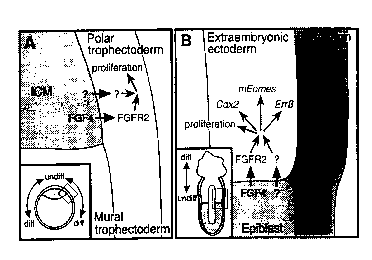
Fig. 4. A model for embryonic-trophoblast interactions and the maintenance of
TS cells in
2 0 vivo. (A) A schematic drawing of a. 3.Sdpc blastocyst (inset) emphasizing
a region where the polar and
mural trophectoderm meet with thc: ICM. FGF4 and at least one other
unidentified factor produced in
the ICM may signal to the overlying polar trophectoderm, maintaining it in a
proliferative state. As the
trophectoderm cells move away fr~~m the ICM to become mural trophectoderm,
they cease to receive
the ICM-derived signals and conseduently differentiate. (B) A schematic
drawing of a 6.Sdpc conceptus
2 5 (inset) emphasizing the embryonic-extraembryonic boundary. Similar to the
blastocyst scenario, this
suggested that FGF4 and an unkno~,~m factors) from the epiblast signal to the
extraembryonic ectoderm
(ExE) and directly or indirectly mediate the expression of genes such as
Err~3, Cdx2, and mEomes
(eomesodermin). These signals maintain a trophoblast stem cell population in
the ExE nearest to the
epiblast. As trophoblast cells move away from the embryonic-extraembryonic
border, they no longer
3 0 receive the epiblast signals and initiate a differentiation pathway.
DETAILED DESCRIPTION Oli A PREFERRED EMBODIMENT
TS cell lines were tirst derived from early postimplantation embryos. ExE
cells were isolated
from 6.Sdpc conceptuses as previously described (4), disaggregated by trypsin,
and cultured on a feeder
layer of primary mouse embryonic fibroblast (EMFI) cells in the presence of
various combinations of
3 5 growth factors (data not shown). The combination of FGF4 (25ng/ml) and
heparin (ltrg/ml) in TS cell
medium (9) proved successful m allowing the passage of colonies with a tight
epithelial morphology
CA 02345397 2001-03-23
WO 00/17325 PCT/CA99/00867
(Fig. lA). Removal of either FGF4, heparin, or the EMFI cells resulted in a
rapid decline in
proliferation with subsequent differentiation into a giant cell-like phenotype
(Fig. IB). Some giant cells
also consistently appeared at the edges of colonies after each passage even
under optimal conditions
suggesting that a small percentage of the cells underwent spontaneous
differentiation (Fig. lA). Since
the giant cells were relatively trypsiv-resistant, they were left behind after
each passage and so remained
at a relatively constant level in the cultures.
Under the identical culture. conditions used for isolating TS cell lines from
ExE, cell lines were
derived from 3.Sdpc blastocysts which exhibited a morphology and behaviour
indistinguishable from
that of ExE-derived TS cell lines (:i2).The blastocyst-derived and ExE-derived
lines are referred to as
TS3.5 and TS6,5 cell lines, respectively, to distinguish their tissues of
origin. Generation of TS3,5 and
TS6,5 cell lines was efficient and reproducible; 58 clonal TS3,5 cell lines
were obtained from 91
blastocysts (64%) and 17 TS6,5 cell lines from 39 ExEs of 6.Sdpc embryos
(44%); they were derived
from different strain backgrounds ( l29/sv and ICR) and of both sexes Some of
these TS cell lines were
stably maintained for more than 50 passages over a period of more than six
months with no apparent
change in their morphology or viability.
To address the possibility that FGF4 stimulated the proliferation of TS cells
indirectly by
inducing the secretion of mitotic factors from the feeder cells, conditioned
medium from EMFI cells
(EMFI-CM) was prepared in the absence of FGF4. TS cells were maintained in an
undifferentiated state
on gelatin-coated plates in medium supplemented with 70% EMFI-CM and
FGF4/heparin; lower
2 0 concentrations of EMFI-CM were not effective (13). Leukemia inhibitory
factor (LIF), the critical factor
produced by EMFI cells that maintains ES cells undifferentiated, could not
substitute for EMFI-CM
even at five-times the concentration used in ES cell medium. These results
suggest that a) EMFI cells
secrete an unidentified factors) (E,MFI-factor) that acts along with FGF4 to
maintain the TS cells in
a proliferative and undifferentiated state, b) secretion of this factors) is
not a result of the addition of
2 5 FGF4 to the media, and c) FGF4 acts directly on the TS cells.
Chromosome spreads from two TS cell lines passaged over 20 times revealed an
apparently
normal euploid karyotype. The ploidy of the stem cells and differentiated
giant cells were determined
by FACS analysis of cells stained with propidium iodide ( 14). The profile for
cells maintained in EMFI-
CM supplemented with FGF4/hep;~rin (13) revealed prominent peaks at 2N and 4N
indicative of the
3 0 Gl and G2/M DNA content of a diploid cell line (Fig. 1C). A small shoulder
of higher ploidy cells
(>4N) was also observed and was Likely due to the presence of spontaneously
differentiating giant cells
in the culture. Upon removal of FCiF4 and EMFI-CM a distinct 8N peak appeared
within 4 days. The
2N peak was reduced and the 4N peak, which would include diploid G2/M cells
and tetraploid G1 cells,
increased in size. By day 6, cells of higher than 8N ploidy were seen in the
analysis. These observations
3 5 are consistent with the morphological differentiation of TS cells to giant
cells.
Several genetic markers N~ere analyzed during stem cell and difterentiative
culture conditions
CA 02345397 2001-03-23
WO 00/17325 PCT/CA99/00867
_ g _
to confirm the trophoblast identity of the TS3,5 and TS6,5 cell lines and
characterize their
differentiation in the absence of FGF4 ( 15). Markers of the diploid ExE were
highly expressed in TS
cells. Err,Q, an orphan nuclear receptor, is specifically expressed in the ExE
nearest to the
extraembryonic-embryonic boundlary at early postimplantation stages and later
in the chorionic
ectoderm ( 16). This gene was highly expressed in TS cells grown in the
presence of FGF4 and 70%
EMFI-CM, but was down-regulated when differentiation was induced by removing
FGF4 and EMFI-
CM (Fig. 2A). This was also the ease for other genes known to be highly
expressed in the ExE, such
as Cdx2 ( 17), Fgfr2 (6), and the mouse homologue of eomesodermin ( 18) (Fig.
2A). In contrast to the
ExE-specific genes, 4311, an EPC-specific gene ( 19), was not detected in the
undifferentiated cells, but
was induced 4 days after the removal of FGF4 and EMFI-CM. Mash2, encoding a
basic helix-loop-
helix (bHLH) transcription factor, has been shown to be required in diploid
trophoblast cells of the EPC
to allow development of the spongiotrophoblast layer (20). Consistent with
this, Mash2 was upregulated
in differentiating TS cells prior to the expression of 4311 {Fig. 2A). Mash2
transcripts were also
progressively induced in TS cells cultured in stem cell conditions. Placental
lactogen 1 (Pl-I), a
specific marker for giant cells (c;l), is progressively induced in cultures
after removal of FGF4,
consistent with the predicted increase in giant cell content. As observed for
the Mash2 gene, the
increasing expression of Pl-1 during stem cell culture conditions may reflect
the presence of
spontaneously differentiating cells that accumulate after each passage (Fig.
2A). Hand7, another bHLH
transcription factor that is known to play an important role in the
development of giant cells but is not
2 0 expressed in the ExE (22), was detected throughout the culture periods
analyzed regardless of the
presence of FGF4 and EMFI-CM (Fig.2A). Oct3/4, Brachyury, and Hnf4, genes
specific for
ICM/epiblast (23), mesoderm (24).. and primitive endoderm (25), respectively,
were not detected in TS
cells (Fig. 2). Thus, these established cell lines conserve a gene expression
profile largely characteristic
of trophoblast cells in the ExE anti they express distinctive markers of other
trophoblast cell lineages
2 5 upon differentiation.
The most definitive test l:or the trophoblast identity and stem cell capacity
of TS cells is to
investigate their potential to incorporate into trophoblast lineages in vivo.
Rossant et al. (2) have shown
that the cells isolated from the Ex)=? of 6.Sdpc embryos can contribute to the
EPC and giant cells when
directly injected into blastocysts, despite temporal asynchrony between donor
and host cells. To
3 0 investigate the potency of TS cells to contribute to trophoblast lineages
in vivo, chimeric embryos were
made by the aggregation method (26) and blastocyst injection. A TS3.5 and a
TS6,5 cell line were
derived from BS/EGFP transgeni~~ mice (27) that ubiquitously express enhanced-
green fluorescent
protein (EGFP, Clontexh) in all embryonic and extraembryonic tissues. These
lines were passaged more
than 20 times (two months) before they were used for the chimera experiments.
Chimeras were obtained
3 5 from each cell line using both methods (Table 1). EGFP-positive cells were
only observed in tissues
of the trophoblast lineage in the 61 chimeric embryos analyzed (Fig. 3). TS
cells contributed to the ExE,
CA 02345397 2001-03-23
WO 00/17325 PCT/CA99/00867
- 9 -
EPC, and giant cells, but were never observed in the epiblast, primitive
endoderm, or other ICM-
derived extraembryonic tissues, such as the allantois, yolk sac, and amnion
(Table 2; Fig. 3). High
contributions to chimeric placentae at term were also observed, indicating
that these cells could
functionally support fetal development (Figure 3K, L). There was no
significant difference between the
EGFP-TS3.5 and EGFP-TS6.5 cell lines in their ability to contribute to
trophoblast subtypes. However,
blastocyst injections gave a higher frequency of chimeras than the aggregation
method (Table 1). These
results clearly show that TS cells retain the potency to differentiate into
all trophoblast cell types in vivo
despite being cultured in vitro for extended periods of time. Taken together
with the results of the
Northern analyses it was concluded that a stable pluripotent mouse trophoblast
stem (TS) cell line was
established.
It has been proposed that the ExE is the first tissue to be formed from the
polar trophectodetm
and that it may act as a stem cell population that subsequently gives rise to
the EPC which generates
new secondary giant cells (2, 3). Successful derivation of TS cell lines
expressing trophoblast markers
from the ExE of 6.Sdpc embryos and 3.Sdpc blastocysts is consistent with this
model. FGF4 produced
by the ICM and later by the epiblast is one of the critical signals required
for the maintenance of the
proliferative undifferentiated state of ExE (Fig.4). From the expression
pattern and null phenotype of
the Fgfr2 gene, this receptor tyrosine kinase is the best candidate to
functionally receive the FGF4
signal in the trophoblast. This model predicts that the lethality observed in
homozygous null mutants
for both Fgf4 and Fgfr2 (6, 7) may in part be caused by the loss of the
proliferating population of the
2 0 ExE soon after implantation. Durin;; normal implantation the blastocyst
first adheres to the uterine wall
through its mural trophectoderm at the abembryonic pole; however, Fgfr2 -/-
blastocysts implanted
randomly implying that the trophectoderm surrounding the embryo is not
polarized. The components
downstream of the trophoblast FGF response are not known, but the T-box gene,
mouse eomesodermin,
and the caudal-related gene. Cdx2, ;ue good candidates since they are
expressed in the appropriate cells
2 5 and members of these gene familiea have been shown to be regulated by FGF-
signaling (28, 29). As
trophoblast cells continue to proliferate and move distally from the
ICM/epiblast, they cease to receive
the mitotic and differentiation-inhibitory signals from the embryo proper.
This would result in
differentiation into EPC and finall:~ to giant cells.
The above model makes a number of testable predictions about the involvement
of FGF-
3 0 signaling in trophoblast development. For example, the model predicts that
TS cell lines could be
derived from Fgf4, but not Fgfr2 mutant blastocysts. Establishing TS cell
lines from other mouse
mutants will reveal the genes essential for this stem cell lineage, while in
vitro differentiation of mutant
lines will identify genes important for other trophoblast subtypes. In
summary, the establishment of
FGF4-dependent TS cell lines from blastocysts and the ExE of 6.Sdpc embryos
has revealed that a stem
3 5 cell population exists within the trophoblast lineage for at least a 3-day
window during early
development and that the essential embryo-derived signals for trophoblast
proliferation include FGF4.
CA 02345397 2001-03-23
WO 00/17325 PCT/CA99/00867
- 10 -
These cell lines are an invaluable tool to further dissect the function of
genes and signaling pathways
important to the development of the mammalian trophoblast lineage and its
interactions with the
embryo. The ability of wild type'CS cells to make high contributions in
chimeras indicates that these
cells have the potential to rescue nwtant embryos with placental defects. Such
"TS cell rescue" analysis
could be an alternative to the "te.traploid rescue" technique (27) currently
used. Finally, obtaining
similar trophoblast stem cell lines from human embryos opens up new avenues to
future cell-based
therapies for placentalinsufficiencies.
CA 02345397 2001-03-23
WO 00/17325 PCT/CA99/00867
- 11 -
Table 1. Frequency of obtaining implanted embryos and chimeric conceptuses
from diploid
aggregations and blastocyst injections of EGFP-TS3.5 and EGFP-TS6.5 cell
lines. Significant
differences were not observed between the: two cell lines analyzed. However,
blastocyst injections
(blast. inj.) yielded a higher percentage of implanted embryos and a higher
percentage of chimeras than
diploid aggregations (dip. agg.). TS cells were not viable in the culture
medium (KSOM) routinely used
for diploid aggregations with embryonic stem cells. Altering the aggregation
medium to 90% KSOM,
10% FBS, 25ng/ml FGF4, and lmg/ml heparin increased the viability of the TS
cells, but decreased the
fitness of the embryos. Consequently, blastocyst injections of TS cells were
routinely performed since it
avoids the overnight culture required for aggregations.
Cell line No. Transferred No. Embryos No. Chimeras
(technique) (% transferred)(% embryos)
EGFP-TS3.5 176 100 47
(blast. inj.) (57%) (47%)
EGFP-TS6.5 42 21 9
(blast. inj.) (50%) (43%)
EGFP-TS3,5 177 29 4
(dip. agg.) (16%) (14%)
EGFP-TS6.5 112 17 1
(dip. agg.) (15%) (6%)
Total 507 167 61
(33%) (37%)
CA 02345397 2001-03-23
WO 00/17325 PCT/CA99/00867
- 12 -
Table 2. Location of TS cell contributions. ExE, extraembryonic ectoderm; EPC,
ectoplacental cone;
GC, iant cells; ChE, chorionic ectoderm; Spong, spongiotrophoblast; Lab,
labyrinthine trophoblast.
Stage No. Chimeras Cell Type
6.5dpc 4 ExE, EPC, GC
(n=15) 3 EPC, GC
4 ExE, EPC
1 Exe
1 EPC
2 GC
7.5dpc 1 EPC, GC
(n=2) 1 GC
8.5dpc 1 ChE, EPC, GC
(n=11) 1 EPC, GC
2 ChE, GC
1 ChE, EPC
4 EPC
2 GC
9.5dpc 1 ChE, EPC, GC
(n=g) 1 EPC, GC
1 ChE, GC
2 EPC
3 GC
10.5dpc 1 Lab, Spong, GC
(n=9) 2 Spong, GC
1 Lab, Spong
2 Spong
3 GC
11.5dpc 2 Lab, Spong, GC
(n=g) 2 Lab, Spong
1 Spong, GC
3 Spong
18.5dpc 1 Lab, Spong, GC
(n=g) 5 Lab, Spong
2 Spong
CA 02345397 2001-03-23
WO 00/17325 PCT/CA99/00867
- 13 -
While the present invention has been described with reference to what is
presently considered
to be a preferred embodiment, it is to be understood that the invention is not
limited to the disclosed
embodiment. To the contrary, the invention is intended to cover various
modifications and equivalent
arrangements included within the spirit and scope of the appended claims.
All publications, patents and patent applications are herein incorporated by
reference in their
entirety to the same extent as if each individual publication, patent or
patent application was specifically
and individually indicated to be incorporated by reference in its entirety.
CA 02345397 2001-03-23
WO 00/17325 PCT/CA99/00867
- 14 -
References and Notes
1. J. Rossant, in Experiment~~l Approaches to Mammalian Embryonic
Developme~:t, J. Rossant
and R. A. Pederson, Eds. (Cambridge Univ. Press, London, 1986), pp. 97-120; J.
Rossant, Sem. Dev.
Biol. 6, 237 (1995).
2. J. Rossant, R. L. Gardner, H. L. Alexandre, J. Embryol. Exp. Morphol. 48,
239 (1978); M. H.
Johnson and J. Rossant, ibid. 61, 103 (1981).
3. R. L. Gardner and M. H. :lohnson, ibid. 28, 279 ( 1972); J. Rossant and W.
Tamura-Lis, ibid.
62, 217 (1981); E. B. Ilgren, Placenta 4, 307 (1983).
4. J. Rossant and L. Ofer, J. Embryol. Exp. Morphol. 39, 183 (1977); E. B.
Ilgren, ibid 62,183
(1981).
5. L. Niswander and G. R. Martin, Development 114, 755 (1992); D. A. Rappolee,
C. Basilico,
Y. Patel, Z. Werb, ibid. 120, 2259 (1994).
6. A. Orr-Urtreger et al., Dew. Biol. 158, 475 (1993); E. Arman , R. Haffner-
Krausz, Y. Chen,
J. K. Heath, P. Lonai, Proc. Natl. Acad. Sci. U.S.A. 95, 5082 (1998); J.
Partanen and J. Rossant,
unpublished data.
7. B. Feldman, W. Poueymuou, V. E. Papaioannou, T. M. DeChiara, M. Goldfarb,
Science 267,
246 ( 1995).
2 0 8. N. Chai et al., Dev. Biol. 198, 105 ( 1998).
9. TS cell medium is RPMI 1640 supplemented with 20% fetal bovine serum
(HyClone), sodium
pyruvate ( lmM, GibcoBRL), (3-m~rcaptoethanol ( 100N.M, Sigma), L-glutamine
(2mM, GibcoBRL),
and penicillin/streptomycin (SOftg/ml each). Human recombinant FGF4 (25ng/ml,
Sigma) and heparin
(lp,g/ml) were added to aliquots of TS cell medium and used immediately.
2 5 10. B. Hogan, R. Beddington, F: Costantini, E. Lacy, Manipulating the
Mouse Embryo (Cold
Spring Harbor Laboratory Press, Cold Spring Harbor. NY, ed. 2, 1994), pp. 265-
272; E. J. Robertson,
in Teratocarcinomas and Embryonic Stem Cells, E. J. Robertson, Ed. (IRL Press,
Oxford, 1987), pp.
71-112.
11. J.-E. Flechon, S. Laurie, :E. Notarianni, Placenta 16, 643 (1995).
3 0 12. TS3.5 cell lines were obtained using similar techniques for ES cell
line derivation (10). Briefly,
3.Sdpc blastocysts were individually plated into 4-well plates on EMFI cells
and cultured in TS media
with FGF4 and heparin (9). The medium was changed after two days and the
blastocyst outgrowth was
trypsinized on the third day. On d~iy 5 or 6, flat colonies, referred to as
"epithelial-like cells" in (10),
were picked and passaged. Once established, the cell lines were grown without
EMFI cells, but in the
3 5 presence of EMFI conditioned medium ( 13). Under the current culture
conditions ES cell colonies were
not observed.
CA 02345397 2001-03-23
WO 00/17325 PCT/CA99/00867
- 15 -
13. Conditioned medium from EMFI cells (EMFI-CM) was prepared by incubating TS
medium
(9) without FGF4 or heparin on confluent plates of mitomycin-treated EMFI
cells for 72 hours. The
conditioned medium was filtered (C~.451.trn) and stored at -20°C.
Established TS cell lines were routinely
cultured in 70% EMFI-CM, 30% 7.'S medium, 25ng/ml hrFGF4, and 1 ftg/ml heparin
on gelatin-coated
plates. The medium was changed every two days and the cells were passaged (1
in 25) every fow days
or at 80%-90% confluency.
14. TS cells were grown in the absence of EMFI cells ( 13) and collected by
cell scraping at 0, 2,
4, and 6 days after the removal of F~GF4, heparin, and EMFI-CM. The cells were
fixed and stained with
propidium iodide (Molecular Probc;s) as described [Z. Darzynkiewicz and G.
Juan, in Current Protocols
in Cytometry (John Wiley & Sons, Inc., New York, 1997), pp. 7.5.2-7.5.3]. Cell
fluorescence was
measured by a flow cytometry with an argon ion laser (488nm). The data was
analyzed with Coulter
EXPO Cytometer Software version 2.0 by Applied Cytometry Systems, 1998.
15. Total RNA was prepared from cells and embryos with TRIzoI (GibcoBRL)
according to the
manufacturer's instructions. Northern blotting was performed by a standard
protocol. Antisense RNA
probes for Err~3 ( 16), eomesodermin ( 18), Cdx2 [E. Suh, L. Chen, J. Taylor,
P. G. Traber, Mol. Cell.
Biol. 14, 7340 (1994)], Fgfr2, Mash2 (20), 4311 (19), Handl (22), Pl-l [P.
Colosi, F. Talamantes, D.
I. H. Linzer, Mol. Endocrinol. 1, 767 (1987)], Oct-3/4 {23), Brachyury (24),
and GAPDH [P. Fort et
al., Nucleic Acids Res. 13, 1431 (1985)] were labeled with either [a-32P]UTP
or DIG-11-UTP
2 0 (Boehringer Mannheim) by using f~trip-EZ RNA kit (Ambion). Blots were
hybridized overnight at 65°C
in NorthernMax Prehybridization/hybridization Buffer (Ambion) and were finally
washed in O.lx
SSC/0.1% SDS at 65°C. DIG-labeled probes were detected with the DIG
Luminescent Detection Kit
(Boehringer Mannheim). Remova'I of hybridized RNA probes was performed with
the Strip-EZ RNA
kit (Ambion) according to manuf2,cturer's recommendations. To assess the
expression of Hnf4 in the
2 5 TS cell lines, first strand cDNA s~rnthesized from 0.5 pg total RNA of TS
cells and 7.Sdpc embryos
with random hexamers was subjected to 35 cycles of PCR (62°C annealing
temperature) by using 0.2
pM each of Hnf4-specific primers (5'-CACGTCCCCATCTGAAGGTG-3' and 5'-
CTTCCTTCTTCATGCCAGCCC-3') and 0.1 pM each of JJ-actin-specific primers (5'-
GACAACGGCTCCGGCATGTGCAAAG-3'and 5=TTCACGGTTGGCCTTAGGGTTCAG-3~. The
3 0 primer sequences were adapted fr~~m D. Ioannis et al., Development 125,
1529 (1998).
16. K. Pettersson et al., Mec,h. Dev. 54, 211 (1996); J. Luo et al., Nature
388, 778 (1997).
17. F. Beck, T. Erler, A. Russell, R. James, Dev. Dyn. 204, 2 t 9 ( 1995).
18. B. G. Ciruna and J. Rossant, Mech. Dev. 81, 199 ( 1999).
19. K. R. Lescisin, S. Varmu;za, J. Rossant, Genes Dev. 2. I 639 ( 1988).
3 5 20. F. Guillemot> A. Nagy, ~~. Auerbach, J. Rossant. A. L Joyner, Nature
371, 333 (1994); M.
Tanaka, M. Gertsenstein. J. Rossamt, A. Nagy, Dev. Biol. 190. ~5 ( 1997).
CA 02345397 2001-03-23
WO 00/17325 PCT/CA99/00867
- 16 -
21. T. N. Faria, L. Ogren, F. 'Calamantes, D. I. Linzer, M. J. Soares, Biol.
Reprod. 44, 327 ( 1991 ).
22. J. C. Cross et al., Development 12I, 2513 (1995); P. Riley, L. Anson-
Cartwright, J. C. Cross,
Nat. Genet. 18, 271 ( 1998).
23. S. L. Palmieri, W. Peter, H. Hess, H. R. Scholer, Dev. Biol. 166, 259 (
1994); H. R. Scholer,
G. R. Dressier, R. Balling, H. Rohdewohld, P. Gruss, EMBO J. 9, 2185 ( 1990).
24. D. G. Wilkinson, S. Bhatt, B. G. Hetrmann, Nature 343, 657 (1990).
25. S. A. Duncan et al., Proc. Natl. Acad. Sci. U.S.A. 91, 7598 (1994).
26. A. Nagy and J. Rossant, in Gene Targeting, A. L. Joyner Ed. (IRL. Press,
Oxford, 1993), pp.
147-180; S. A. Wood, N. D. Allen, J. Rossant, A. Auerbach, A. Nagy, Nature
365, 87 (1993).
27. A.-K. Hadjantonakis, M. Gertsenstein, I. Ikawa, M. Okabe, A. Nagy, Mech
Dev. 1998
Aug;76( 1-2):79-90.
28. K. J. P. Griffin, S. L. Am;tcher, C. B. Kimmel, D. Kimelman, Development
125, 3379 (1998);
K. Griffin, R. Patient, N. Holder, .ibid. 121, 2983 (1995).
29. M. E. Pownall. H. V. Isaacs, J. M. Slack, Curr. Biol. 8, 673 (1998); H. V.
Isaacs, M. E.
Pownall, J. M. Slack, EMBO J. 1 ~~, 3413 ( 1998).