Note: Descriptions are shown in the official language in which they were submitted.
CA 02345669 2007-08-14
79214-5
-1-
SELECTIVE ADHERENCE OF STENT-GRAFT COVERINGS, MANDREL
AND METHOD OF MAKING STENT-GRAFT DEVICE
BACKGROUND OF THE INVENTION
The present invention relates generally to endoluminal stent-graft devices
suitable for percutaneous delivery into a body through anatomical passageways
to treat
injured or diseased areas of the body. More particularly, the present
invention relates to
a method of bonding microporous polytetrafluoroethylene ("PTFE") coverings
over a
stent scaffold in a manner which maintains unbonded regions to act as slip
planes or
pockets to accommodate planar movement of stent elements. In one embodiment of
the
present invention bonded and unbonded regions are formed by means of a mandrel
which has a pattern of either raised projections or recesses in its surface
which are either
synchronous or asynchronous, respectively, with stent elements.
The use of implantable vascular grafts comprised of PTFE is well known in the
art. These grafts are typically -used to replace or repair damaged or occluded
blood
vessels within the body. However, if such grafts are radially expanded within
a blood
vessel, they will exhibit some subsequent retraction. Further, such grafts
usually require
additional means for anchoring the graft within the blood vessel, such as
sutures,
clamps, or sirriilarly functioning elements. To minimize the retraction and
eliminate the
CA 02345669 2001-03-28
-
WO 00/18328 PCTIUS99/22808
-2-
requirement for additional attachment means, those skilled in the art have
used stents,
such as those presented by Palmaz in U.S. Patent No. 4,733,665 and Gianturco
in U.S.
Patent No. 4,580,568 which patents are herein incorporated by reference,
either alone or
in combination with PTFE grafls.
For example, the stent described by Pahnaz in U.S. Patent No. 4,733,665 can be
used to repair an occluded blood vessel. The stent is introduced into the
blood vessel via
a balloon catheter, which is then positioned at the occluded site of the blood
vessel. The
balloon is then expanded thereby expanding the overlying stent to a diameter
comparable to the diameter of an unoccluded blood vessel. The balloon catheter
is then
deflated and removed with the stent remaining seated within the blood vessel
because
the stent shows little or no radial retraction. Use of radially expandable
stents in
combination with a PTFE graft is disclosed in U.S. Patent No. 5,078,726 to
Kreamer.
This reference teaches placing a pair of expandable stents within the interior
ends of a
prosthetic graft having a length that is sufficient to span the damaged
section of a blood
vessel. The stents are then expanded to secure the graft to the blood vessel
wall via a
fi-iction fit.
Although stents and stent/graft combinations have been used to provide
endovascular prostheses that are capable of maintaining their fit against
blood vessel
walls, other desirable features are lacking. For instance, features such as
increased
strength and durability of the prosthesis, as well as an inert, smooth,
biocompatible
blood flow surface on the luminal surface of the prosthesis and an inert,
smooth
biocompatible surface on the abluminal surface of the prosthesis, are
advantageous
characteristics of an implantable vascular graft. Some of those skilled in the
art have
recently addressed these desirable characteristics by producing strengthened
and
reinforced prostheses composed entirely of biocompatible grafts and graft
layers.
For example, U.S. Patent No. 5,048,065, issued to Weldon, et al. discloses a
reinforced graft assembly comprising a biologic or biosynthetic graft
component having
a porous surface and a biologic or biosynthetic reinforcing sleeve which is
concentrically fitted over the graft component. The reinforcing sleeve
includes an
internal layer, an intermediate layer, and an external layer, all of which
comprise
CA 02345669 2001-03-28
-
WO 00/18328 PCTIUS99/22808
-3-
biocompatible fibers. The sleeve component functions to provide compliant
reinforcement to the graft component. Further, U.S. Patent No. 5,163,951,
issued to
Pinchuk, et al. describes a composite vascular graft having an inner
component, an
intermediate component, and an outer component. The inner and outer components
are
preferably formed of expanded PTFE while the intermediate component is formed
of
strands of biocompatible synthetic material having a melting point lower than
the
material which comprises the inner and outer components.
Another reinforced vascular prosthesis having enhanced compatibility and
compliance is disclosed in U.S. Patent No. 5,354,329, issued to Whalen. This
patent
discloses a non-pyrogenic vascular prosthesis comprising a multilamellar
tubular
member having an interior stratum, a unitary medial stratum, and an exterior
stratum.
The medial stratum forms an exclusionary boundary between the interior and
exterior
strata. One embodiment of this prosthesis is formed entirely of silicone
rubber that
comprises different characteristics for the different strata contained within
the graft.
The prior art also includes grafts having increased strength and durability,
which
have been reinforced with stent-like members. For example, U.S. Patent No.
4,731,073,
issued to Robinson discloses an arterial graft prosthesis comprising a multi-
layer graft
having a helical reinforcement embedded within the wall of the graft. U.S.
Patent No.
4,969,896, issued to Shors describes an inner elastomeric biocompatible tube
having a
plurality of rib members spaced about the exterior surface of the inner tube,
and a
perforate flexible biocompatible wrap circumferentially disposed about, and
attached to,
the rib members.
Another example of a graft having reinforcing stent-like members is disclosed
in
U.S. Patent No. 5,123,917, issued to Lee which describes an expandable
intraluminal
vascular graft having an inner flexible cylindrical tube, an outer flexible
cylindrical tube
concentrically enclosing the inner tube, and a plurality of separate scaffold
members
positioned between the inner and outer tubes. Further, U.S. Patent No.
5,282,860, issued
to Matsuno et al. discloses a multi-layer stent comprising an outer resin tube
having at
least one flap to provide an anchoring means, an inner fluorine-based resin
tube and a
mechanical reinforcing layer positioned between the inner and outer tubes.
CA 02345669 2001-03-28
WO 00/18328 PCTIUS99/22808
-4-
Still another stent containing graft is described in U.S. Patent No. 5,389,106
issued to Tower which discloses an impermeable expandable intravascular stent
including a dispensable frame and an impermeable deformable membrane
interconnecting portions of the frame to form an impermeable exterior wall.
The
membrane comprises a synthetic non-latex, non-vinyl polymer while the frame is
comprised of a fine platinum wire. The membrane is attached to the frame by
placing
the frame on a mandrel, dipping the frame and the mandrel into a polymer and
organic
solvent solution, withdrawing the frame and mandrel from the solution, drying
the frame
and mandrel, and removing the mandrel from the polymer-coated frame.
Microporous expanded polytetrafluoroethylene ("ePTFE") tubes may made by
any of a number of well-known methods. Expanded PTFE is frequently produced by
admixing particulate dry polytetrafluoroethylene resin with a liquid lubricant
to form a
viscous sluiTy. The mixture is poured into a mold, typically a cylindrical
mold, and
compressed to form a cylindrical billet. The billet is then ram extruded
through an
extrusion die into either tubular or sheet structures, termed extrudates in
the art. The
extrudates consist of extruded PTFE-lubricant mixture called "wet PTFE." Wet
PTFE
has a microstructure of coalesced, coherent PTFE resin particles in a highly
crystalline
state. Following extrusion, the wet PTFE is heated to a temperature below the
flash
point of the lubricant to volatilize a major fraction of the lubricant from
the PTFE
extrudate. The resulting PTFE extrudate without a major fraction of lubricant
is known
in the art as dried PTFE. The dried PTFE is then either uniaxially, biaxially
or radially
expanded using appropriate mechanical apparatus known in the art. Expansion is
typically carried out at an elevated temperature, e.g., above room temperature
but below
327 C, the crystalline melt point of PTFE. Uniaxial, biaxial or radial
expansion of the
dried PTFE causes the coalesced, coherent PTFE resin to form fibrils emanating
from
nodes (regions of coalesced PTFE), with the fibrils oriented parallel to the
axis of
expansion. Once expanded, the dried PTFE is referred to as expanded PTFE
("ePTFE")
or microporous PTFE. The ePTFE is then transferred to an oven where it is
sintered by
being heated to a temperature above 327 C, the crystalline melt point of PTFE.
During
the sintering process the ePTFE is restrained against uniaxial, biaxial or
radial
contraction. Sintering causes at least a portion of the crystalline PTFE to
change from a
CA 02345669 2001-03-28
WO 00/18328 PCT/US99/22808
-5-
crystalline state to an amorphous state. The conversion from a highly
crystalline
structure to one having an increased amorphous content locks the node and
fibril
microstructure, as well as its orientation relative to the axis of expansion,
and provides a
dimensionally stable tubular or sheet material upon cooling. Prior to the
sintering step,
the lubricant must be removed because the sintering temperature of PTFE is
greater than
the flash point of commercially available lubricants.
Sintered ePTFE articles exhibit significant resistance to further uniaxial, or
radial expansion. This property has lead many in the art to devise techniques
which
entail endoluminal delivery and placement of an ePTFE graft having a desired
fixed
io diameter, followed by endoluminal delivery and placement of an endoluminal
prosthesis, such as a stent or other fixation device, to frictionally engage
the
endoluminal prosthesis within the lumen of the anatomical passageway. The
Kreamer
Patent, U.S. Patent No. 5,078,726, discussed above, exemplifies such use of an
ePTFE
prosthetic graft. Similarly, published International Applications No.
W095/05132 and
W095/05555, filed by W.L. Gore Associates, Inc., disclose balloon expandable
prosthetic stents which have been covered on inner and outer surfaces by
wrapping
ePTFE sheet material about the balloon expandable prosthetic stent in its
enlarged
diameter, sintering the wrapped ePTFE sheet material to secure it about the
stent, and
crimping the assembly to a reduced diameter for endoluminal delivery. Once
positioned
endoluminally, the stent-graft combination is dilated to re-expand the stent
to its
enlarged diameter returning the ePTFE wrapping to its original diameter.
Thus, it is well known in the prior art to provide an ePTFE covering which is
fabricated at the final desired endovascular diameter and is endoluminally
delivered in a
folded or crimped condition to reduce its delivery profile, then unfolded in
vivo using
either the spring tension of a self-expanding, thermally induced expanding
structural
support member or a balloon catheter. However, the known ePTFE covered
endoluminal stents are often covered on only one surface of the stent, i.e.,
either the
lumenal or abluminal wall surface of the stent. Where the stent is fully
covered on both
the luminal and abluminal wall surfaces of the stent, the covering completely
surrounds
the stent elements and fills the stent interstices. When the encapsulated
stent is
comprised of shape memory alloy, characteristics of the stent make it
necessary to
CA 02345669 2001-03-28
WO 00/18328 PCT/US99/22808 -
-6-
encapsulate in the "large" state and then compress the encapsulated stent for
delivery. In
this case encapsulation either increases the device's resistance to
compression, or
increases the delivery profile of the device as compression causes the
polymeric material
to fold or buckle around the stent. Perhaps the most serious problem is that
the folding
during compression actually encompasses folding of the stent itself, which
unduly
stresses the stent material and may result in structural failure.
In contrast to the prior art, the present invention provides a method to
encapsulate a stent in ePTFE whereby the structure contains pockets or regions
where
the ePTFE layers are not adhered to one another allowing the stent to contract
or expand
without being encumbered by ePTFE and without folding or stressing the stent
itself.
As use herein, the following terms have the following meanings:
"Fibril" refers to a strand of PTFE material that originates from one or more
nodes and terminates at one or more nodes.
"Node" refers to the solid region within an ePTFE material at which fibrils
originate and converge.
"Internodal Distance" or "IND" refers to a distance between two adjacent nodes
measured along the longitudinal axis of fibrils between the facing surfaces of
the
adjacent nodes. IND is usually expressed in micrometers ( m).
"Node Length" as used herein refers to a distance measured along a straight
line
between the furtherrnost end points of a single node which line is
perpendicular to the
fibrils emanating from the node.
"Nodal Elongation" as used herein refers to expansion of PTFE nodes in the
ePTFE microstructure along the Node Length.
"Longitudinal Surface" of a node as used herein refers to a nodal surface from
which fibrils emanate.
"Node Width" as used herein refers to a distance measured along a straight
line,
drawn parallel to the fibrils, between opposing longitudinal surfaces of a
node.
CA 02345669 2001-03-28
WO 00/18328 PCT/US99/22808
=7-
"Plastic Deformation" as used herein refers to the deformation of the ePTFE
microstructure under the influence of a expansive force which deforms and
increases the
Node Length and results in elastic recoil of the ePTFE material less than
about 25%.
"Radially Expandable" as used herein to describe the present invention refers
to
a property of the ePTFE tubular member to undergo radially oriented Plastic
Deformation mediated by Nodal Elongation.
"Structural Integrity" as used herein to describe the present invention in
terms of
the ePTFE refers to a condition of the ePTFE microstructure both pre- and post-
radial
deformation in which the fibrils are substantially free of fractures or breaks
and the
ePTFE material is free of gross failures; when used to describe the entire
device
"Structural Integrity" may also include delamination of the ePTFE layers.
Endoluminal stent devices are typically categorized into two primary types:
baUoon expandable and self-expanding. Of the self-expanding types of
endoluminal
stent devices, there are two principle sub-categories: elastically self-
expanding and
thermally self-expanding. The balloon expandable stents are typically made of
a ductile
material, such as stainless steel tube, which has been machined to form a
pattem of
openings separated by stent elements. Radial expansion is achieved by applying
a
radially outwardly directed force to the lumen of a balloon expandable stent
and
deforming the stent beyond its elastic limit from a smaller initial diameter
to an enlarged
final diameter. In this process the slots deform into "diamond shapes."
Balloon
expandable stents are typically radially and longitudinally rigid and have
limited recoil
after expansion. These stents have superior hoop strength against compressive
forces but
should this strength be overcome, the devices will deform and not recover.
Self-expanding stents, on the other hand, are fabricated from either spring
metal
or shape memory alloy wire which has been woven, wound or formed into a stent
having interstices separated with wire stent elements. When compared to
balloon-
expandable stents, these devices have less hoop strength but their inherent
resiliency
allows them to recover once a compressive force that results in deformation is
removed.
CA 02345669 2001-03-28
WO 00/18328 PCT/US99/22808
-8-
Covered endoluminal stents are known in the art. Heretofore, however, the
stent
covering has been made of a polymeric material which has completely subtended
the
stent interstices, that is, the stent was completely embedded in the polymeric
material.
This has posed difficulty particularly with the self-expanding stents. To
preserve their
self-expanding property, all covered self-expanding stents have been covered
with a
polymeric covering while the stent is in its unstrained dimensional condition,
i.e.; its
native enlarged diameter. Yet to delivery a covered stent it must be
constricted to a
smaller delivery diameter. Radial compression of a stent necessarily causes
the
individual stent elements to traverse the stent interstices and pass into
proximity to a
to laterally adjacent individual stent element, thereby occupying the
previously open
interstitial space. Any polymeric material which subtends or resides within
the
previously open interstitial space will necessarily be displaced, either
through shearing,
fracturing or otherwise responding to the narrowing of the interstitial space
as the stent
is compressed from its enlarged unstrained diameter to its strained reduced
diameter.
Because the struts of the stent are completely encapsulated, resistance of the
polymer
may cause folding or stressing of the struts during compression.
It was recognized, therefore, that a need has developed to provide an
encapsulating covering for a stent which is permanently retained on the stent,
substantially isolates the stent material from the body tissue forming the
anatomical
passageway or from matter within the anatomical passageway, and which permits
the
stent to deform without substantial interference from the covering material.
It is, therefore, a primary objective of the present invention to provide a
method
for encapsulating an endoluminal stent such that the encapsulating covering
forms non-
adhered regions which act as slip planes or pockets to permit the individual
stent
elements to traverse a substantial surface area of interstitial space between
adjacent stent
elements without resistance or interference from the encapsulating covering,
thereby
avoiding damage or stress to the stent elements.
It is a further object of the present invention to use the pockets between the
bonded regions to contain and deliver therapeutic substances.
CA 02345669 2001-03-28
WO 00/18328 PCT/11S99/22808 "
-9-
It is another objective of the present invention to provide an apparatus for
applying to and selectively adhering sections of the encapsulating covering
about the
stent, and to provide a selectively adhered encapsulated covered stent-graft
device.
SUMMARY OF THE INVENTION
These and other objectives of the present invention are achieved by providing
an
encapsulated stent-graft device in which an endoluminal stent having a
plurality of
individual stent elements separated by interstitial spaces is
circumferentially covered
along at least a portion of its longitudinal axis by at least one luminal and
at least one
abluminal covering of a polymeric material, the luminal and abluminal
coverings being
selectively adhered to one another at discrete portions thereof in a manner
which forms a
plurality of open pockets surrounding a plurality of stent elements. A
radially
expandable reinforced vascular graft that includes a first layer of
biocompatible flexible
material, a second layer of biocompatible flexible material, and a support
layer
sandwiched between the first and second layers of biocompatible flexible
material. In
addition, the selective bonding system disclosed herein can be advantageously
used to
produce inflatable pockets by bonding the first layer to the second layer in
defined
patterns. The resulting structure can then be inflated and stiffened by
injection of a fluid
resulting in a supporting structure without inclusion of a stent. A crude
analogy might be
the construction of an air mattress that is composed of flexible polymeric
layers bonded
to each other in a predetermined pattern.
The at least one luminal and at least one abluminal covering of a polymeric
material are preferably comprised of expanded PTFE, unexpanded porous PTFE,
woven
polyester or expanded PTFE yams, polyimides, silicones, polyurethane,
fluoroethylpolypropylene (FEP), polypropylfluorinated amines (PFA), or other
related
fluorinated polymers.
The stent preferably comprises a stent and may be made of any strong material
which can undergo radial expansion but which is also resistant to non-elastic
collapse
such as silver, titanium, nickel-titanium alloys, stainless steel, gold, or
any suitable
plastic material capable of maintaining its shape and material properties at
sintering
CA 02345669 2001-03-28
WO 00/18328 PCT/US99/22808
-10-
temperatures and having the necessary strength and elasticity to enable radial
expansion
without collapse due to the presence of the polymer coverings.
A preferred embodiment of the radially expandable reinforced vascular device
comprises a tubular stent, composed of a plurality of stent elements and stent
interstices,
the tubular stent is concentrically covered along at least a portion of its
longitudinal
length by a luminal polymeric covering and an abluminal polymeric covering.
The
luminal and abluminal polymeric coverings are discontinuously joined to one
another
through some of the stent interstices. The luminal and abluminal polymeric
coverings
may be shorter in length than the stent member to permit opposing stent ends
to flare
io outwardly upon radial expansion of the stent member. Alternatively, the
ends of the
stent member may be completely encased by the luminal and abluminal polymeric
coverings.
The stent member is preferably a self-expanding stent, which may be either an
elastic spring material stent, such as a stainless steel stent as disclosed in
Wall, U.S.
Patent No. 5,266,073or a non-woven stainless steel self-expanding stent as
disclosed in
Gianturco, U.S. patent No. 5,282,824, or a thermoelastic stent made of a shape
memory
alloy, e.g., a nickel-titanium alloy commonly known as NITINOL, such as that
disclosed in U.S. Patent No. 5, 147, 370. Tubular shaped support member
preferably
comprises a stent made of silver, titanium, stainless steel, gold, or any
suitable plastic
material capable of maintaining its shape and material properties at sintering
temperatures and having the strength and elasticity to permit radial expansion
and resist
radial collapse.
In accordance with the present invention, selective bonding of expanded PTFE
luminal and abluminal layers encapsulates the endoluminal stent and isolates
the stent
from both the tissue fonning the anatomical passageway as well as any fluid,
such as
blood, bile, urine, etc. which may pass through the anatomical passageway. The
presence of slip planes or pockets formed by the selectively adhered regions
of ePTFE i)
permits freedom of movement of stent elements within the encapsulating
covering
during both during expansion and contraction of the stent along either its
radial or
longitudinal axes; ii) permits uniform folding of the ePTFE stent covering
material
CA 02345669 2001-03-28
WO 00/18328 PCT/US99/22808 -
-11-
which is complementary to the structure of the stent element lattice; iii)
permits
movement of the stent relative to the ePTFE encapsulating layers; iv) reduces
forces
required to compress or dilate the stent in the case of elastically or
thermally self-
expanding stents; v) reduces radial expansion pressures required to balloon
expand an
ePTFE encapsulated stent; and vi) provides void regions which may be used in
conjunction with the microporous microstructure of the ePTFE covering material
to
retain and release bioactive substances, such as anticoagulant drugs, anti-
inflammatory
drugs, or the like.
Alternative arrangements of the stent member or other suitable structural
support
lo sufficient to maintain the lumenal patency of the lumenal and abluminal
polymer
coverings may be employed. For example, a radially expandable, articulated
reinforced
vascular graft may be formed by concentrically interdisposing a structural
support
assembly comprising multiple stent members spaced apart from one another
between
two tubular polymer covering members, then partially joining the two tubular
polymer
covering members by circumferentially compressing selected regions of the two
tubular
polymer covering members and thermally bonding the selectively compressed
regions to
one another.
The present invention also encompasses selective bonding of multiple polymeric
layers to create an inflatable structure. Such a structure can be inflated by
fluids
delivered through lumens within the delivery catheter. The selective bonding
method
allows creation of devices with multiple adjacent channels or pockets. Some of
these
pockets can be prefilled with a therapeutic drug to prevent restenosis or
local
thrombosis. Altemate pockets can be arranged for fluid inflation after the
device is
inserted.
One method of making the foregoing encapsulated stent-graft is to join
concentrically a luminal polymeric tube, an endoluminal stent, and an
abluminal
polymeric tube and to place the assembly onto a mandrel having a plurality of
raised
projections separated by land areas, or by a plurality of land areas separated
by a
plurality of recesses. Either the raised projections or the land areas are
patterned to
match a pattern of either the stent elements of the stent interstices, both
the stent
CA 02345669 2001-03-28
WO 00/18328 PCT/US99/22808
-12-
elements and stent interstices or portions of each. In this way the
projections or the
landed areas exert pressure, respectively on select regions of the PTFE
resulting in
limited regions of adherence or fusion when the device is heated to sintering
temperatures. With a mandrel luminal pressure is selectively applied to
produce
selectively placed bonds. As will become clear, bonding pressure can be
applied from
the luminal or the abluminal or both surfaces of the device.
The present invention is also directed to a process for making a radially
expandable reinforced stent-graft device by the steps of
a) positioning a radially expandable stent member composed of a
plurality of interconnected stent elements and a plurality of interstices
between
adjacent interconnected stent elements, concentrically over a first polymeric
cover member;
b) positioning a second polymer cover member concentrically over the
radially expandable stent member and the first polymeric cover member;
c) selectively joining portions of the first polymeric cover member and
the second polymeric cove member through a plurality of the interstices of the
stent member, while leaving portions of the first and second polymeric cover
members unjoined and forming slip planes or pockets to accommodate
movement of at least a portion of the interconnected stent elements
therethrough;
d) fully joining opposing end regions of the first and second polymer
cover members through the interstices of the stent member proximate to
opposing ends of the stent member
The step of fixing the support layer to the biocompatible graft layers
comprises
selectively applying pressure to the portions of the luminal and abluminal
polymer
covers after they are loaded onto a mandrel and then heating the resulting
assembly at
sintering temperatures to form a mechanical bond at the selected areas of
applied
pressure. Alternatively, a pattern of at least one of an adhesive, an aqueous
dispersion of
polytetrafluoroethylene, a polytetrafluoroethylene tape,
fluoroethylpolypropylene (FEP),
or tetrafluoroethylene (collectively the "adhesive") may be introduced between
the
CA 02345669 2007-08-14
79214-5
-13-
luminal and abluminal polymer covers at selected positions,
followed by heating the assembly to the melt temperature of
the adhesive to bond the luminal and abluminal polymer
covers while leaving unbonded slip plane regions to
accommodate movement of the stent elements. If ultraviolet
curable adhesives are used, a UV laser or a photolithography
system can be used to create the bond pattern. Many
thermoplastic polymers such as polyethylene, polypropylene,
polyurethane and polyethylene terephthalate can also be
used. If pieces of one of these or similar polymers are
placed or attached to one of the polymer covers in the
region to be bonded, heat and pressure will melt the
thermoplastic causing it to flow into the pores of the
ePTFE, thereby bonding the ePTFE layers together.
In accordance with an aspect of the invention,
there is provided a method of making an endoluminal stent-
graft, comprising the steps of: placing a first covering
member composed of a biocompatible polymer on a surface
having a pattern of elevated regions; placing a radially
expandable stent over said first covering member in
alignment with said pattern; placing a second covering
member composed of a biocompatible polymer over said
expandable stent; applying pressure to force said first
covering member and said second covering member into
intimate contact through openings in the stent and in
registration with the pattern; and heating the first and
second covering members to form a pattern of bonds between
the covering members, said pattern of bonds corresponding to
the pattern of elevated regions.
In accordance with an aspect of the invention,
there is provided a method for making an endoluminal
prosthesis, comprising the steps of: positioning a first
CA 02345669 2007-08-14
79214-5
-13a-
covering member composing a biocompatible polymer material
over a mandrel; positioning at least one radially expandable
support member, having a plurality of openings passing
through a wall thereof, over said first covering member;
positioning a second covering member comprising a
biocompatible polymer material over said support member and
said first covering member; and attaching said first
covering member to said second covering member at a
plurality of predetermined bonding locations, wherein said
bonding locations are positioned in said openings in said
support member, and wherein a plurality of unbonded regions
are formed between said first and second covering members.
In accordance with an aspect of the invention,
there is provided an endoluminal prosthesis, comprising: at
least one radially expandable support member, having a
plurality of openings passing through a wall thereof, a main
portion with a first outer diameter, and at least one flared
end portion with a second outer diameter greater than the
first outer diameter; a first covering member comprising a
biocompatible material, positioned about an interior surface
of said support member; and a second covering member
comprising a biocompatible material, positioned about an
exterior surface of said support member; wherein said first
covering member is bonded to said second covering member
along two or more spaced apart bonding regions positioned in
said openings in said support member, such that a pocket is
formed between adjacent bonding regions, the pocket being
sized greater than a thickness of the support member wall to
permit movement of the support member wall within the
pocket.
In accordance with an aspect of the invention,
there is provided an endoluminal prosthesis, comprising: a
first tubular structure comprising expanded
CA 02345669 2007-08-14
79214-5
-13b-
polytetrafluoroethylene; a second tubular structure
comprising expanded polytetrafluoroethylene positioned
concentrically about the first tubular structure, the first
and second tubular structures having a length; and at least
one radially expandable support member positioned between
the first and second tubular structures, the support member
comprising a wall with openings passing therethrough,
wherein the length of the first and second tubular
structures is less than a length of the radially expandable
support member, and wherein the first tubular structure is
bonded to the second tubular structure along two or more
spaced apart bonding regions positioned within the openings
of the support member, the spacing of adjacent bonding
regions resulting in a pocket that permits movement of the
support member therewithin.
These and other objects, features and advantages
of the present invention will become more apparent to those
skilled in the art when taken with reference to the
following more detailed description of the preferred
embodiments of the invention in conjunction with the
accompanying drawings.
BRIEF DESCRIPTION OF THE DRAWINGS
Figure 1 is a process flow diagram illustrating a
preferred method of making the inventive stent-graft device
in accordance with the present invention.
Figure 2 is a perspective view of a mandrel having
longitudinal ridges or splines.
Figure 3 is a cross-section view of the mandrel
shown in Fig. 2.
CA 02345669 2007-08-14
79214-5
-13c-
Figure 4 is a perspective view of a stent-graft
device illustrating selected regions of bonding between the
luminal and abluminal stent covers and a plurality of slip
plane pockets intermediate the luminal and abluminal stent
covers.
Figure 5 is a cross-sectional view taken along
line 5-5 of Figure 4.
Figure 6 is a scanning electron micrograph
illustrating a selectively bonded region and a slip plane
pocket with a stent element residing therein, of the
inventive stent-graft device.
Figure 7 is a perspective view of a mandrel having
circumferential ridges (as opposed to longitudinal splines).
CA 02345669 2001-03-28
WO 00/18328 PCT/US99/22808
-14-
Figure 8 is a flow diagram showing a method of using adhesives to create
selective adherence..
Figure 9 is a flow diagram of an alternative method of using adhesives to
create
selective bonds.
s DETAILED DESCRIPTION OF THE PREFERRED EMBODIMENTS
The selective adherence encapsulation of the present invention is an
improvement of the total adherence method taught in U.S. Patent 5,749,880 that
is
incorporated herein by reference. That patent discloses a method for
encapsulating a
support stent by placing the stent over a first tubular member of unsintered
ePTFE and
then placing a second tubular member of unsintered ePTFE coaxially over the
stent so
that the stent is sandwiched between two layers of ePTFE. Radial force is
applied either
internally or externally to force the first tubular member into contact with
the stent and
into contact with the second tubular members through openings in the stent or,
respectively, to force the second tubular into contact with the stent and into
contact with
the first tubular member through openings in the stent. Finally, the compound
structure
is exposed to an elevated temperature to bond the first tubular member to the
second
tubular member wherever they are pressed into contact. In one embodiment an
adhesive
spread between the tubular members achieves the bonding. In a preferred
embodiment
the elevated temperature is a sintering temperature (above the crystalline
melting point
of PTFE) and direct PTFE to PTFE bonds form.
As mentioned above, a potential drawback of this approach is that when the
radial dimensions of the stent change, movement of components of the stent
(necessary
for radial dimensional changes) may be impeded by surrounding ePTFE. If the
stent is
encapsulated in an expanded form and then reduced in diameter prior to
insertion into a
patient, the encapsulating ePTFE may significantly increase the force needed
to
compress the stent and may fold in a manner so as to increase the profile of
the
collapsed device. If the bonding of the first member to the second member is
selective,
i.e., does not occur through all available openings in the stent, slip planes
or pockets will
CA 02345669 2001-03-28
WO 00/18328 PCT/US99/22808 -
-15-
be left in the structure so that stent components can reorient within these
pockets
without encountering resistance from the ePTFE. Without the slip planes formed
by the
selective bonds of the present invention crimping a shape memory stent may
cause the
stent members to fold or otherwise become stressed. This can result in
permanent
damage to the stent.
There is a considerable possible range of extent for the selective adherence
of the
instant invention. At one extreme is a fully encapsulated stent as provided by
the '880
patent in which there is fully bonding between all areas of the two tubular
members in
which the stent struts do not block contact. At the other extreme would be a
"spot
1o welded" device where only tiny areas, probably in the middle of the open
areas of the
stent structure, are bonded. At that extreme there might be a tendency for the
PTFE
members to separate from the stent should the spot weld bond strength be
exceeded;
however, the spot weld structure would provide virtually no impedance to
radial
deformation of the stent.
The optimum extent of selective adherence as well as the geometric position of
the bonds in relation to the stent depends on the structure of the stent as
well as the
desired properties of the completed device. Complete control of the bond
positions can
be achieved by a numerically controlled (NC) machine in which the two-ePTFE
members with the interposed stent are mounted on a mandrel that is attached to
the
spindle drive of a modified NC lathe. In this device a heated tool whose tip
is equal to
the desired spot weld area is automatically pressed onto the mandrel-mounted
ePTFE-
stent sandwich in proper registration to create a bond in an open region
between
components or struts of the stent. The tool moves away slightly as the mandrel
turns to
expose another open region and the tool then moves in to create a second bond
and so
on. Depending on the distance that the mandrel turns, the spot welds may be in
adjacent
open spaces or may skip one or more open spaces. As the mandrel is turned, the
tool
advances along the longitudinal axis of the mandrel so that virtually any
patterns of spot
welds can be created on the ePTFE-stent device. The precise pattern is under
computer
control and an entire stent can be treated quite quickly. If the design calls
for spot welds
of different surface areas, the stent can be treated with different tools
(e.g., different
areas) in several passes. An ultrasonic welding tip can readily be substituted
for the
CA 02345669 2001-03-28
WO 00/18328 PCT/US99/22808
-16-
heated tool. It is also possible to use radiant energy, as with a laser, to
effect similar
results. However, the inventors presently believe that pressure as well as
heat are needed
for the best bonds. Currently, laser-induced bonds do not appear to be as
strong as bonds
that are made with heat and pressure unless a curable adhesive system (as with
a UV
laser) is employed.
Splined or textured mandrels can also be used to apply selective heat and
pressure to create selective adherence between the ePTFE members. By "spline
or
splined" is meant an cylindrical structure with longitudinally oriented ridges
equally
spaced about the structure's circumference. Wherever the first and second
ePTFE
tubular members come into contact a bond can be formed if heat and pressure
are
applied. If the ePTFE tubular members and support stent are placed over a
mandrel
whose surface is patterned with elevated and depressed regions, (hills and
valleys) the
elevated regions or ridges will apply pressure to the overlying stent-ePTFE
regions
allowing selective bonding of those regions. Regions of ePTFE overlying
valleys will
not be pressed together and no bond will form there. That is, the pattem of
the mandrel
will be translated into an identical pattem of bonded regions in the stent-
graft device. To
make this translation the process diagram of Fig. 1 is followed.
In a first step 32, a first ePTFE tubular member is placed on a mandrel.
Preferably the first tubular member is composed of unsintered ePTFE. In a
second step
34, a stent device is placed over the first tubular member. In a third step
36, a second
ePTFE tubular member is slid coaxially over the stent. The second tubular
member may
be unsintered or partially sintered. Use of a partially sintered second
tubular member
reduces the chance of tearing the member while pulling it over the stent. It
will be
apparent to one of skill in the art that there is an advantage to using a
second tubular
member with a slightly larger diameter than the first tubular member. However,
if the
second tubular member is too large, folds or creases may develop during the
bonding
process.
This entire process may use one of the textured mandrels that will be
described
below. However, it is also possible to assemble one or both tubular members
and the
stent on a smooth mandrel and then slip the assembly off the smooth mandrel
and onto
CA 02345669 2001-03-28
WO 00/18328 PCT/US99/22808 -
-17-
the textured mandrel. If the fit is fairly tight, it may be easier to place
the stent over the
first tubular member when that member is supported by a smooth mandrel. Also,
there
may be a limited number of textured mandrels available for production so that
making a
number of ePTFE-stent assemblies on less expensive smooth mandrels may result
in a
significant savings of time. If a smooth mandrel is used, the stent assembly
is transferred
to a textured mandrel before the next step (wrapping) occurs.
In a fourth step 38, the ePTFE-stent assembly is helically wrapped with PTFE
"tape." This tape is actually a long, thin strip of PTFE of the type generally
known as
"plumber's tape." The tape is evenly wound over the stent device so that the
device is
covered from end to end. The tape is wound so that the long axis of the tape
is
approximately normal (offset by 10-15 ) to the long axis of the stent device.
Ideally,
there should be some overlap of the tape covering the device so that coverage
is even
and complete. In fact an overlap ratio wherein five revolutions is needed to
progress one
tape width has proven effective. The tape should be applied with a controlled
and even
tension so that it is sufficiently tight to apply pressure at right angles to
the surface of the
stent device. One way of achieving this is to use a force clutch on the tape
spool to
ensure a reproducible tension in the tape as it is wound over the stent
device. While this
process can be performed by hand, it is fairly easy to automate the winding
process by
having the mandrel mounted in a modified lathe. As the lathe spindle turns,
the spool of
tape automatically advances along the tuming mandrel ensuring an even and
reproducible wrapping.
In a fifth step 42, wrapped assembly is then placed into an oven at a
temperature
above or nearly equal to the crystalline melting temperature of ePTFE. The
wrapping
applies pressure to regions of ePTFE that are underlaid by raised portions of
the textured
mandrel. The oven provides the necessary heat to cause a strong ePTFE-ePTFE
bond to
form in these regions. The sintering time can vary from a few minutes to a few
tens of
minutes. The overall time depends to some extent on the mass of the mandrel.
If the
mandrel is solid, it may take a considerable time for the surface of the
mandrel to reach
sintering temperatures. The process can be speeded up by using a hollow
mandrel or
even a mandrel containing a heating element so that the ePTFE is rapidly
brought to a
sintering temperature. A thermistor or similar temperature sensor is
advantageous
CA 02345669 2001-03-28
WO 00/18328 PCT/US99/22808
-18-
embedded into the surface of the mandrel so that it is possible to determine
when the
ePTFE reaches sintering temperature. In this way the process can be accurately
timed.
In the final step 44, the tape is removed from the mandrel (after cooling) and
the
finished device is removed. Results in this step indicate the success of the
sintering step
42. If sintering time or temperature is excessive, there may be some bonding
of the
PTFE tape to the stent device. The solution is to reduce the sintering time
and/or
temperature in future sintering. This is one reason that time, temperature and
wrapping
force should be carefully controlled. This problem can also be avoided by
using means
other than PTFE wrapping to apply pressure to the device during the sintering
process.
At first glance it would appear that the radial pressure can be applied by a
"clam shell"
heating device that clamps around the stent device and mandrel. However, such
a device
is not capable of applying even radial pressure. One possible solution is to
divide the
clam shell into a number of segments, preferably at least six, each of which
is equipped
with pressure means to force the segment radially towards the center of
textured
mandrel. Similarly, the mandrel can be divided into segment or otherwise be
capable of
an increase in diameter (e.g. by formation from a material having a large
coefficient of
expansion upon temperature increase) to create radial pressure between the
surface of
the mandrel and the surrounding clamshell.
An additional method of achieving bond pressure without wrapping is to use a
clamshell having an inner surface relief mirroring the textured mandrel. That
is, there
would be ridges and valleys that would exactly register with the ridges and
valleys on
the mandrel when the shell is closed. Similarly, a flat surface could be
provided with
ridges and valleys matching the mandrel surface if that surface were unrolled
onto a flat
plane. With such a surface it is possible to roll the mandrel in contact and
registration
with the flat pattern so that defmed pressure is applied to the raised mandrel
regions.
The downward force applied to the mandrel controls the bond pressure while the
rate of
rolling controls the time a given bond is under pressure. This process can be
carried out
in an oven or the mandrel and surface can contain heating elements. One method
of
ensuring registration between the mandrel pattern and the flat surface pattern
is to have
gears attached to one or both ends of the mandrel mesh with a toothed rack
that runs
along one or both edges of the patterned surface. Contact pressure is
controlled by
CA 02345669 2001-03-28
WO 00/18328 PCT/US99/22808
-19-
weight of the mandrel or by a mechanical linkage that applies a controlled
downward
force to the mandrel.
To this point no mandrel patterns or textures have been described. It will be
clear
to one of skill in the art that this invention permits a complex pattern
wherein the entire
stent structure is mirrored by the valleys and ridges of the mandrel with the
structural
members of the stent fitting into the valleys and the apices of the ridges or
raised
portions falling at discrete points within the open areas of the stent. What
may be
somewhat less obvious is that far simpler patterns can also produce excellent
results in
the present invention. One simple mandrel design is a "splined" mandrel
wherein the
mandrel has a number of longitudinal ridges (splines) so that a cross-section
of the
mandrel looks something like a toothed gear. Fig. 2 shows a perspective view
of such a
mandrel 20 with longitudinal splines 22. Fig. 3 shows a cross section of the
mandrel 20
wherein it is apparent that the splines 22 have rounded edges to avoid
damaging or
cutting the surface of the ePTFE.
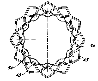
Fig. 4 shows a perspective view of an encapsulated stent 30 made on the
splined
mandrel 20. The stent 46 is composed of struts 48 arranged in a diamond
pattem.
Regions 52 at the ends of the device (marked by cross-hatching) have complete
bonding
between the two-ePTFE tubular members. This region is produced by smooth, non-
splined regions of the mandrel. Dotted lines 54 marks the position of the
splines and the
resulting regions of selective bonding. That is, the device has spaced apart
bonded
regions running the length of the open diamond regions 56. Because of this
orientation
successive tiers of diamond regions 56 along the longitudinal axis of the
device are
alternately bonded and unbonded. Fig. 6 shows a scanning electron micrograph
of an
oblique section through a longitudinally selectively bonded stent 44. A cross-
section of
the strut 48 is shown as well as a bonded region 54 and an unbonded slip
pocket 62. The
unbonded pockets 62 allow free movement of the stent struts 48. However, even
those
diamond regions 56 containing bonds 54 allow relatively unimpeded movement of
the
struts 48 because the bond 54 is only down the central part of the diamond
region 56-
relatively distant from the struts 48. Tests show that the selectively bonded
stent 30 can
be radially compressed with considerably less force than a stent that is
encapsulated by
uniformly bonding all regions were the ePTFE tubular members contact each
other. The
CA 02345669 2001-03-28
WO 00/18328 PCT/US99/22808
-20-
longitudinal bonds somewhat restrict longitudinal compression of the device as
the
bonded regions buckle less readily than unbonded ePTFE.
The longitudinal bonds 54 do restrict the side to side flexibility or
bendability of
the device to some extent. In some applications this stiffening of the device
is desirable
while in other applications one needs a stent device that is able to bend more
freely.
Increased lateral flexibility can be achieved by using a mandrel with radial
ridges rather
than longitudinal ridges as shown in Fig 7. Again the ridges 58 are spaced
apart in
relation to the strut 48 spacing in the stent to be encapsulated. If the stent
46 shown in
Fig. 4 is used, the radial ridges 58 can be spaced apart to place
circumferential bonds
through alternate tiers of diamond regions 56. The resulting device is more
bendable
laterally than the version with longitudinal bonds. In addition, the
circumferential bonds
result in a device that is more easily compressed longitudinally.
It is clear that the area and orientation of the bond regions influence the
properties of the final device. For example, a helical pattern of ridges
produces a device
with intermediate properties: it is more laterally bendable that the
longitudinally bonded
device of Fig. 4, but it has more resistance to longitudinal compression than
does a
device with circumferential bonds. The pitch of the helical pattern controls
the overall
effect with shallow pitches acting more like circumferential ridges and steep
pitches
acting more like longitudinal ridges. Multiple helices can be used with
opposing (e.g.,
clockwise and counter clockwise) producing a device that is more resistant to
lateral
bending. Virtually any combination of the described patterns can be used to
produce
devices having a preferred direction of bendability or devices that resist
longitudinal
compression in one region while permitting such compression in another.
The stent device illustrated in the above-figures is one in the stent struts
form
courses or diamond-shaped spaces in which the struts continue from course to
course to
create an extended tubular device. Stents are also available which consist of
only a
single course (or segment) of diamond-shapes. The current method can
advantageously
be used to combine a number of these segments together to make an extended
tubular
device. Frequently these single segment stents consist of an alternation of
larger and
smaller diamond shapes. For example, the segments can be arranged with large
CA 02345669 2001-03-28
WO 00/18328 PCT/US99/22808 "
-21-
diamonds touching large diamonds. Other arrangements included a "twisted"
design
wherein each successive segment is rotationally offset and an "alternating"
design
wherein altemate segment are rotated so that a given large diamond is bounded
on either
side by a small diamond. The precise properties of the resulting encapsulated
device
depend on these factors. However, the significant thing about the prior art
encapsulation
is that it produced a device that is relatively stiff and unbending.
Various adhesives (as opposed to directly adhering PTFE to PTFE)can also be
used to create the pattern of bonded regions. Fig. 8 shows a diagram of one
method for
using adhesives to create selective bonds. In a first step 32 a tubular graft
member is
placed on a support such as a mandrel. In a second step 34 a stent (or stents)
is placed
over the first graft member. In the third step 64 a coating of adhesive is
placed over the
stent graft combination. This adhesive is one that is "activatable" meaning
that the
material is not inherently sticky as it is applied. However, it can be
activated by applying
heat, light or some other energy so that it hardens or otherwise changes to
form a
permanent bond. In the next step 64 a second tubular member is placed over the
adhesive-coated stent. In the final step 66 a pattern of desired bonds is
inscribed on the
device with, for example a laser or a heated probe or a photolithographic mask
image.
The inscribing process provides energy to local regions of the structure to
activate the
adhesive and create selectively bonded regions. A number of different
activatable
adhesive materials can be used in the present invention. One such material
might be a
layer or coating of a thermoplastic such as polyethylene. This material can be
activated
by heat that melts it so that it flows into the pores of the ePTFE. After
cooling the plastic
hardens so that the PTFE of one tubular member is bonded to the other tubular
member.
Fig. 9 shows a second adhesive-based method of creating selective bonds. The
initial steps are the same as in the previous method. However, in step 68 the
adhesive
material is applied selectively to form the future pattern. This can be done,
for example,
by a screening or offset printing method. An inherently sticky adhesive can be
used or
an activatable adhesive (as in the previous method) can be employed. The
second
tubular member is applied (step 36) and the adhesive pattein is formed either
by
applying pressure (when using an inherently sticky adhesive) or by applying
pressure
followed by an activation step-for example heating to melt a thermoplastic
adhesive.
CA 02345669 2001-03-28
WO 00/18328 PCT/US99/22808
-22-
The words used in this specification to describe the invention and its various
embodiments are to be understood not only in the sense of their commonly
defined
meanings, but to include by special definition in this specification
structure, material or
acts beyond the scope of the commonly defined meanings. Thus if an element can
be
understood in the context of this specification as including more than one
meaning, then
its use in a claim must be understood as being generic to all possible
meanings
supported by the specification and by the word itself. The definitions of the
words or
elements of the following claims are, therefore, defined in this specification
to include
not only the combination of elements which are literally set forth, but all
equivalent
structure, material or acts for performing substantiallv the same function in
substantially
the same way to obtain substantially the same result.