Note: Descriptions are shown in the official language in which they were submitted.
CA 02345960 2001-03-29
WO 00/19262 PCT/US99/19086
HIGH THROUGHPUT MICROSCOPY
Technical Field
The present invention relates generally to high resolution, three dimensional,
fluorescence microscopy systems and methods for their use. More specifically
the
invention represents specific improvements to existing wide-field, optical
sectioning
microscopes systems designed for acquisition and analysis of multi-dimensional
fluorescence images.
Background Art
The field of optical microscopy has been revolutionized in recent years by the
widespread use of confocal and fluorescent microscopes. Combining laser
illumination
and digital image processing, these optical instruments allow biologists to
obtain high-
resolution, three dimensional fluorescent images.
Deconvolution microscopy, an alternative approach to laser-scanning cofocal
microscopy, is gaining in popularity. This technology avoids the high costs
and
limitations of laser illumination and is ideal for live-cell studies requiring
high resolution
and multiple wave lengths. Typical of the devices of this type is the Delta
Vision~
microscope system. Delta Vision~ system is a wide-field optical sectioning
microscope
system. See, for example, info@api.com and U.S. Patent No. 5,684,628. The
basic
concepts embodied in the Delta Vision~ system include: collection of imaging
data
directly in digital form using a high quality charge coupled device, i.e., CCD
(silicon
chip) area detector; computation removal of noise arising from light leakage
into the
focal plane from adjacent planes, provision of a facile user interface for
data capture and
analysis.
Disclosure of the Invention
T. he invention is directed to improvements to a standard wide field
microscope
system, e.g. the Delta Vision~ system. The improvements permit imaging of more
than
1,000, preferably more than 10,000 samples, e.g. antibody/antigen reactions,
per day.
The basic system to which these improvements are applied share generally the
features
of a fluorescent microscope -- i.e., excitation and emission filters, an
objective lens, a
CA 02345960 2001-03-29
WO 00/19262 - 2 - PC'T/US99/19086
movable stage, and image recording by CCD. Such basic instruments also are
able to
image transmitted light.
The features which constitute improvements to this basic design fall into
three
general categories:
tical features which constitute the improvements of the invention include a
pulsed light source, a fiber-optic light source, a computer-controlled
condenser, infinity
focusing, polychromatic beam splitting and multiple CCD detectors rather than
a single
CCD detector.
Mechanical features which comprise the improvements of the invention include a
rotatable circular disc in place of a conventional sample holder, wherein the
disc may
also contain further design features creating separate small wells; reagent
dispenser and
readout stations positioned around the disc, an anm that moves the dispenser
and readout
heads radially; a stage-tilting device, and a temperature-controlled specimen
chamber.
In addition, the improvements comprise computational features including a
control and calibration program, a nonhomogeneous illumination compensator,
chromatic aberration compensator, a Fourier space feature detector, and a real
space
feature detector.
One object of the innovation is to minimize photodamage to the samples by
pulsing the light source at a speed consistent with the rate of data
collection. Fiber optics
plumbing, pre- and post-specimen, with a rotating dichroic filter in the
illumination
path, enables more efficient automated operation. An automated condenser (e.g.
to slide
in masks for differential interference and polarization imaging) is also
advantageous.
Another aspect of the invention relates to infinity imaging. This employs an
objective lens with a sliding element to allow for switching between high and
low
magnification, a more efficient system than conventional rotating head
microscopes.
The image is further focused by sliding a secondary lens into the light path
downstream
from the objective lens. This simplifies positioning of the objective lens
since the
computer controlled motor for focusing can be placed physically out of the
way.
"Infinity imaging" also enhances the range of magnification possibilities, and
eliminates
moving parts from the proximity of the sample. This simplifies temperature
control, for
example permitting cooling of the specimen. Cooling the specimen sharpens the
emission line widths enhancing the ability to recognize differences in hues.
CA 02345960 2001-03-29
WO 00/19262 - 3 - PCT/US99/19086
A still further aspect of the invention is the use of beam splatters in
conjunction
with filters to allow the same primary light to be imaged onto multiple CCD
detectors.
Multiple detectors allow faster collection of data and more reliable image
registratic:i for
detection of multiple colors.
In mechanical features, in one aspect, the invention employs rotatable
circular
discs in place of the typical microscope slides or 96-well plates. These discs
are placed
on a rotatable stage. Typically, the disc can range in size from a 4-inch
diameter to 9 or
12 inches. The exact size of the disc is not critical. These discs are
available from the
computer chip industry. The discs can be painted with hydrophobic materials to
form
barriers which would define sample wells using lithographic techniques. The
lithographic techniques employed are similar to those taught in U.S. Patent
No.
5,212,028, the contents of which are incorporated herein by reference. Using
these
techniques, a 4-inch disc can be painted, for example, to form 2 mm segments
which
creates 1,000 sample wells and to form 1 mm segments which creates 2,500
wells.
Alternatively, adhesive lines that allow the deposit of small specimen
carriers can be
used.
Other advantageous mechanical features of the invention are related to the use
of
these multispximen discs. Thus, reagent dispensers and readout stations may be
positioned around the disc and may be fitted with arms that move the
dispensers and
read-heads radially. Other mechanical features include a stage-tilting device
and a
temperature controlled specimen chamber, facilitated by the infinity focusing
feature.
Computational aspects of the invention include dedicated circuitry and
equivalent
soffiware for computational processing in areas which include data capture,
photon
reassignment, feature detection, counting and presentation. These measures
facilitate
control and coordinate data handling. Further, the circuitry and software
equivalent
permit the recognition of signature curves of particulate labels such as latex
beads. This
would permit the system to count and identify individual tags. An alternative
filter can
be designed to read microbar codes for sample tracking.
Other filters can also be defined computationally to improve the accuracy of
cell
counting. Cell borders can be recognized by virtue of lipid soluble dye, while
definition
of the nucleus can be achieved using a DNA stain like DAPI.
CA 02345960 2001-03-29
WO 00/19262 - 4 - PCTNS99/19086
Brief Description of the Drawings
Figure 1 illustrates a high resolution fluorescence microscope of the prior
art
which includes a light source (1), objective lens (3), eye pieces (5) and
other enume:ated
elements.
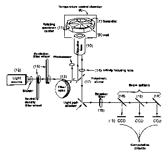
Figure 2 illustrates one embodiment of the high throughput microscope (HTM)
system of the invention.
Detailed Description of the Invention
The present invention represents an improvement over systems which include
elements, for example, present in the devices described in U.S. Patent No.
5,684,628,
incorporated herein by reference. Such fluorescent microscopes provide the
basis of
image collection. A conventional fluorescent microscope is shown in Figure 1.
A
specimen is labeled with a fluorescent tag and placed on the microscope stage
( 1 ) which,
in this conventional embodiment, is movable in an Cartesian coordinate system.
The
irradiating light is supplied from an arc lamp (2) through appropriate filters
and shutters
and through a fiber-optic system to an objective lens (3). The emitted light
is filtered
through a polychroic mirror (4) and deflected to the eye for visual inspection
(5) or to a
CCD array for detection using appropriate circuitry (6).
A modified form of the conventional system containing the improvements that
comprise the invention is shown in Figure 2. The XYZ microscope stage is
replaced by
a rotatable stage {11) (specimen carrier) which supports a circular disc (7)
specimen
holder (disc). The specimen disc is divided into individual sample
compartments (8)
(well) which may be wells separated by hydrophobic barriers or adhesive
segments.
Disc technology is now available in the semiconductor chip industry for
inspection,
handling and automation of chip treatment. The system shown in Figure 2 can
also be
modified so that the disc can be placed on multiple positioning stages and/or
on stages
provided with stage tilting devices that permit the study of the microscopy
samples at a
variety of angles reducing certain kinds of light scattered noise. The
circular specimen
disc allows for more quantitative channels for faster reading, tracking of
temporal
phenomena, and the processing of large numbers of samples in parallel. The
system can
be designed to include one or more disc cassette towers which increases
automation
possibilities. The discs can be selectively moved from a cassette tower to the
inspection
stage, selectively rotated at a variety of angles, if desired, removed from
the inspection
CA 02345960 2001-03-29
WO 00/19262 - 5 - PCT/US99/19086
stage and selectively placed in a separate cassette tower or returned to the
same position
in the original cassette tower for storage or for subsequent viewing. This can
be done
manually or automatically. See for example U.S. Patent Nos. 4,938,654;
5,096,291;
5,119,434; 5,129,009; and 5,471,066, respectively, incorporated herein by
reference.
The instrument may also be provided with a reagent dispenser and multiple
readout
stations around the circle described by the stage and disc. The dispensers and
readout
stations may be moved radially and the stage and disc rotated to provide
multiple
sampling opportunities.
The entire specimen compartment may be enclosed in a temperature controlled
environment (9), schematically indicated in Figure 2. For example, to improve
emission
spectrum resolution, very low temperatures such as those of liquid nitrogen or
liquid
helium may be used. A detailed description of a labeling system which permits
a
multiplicity of hues to be generated on a particulate support, such as latex
beads, is
described in copending application U.S. Serial No. 09/146,984 filed 3
September 1998
and incorporated herein by reference. Briefly, by varying the ratio of dyes of
primary
colors on the particulate supports, or by varying the intensity of individual
dyes, a large
number of hues can be created. Cooling to very low temperatures permits more
than ten
primary colors for use in creating these individual hues, defined as the ratio
of the
primary colors attached to the beads. At present, latex beads are made with a
doping
precision better than ~ 5%, so that even specifying only ten gray scale levels
for each of
ten primary colors provides ten billion distinguishable tags.
Of course, it is still within the invention to use conventional fluorescent
labeling
as well as the multihued fluorescent tags described in the above-referenced
copending
application.
In the embodiment wherein a closed chamber (9) encasing the sample is used,
the
objective lens (10) will generally protrude into the chamber. Eliminating the
axis
movement is the objective and thus desirable to maintaining a tight seal . The
stage
containing the disc (7} is movable independent of the objective lens. After a
specimen is
loaded with any desired fluorescent label, it is placed in a sample well (8)
of the disc and
positioned on the stage ( 11 ) and the area of interest is determined using
ordinary visual
inspection. Excitation light is then introduced to the sample through the
objective lens
(10) or through an optical fiber(s). As stated above, the temperature may be
controlled.
Warming permits one to maintain the samples within physiologically desirable
CA 02345960 2001-03-29
WO 00/19262 - 6 - PCT/US99/19086
temperature ranges. Cooling of the disc or individual sample can sharpen
emission band
widths which enhance hue resolution.
In one embodiment of the present invention the light source (12) is pulsed so
as
to control any photodamage of the sample and the excitation beam is conducted
through
the objecti~~e lens by fiber-optic conduits (13).
The the automated image collection features of the system are used to acquire
a
series of images. After deconvolution, the images are combined to form a three-
dimensional view of the specimen.
The series of images used to foam the three-dimensional view is collected by
moving the focal plane of the objective lens (10) through the specimen. This
movement,
usually called a z-scan, can be accomplished by moving either the sample or
the
objective lens. Alternatively, a secondary lens (14), in an infinity focusing
system may
be employed. The typical distance between sections ranges between 0.1 um to 5
um,
with a total range of 5 mm. In the embodiments that include infinity focusing,
the
objective lens is coupled with a secondary sliding element lens (14). This
secondary lens
is located later in the light path. This combination also facilitates the
controlled shift
between low and high magnification. Extremely precise microstepping motors are
used
to achieve the small movements necessary.
In one embodiment, the light source is a mercury light source, which provides
the
illumination from the LJV to the near-IR. This light source is capable of
being pulsed at
a speed consistent with the rate of data collection to minimize photodamage to
microscopy sample. An optical fiber optic light scrambler homogenizes the
illumination
light, smoothing out arc lamp wander and evenly filling the back aperture of
the
objective lens. Excitation and emission wave lengths appropriate for the
fluorescent
probes being used are selected by multi-cavity interference filters. These
filters transmit
the desired wavelengths, while blocking more than 99% of the light from other
regions
of the spectrum. Between the excitation (15) (excitation filter wheel; neutral
density
filter wheel) and emission filter (16) is a movable custom polychroic mirror
(17), which
is a key component for wave length selection. The mirror consists of carefully
selected
reflection and transmission bands, designed to provided optimal performance
for a wide
variety of commonly used fluorescent probes. The use of a single mirror to
image all
probes means that no artifacts are introduced from components moving within
the
critical path. This is significant since artifacts resulting from optical
component motion
CA 02345960 2001-03-29
WO 00/19262 - ~ - PCT/US99/19086
are often indistinguishable from real information and can cause
misinterpretation of the
specimen characteristics.
Precise positioning of the emission filters (15) minimizes artifacts from
wedging,
dust, and filter variation. Although such artifacts can sometimes be minimized
with
digital processing techniques, details obscured by dirty optics will simply
not be present
in the final image. Superior images will yield superior results after
deconvolution.
Precise filter positioning allows different fluorescent probes to be imaged
before moving
the specimen. This approach provides the extremely accurate image alignment
that is
critical for co-localization studies. Systems that conduct a complete z-scan
for each
probe being imaged may suffer from image registration problems.
At least one CCD detector, typically cooled and scientific grade, collects the
fluorescent images. This type of detector provides very sensitive light
detection, low
noise, and an extremely linear response to light intensity. Beam splitters
(18) in
conjunction with filters can be placed earlier in the light path relative to
the CCD
detector. This allows the same primary light to be imaged onto multiple CCD
detectors
(19). Multiple detectors permit faster collection of data and more reliable
detection of
mix-and-match colors. The use of multiple CCD detectors (19) is shown in
Figure 2.
This permits better discrimination between multihued particles and facilitates
the use of
multihued particle detection schemes.
5ma11 photon detector elements allow the image to be oversampled, increasing
the resolution possible with the system. Images acquired by the detectors are
collected
by suitable circuitry and the data managed by appropriate software to be
displayed and/or
stored in digital format.
After the images have been collected, they can be viewed and measured using a
variety of powerful tools. These tools include the capacity to rotate,
measure, highligh~
selected regions, and modify the image contrast. Deconvolution circuitry
computationally reassigns blur present in the image. After correcting for
variations due
to arc flicker, an automated deconvolution process is triggered dedicated
circuitry speed
up the computational work. Equivalent circuit can be created by programming a
more
flexible circuit or by using software to create temporary circuits.
In the data capture aspect of the invention, each CCD chip has the accessory
circuitry for data transfer and reinitialization. These need to be
synchronized with the
illumination system and specimen identifier as well as with control and
calibration
CA 02345960 2001-03-29
WO 00/19262 - g - PCT/US99/19086
functions such as checking and correcting for bad pixels or dirt on the lens.
The
associated circuitry also performs photon reassignment -- i.e., Fourier space
deconvolution. The actual photon counts are the result of a convolution of
point-spread
function and the real sample. Con: ection for the differences in focus of
different
wavelengths -- chromatic aberration -- can be most conveniently corrected
after this step.
Thus, the computation system refracts streams of photons into image pixels and
adjusts
gain on a pixel-by-pixel basis.
The use of deconvolution in Fourier space permits the identification of
certain
features such as spherical latex beads, and permits a "bar code" to be
embedded into a
specimen for readout at this step. Photolithography can be used to create one
or two
dimensional patterns on a rod or chip of plastic which could be on the sample
carrier or
added to the sample. Thus, the identification of the sample being viewed is
greatly
aided. Similarly, in a deconvoluted real-space image, particular objects can
be identified.
Cell nuclei, for example, have characteristic dimensions and are typically
separated from
each other by cell membrane.
The plethora of information generated by the HTM can be managed by the
circuitry. For example, in tissue typing, the deconvoluted image can be
collapsed into a
cartoon wherein multihued beads, each carrying a distinct typing antibody, are
only
counted if they are on the surface of white cells which are recognized as
objects with
having an outer membrane stained with a lipophilic dye, said membrane
surrounding a
nucleus stained, for example, with DAPI. They can be distinguished from beads
not on
the surface of white cells as well as those associated with nonnucleated red
cells. In this
application, the hue of the beads on a single cell surface should be
consistent.
Thus, the multiplicity of improvements provided the present invention permits
a
truly high throughput system for analysis of large numbers of samples, each
probed with
a large number of detection agents. The many applications include tissue
typing,
identification of antigen-antibody interactions, real-time observation of
intracellular
movement of components and the like. Particular applications of this
technology are
fiu~ther described in copending U.S. Serial No. 09/144,609 filed 31 August
1998 and
incorporated herein by reference.