Note: Descriptions are shown in the official language in which they were submitted.
CA 02347391 2001-01-18
WO 00/10636 PCT/US99/18878
-1-
PREFORMED WIRE GUIDE
Description
Technical Field
This invention relates generally to medical devices and, in particular, to a
wire guide.
Background of the Invention
Balloon angioplasty, a medical procedure by which an occluded or
narrowed blood vessel is dilated and reopened using an inflatable balloon
mounted
on a catheter, was pioneered by Andreas Greuntzig in the 1 970's. The coronary
version of this new procedure, Percutaneous Transluminal Coronary Angioplasty
(PTCA), soon became recognized as a highly effective method of treating
diseased
coronary artery disease. More recently, angioplasty has become a standard
approach
for treatment of renal artery stenoses. Percutaneous Transluminal Renal
Angioplasty
(PTRA), with its low rate of complications, has now largely replaced surgery
as
treatment for renal artery stenoses, which are common contributing factors in
patients diagnosed with arterial hypertension, renal insufficiency, or cardiac
insufficiency.
The basic angioplasty procedure usually involves percutaneously
introducing a guiding catheter through an introducer sheath to the target site
and
then engaging the ostium of the vessel. A wire guide is fed through the
guiding
catheter and ostium where it is placed across the lesion in the vessel.
Finally, a
balloon catheter is introduced over the wire guide and positioned at the
lesion to
dilate the vessel. Increasingly more often, a stent is also placed following
balloon
dilatation to prevent restenoses of the lesion. One procedure for placing the
balloon
catheter at the treatment site is known as the "Push-Pull" Technique whereby
the
physician advances the balloon catheter through the guiding catheter ("push")
while
applying slight forward pressure to the latter. At the same time, an assistant
holds
the proximal end of the wire guide, providing gentle traction ("pull"). Care
must be
taken during the advancement of the catheter to avoid dislodging the wire
guide from
25-09-2000 CA 02347391 2001-01-18
PCT/US99/18878
PCT REPLACEMENT
-2-
the treatment site. This is especially of concern during a renal procedure due
to the
relatively short length of the renal artery and the acute angle of the artery
relative
to the aorta.
The unique anatomy of the renal vessels presents difficulties when using
existing wire guides for PTRA. Many physicians select wire guides developed
for
coronary procedures which are designed to facilitate negotiation of tortuous
vessels
and minimize trauma to small delicate coronary arteries. Because of their
required
flexibility, coronary wire guides usually lack the desired stiffness for PTRA.
A stiffer
wire guide permits better tracking by the catheter over the wire. However, a
stiff
wire guide can also subject the vasculature to forces during manipulation that
are
capable of perforating the vessel or injuring the ostial takeoff from the
aorta into the
renal vessel. The wire guide receives much of the up and down stresses during
the
procedure and transfers them to the vessel wall. These same stresses are often
responsible for dislodging the distal end of the wire guide from the orifice,
necessitating withdrawal of the catheter and reintroduction of the wire guide.
If the
wire guide enters the ostium of the vessel at the correct angle, the stresses
are
instead received by the catheter, thus protecting the fragile vessel.
Furthermore, the
typical stresses at that site during manipulation of a straight wire can also
cause
thrombus to shear from the vessel wall, often leading to an embolus and
related
serious complications.
One prior art wire guide is disclosed in US-A-5295493 to be of solid
flexible wire such as of stainless steel, that is formed to have a preformed
shape
conforming when unstressed to the general anatomical shape of the particular
segment of a vessel that has a stenosis to be removed in an atherectomy.
Another
prior art guidewire is disclosed in US-A-5238004 and at least the distal
portion is
formed from a precursor of a superelastic alloy (e.g., nitinol) by cold
drawing; the
guidewire has a solid core with a tapered elongate distal tip portion that is
elastic
and deformable, and a highly flexible spring coil wire is secured about the
distal tip
portion, such that the distal tip portion can be manually shaped into a
curvature to
complement the curvature of the lumen of the patient.
AMENDED SHEET
CA 02347391 2007-08-20
-2a-
Summary of the Invention
Certain exemplary embodiments can provide a wire guide comprising a
mandril, and a tip portion disposed at an end portion of the mandril, wherein
the
mandril includes at least one preformed bend disposed along the end portion
for
anchoring the wire guide in a vessel, wherein the mandril is of superelastic
material, and that the preformed bend includes a localized martensitic region.
CA 02347391 2007-08-20
-3-
Other embodiments provide a preformed wire guide having a mandril with a
flexible tip
portion that is atraumatic to the vessel as the wire guide is advanced, the
flexible tip portion
having a distal tip and a proximal portion that includes a preformed bend
approximating the takeoff angle of a vessel, for example, a renal artery
relative to
the aorta from which it branches. By producing a wire guide with the correct
anatomical preformed bend, there is much less risk of trauma to the vessel. A
related benefit of the present invention is lowering the risk of displacing
thrombus
that often forms just inside the ostium, especially in the presence of a
stenotic
lesion. A straight wire would receive much of the force at the turn into the
ostium
created by the advancing catheter and potentially transfer much of that force
to the
wall of the vessel. By forming the bend in the wire guide, the forces created
from
the catheter tracking over the wire are exerted on the catheter itself and not
to the
vessel wall where injury or disruption of thrombus can occur. Nitinol can be
permanently shaped by annealing with extreme heat, or by cold-working which
involves overstressing the wire. To produce a more rigid bend segment for
protecting the vessel, cold working the nitinol mandril is preferred over the
annealed
embodiment which exhibits less resistance to the tracking forces of the
catheter.
The second major benefit of having an anatomically shaped preformed
bend is providing a portion of the wire guide to serve as an anchor to
maintain the
device within the vessel during advancement of a catheter over the wire. A
straight
wire guide would be much more likely to become dislodged during the course of
tracking the catheter to the treatment site.
In a preferred embodiment of the illustrative invention, the flexible tip
portion includes a spring coil wire that is attached over a solid wire
mandril. The
transition between the highly-flexible atraumatic tip and the stiffer mandril
is
relatively abrupt, compared to typical wire guides, due to the short available
length
of vessel in which the anchoring portion of the mandril can reside and the
need for
that mandril to be of sufficient stiffness to maintain a proper anchor. A bend
having
a preferred range of 30 to 150 formed in the mandril wire allows the wire
guide
25-09-2000 RCT CA 02347391 2001-01-18 REPLACEMENT PCT/US99/18878
-4-
to more easily enter the ostium of the renal artery or vein, depending on the
particular anatomy of the patient, and whether a superior or inferior approach
is
used. A more preferred range of bend angles is 450 to 1350, with the most
preferred range being 600 to 120 . The improved ability to access the renal
vessel
can reduce the need for using a guiding catheter to place the wire guide,
thereby
eliminating a step of the procedure and the attendant risks.
The solid mandril wire is of sufficient stiffness to retain the anatomical
preformed bend and allow the wire guide to remain anchored in the vessel while
a
catheter is being fed over the wire. In the preferred embodiment of the
invention,
the mandril wire is made of a superelastic material such as a nickel-titanium
(Ni-Ti)
alloy (commercially available as nitinol). The bend in the mandril is formed
by
mechanically stressing (cold working) and plastically deforming the wire while
in its
austenitic state to create at least a partial localized zone of martensite.
The nitinol
wire can be made relatively thin while still retaining the preformed bend and
the
requisite stiffness. Other possible materials for the mandril include elastic
biocompatible metals such as stainless steel, titanium, or tantalum. While the
potential benefits of cold working nitinol wire to plastically deform the
original shape
have not been fully appreciated by manufacturers of wire guides and other
medical
devices, there are two primary advantages over the standard annealing method.
The
first involves the differences in how the device behaves as bending stresses
are
applied. In the absence of applied stress, the annealed wire guide is
completely in
an austenitic state, even in the curved regions. When sufficient stress is
applied
anywhere upon the length of the device, the face-centered crystals of the
austenitic
material shift to martensite until the stress is removed. Thus, the bend and
straight
portions of the annealed wire guide have very similar flexural properties. In
contrast,
the cold-worked wire guide is comprised of regions of both austenite and
martensite
along its length. Consequently, the preformed bend of a cold-worked renal wire
guide remains in at least a partial martensitic state and does not exhibit the
unusual
superelastic phenomenon that occurs during an austenitic to martensitic
transformation.
AMENDED SHEET
25-09-2000 CA 02347391 2001-01-18 PCT/US99/18878
PCT REPLACEMENT rr1Qr-
-5-
To provide maximum protection to the renal vessels during a procedure,
the flexible tip portion of the preferred embodiment has a curved shape. The
"J"-tip
of the illustrative embodiment protects the vessel and delicate tissues as the
wire
guide is advanced into the renal vein. A curved shape tip is more easily
deflected
and prevents the stiff mandril wire from exerting a dangerous amount of force
against the vessel wall. The transition from a flexible tip to the stiffer
mandril is
achieved by soldering the spring coil tip to the tapered end of the mandril at
the
point where the taper begins. The tapered distal end of the mandril provides
the
overlapping coiled portion with a diminishing degree of stiffness toward its
distal
end.
In the illustrative embodiment, a polymer coating is added to the mandril
of the wire guide for improved lubricity. Polytetrafluoroethylene (PTFE) is
the
preferred material; however, hydrophilic coatings such as SLIP-COATTM'
(Sterilization
Technical Services, Inc., Rush, NY) can be used as an alternative material as
well as
other lubricious coatings or coating materials.
Brief Description of the Drawing
FIG. 1 depicts a side view of the illustrative wire guide of the present
invention;
FIG. 2 depicts an alternative embodiment of the flexible tip portion of the
wire guide of FIG. 1;
FIG. 3 depicts a cross-sectional view of the embodiment of FIG. 1 along
line 3-3;
FIG. 4 depicts a second preferred embodiment of the illustrative wire guide
of the present invention;
FIG. 5 depicts a schematic view of a third embodiment of the wire guide
of the present invention located within the renal system of a patient;
FIG. 6 depicts an enlarged, partially sectioned side view of the distal
portion of the wire guide of FIG. 1; and
AMENDED SHEET
25-09-2000 CA 02347391 2001-01-18 PCT/US99/18878
rr+-: i i v PCT . REPLACEMENT rr~c
-6-
FIG. 7 graphically depicts stress-strain curves for cold-worked nitinol wire
and for annealed nitinol wire.
Detailed Description
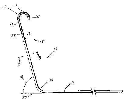
FIG. 1 depicts a side view of an illustrative embodiment of wire guide 10
of the present invention. The wire guide 10 includes both a mandril 11 and a
tip
portion 12, preferably a flexible tip portion 12, extending proximally from
the distal
tip 30 of the wire guide. In the preferred embodiment, the mandril 11 extends
the
entire length of the wire guide with distal end 25 of the flexible tip portion
12
extending from distal tip 30 of the wire guide to proximal end 26 of the
flexible tip
portion 12 and to solder joint 13. The mandril 11 includes a preformed bend 14
that
marks the beginning of a distal portion 27 of the wire guide. Angling the
distal
portion 27 facilitates entry of the wire guide into the ostium of the renal
artery. The
distal portion 27 becomes an anchor to help prevent dislodgment of the wire
after
it has been placed. The wire guide is also anatomically shaped for procedures
involving the renal vein, however these are far less common. The takeoff of
the
renal artery from the aorta varies in its angle. Therefore, it is contemplated
that the
wire guide be made available with different bend angles to accommodate the
normal
variation in patient anatomy. An additional factor is that the wire guide can
be
introduced using either an inferior approach via the femoral artery
(preferred) or a
superior approach, typically via a brachial access site. The wire guide bend
angles
can range from 300 to 1501, with a more preferred range of 450 to 1350. The
distal portion 27 of the first illustrative embodiment is bent at an angle 15
of
approximately 600 relative to the longitudinal axis 28 of the wire guide 10. A
second embodiment depicted in FIG. 4 has a preformed bend 14 with an angle 15
of approximately 120 . Together, these two embodiments represent the most
common, and therefore, most preferred range of angles for accessing the renal
artery. A third preferred embodiment is depicted in FIG. 5 whereby the distal
portion
27 of the wire guide 10 is formed at a 90 angle.
In the preferred embodiment, the portion of the mandril 11 proximal to the
flexible tip portion 12 is comprised of a mandril core 18 and a microthin
polymer
AMENDED SHEET
25-09-2000 CA 02347391 2001-01-18 PCT/US99/18878
rH-;. i i a PCT REPLACEMENT t'ivvt
-7-
outer coating 19 such as polytetrafluoroethylene (PTFE) as depicted in FIG. 3.
Alternative coatings include hydrophilic materials such as SLIP-COATTM'
polymers
(Sterilization Technical Services, Inc., Rush, NY) or other polymers that have
been
surface treated to increase lubricity. Preferably, the mandril core 18
includes
material having superelastic properties such as the Ni-Ti alloy commercially
known
and available as nitinol. Nitinol is comprised of nearly equal parts of nickel
and
titanium and can also include small amounts of other metals such as vanadium,
chromium, or iron to affect the physical properties of the alloy. The
preferred nitinol
formulation for this application has a martensitic to austenitic
transformation
temperature below body temperature, and most preferably, below normal room
temperature. The remarkable ability of a superelastic alloy to return to its
predetermined shape when subjected to stress, makes it an excellent material
for
this application. Although stainless steel and other non-superelastic
materials can
be used, they are less resilient. In the case of the present invention where
the shape
of the wire guide is matched to the anatomical site in which it is used, the
plastic
deformation that can occur with ordinary metal wires during manipulation can
affect
the efficacy of the device. In addition to nitinol, superelastic or
pseudoelastic copper
alloys, such as Cu-Al-Ni, Cu-AI-Zi, and Cu-Zi are available as alternative
wire guide
materials. The preferred diameter for the wire guide ranges from about .010 to
.035
in (0.254 to 0.889 mm) with a diameter of approximately .018 in (0.457 mm),
mostly comprised of the nitinol metallic core 18, being generally preferred
when
using a single diameter wire guide. Another embodiment includes making the
mandril 11 larger in diameter, e.g., .023 in (0.584 mm), and attenuating the
tip 12
to .018 in (0.457 mm). The larger mandril provides better positional support
for
placement in the renal vessel, while attenuation of the distal portion 27
advantageously provides a substantially atraumatic tip. The coating 19, which
is
approximately .003 .001 in (0.0762 0.0254 mm) thick in the illustrative
embodiment, serves to lower the coefficient of friction and ease manipulation
of the
wire guide within the vessel or guiding catheter, if the latter is used.
Because of the superelasticity of nitinol, permanently deforming the
material to produce the desired bend in the wire requires special
manufacturing
AMENDED SHEET
CA 02347391 2007-08-20
-7a-
techniques. The standard method of forming nitinol into a desired shape is
disclosed in U.S.
Patent No. 5,597,378 and 4,665,906 to Jervis, both entitled "Medical Devices
Incorporating
SIM Alloy Elements". The basic procedure involves maintaining the device in
the desired final
shape while subjecting it to extreme heat for a prescribed period of time.
Stressing
the wire guide under annealing temperatures "locks" the curve in an austenitic
state.
When the annealed wire guide is deflected, there is a localized, transient
shift of the
austenitic material to martensite, known as stress-induced martensite (SIM).
While
annealing represents a viable method of producing the specific bend in the
present
invention, the preferred method involves cold working the wire guide, i.e.,
reshaping
the wire guide by the application of sufficient mechanical force to
permanently shift
a portion of the crystalline structure of the nitinol from austenite to
martensite within
the region of the preformed bend. Given the high degree of resilience of the
austenitic nitinol, the stress required to permanently deform the device to
the degree
required is considerable. One method of cold working the nitinol wire involves
using
a fixture or forming tool which holds the wire and includes a pin around which
the
wire is deformed into a much tighter angle than the final angle. The diameter
of the
pin, the position of wire within the fixture, and the degree of force applied
determine
CA 02347391 2001-01-18
WO 00/10636 PCT/US99/18878
-8-
the tightness of the resulting bend. By using predetermined wire and fixture
parameters, it is possible to achieve a predictable angle of bend using such a
forming
tool to overstress the nitinol wire.
FIG. 7 graphically depicts the generalized stress-strain curves 35 and 36
for similar wires made from cold-worked nitinol and annealed nitinol 35 and
36,
respectively. As stress 37 is applied to the cold-worked nitinol wire 35,
there is an
initial resistance 38 to the increase in strain 39. At a point 40 in the cold-
worked
nitinol curve, further stress produces a more linear increase in strain. The
annealed
nitinol curve 36 exhibits the traditional SIM stress-strain curve whereby
following an
initial resistance to strain exhibited by portion 41 of the curve, the
material enters the
stress-induced martensitic phase, depicted by portion 42 of the curve. During
this
SIM phase, the device can continue to deflect (strain) with minimal
application of
additional stress. At a certain point in the curve 43, the stress-strain
relationship for
the material becomes much more linear. Both processes produce a device with
nitinol's superelastic properties, yet the preformed bend of the annealed
device
becomes highly flexible when subjected to stress and undergoes the phase
change
The stiffer preformed bend of the cold-worked device is ideal for the renal
wire guide
because of its dual function as an anchor into the renal artery and a track
over which
a catheter is guided. While increased flexibility can be an advantage for
certain
medical applications, a more flexible annealed wire guide would be more likely
to
dislodge from the vessel as the PTRA balloon catheter is tracking over the
guide.
The second advantage of cold working the bend of the wire guide of the present
invention is that stock polymer-coated nitinol wire can be used to manufacture
the
finished device. The high temperatures required to produce the annealed wire
guide
preclude using the pre-coated wire stock since the polymer coating cannot
withstand
the temperatures used in the annealing process. This means that virtually any
coatings or treatment must be performed by the manufacturer as a final step.
Cold
working allows a manufacturer the flexibility to purchase pre-coated nitinol
wire
stock, easily customizing the shape of the stock or existing straight wire
guides for
a given application, and doing so at a lower cost.
CA 02347391 2007-08-20
-9-
The fiexible tip portion 12 of the wire guide 10 provides a distal tip 30 that
is atraumatic to the vessel and far less likely to damage delicate tissues
during
introduction and positioning of the wire guide. In the illustrative
embodiment, the
flexible tip portion 12 comprises a segment of spring coil wire 16 with
closely
adjacent turns. Platinum wire is used to make the distal end of the device
highly
visible under fluoroscopy. Other possible radiopaque materials include gold,
tantalum, or tungsten. Radiolucent materials such as stainless steel can also
be
used. The poor imaging disadvantage can be overcome if a second radiopaque
material is used in conjunction with the stainless steel such as at the tip or
being
interwound with the stainless steel coil. A surface treatment can also be used
to
make the coil radiopaque or echogenic. The distal tip 30 of the coiled
flexible portion
terminates in a solder tip that is ground into a rounded shape and then buffed
to
minimize potential trauma. The solder joint 13 that joins the coiled, flexible
portion
to the mandril is made through a process that is fully described in U.S.
Patent No.
5,242,759 to Hall entitled, "Joint, a Laminate, and a Nickel-Titanium Alloy
Member
Surface for Bonding to Another Layer of Metal".
Preferably, the distal end 25 of the flexible tip portion 12 includes a curve
31 to reduce the likelihood of trauma caused from the advancing wire guide. In
the
illustrative embodiment, the curve 31 comprises a hook-shaped tip 29, such as
a "J"
or "Shepard's crook". Directing the distal tip 30 of flexible portion away
from the
distal end 25 of the wire guide provides a higher degree of protection against
damaging tissue compared to the concentrated force that is potentially exerted
by
a forward-directed tip, even though the tip is made to flex with contact. FIG.
2
depicts an alternative atraumatic flexible tip portion 12 that contains a
curve 31 of
approximately 45 that causes the distal tip 30 to laterally deflect when it
encounters resistance.
FIG. 6 depicts an enlarged, partially sectioned side view of the flexible tip
portion 12 of the illustrative wire guide 10 of FIG. 1. In the preferred
embodiment
shown, one end portion 20 of the mandril 11 includes a tapered distal portion
20
wherein the taper begins at the point 13 at where the coiled, flexible tip
portion 12
CA 02347391 2001-01-18
WO 00/10636 PCT/US99/18878
-10-
is soldered to the mandril. The taper continues to soldered distal tip 30 at
the distal
end of the mandril. The taper is produced by performing a centerless grind of
the
nitinol core 18, a process which also removes the existing PTFE coating. In
the
preferred embodiment, the reduction in diameter of the tapered distal portion
20 is
gradual across its entire length. Alternatively, the overall taper can be
accomplished
in a stepped manner with an alternating series of tapered and straight
portions. The
taper both permits the flexible portion to attach relatively flush to the
coated mandril
wire such that the outside diameter of the wire guide remains constant across
its
entire length, and imparts an increasing degree of flexibility to the flexible
portion of
the wire guide. In an embodiment in which the flexible portion has a smaller
diameter
than the mandril core, the taper of the mandril normally begins prior to the
attachment point of the flexible portion. While the flexible portion can be
soldered
to the distal end of the mandril, usually making a standard safety wire
necessary so
that the flexible portion remains secured to the mandril, the result would be
a tip of
uniform flexibility that would provide less protection to the patient from the
much
stiffer advancing mandril wire. The coiled wire 16 of the flexible tip portion
12
assumes the shape of the shaped tapered distal portion 20 and would otherwise
comprise a straight segment at the distal end of the device. Creating a curve
31
such as the "J"-shaped hook 29 at the distal end 25 of the wire guide is
accomplished in similar manner as the anatomical preformed bend 14 of the
mandril
(depicted in FIG. 1). If the core comprises nitinol, the distal tapered
portion 20 is
formed into a curve 31 by overstressing the wire over a forming tool to
produce the
desired final preformed bend. As with the more proximal anatomical preformed
bend
14, the distal bend 32 in the tapered nitinol portion 20 undergoes at least a
partial
localized phase shift to martensite due to the mechanical stress. Similarly,
the distal
bend 32 in the tapered portion differs in structure from stress-induced
martensite
produced by the combination of heat and mechanical stress, although the latter
technique is also an alternative method of forming the distal bend 32.
Although
there are benefits to having a coiled, flexible tip at the distal end, namely
providing
radiopacity and allowing the distal portion of the device to have the same
diameter
as the mandrii portion, a wire guide that lacks the coiled portion would
represent a
CA 02347391 2001-01-18
WO 00/10636 PCTIUS99/18878
-11-
viable alternative embodiment. The primary requirement is that the distal
portion is
sufficiently flexible to be atraumatic to tissue, whether by tapering or other
structural
modifications.
FIG. 5 depicts partially-sectioned view of the wire guide 10 of the present
invention placed within the renal anatomy of a patient to illustrate its use.
As
shown, the distal portion 27 of the wire guide is anchored within the renal
artery 23
which supplies the right kidney 24. The preformed bend 14 of mandril portion,
which is at a 90 angle in this particular embodiment, is situated at the
ostium 22
where the aorta 33 feeds into the renal artery. The flexible tip portion 12 of
the wire
guide lies distal to the ostium 22 within the renal artery 23 and usually
extends to
a point proximal to where the renal artery branches to form the interlobar
arteries 34.
The distal portion 27 of the wire guide, approximately 3 to 13 cm length and
most
preferably around 7 cm for most patients, provides a firm anchor to resist
dislodgment when a PTRA catheter 21 is fed over the wire to dilate a stenosis
17 of
the renal artery. This is especially critical as the advancing catheter nears
the ostium
22.
Any undisclosed or incidental details of the construction or composition of
the various elements of the disclosed embodiment of the present invention are
not
believed to be critical to the achievement of the advantages of the present
invention,
so long as the elements possess the strength or flexibility needed for them to
perform
as disclosed. The selection of these and other details of construction are
believed
to be well within the ability of one of even rudimentary skills in this area,
in view of
the present disclosure.