Note: Descriptions are shown in the official language in which they were submitted.
CA 02347828 2001-04-23
WO 00/24419 PCT/I>S99/24182
- 1 -
METHODS FOR ENHANCING WOUND HEALING
Technical Field
The invention relates to a method for enhancing
wound healing.
Background of the Invention
Immobilization is a basic therapeutic principle in
wound healing, common to the treatment of lesions of all
kinds. Casts, plate , and sutures minimize the negative
effects of muscle tension on healing tissues. Since
tension is one of the chief factors determining the
degree of scar formation, this principle also holds true
in skin lesions. The carefully-planned execution of an
elective skin incision frequently achieves the best
aesthetic result.
Surgeons have been seeking techniques and methods
to reduce excessive scar formation, especially in the
face. Many approaches have been undertaken to overcome
the negative influence of muscular tension on the wound
healing process, including various suture techniques,
steroid injections, undermining wound edges, and placing
incisions in a line parallel to relaxed skin tension
lines (RSTLs).
The etiology of skin tension lines, first
described more than a century ago, has been subject to
controversy over the years. There is general agreement,
however, that skin tension lines influence the healing of
incisions according to their relative positions. There
is evidence that the formation of RSTLs is a dynamic
process over time. Studies on fetal calves and human
fetal skin suggest that RSTLs are not genetically
determined, but represent a r_hange of texture of the skin
CA 02347828 2001-04-23
WO 00/24419 PCT/US99/24182,
- 2 -
secondary to extrinsic and/or intrinsic forces. Lorenz,
H.P. et al., Development, 114(1):253-259, (1992). This
change in texture gives skin certain mechanical
characteristics that are retained even when excised.
Muscle tension is thought to be a major factor in the
formation of RSTLs.
Increased skin tension has a negative effect on
wound healing, causing hypertrophic scars or wound
dehiscence. See, for example, Sherris, D.A. et al.,
Otolarynctologic Clinics of North America, 28(5):1957-
1968, 1995. Repeated microtrauma, caused by continuous
displacement of injured tissue, induces a prolonged
inflammatory response and an increased metabolic activity
during the healing process. As a consequence,
extracellular deposition of collagen and
glycosaminoglycans can intensify and lead to hypertrophic
scars. The incidence of hypertrophic scars is higher in
certain anatomic areas where there is increased muscular
movement. McCarthy, J.G., Plastic Surgery, 1990, Vol. 1,
Philadelphia, WB Saunders, page 44.
Summary of the Invention
The invention is based, in part, on a new therapy
for management of both traumatic and iatrogenic wounds,
which includes the elimination of the tension acting on
the wound. The new therapy includes injection of a
chemodenervating agent to paralyze muscles capable of
exerting tension on such wounds, providing better wound
healing with minimal scar development. In addition,
early immobilization in elective procedures also allows a
surgeon to use finer sutures, further improving the
cosmetic result.
In one aspect, the invention features a method for
treating a patient having a wound (e. g., a facial wound).
The method includes locally administering an amount of a
CA 02347828 2001-04-23
WO 00/24419 PCTlUS99/24182
- 3 -
chemodenervating agent such that healing of the wound is
enhanced. The chemodenervating agent can be, for
example, a botulinum toxin, saxitoxin, tetanus toxin, or
tetrodotoxin, and is typically administered by injection.
The botulinum toxin can be botulinum toxin A, B, C, D, E,
F, or G, and in particular botulinum toxin A or B. The
method further can include administering an amount of a
local anesthetic agent and/or a local vasoconstrictive
agent effective to enhance wound healing. Local
anesthetic agents such as lidocaine, bupivacaine, or
mepivacaine, or local vasoconstrictive agents can be
administered prior to injection with the chemodenervating
agent or simultaneously with the chemodenervating agent.
A composition having a chemodenervating agent, a
1.5 local anesthetic, and a local vasoconstrictive agent also
is featured.
In another aspect, the invention features an
article of manufacture that includes packaging material
and an amount of a chemodenervating agent. The packaging
~:0 material includes a label that indicates the
chemodenervating agent is useful for treating a patient
having a wound. Administration of the chemodenervating
agent enhances healing of the wound. The
chemodenervating agent can be a botulinum toxin such as
a?5 botulinum toxin A. The article of manufacture also can
include a local anesthetic agent or a vasconstrictive
agent.
Unless otherwise defined, all technical and
scientific germs used herein have the same meaning as
:30 commonly understood by one of ordinary skill in the art
to which this invention belongs. Although methods and
materials similar or equivalent to those described herein
can be used to practice the invention, suitable methods
and materials are described below. All publications,
:35 patent appl_Lcations, patents, and other references
CA 02347828 2001-04-23
WO 00/24419 PCT/US99/24182
- 4 -
mentioned herein are incorporated by reference in their
entirety. In case of conflict, the present
specification, including definitions, will control. In
addition, the materials, methods, and examples are
illustrative only and not intended to be limiting.
Other features and advantages of the invention
will be apparent from the following detailed description,
and from the claims.
Brief Description of the Drawings
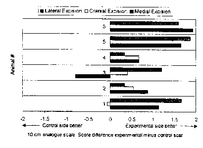
Figure 1 is a graph that indicates the mean
differences of the scores of the paired experimental and
control scars across three observers.
Detailed Description
As described herein, the cosmetic appearance of a
scar is influenced by underlying muscle activity during
the wound healing process. Paralysis of the underlying
muscle activity increases the rate of healing and yields
a better cosmetic result. Without being bound by a
particular mechanism, locally induced paralysis of the
musculature subjacent to a cutaneous defect is thought to
minimize the repetitive tensile forces on the wound
edges, resulting in superior cosmetic outcome in the
resultant scar.
Thus, the invention provides a method for treating
a patient having a wound that includes locally
administering an amount of a chemodenervating agent
effective to enhance wound healing in the patient. As
used herein, "chemodenervating agent" refers to any agent
that interrupts nerve impulse transmission across the
neuromuscular junction, blocks the release of
neurotransmitters, or alters the action potential at the
voltage gated sodium channel of neurons, sufficient to
reduce tension within muscles in and near a wound site.
CA 02347828 2001-04-23
WO 00/24419 PCT/US99/24182
As used herein, "wound'" refers to skin, tendon, or bone
wounds, and can include inflammatory lesions or other
lesions adversely affected by muscle tension or movement.
Skin wounds :include, for example, facial lacerations such
as those introduced by trauma (i.e., a car accident), or
iatrogenic, such as surgically introduced incisions. In
particular, ;surgically introduced incisions include scar
revision excision surgery. As such, a skin wound
includes elective incisions and nonelective incisions.
Skin wounds may be relatively favorable or unfavorable.
As used herein, "favorable wound" refers to an incision
or laceration that is relatively parallel to RSTLs,
whereas "unfavorable wound" refers to an incision
relatively perpendicular to RSTLs. Both favorable and
unfavorable wounds benefit from the methods described
herein. Tendon wounds include, for example, ruptured or
injured tendons and tendinitis.
Bone wounds include favorable and unfavorable
fractures. .A "favorable fracture" refers to a fracture
that is not :prone to displacement of one or more
fragments of the fracture by muscle pull, whereas an
"unfavorable fracture" refers to a fracture that is prone
to displacement of one or more fragments by muscle pull.
The treatment for a fracture ~~an be facilitated if muscle
tension on the affected fracture is minimized. Thus, the
treatment becomes less invasive, less time consuming
and/or less costly. For example, with a fractured elbow,
the triceps muscle can displace the bone fragments. An
alternative to surgical repair includes use of
percutaneous wires to hold the bones in place, and
relaxation of the triceps muscle by paralysis with a
chemodenervating agent. Use of wires and a
chemodenervating agent may reduce or avoid surgery and/or
the accompanying general anesthesia.
CA 02347828 2001-04-23
WO 00/24419 PCT/US99/24182
- 6 -
The methods described herein enhance wound healing
by minimizing the adverse effect of muscle tension and
movement on the wound, as well as improving cosmetic
appearance through reduced scar development. In
addition, inflammation may be reduced during the healing
process.
Chemodenervating Agents
Non-limiting examples of chemodenervating agents
include botulinum toxin, saxitoxin, tetanus toxin, and
tetrodotoxin. Suitable botulinum toxins include, for
example, botulinum toxins A, B, C (Cl and C2), D, E, F,
or G. Botulinum toxins A, B, and F are particularly
useful. Botulinum toxin A is a potent drug that produces
temporary muscular paralysis when injected locally.
Botulinum toxin A has been used in the treatment of a
wide range of disorders associated with involuntary
muscle contraction. It has been demonstrated to be
effective in treating focal dystonias such as
blepharospasm, nondystonic disorders such as hemifacial
spasms, disorders of conjugate eye movement such as
strabismus and nystagmus, spasticity disorders such as
multiple sclerosis and cerebral palsy, and for disorders
of localized muscle spasm. In addition, botulinum toxin
A has been used to treat age related rhytids of the upper
face. Botulinum toxin A is safe and effective to use,
and is relatively painless with rare side effects
characterized as mild and transient. Onset of action
takes place within 24 to 72 hours after injection and
lasts 2 to 6 months. Botulinum toxin A is available
commercially, e.g. from Allergan, Inc. (Irvine, CA,
Botox~) and Speywood Pharmaceuticals (England, Dysport°).
Dosages of botulinum toxin A required for local
immobilization typically do not exceed 1 unit toxin per
kg body weight and are safe. Primate studies have
CA 02347828 2001-04-23
WO 00/24419 PCT/I,fS99/24182
7 _
indicated that no systemic effects are observed at
dosages below 33 units/kg body weight. See, for example,
Scott and Suzuki, Mov. Disord., 1988, 3:333-335.
Botulinum toxins B and F also have been used for
dystonia patients. Greene, P.:~. et al., Mov. Disord.,
1996, 11(2):181-184; and Truong, D.D. et al., Mov.
Disord., 1997, 12(5):772--775. Botulinum toxin B is
available from Elan Corporation (Dublin, Ireland,
Neurobloc~).
Botul~.num toxins also can be obtained by purifying
the toxins from strains of Clostridium botulinum, using
standard techniques. For example, botulinum toxin A can
be produced in a Hall strain using a nutritive medium
containing casein digest, yeast extract, and dextrose.
After lysis of the culture, the toxin is released into
the medium and activated by proteases, and then is acid
precipitated. Further purification can include
extraction with a sodium phosphate buffer, ethanol
precipitation., and crystallization in ammonium sulfate.
2c) See, for example, Schantz, E.J. and Johnson, E.A.,
Microbiol. Rev., 1992, 56(1):80-99.
Other chemodenervating agents such as saxitoxin,
tetanus toxin., and tetrodotoxin are also suitable. The
paralysis induced by saxitoxin, however, does not last as
long as that induced by botulinum toxin. Consequently,
repeated injections of saxitoxin may be needed.
Saxitoxin can be purified by known procedures. See, for
example, Schantz, E.J. et al., J. Am. Chem. Soc., 1975,
97:1238-1239. Tetanus toxin can decrease acetylcholine
3~ release in cholinergic peripheral nerves when injected
locally. Dreyer, F., Peripheral actions of tetanus
toxin, p. 179-202, In: Botulinum neurotoxin and tetanus
toxin. Academic Press, Inc., San Diego. L.L. Simpson
(ed.). Tetanus toxin also can enter the central nervous
system where it causes uncontrolled muscle spasms. When
CA 02347828 2001-04-23
WO 00/24419 PCT/US99124182
_ g _
tetanus toxin is employed in the methods described
herein, precautions must be taken to ensure local
response. Matsuda, M. et al., Biochem. Biophys Res.
Commun., 1982, 104:799-805; and Habermann, E. et al.,
Naunyn-Schmiedeberg's Arch. Pharmacol , 1980, 311:33-40.
Tetanus toxin can be purified by standard procedures.
See, for example, Robinson, J.P., Methods Enzymol., 1988,
165:85-90. Tetrodotoxin blocks the sodium channel of
excitable membranes of nerve and muscle tissues, and can
be purified using routine techniques. See, for example,
Yotsu, M. et al., Toxicon, 1987, 25:225-228.
Local administration of the chemodenervating
agents typically occurs by subcutaneous (SQ),
intramuscular (IM), perimuscular injection, or
percutaneous instillation (e. g., air gun or skin patch).
When chemodenervating agents are injected SQ, the agent
reaches the muscle by perfusion. For elective incisions,
the chemodenervating agent can be administered prior to
making an incision, while making an incision, or after an
incision has been made.
Administration of Local Anesthetics and Local
Vasoconstrictive Agents
The method of treatment further can include
administering either a local anesthetic agent or a local
vasoconstrictive agent, or both. Such agents can be
administered prior to injection of the chemodenervating
agent or simultaneously with the injection of the
chemodenervating agent. Local anesthetics block nerve
conduction, and can cause sensory and motor paralysis in
a localized area. Local anesthetics have a rapid onset
of action, and therefore reduce muscle tension on the
wound almost immediately as well as reduce pain
associated with the injection. The extent of muscular
paralysis achieved by a local anesthetic agent is helpful
in predicting the extent of paralysis that can be
CA 02347828 2001-04-23
WO 00/24419 PCT/US99/24182
_ g _
achieved by subsequent injection of a chemodenervating
agent into the same injection site. Thus, possible local
side effects, such as diffusion of the chemodenervating
agent to adjacent muscle groups, is prevented. Non-
limiting examples of local anesthetic agents include
lidocaine, bupivacaine, chloroporcaine etidocaine, or
mepivacaine, and are available commercially. In
addition, other amide types of local anesthetics can be
used in the method. Suitable amounts of local
le) anesthetics c:an be readily determined by a physician.
For example, about 1 to 5 mls of lidocaine at a
concentration of about 0.5%-about 2% can be injected.
Administration of local anesthetics is particularly
useful when incisions are introduced surgically, such as
1!~ during scar reversion excision surgery.
Administration of a local vasoconstrictive agent
results in a decreased hemoperfusion of the injected
tissue. Thus, administration of a local vasoconstrictive
agent can he:Lp prevent or control diffusion of the
20 chemodenervat~ing agent and minimize possible side
effects, such as brow ptosis ar incomplete eye closure
from injection into the frontalis and/or corrugator
supercilii muscles. Non-limiting examples of local
vasoconstrictive agents include epinephrine and
25 phenylephrin~~, and are available commercially. A
suitable amount of a local vasoconstrictive agent can be
readily determined by a physician. For example, 5 mls of
epinephrine 1:100,000 or 1:200,000 typically is used for
local vasoconstrictive action.
30 Compositions containing a chemodenervating agent
and a local anesthetic, and/or a local vasoconstrictive
agent, can be produced for applications in which it is
desired to introduce chemodenervating agents and one or
more other components simultaneously. Such compositions
35 can be prepared, for example, by reconstituting a
CA 02347828 2001-04-23
WO 00/24419 PCT/US99/24182
- 10 -
lyophilized component with a solution of another
component. For example, lyophilized botulinum toxin can
be reconstituted in a solution containing a local
anesthetic and a local vasoconstrictive agent, or in a
solution containing either a local anesthetic or a local
vasoconstrictive agent. A composition containing
lidocaine and epinephrine is commercially available, for
example, from Astra. Typically, lidocaine is present at
0.5-2% and epinephrine is present at 1:100,000 to
1:200,000.
The invention will be further described in the
following examples, which do not limit the scope of the
invention described in the claims.
Example 1 - Enhanced wound Healing By Injection of
a Chemodenervatina A4ent in Monkeys In order to closely
mimic the effects of muscle activity on human facial skin
wounds, the use of an appropriate animal model was
mandatory. Due to extensive skin laxity and inadequate
mimetic musculature, established models like rats, pigs,
and horses, were not ideal for this purpose. Cynomolgus
macaque monkeys (Macaca fascicularis) were chosen as a
model since the anatomy of their cranio facial and
cutaneous anatomy resembles that of humans.
The study was approved by the Institutional
Committee of Animal Care and Use at the Mayo Clinic and
the animals were housed, cared for, and fed in compliance
with the institutional guidelines. No animal was
sacrificed. All procedures were performed with
anesthesia consisting of Ketamine at 20 mg/kg IM
(Ketaset°, Fort Dodge), Xylazine at 0.5 mg/kg IM
(Rompun°, Bayer), and Isoflurane at 1% (Isoflurane~,
Abbott).
The forehead was chosen for the excision site in
the monkeys as the frontalis, procerus and corrugator
CA 02347828 2001-04-23
WO 00/24419 PCT/US99/2418?
- 11 -
supercilii muscles constantly exert tension on the
forehead skin. and paralysis of these muscles leads to no
functional deficit. In order to minimize local
variables, tr~.e experimental and control excisions were
each planned in symmetric anatomic location in the same
individual animal. Three Y-shaped excisions with their
main axis perpendicular to the RSTLs were planned
symmetrically in relation to the midline on each side of
the forehead.
A template was used to determine the location and
outline of the excisions to ensure maximal precision. An
experienced facial plastic surgeon, blinded to the
experimental conditions, performed all excisions. Using
standard surgical technique, the skin and subcutaneous
tissue was excised and the frontalis muscle was preserved
in the base of the defects. Subsequently, one side of
the forehead was randomly determined as experimental and
the mimetic musculature adjacent to each excision on that
side was injected under direct vision with 7 units of
Botulinum Toxin A (Botox ~, Allergan) in 0.9% saline (25
units/ml), resulting in a total dose of 21 units of
Botulinum to3cin A per half forehead. The control side
was injected in the same fashion with an equal volume of
0.9% saline alone. All wounds were closed with a single
6-O Chromic Ciut (Chromic Gut°, Ethicon) buried suture and
multiple 5-O black monofilament Nylon (Ethilon°, Ethicon)
superficial sutures. From the third day postoperatively,
marked paralysis of the Botulinum toxin A treated side
was observed in all six animals. Extraocular muscle
movement and eyelid closure were not compromised.
Three experienced facial surgeons, who were not
present during the surgical procedures, were used as
blinded observers to evaluate the cosmetic appearance of
the scars at 1, 4, and 12 weeks postoperatively. Care
was taken to sedate the animals deeply for each
CA 02347828 2001-04-23
WO 00/24419 PCT/US99/24182
- 12 -
assessment so the evaluators were not able to recognize
the paralyzed side of the forehead.
First, the evaluators were asked to score each
single scar on a 10 cm visual analogue scale. The
36 forehead scars (3 experimental scars and 3 control
scars per animal) were evaluated by each assessor
independently. In this scale, scars were rated from 1 to
10, with 0 being the worst and 10 being the best. At 1
and 4 weeks postoperatively, none of the blinded ratings
revealed a significantly better cosmetic appearance of
the experimental or the control wounds. The mean ratings
of the three assessors at 12 weeks postoperatively
reached a higher score on the experimental side in 16 of
18 of the symmetric pairs of scars (Figure 1). The bars
in Figure 1 represent the mean differences of the scores
of the paired experimental and control scars across the
three observers. The mean score by assessor #1 was 9.4
for the experimental scars and 8.1 for the control scars;
the mean score by assessor #2 was 8.0 for the
experimental scars and 7.3 for the control scars; and the
mean score by assessor #3 was 7.9 for the experimental
scars and 7.3 for the control scars. The mean scores
across the three assessors were 8.4 (SD 1.0) for the
experimental side and 7.6 (SD 0.9} for the control side.
The statistical assessment of an intervention effect was
based on using the average rating across the three
evaluators and fitting a two-factor (intervention, site)
repeated measures analysis of variance model, taking into
account the correlation of measurements obtained on the
same animal. Based on this analysis, the scars on the
experimental side were rated significantly better than
the scars on the control side (p<0.01).
Secondly, the assessors were asked to examine the
groups of 3 scars on either side of each animal's
forehead (12 weeks postoperatively) and to rate each scar
CA 02347828 2001-04-23
WO 00/24419 PCT/US99/24182
- 13 -
as better, equal to, or worse than its symmetric
counterpart. A consensus sccre was derived from the
majority of the votes. The experimental sides were
assessed as better than the control sides in 6 of the E>
animals. Based on a two-tailed, one-sample binomial
test, this result was statistically significant (p<0.031)
(Table 1) .
TABLE 1
Assessmenr_ of Scars
.LOAnimal Assessor Assessor 2 Assessor Consensus
1 3 Score
1 + ? + +
2 + ? + +
3 + + - +
4 + + + +
:L 5 + + + +
5
6 + + + +
+ = Assessment of experimental side as better
- - Asses:ament of experimental side as worse
? - Assessment of both sides equal
20 Representative sections of the scars were excised
12 weeks postoperatively, using a 4mm punch. The biopsy
specimens were embedded in formalin, cut in 25 ~.m thick
sections, a:nd hematoxylin and Eosin stained for
evaluation. Scars were classified as mature with no sign
25 of inflammation or ongoing remodeling.
Example 2 - Enhanced Wound Healing Bv Botulinum
Toxin A Injection In Humans: A male patient (26 years of
age, 82 kg) underwent scar revision excision surgery.
The scar was located on the forehead approximately 2 cm
30 lateral of the midline on the left, and approximately 3
cm cranial to the most superior extension of the orbital
rim. Its direction was horizontal, giving it a favorable
CA 02347828 2001-04-23
WO 00/24419 PCT/US99/24182
- 14 -
position relative to the wrinkle lines. The scar was a
result of a trauma at age seven, and was closed at a
tertiary referral center at the time.
The patient was placed in a supine position, and 5
S ml of 0.5°s lidocaine with 1:200,000 epinephrine was
locally injected. The scar was excised and bleeding was
controlled with monopolar cautery. Botulinum toxin A was
injected (10 units) into the frontalis muscle under
direct vision fanning out from the wound. The wound was
closed using 6-0 Vicryl for deep and 6-0 Nylon for
superficial sutures. An additional 7.5 units or
botulinum toxin A were injected into the procerus and
corrugator muscles bilaterally, as frowning caused
distortion of the wound.
Approximately 24 hours after surgery, the patient
developed marked paralysis of the injection muscles, and
had lost the ability to wrinkle the forehead skin in an
area of about 4 cm in diameter around the excision. The
wound healed well in the early postoperative period. It
was apparent that there was decreased movement and
tension on the wound edges. The forehead wound of the
patient healed without complications. Compared to the
preoperative scar, the cosmetic appearance of the
resulting scar 12 months postoperatively was excellent
and superior to the initial scar.
Example 3 - Evaluation of scars from patients
infected with a chemodenervating agent alone or in
combination with a local anesthetic Healthy volunteers
were informed about potential risks and side effects of
the treatment. Formal written informed consent was
obtained in accordance with the Mayo Institutional Review
Board regulations. Prior to enrollment in the study,
symmetry of frontalis, procerus, and corrugator
supercilii function was assessed and subjects were only
CA 02347828 2001-04-23
WO 00/24419 PCTlUS99/24182
- 15 -
included in t:he study if there was no functional
asymmetry present. The forehead of the subjects was
divided by the midline into two symmetric sides, one
serving as the control and the ether as the experiment
~ side. The side of the forehead which was to serve as
control was determined randomly, and was injected with
Botulinum Toxin A (Botox) reconstituted in 0.9% saline.
The experimental side was injected with Botulinum Toxin A
reconstituted in 1% or 2% lidccaine with 1:200,000
epinephrine. The combination of these agents with
Botulinum toxin A was achieved. by reconstituting 100
units of freeze dried Botulinum toxin A in 5m1 of 1% or
2% Lidocaine with 1:100,000 epinephrine solution
(Xylocaine at: 1% or 2% with epinephrine 1:100,000,
Astray. This resulted in a dosage which is commonly
utilized for each of these substances in routine clinical
use (20 units Botulinum toxin per ml of 1% or 2%
lidocaine wit:h 1: 100,000 epinephrine).
In order to assure symmetry and equality of the
injections, the sites of injection were predetermined
with a templ<~te. A predetermined amount and volume of
toxin was injected into each location. After the
injection, subjects were asked to evaluate the intensity
of the pain :resulting from the percutaneous injections
for both sida_s of the forehead separately. This was done
with a standardized questionnaire approximately 10
minutes after the injection. The pattern of muscular
paralysis ac?nieved by the local anesthetic plus Botox was
compared to 'the pattern of paralysis resulting from Botox
A alone at o:ne week after the injection. The potency and
duration of .action of Botox A reconstituted in the
vasoconstrictive and anesthet_~c agent was compared to
Botox A reconstituted in 0.9% saline by serial
observation until the return of facial muscular function.
Subjects were photographed 5-15 minutes after injection,
CA 02347828 2001-04-23
WO 00/24419 PCT/US99/24182
- 16 -
one week after injection, and monthly thereafter
attempting maximal forehead muscle contracture.
Two particular examples of such injections are
provided. A white female was injected with 20 units
Botox in 1 ml 1% lidocaine with x:100,000 epinephrine in
the right side of the forehead and in exactly the same
fashion with 20 units Botox, reconstituted in 0.9% saline
in the left side of the forehead. A second white female
was injected in the same manner, except that 2% lidocaine
was used. Eight portions of 0.125 ml were injected into
each side of the forehead and the sites of injection were
determined by a template. Each subject immediately
developed paralysis of the frontalis, procerus, and
depressor supercilii muscles on the right side of the
forehead. The pattern and extent of immediate muscular
paralysis resulting from the immediate action of the
local anesthetic drug (Lidocaine 1% or 2%) was
predictable of the pattern and extent of delayed
paralysis achieved by Botox one week later. The effect
of the Botox-induced muscular paralysis faded in a
symmetric fashion, indicating that the duration of Botox
induced muscular paralysis was not affected by the
addition of Lidocaine or epinephrine.
Other Embodiments
It is to be understood that while the invention
has been described in conjunction with the detailed
description thereof, the foregoing description is
intended to illustrate and not limit the scope of the
invention, which is defined by the scope of the appended
claims. Other aspects, advantages, and modifications are
within the scope of the following claims.