Note: Descriptions are shown in the official language in which they were submitted.
CA 02348558 2001-04-25
ENDOCARDIAL CATHETER SYSTEM FOR WAVELENGTH
MEASUREMENT , MAPPING AND ABLATION
1 Background of the invention
The present invention relates generally to the field of the diagnosis and
ablation using steerable
vascular cathet~,~rs .The invention is particularly directed to cardiac
wavelength measurement and
an ablation catheter system capable of creating linear lesions in the atrial
wall .
2) Discussion ofthe related art:
Dr Cox & al ,have described an empirical surgical technique called the «MAZE
operation »in which
a series of incisions produce a segmentation of atrial tissue which could cure
the majority of
patients , however , the percentage of parients who do not benefit from this
operation is far from
negligible .
The eW ciency of this operation is explained by the relation between the
critical mass (the minimum
width of a given atrial tissue under which the fibrillation is impossible) and
the wavelength .The
critical mass itself depends on the wavelength of the atrial tissue .In other
words the greater the
wavelength measured, the greater the width of tissue in which the fibrillation
is impossible , this
principal is represented by the following formula RWL = K'. WL / CM ( where WL
= wavelength ,
CM = critical mass , defined as the width of tissue below which the
fibrillation is impossible ,
RWL = relative wavelength , K' is a tissue constant related to the anisotropy
of conduction and
spatial dispersion of the refractory periods ). As we can see there is a
linear relation between the
critical mass and the wavelength , the limit of the normal and pathological
zone is defined
by this line and the determinant Element is the RWL, .
This means that: each time the width of the tissue is below the critical mass
, the fibrillation stops
and on the contrary , when it is above this value , it continues .This
explains the failure of the
MAZE operation in some patients .'The MAZE operation is a serious surgical act
with pain ,a long
stay in hospital and multiple complications.Many have tried to use this
technique by radiofrequency
catheter ablation , however , the results have been disappointing . Not only
does the procedure
remain empiric~~l ( without measurement of the wavelength and the mass), but
also, the creation of
complete ablation lines is very difficult .Undoubtedly , this is the result of
a mediocre stability of
ablation catheters actually available on the one hand , and the impossibility
of effecting transmural
lesions on the whole route with the same catheter and in the same conditions
on the other .
It is obvious that the number of lines to be created by radio frequency
ablation is dependent on the
above parameters , so the measurement of the size of the atria ( by
echocardiography or
by angiography ) , the wavelength and the appreciation of the anisotropy seems
essential for
appropriate ablation of the fibrillation and some types of flutter.
~~,>1~~~~~~ ~~~
CA 02348558 2001-04-25
_7_
The measurement of these parameters can also be helpful in the diagnosis of
these arrhythmias
when they are undiagnosed by classical means .For example rare episodes and
,or,occurrence in
some special circumstances . Also , for the evaluation of the mechanism of
action of the drugs or
other research programs this can be helpful .
The major problem for the rr~easurement of the WL is the conduction velocity
because the
knowledge of the direction of thc; conduction velocity is essential for it's
appropriate evaluation .
The WL has been measured in dog atria after thoracotomy by SMEETS & al using
the parallel
electrodes and adjusting these electrodes in a manner to have parallel
recordings to confirm the
direction of the propagation .
In clinical settings there is no lmown techniques for the measurement of the
wavelength . The
provision of a WL measuring and ablation catheter system that can successfully
treat atrial
fibrillation and some types of a>'ial flutters , by readily creating linear
continuous lesions in the
atria ,would represent a definite advance in the treatment of these conditions
.
Accordingly, it is a primary object of the invention to provide a
multielectrode catheter shape,easily
deployed directly ,or from main catheters or sheaths that provides shapes
capable of adapting to
varying contours of the atria and keeping a full stability until the end of
the linear ablation
procedure.Another object is to provide catheter shapes , which are easily
deployed directly ;or
from main catheters or sheaths that provide shapes capable of adapting to
varying contours of the
atria and manoeuvred to contact desired inner wall surfaces of the atria and
sustain contact so that
the WL measurement can be taken .
A further object is to provide ;~ catheter system , which is easily deployed
directly or from main
catheters that provide shapes capable of adapting to varying contours of the
heart chambers and
sustaining contact so that the anisotropy of conduction can be appreciated .
Other objects and advantages of the invention will become apparent to those
skilled in the art with
the descriptions and figures of this specification .
31 Summary of the invention:
By means of the; present invention,an array of distal working catheter shapes
is provided ,which are
easily deployed to contact the inner wall surfaces of the cardiac chambers in
a manner that allows
them to adapt completely to endocardial surfaces of the cardiac chambers and
enables easy
recording of impulses or the ablation procedures .
In one aspect , the invention features an ablation system including:
- an elongated , flexible , hollow catheter shaft having a plurality of lumens
extending
longitudinally from the proximal to the distal extremity. At least one of the
lumens is connected to
a screw syringe by an inflation port .One of the lumens is a large one
allowing a special working
catheter with radiofrequency delivering electrodes to move slidably therein .
~~~~L'.~~~~ ~~',~Cl'
CA 02348558 2001-04-25
- J ..
- an ablation head comprising a hollow catheter in continuity with the said
catheter shaft
wherein a plurality of longitudinally spaced-apart incomplete ring-form
electrodes are incorporated.
- a stabilising system which is a sing-like thin balloon made of a very
compliant material
attached to the working head of said catheter and connected to an inflation
lumen .
The operator c,an stabilise the working head segment by inflating the ring
form balloon in order to
fix the head portion against the atrial wall . After the stabilisation of the
tip portion by changing the
position of the: radio frequency delivering electrodes , one can realise a
linear ablation without
moving the head portion of the main catheter .It is also possible to verify
the perfect continuity of
the ablation line before moving the so-called head .
In another embodiment the stabilising system includes
- a cylindrical balloon attached to the distal and proximal extremities of the
head portion
which forms an arc when expanded , containing a plurality of cylindrical
structures ( bridges )
extending between the ablation head and the arc-form balloon.These cylinders
are linked separately
to the inflation ports so that the operator can improve the contact between
the ablating head and the
atrial wall by increasing the pressure of each individual bridge as necessary
in order to compensate
for any bulges :in the wall .
Association of each of the catheter systems described above with a cooling
system which includes
two lumens in the catheter shaft by one of which the cooling solution is
introduced to the head
portion and by the other one it is drained.
In another embodiment the invention features a catheter system including
-an elongated , flexible catheter shaft having a plurality of lumens ,one of
which is a large
one and wherein an ablation catheter can slidably move and the others are
inflation Lumens .
-an ablation head comprising a catheter in continuity with the catheter shaft
which is open
on the opposite side to the balloon so as to allow the inner catheter to be
directly in contact with the
cardiac wall.
- a stabilising system which is a ring - form very expandable thin balloon
connected to
s
screw syringe by an inflation lumen and attached to the working head in the
opposite side of the
opening .
In another embodiment the ablation catheter includes
- an elongated , flexible catheter shaft having an inflation lumen extending
from the proximal
inflation port to the distal inflation port with a plurality of electrical
conductors extending from the
proximal extremity to the electrodes on the head portion situated on the
distal tip.
- an ablation head comprising of a catheter in continuity with the catheter
shaft wherein a
plurality of longitudinally spaced-apart ring-form electrodes are incorporated
and each electrode is
connected by an insulated conductor to a radiofreqency delivering source .
~sRs;~r_~~~1~~ ~~
CA 02348558 2001-04-25
-4-
- a stabilising system , which is the same as described above .
In another embodiment
There are two expandable arms provided on each side of the ablation head with
sensing electrodes
in their tips.In the contracted position these aims are very thin and are
parallel to the axis of the
head .In the expanded position they are perpendicular to the direction of the
ablating head , and in
this position the sensing electrodes are equidistant on each side.There is
also a special catheter
provided , having a plurality of electrodes with sensing capability which can
move slidably in the
head portion .
This assembly enables to find the direction of the conduction velocity by
changing the position of
the catheter and evaluating the velocity between the recording of the
electrodes of the head portion
and the lateral arms .
In another aspect , the invention features an atrial wavelength measuring
system which includes
- an elongated , flexible catheter shaft incorporating a plurality of
electrical conductors ,
extending from the proximal extremity of the elongated , flexible catheter
shaft to the distal
extremity.There are a plurality of inflation lumens extending longitudinally
through it , coupling
screw syringes through proximal inflation ports to the distal inflation ports
which are connected
to inflatable s>zuctures of the stabilising system .
- a wavE;length measuring head composed of : three arms having an expanded
position and a
contracted position .In the expanded position , the three arms are parallel
and have a predetermined
distance between them .
The expanded position is secured by thin cylindrical balloons of approximately
one mm diameter
made of a very strong , non-stretchable resinous material extended between the
arms . Thereby
providing a plurality of longitudinally spaced-apart electrodes on each ann
and a plurality of
conductors threaded wherein .
- a stabilising system which is a ring-like balloon made of a very compliant
material bonded
to the central arm .In an expanded position the free part of the ring-like
balloon backs up against
the atrial wall and applies the Wlr measuring head to the opposite atrial wall
under investigation.
In one embodunent the so-called arms are similar and constructed like
classical catheters .
In another embodiment the wavelength measuring head includes
a central rigid arm and a plurality of inflatable arms made of a very strong ,
non-stretchable
material in which the precise interelectode distance is possible by accurate
dosing of the inflation
pressure .
In another embodiment the wavelength measuring head is composed of
- one central classical catheter, whereon a plurality of longitudinally spaced-
apart electrodes
are provided .
CLI,r~T
~,~ P~. f,.~:-_n
c..,~,~ .
CA 02348558 2001-04-25
-5-
a plurality of inflatable arms on each side of the central anrn having an
extremely
contracted position and an expanded position in which the lateral alms are
perpendicular to the
central arm . 7f here are a plurality of electrodes on each arm having a
predetermined distance
between one another and the electrodes of the central arm in the expanded
position .Each arm is
connected to a screw syringe by an inflation lumen threaded in the elongated
flexible member .
-a stabilising system which is a ring-form thin balloon made of a highly
elastic material
connected to a screw syringe by an inflation lumen.
In another embodiment the wavelength measuring head includes
- three aums each having a determined number of electrodes and an
interconnected proximal
extremity .1n the contracted position, the arms are parallel and in the
expanded position they form
a fan-like structure .The expanded position is secured by fine , expandable ,
cylindrical balloons
which connect the distal extremity of the alms .This system , when expanded ,
changes the position
of the lateral arms and extends them more and more . A very flexible part is
provided in the
proximal extremity of each arm for easy bending. Each ann can realise the
measurement of the
CV in all directions in one quadrant (90°) so the provision of a
complete mapping of the WL for
180° in one direction is possible (i.e. by stimulation of the proximal
electrodes and recording of
the distal electrodes ) and 180° in the other direction ( Le. by
stimulation of the distal electrodes
and recording of the proximal electrodes ) .
In another embodiment
the arms are auto- expandable with a plurality of electrodes . The main
catheter supporting the
terminal part is applied to the cardiac wall by the stabilising system
described above .
In another embodiment the working head is a mapping head which is composed of
- a plurality of longitudinally spaced-apart inflatable arms having an
interconnected
proximal extremity . These arms have a contracted very thin diameter and an
expanded position.
There are a plurality of longitudinally spaced-apart electrodes provided on
each arm which are
connected by a plurality of insulated conductors threaded in the arms and the
elongated catheter
shaft to a console . These arms are connected to screw syringes by inflation
lumens .
- an expanding system which is a very thin cylindrical balloon made of a
highly elastic
material interconnecting the distal extremities of the arms . The expanding
system is connected to a
screw syringe by an inflation lumen .
By the infusion of a solution the expanding system is progressively inflated
and the arms are
progressively extended .
- a stabilising system (a ring-like,very thin balloon linked to the central
anm and connected to
a screw syringe by an inflarion lumen . In the expanded position the
stabilising system backs up on
the opposite wall of the cardiac chamber and applies the working head firmly
to the wall under
/!,nf ~ivt'~~p S~-IEEI'
CA 02348558 2001-04-25
-6-
investigation .
In another embodiment there is a separate stabilising system provided composed
of
- an elongated , flexible , c:atheter shaft having proximal and distal
extremities .There are a
plurality of lumens provided in the catheter shaft , one of which is a large
one and in which a
relatively rigid central arm can slide between a shortened and an elongated
position .
- a plurality of arms having proximal and distal extremities .The proximal
extremity of the
arms is connected to the distal extremity of the elongated catheter ( main
catheter) and their distal
extremities to the distal extremity of the central arm . The arms have a
contracted and an expanded
position .The aims are thin , cylindrical balloons made of a relatively rigid
low-elasticity material
connected to screw syringes by inflation lumens threaded in the main catheter
shaft . By changing
the position of the central arm , the operator can define the shape of the
head . A perfect application
of the catheter head against the wall under study can be achieved by altering
the position of the
central arm and simultaneously regulating the pressure on the appropriate arm
of the head portion .
In another embodiment the separate stabilising system is composed of
- an elongated , flexible , catheter shaft having proximal and distal
extremities . There are a
plurality of lumens provided in the catheter shaft , one of which is a large
one and in which a
relatively rigid central arm can slide between a shortened and an elongated
position .
- three arms having proximal and distal extremities . The proximal extremity
of the arms are
connected to ttze distal extremity of the elongated catheter shaft and their
distal extremity to the
distal extremity of the central ann . Two of the arms are linked by a highly
elastic sheet ; allowing
easy application of catheters having a unique member .
This stabilising system can be associated to any of the working heads
described above or can be a
network of branches carrying a plurality of electrodes allowing any kind of
measurements and
ablations .
In another embodiment the ablation system is composed of
- an elongated ,flexible, catheter shaft having an inflation lumen extending
from the proximal
inflation port to the distal inflation port with a plurality of electrical
conductors extending from the
proximal extremity to the electrodes on the head portion situated on the
distal tip.
- an ablation head comprising of a catheter in continuity with the catheter
shaft wherein a
plurality of longitudinally spaced-apart ring-form electrodes are incorporated
and each electrode
is connected by an insulated conductor to a radio-frequency delivering source
. The head portion
is preshaped to take oblique or transversal position , once the guiding
catheter is withdrawn inside
the atrium .
- a stabilising system , which is the same as described above .
R C~~~s=
A~t~.nv,~D S~~~f
CA 02348558 2001-04-25
_7_
Brief Description of the Drawings
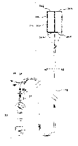
FIG.1 is a schematic illustration of an ablation system with a hollow catheter
probe incorporating
the present invention .
FIG. 2 is a side view of the distal portion of the ablation head with a
stabilising system in expanded
position .
FIG. 3 is an enl~~rged cross-sectional view taken along the line 7C -7C of
FIG.1 .
FIG. 4 is an enl<~rged cross -sectional view taken along the line 7B -7B of
FIG.1.
FIG. 5 is an enlarged cross-sectional view taken along the line 7A -7A of
FIG.1.
FIG .6 is a schematic illustration of the working head of an ablation system
having a wavelength
measuring capacity , in which a plurality of expandable lateral arms allow the
determination of the
direction of the conduction velocity .
FIG. 7 is an enlarged side view of the ablation head with the stabilising
system having a plurality
of bridges in an expanded position .
FIG. 8 is an enlarged cross sectional view taken along the line 7C-7C of a
catheter shaft having a
plurality of conductors connected to the electrodes of the working head .
FIG. 9 is an enlarged cross - sec;tional view taken along the line 12-12 of a
catheter shaft having
a large lumen in which an ablation catheter can move slidably with wavelength
measuring
capacrty .
FIG. 10 is an enlarged cross-sectional view taken along the line 14-14 of an
ablation system having
a hollow catheter and a working head with a stabilising system including a
plurality of bridges .
FIG.11 is a pictorial representation of the catheter of FIG 1 in the right
atrium with the stabilising
system in an expanded position taking back up on the sept<un and pushing the
ablation head to the
wall under ablation .
FIG .12 is an enlarged side view of a catheter system having two ablation
catheters with the
stabilising system in an expanded position .
FIG. 13 is an enlarged side view of the ablation head with an opening on the
opposite side of
the stabilising s<rstem in an expanded position .
FIG.14 is an enlarged cross-sectional view taken along the line ;0-30 of an
ablation system having
a hollow catheter shaft and a double ablation catheter .
FIG.15 is an enlarged cross-sectional view taken along the line 42-42 of the
FIG 17 .
FIG.16 is an enlarged view of the working head ( part 31 of FIG 17 ) in a
contracted position.
FIG.17 is a schematic illustration of a wavelength measuring system with a
working head
constnicted of three longitudinally spaced-apart arms which are parallel in an
expanded position.
FIG. 18 is an enlarged cross-sectional view taken along the line 54-54 of FIG
20 .
FIG. 19 is an enlarged lateral view of the head portion (31 of FIG 17 ) in a
partly expanded
~t4r_~'~..}i_~ J
CA 02348558 2001-04-25
-$-
position .
FIG. 20 is a schematic illustration of a wavelength measuring system with a
working head having a
central classical arm and two lateral inflatable arms .
FIG. 21 is an enlarged CTpSS-SeCtipnll view taken along the line 65 -65 of FIG
22 .
FIG. 22 is a schematic illustration of a wavelength measuring system with a
working head having
a central classical arm and a ;plurality of inflatable lateral arms which are
perpendicular to the
central arm in an inflated positiov .
FIG. 2s is a pictorial representation of the catheter of FIG 1 in the left
atrium with the stabilising
system in an inflated position taking back up on the septum and pushing the
ablation head to the
wall under ablation .
FIG. 24 is a schematic presentation of the position of the electrodes on the
catheters of FIG 17
-20-22.
FIG. 25 is an enlarged cross-sectional view taken along the line 79 -79 of the
FIG 26 .
FIG. 26 is a schematic illustration of a wavelength measuring system with a
working head having
three parallel arms which are fan-shaped in an expanded position .
FIG. 27 is a schematic illustration of a mapping head .
FIG. 28 is a schematic illustration of a wavelength measuring system with a
working head having
9 parallels arms which are fan-shaped in an expanded position .
FIG. 29 is an enlarged cross-sectional view taken along the line 85-85 of FIG
27 .
FIG. 30 is an enlarged cross-sectional view taken along the line 94-94 of FIG
28 .
FIG. 31 is a schematic view of a separate stabilising catheter system with a
head comprising three
lateral arms with a membrane 102 between two of them . The lateral arms are
represented in an
expanded position .
FIG. 32 is an enlarged longitudinal view of the head portion of the catheter
of the FIG 31, showing
the different positions of the l;~teral arms 103 by dotted lines depending on
the position of the
central member 101 .
FIG. 33 is an enlarged longitudinal cross-sectional view of the elongated
flexible member of the
catheter of FIG.31 showing the central member 101 and inflation lumens 106 and
the large central
lumen 107 .
s0 FIG. ~4 is the head portion of a separate stabilising system with a
phuality of lateral arms in an
expanded position .
FIG. ~5 is an enlarged cross-sectional view taken along the line 104-104 of a
separate stabilising
system having 4 lateral arms .
FIG. a6 is a schematic view of a wavelength measuring system with both the
expanding system and
the stabilising system in an expanded position .
y'a~~ ,'1~p SHE --~T
CA 02348558 2001-04-25
-9-
FIG . 37 is a pictorial representation of an ablating system in the right
atrium which is preshaped to
take a transversal position .
Detailed description of the invention
The present invention relates to catheter probes for introduction into a
chamber of the heart having
blood therein and formed by a wall through a lumen leading to the chamber .
Generally, the catheter system is comprised of three main co-operating
components including
- a flexible elongated tubular member having proximal and distal extremities.
- a distal working catheter portion ( called the working head ).
- a stabilising system which assures a high stability profile to the working
head .
FIG. 1 shows generally one embodiment having a working head with ablation
capabilities
comprising
- an elongated hollow catheter shaft 4 having two lumens 8 , 9 ( FIG 3 )
extending
longitudinally from the proximal to the distal extremity . One of the lumens 9
is connected to a
screw syringe 5 by an inflation port and the other lumen 8 is a large
one,allowing a special working
catheter 6 with ablation capability to move slidably wherein .
-a working head 1 including a hollow catheter in continuity with the tubular
member wherein
a plurality of «incomplete» ring-form electrodes 10 are incorporated .The
inter electrode distance is
0.5 to 4 mm and the electrodes are 4 mm in length .
- a stabilising system 3 which is a ring-like , very expandable balloon
connected to inflation
lumen 9 and attached to the worldxig head.
After positioning the catheter under fluoroscopy the physician can stabilise
the head by inflating the
ring-like structure with a radiopaque solution and by changing the direction
of the x-ray beam he
can determine 'the maximum diameter of the circle formed by the said ring and
if he desires two ,
three , four....... ablation lines he can determine the same distances between
the lines by the
determination of the angle of the beam and the greatest diameter of the said
ring .
FIG. 7 shows another embodirneat with a working head composed of
i
- a stabilising system with an arc-shaped , cylindrical balloon 13 attached to
the proximal -
extremity and the distal extremity of the head portion and a plurality of
cylindrical bridges 17
between the catheter shaft and tt~e arc-shaped balloon .
This can help tt~e physician stabilise the catheter head perfecfly ,
especially in parts where the atrial
wall has bulges limiting an optimal contact between the catheter and the
atrial wall .
FIG. 6 shows .another embodiment in which the working head includes
- a catheter shaft in continuity with the tubular member wherein a plurality
of incomplete
ring-form electrodes are incorporated .
- a stabilising system which is a ring-form , very expandable structure
connected to an
~~~~~t
CA 02348558 2001-04-25
-10-
inflation port .
- a plurality of lateral arms 11 having an expanded position and a contracted
position . When
contracted they have a very small size and are parallel to the axis of the
catheter shaft . Sensing
electrodes are provided at their tips and when expanded the said electrodes
have equal distances
from the catheter shaft . The physician can find the direction in which the
recording of these
electrodes are parallel .This direction could be considered as the direction
of the propagation of the
depolarisation . At this moment he can measure the velocity by introducing a
working catheter with
a plurality of sensing electrodes each with a predetermined distance matching
the electrodes of the
head portion . 'The same catheter can be employed to ablate by using a
radiofrequency delivering
catheter which can slide in the said catheter shaft .
In another embodiment there is a cooling system provided comprising of an
infusion port extending
through the elongated flexible catheter shaft to the distal infusion port .
The cooling solution either
enters the blood flow (if the catheter head is open) or is pumped by a distal
extraction port to an
extraction lumen extending through the catheter shaft to a proximal extraction
port .
In another embodiment the ablation catheter includes
- an elongated ,flexible , catheter shaft (FIG 8) comprising an inflation
lumen and a plurality
of separated ilisulated leads 15 extending from the proximal extremity to the
distal extremity.
Each lead is separately connected to an electrode in one hand and to a radio
frequency delivering
system on the other hand .
-a working head comprising a catheter shaft in continuity with the elongated
flexible catheter
shaft , with a plurality of « incomplete » ring-form electrodes , which are
connected separately to
insulated leads .
- a stabilising system composed of a ring-form , very thin structure
constructed of a very
expandable material allowing the stabilisation of the head in the manner
described earlier. The
physician , by connecting the leads separately , or in gang , can apply the
ablation and in the end
of the first ro~md of the ablation can verify the perfect continuity of the
created line and can
reablate the parts where the ablation has been incomplete .
FIG . 11 represents schematically the catheter head 19 applied firmly to the
lateral wall of the right
atrium by the stabilising system 18 in an expanded position .
FIG. 12 shows another embodiment in which the ablation head includes
- a stabilising system fornxed of a ring-form , very expandable balloon 29
connected to a
screw syringe by an inflation lumen 27 .
- two hollow catheters 28 encircling the stabilising system in which an
ablating catheter can
slidably move allowing the ablation of two lines at the same time .
FIG. 13 shows another embodiment in which the ablation head includes
;- c,~.;5.'°.
PG' '- ._1~.7
CA 02348558 2001-04-25
-11-
- a hollow catheter in continuity with the elongated tubular member which is
open on
the opposite side of the balloon so as to allow the electrodes 23 of the
ablation catheter 24 to
be applied directly to the cardiac wall .
- a stabilising ring-form balloon 3 bonded to the head portion in the opposite
side of the
opening .
FIG. 17 is a general view of a wavelength measuring catheter comprising
- an elongated , flexible , catheter shaft 32 with a plurality of inflation
lumens 43 and a
plurality of insulated conductors 44 extending from the proximal extremity to
the distal extremity .
- a plurality of longitudinally extending spaced-apart arms having
interconnected proximal
extremities and carried by the distal extremity of the flexible elongated
tubular member .The said
arms are moveable between a contracted position (FIG 16) and an expanded
position (FIG 17). The
means are provided for moving the arms between a contracted and expanded
position . In the
expanded position , the wavelength measuring head 31 has a predetermined form
allowing the
wavelength measurement in a precise manner . A plurality of longitudinally
spaced-apart electrodes
36 are providedl on each arm and are connected to a plurality of conductors 44
extending through a
lumen provided in the flexible a ongated member .The conductors 44 are
connected to cables 38
which are connected to a control console and power supply .
- a stabilising system is provided to apply the WL measuring head firmly to
the heart wall
under investigation . The said means is moveable between a contracted ( small
size ) and an
expanded position (FIG 19) . When expanded the said stabilising means backs up
on the contra
lateral wall of the atrium and applies firmly the so called head to the wall
under investigation .
In one embodiment ( FIG 17 ) the so-called head 31 consist o~:
- three aims , longitudinally spaced- apart , having interconnected proximal
extremities in
which the lateral arms are connected to the central one by cylindrical , thin
, very low elasticity
balloons in proximal 34 A and distal 34 B extremities . Said balloons are
coupled to inflation ports
37 through inflation lumens extending along the length of catheter shaft 43 .
Balloons 34A and
i
34 B are inflatable with fluid prE;ferably by a radiopaque solution which is
injected by a syringe at
the balloon inflation ports 37 . By increasing the volume of the balloons the
arms are separated and
pressures are dosed in such a manner that the structure has three parallel
arms with a distance of
approximately 1 cm between each of the three arms (FIG 17 ) .
- a ring-like , thin balloon 3b having a very high elasticity and attached to
the central arm is
coupled to an iinflalion port through an inflation lumen 43 extending along
the length of catheter
shaft and is inflatable with fluid preferably by a radippaclue solution which
is injected by a syringe
15 33 at balloon inflation port 37 . When the balloon is expanded it backs up
on the opposite wall and
applies the central arm firmly to the wall under investigation .
A~'tfiu'.i :llL~ .~''~ '~E~
CA 02348558 2001-04-25
-12-
In this way , laiowing the distances , the operator can stimulate the
different electrodes and register
the electrodes situated in the direction of the depolarisation .
i.e.( see fig 24 ;1 let us suppose that pairs of electrodes are positioned at
points
D.E,F,G,H,K,L,M,N,C),P,Q,R,S,Twithadistance:
DE = EF = CiH = HK = LM = MN = OP = PQ = RS = ST = 1 cm and
DG = EH = F K = GL = li~i = KN = LO = MP = NQ = OR = PS = QT = 0.5 cm
1n this way , on stimulating at '"I)" we can measure the speed of conduction
between E and F ,
between H and N , between M and T , between H and Q , between M and Q and by
stimulating
different points and combining the different measurements we can find the
speed of conduction in
different directions and on in~easing the number of electrodes we can complete
an extremely
precise mapping of the conduction velocity .
FIG. 20 shows another embodiment in which the wavelength measuring head
comprises
- a central arm 48 resembling classical catheters .
- two lateral arms 47 wluch are thin balloons having a cylindrical form in an
inflated
position . These; balloons are connected to inflation ports 51 by inflation
lumens 55 in which a
plurality of dot - shaped or ring - form electrodes are provided . In the
expanded position this
structure has the same shape as the embodiment described above , but in a
contracted position it is
very thin and e~~sily movable in the cardiovascular system .
FIG. 22 shows .another embodiment in which the wavelength measuring head
includes
- a central arm 64 which is a classical - type catheter with a plurality of
electrodes 63 .
- a plurality of inflatable lateral arms attached to the body of the central
arm 64 .These arms
are perpendicular to the body of the central arm when they are in an expanded
position and the
distance between the tip of these arms and the catheter shaft is precisely
determined by the pressure
applied . There are two electrodes 62 provided in the tip portion of the
lateral anus with a precise
distance betweEn the proximal Electrode and the catheter shaft . In this way ,
the arms would have
a negligible siae when in a retracted position allowing the main body to be
easily manipulated
s
and when the aims are expanded we can see the parallel recordings between the
different arms -
enabling us to determine the direction of the depolarisation in the same way
as the experiments of
Smeet & al , or use the different electrodes in the same manner as described
above to determine
the velocity in different directions .
FIG. 26 shows ;another embodiment in which the head portion includes
- three arms 69,71 longitudinally spaced-apart having an interconnected
proximal extremity.
The distal extremity of the arms are connected to a very expandable ,
cylindrical . thin balloon
(expanding sysl:em ) allowing the physician to extend the arms more and more
by increasing the
pressure in this balloon .
A"J~E~~?ED SH~Et
CA 02348558 2001-04-25
-13-
- a plurality of electrodes 72 , 73 , 74 connected to a console by a plurality
of insulated leads
79 threaded in the catheter shaft are provided in each arm allowing an
extremely precise mapping
in each position .
- a stabilising system which is a ring-like , thin very expandable balloon
co~ected to an
inflation lumen 80 by an inflation port 77. Its function is identical to the
stabilising systems
described above; .
By increasing the movement of die expanding system , the lateral arms are
progressively extended .
1n this way , the alms take the place of the radius of the circle which has as
it's centre , the proximal
end of the arms .
This allows the measurement of the conduction velocity in all directions,
because if the stimulation
comes from the; central electrodes 73 and the measurement of the conduction
velocity between the
median 72 and distal 74 , electrodes , it's possible to measure between
0° and 180° and in the
opposite direction between 180° and 360°
Each lateral arm is further linked to a cylindrical , very rigid balloon 70
which is coupled to an
inflation port 7 7 through an inflation lumen 80 extending along the length of
the catheter shaft 75 .
'This balloon is iinflatable with fluid , preferably radiopaque solution ,
which is injected by a syringe
at balloon inflation port 77 .
By increasing vthe volume of die balloon progressively the arms become very
rigid and keep a
straight position .
FIG. 28 shows another embodiment in which the working head includes
- a plurality of arms 88 in each side of the central arm with three pairs of
electrodes
on each arm .
- a cylindrical , thin balloon 87 (expanding system) interconnecting the
distal extremities of
the arms . The expanding system is connected to a screw syringe by an
inflation lumen 100 . The
expanding system can be inflated by a radiopaque solution giving a fan-shaped
form to the
working head .
- a stabilising system identical to that described above .
FIG. 27 shows another catheter system in which the working head is a mapping
one and includes
- an elongated , flexible catheter shaft 85 with a plurality of inflation
lumens 96 , 97 and
insulated conductors 95 extending from the proximal extremity to the distal
extremity .
- a working head 86 comprising : a plurality of longitudinally extending
spaced-apart arms
83 , with a plurality of longitudinally spaced - apart electrodes 82 on each
arm and a thin ,
cylindrical , very expandable balloon (expanding system ) 81 interconnecting
the distal extremity
of the arms .
- a stabilising system which is a ring-like thin , very expandable balloon
coupled to the
A~~Na~~
CA 02348558 2001-04-25
-14-
central arm .
The electrodes 82 are connected to a console by insulated conductors 95
threaded in the anus and
in the elongated flexible member allowing electrical measurements .The
expanding system 81 is
connected to a screw syringe by an inflation lumen 96 .The stabilising system
is connected to a
screw syringe by an inflation lumen 97 .
After stabilisation of the head portion the operator can realise a complete
mapping of the wall
under study by increasing the pressure in the expanding system progressively
allowing the arms a
progressive expansion .
In another embodiment (FIG. 31;~ there is a separate stabilising system
provided including
- an elongated , flexible ,catheter shaft 105 having a plurality of inflation
lumens 106 and a
large central lumen 107 in which a relatively more rigid member 101 can slide
.
- a head portion comprising three expandable arms 103 having proximal and
distal
extremities . The proximal extremity of the arms is connected to the distal
extremity of the
elongated member 105 and their distal extremities are connected to the distal
extremity of the
central rigid member . Between two arms there is a thin expandable membrane
102. The movement
of the central member in the central lumen changes the radius of the arms and
adapts it to the
cardiac chamber contour . When expanded the arm which is free backs up against
the cardiac
wall and by the membrane the two other arms apply firmly any catheters to the
atrial wall under
investigation . In another embodiment ( FIG. 34 '~ the head portion of the
individual stabilising
system is composed of more arms without a membrane between them allowing the
stabilisation of
the complex , large working heads of the FIG. 17 , 20 , 22 , 26 , 27 , 28 .It
is clear that all the
combinations described above are possible and remain in the scope of this
invention ...
~~' ~ls~~