Note: Descriptions are shown in the official language in which they were submitted.
CA 02348776 2007-03-05
WO 00/26381 PCT/US99/25437
HIGH FIDELITY THERMOSTABLE LIGASE AND USES THEREOF
The present invention was made with support under National Institutes
of Health Grant Nos. GM-41337-09 and PO1-CA65930-02-04). The U.S.
Government may have certain rights.
FIELD OF THE INVENTION
The present invention is directed to a high fidelity thermostable ligase
and uses thereof.
BACKGROUND OF THE INVENTION
DNA ligases, as an essential component of DNA replication,
recombination, and repair systems found from viruses to humans, catalyze the
formation of a phosphodiester bond at single-stranded breaks on duplex DNA
(Lehman, I.R., Science, 186:790-797 (1974)). DNA ligases can be classified
into two
families based on cofactor dependence. ATP-dependent ligases are found in
bacteriophages (Dunn, et al., J Mol Biol., 148(4):303-330 (1981) and Weiss, et
al.,
Proc Nat] Acad Sci USA, 57(4):1021-1028 (1967)), Chloi-ella virus PBCV-1 (Ho,
et
al., J Virol, 71(3):1931-19374 (1997)), Vaccinia virus (Shuman, S.,
Biochemistrv,
34(49):16138-161475 (1995)), Archea (Kletzin, A., Nucleic Acids Res,
20(20):5389-
5396 (1992) and Bult, et al., Science, 273(5278):1058-1073 (1996)), yeasts
(Andaluz,
et al., Yeast, 12(9):893-8988 (1996), Ramos, et al., Nucleic Acids Res,
25(8):1485-
1492 (1997), Schar, et al., Genes Dev, 11(15):1912-1924 (1997)), mammalian
(Tomkinson, et al., ioessa s, 19(10):893-901 (1997), Tomkinson, et al., Mutat
Res,
407(1):1-9 (1998), and Wang, et al., J Biol Chem, 269(50):31923-3192811
(1994)),
and more recently eubacteria (Cheng, et al., Nucleic Acids Res, 25(7):1369-
1374
(1997) and Deckert, et al., Nature, 392(6674):353-358 (1998)). NAD+(i.e.
nicotinamide adenine dinucleotide)-dependent ligases, however, are found
exclusively
CA 02348776 2001-04-27
WO 00/26381 PCT/US99/25437
-2-
in eubacteria. While some higher eucaryotic organisms may use multiple ATP
(i.e.
adenosine triphosphate)-dependent ligases to fulfill diverse biological
functions, some
simple eubacteria genomes could host both an NAD+-dependent ligase and an ATP-
dependent ligase (Deckert, et al., Nature, 392(6674):353-358 (1998) and
Fleischmann,
et al., Science, 269(5223):496-512 (1995)). The origin of the additional ATP-
dependent ligases in these genomes remains to be determined.
Although the ATP-dependent ligases and NAD+-dependent ligases
share little sequence homology, all -the ligases investigated so far use the
same KXDG
motif to form adenylated enzyme intermediate (Tomkinson, et al., Bioe ssavs,
19(10):893-901 (1997), Shuman, et al., Virloav, 211(1):73-83 (1995), and Luo,
et
al., Nucleic Acids Res, 24(15):3079-3085 (1996)). Furthermore, they seem to be
organized by similar domains and structural folds ((Doherty, et al., Nucleic
Acids
$z, 24(12):2281-2287 (1996), Subramanya, et al., _C&U, 85(4):607-615 (1996),
and
Sekiguchi, et al., Nucleic Acids Res, 25(4):727-734 (1997)). The diversity of
ligase
sequences is not only reflected by their different optimal reaction conditions
and
kinetic rates, but more importantly by their different specificities toward
match and
mismatch substrates. Among the viral ATP-dependent ligases, the broad
substrate
tolerance is represented by the T4 enzyme which seals various mismatches on
both
the 3' and 5' side of the nick junction (Wu, et al., -Q=, 76(2):245-254
(1989)).
Vaccinia ligase ligates various mismatches at both 3'-hydroxyl or 5'-phosphate
sides
with the exception of purine-purine mismatch pairs at the 3'-hydroxyl side
(Shuman,
S., Biochemistry, 34(49):16138-161475 (1995)). Mammalian ATP-dependent ligases
show different substrate sensitivity, as ligase I is more sensitive to 3'
mismatches than
ligase III (Husain, et al., J Biol Chem, 270(16):9683-9690 (1995)).
Additionally, both
ligase I and III tolerate a 3'C/T mismatch more than a 3'G/T mismatch. Little
is
known about archeal ATP-dependent ligases which may reveal the nature of the
progenitor of ATP-dependent ligases. Studies on NAD+-dependent DNA ligase from
E. coli, along with T4 ligase, have contributed immensely to understanding of
the
basic biochemical pathway of the DNA ligation reaction (Lehman, I.R., Science,
186(4166):790-797 (1974) and Rossi, et al., Nucleic Acids Res, 25(11):2106-
2113
(1997)). Studies on the NAD+-dependent ligase from Thermus thermophilus HB8
have revealed the highly discriminative power this enzyme possesses (Luo, et
al.,
CA 02348776 2001-04-27
WO 00/26381 PCT/US99/25437
-3-
Nucleic Acids Res, 24(15):3071-3078 (1996)). Although mismatches at 5'-
phosphate
side are tolerated to some degree (5'A/C, 5'A/A, 5'C/A, 5'C/T, 5'G/T, 5'G/A,
5'T/T,
5'T/G), mismatches at the 3'-hydroxyl side essentially abolish nick-closure
activity
except 3'G/T or 3'T/G mismatch (Luo, et al., Nucleic Acids Res, 24(15):3071-
3078
(1996)). Apparently, sequence divergence and subsequent subtle structural
variation
among DNA ligases underlie an enzyme's recognition preferences toward
different
mismatched base-pairs.
The study of ligase biochemistry is not only important for
understanding its biological functions, but also for developing new
technologies. The
single nucleotide discrimination observed on DNA ligases has led to the
development
of ligase-mediated detection techniques (Wu, et al., Ggne, 76(2):245-254
(1989), Wu,
et al., Genomics, 4(4):560-569 (1989), Landegren, et al., Science,
241(4869):1077-
1080 (1988), Landegren, U., ioessav~, 15(11):761-765 (1993), Barany, F., ECR
Methods Annl, 1(1):5-16 (1991), and Barany, F., Proc Natl Acad Sci USA,
88(1):189-
193 (1991)). Ligase-based linear signal amplification known as LDR (i.e.
ligase
detection reaction), combined with PCR (i.e. polymerase chain reaction)-based
gene
specific target amplification, has been proven to be a powerful tool in cancer
and
disease gene mutation detection (Day, et al., Genomics, 29(1):152-162 (1995)).
PCR/LDR technique relies on two properties of a DNA ligase: (i) specificity
and (ii)
thermostability. Tth (i.e. Thermus thermophilus HB8) DNA ligase has been
successfully used in LDR and LCR (i.e. ligase chain reaction) due to its
highly
discriminative nick closure activity toward a perfect match substrate and its
thermostability which makes thermocycling possible (Barany, F., PCR Methods
Appi,
1(1):5-16 (1991) and Barany, F., Proc Natl Acad Sci USA, 88(l):189-193
(1991)).
To date, one more ligase was cloned and sequenced from T. Scot. (i.e. Thermus
scotoductus) (Thorbjamardottir, et al., -Q=, 161(1):1-6 (1995) and Jonsson, et
al.,
Gene, 151(1-2):177-180 (1994)), but the substrate specificity of this ligase
was not
determined.
Despite the existence of a number of ligases from different host
sources, the need remains to identify additional ligases with greater
fidelity. The
present invention is directed to achieving this objective as a result of the
cloning and
CA 02348776 2001-04-27
WO 00/26381 PCT/US99/25437
-4-
expression of a ligase from T. sp. (i.e. Thermus species) AK16D and the
biochemical
characterization of this high fidelity enzyme.
SUMMARY OF THE INVENTION
The present invention is directed to a thermostable ligase having 100
fold higher fidelity than T4 ligase and 6 fold higher fidelity than wild-type
Thermus
thermophilus ligase, when sealing a.ligation junction between a pair of
oligonucleotide probes hybridized to a target sequence where there is a
mismatch with
the oligonucleotide probe having its 3' end abutting the ligation junction at
the base
immediately adjacent the ligation junction.
Another aspect of the present invention is directed to a therrnostable
ligase having 50 fold higher fidelity than T4ligase and 5 fold higher fidelity
than
wild-type Thermus thermophilus ligase, when sealing a ligation junction
between a
pair of oligonucleotide probes hybridized to a target sequence where there is
a
mismatch with the oligonucleotide probe having its 3' end abutting the
ligation
junction at the base penultimate to the ligation junction.
Yet another aspect of the present invention is directed to a
thermostable ligase having, in the presence of a Mn2+ cofactor, a 12 fold
higher
fidelity than wild-type Thermus thermophilus ligase, when sealing a ligation
junction
between a pair of oligonucleotide probes hybridized to a target sequence where
there
is a mismatch with the oligonucleotide probe having its 3' end abutting the
ligation
junction at the base immediately adjacent to the ligation junction.
The present invention also relates to a DNA molecule encoding the
thermostable ligase as well as expression systems and host cells containing
such DNA
molecules.
Another aspect of the present invention relates to the use of the
thermostable ligase in carrying out a ligase detection reaction process or a
ligase chain
reaction process.
The ligase detection reaction process, involves detecting a target
nucleotide sequence which differs from other nucleotide sequences in the
sample by
one or more single base changes, insertions, deletions, or translocations.
This
CA 02348776 2001-04-27
WO 00/26381 PCT/US99/25437
-5-
involves providing a sample potentially containing a target nucleotide
sequence which
differs from other nucleotide sequences in the sample by one or more single
base
changes, insertions, deletions, or translocations.
The method further includes providing one or more oligonucleotide
probe sets, each characterized by (a) a first oligonucleotide probe having a
target
specific portion and (b) a second oligonucleotide probe having a target-
specific
portion. The oligonucleotide probes in a particular set are suitable for
hybridization to
a target nucleotide sequence which differs from other nucleotide sequences in
the
sample by one or more single base changes, insertions, deletions, or
translocations.
The probes are also suitable for ligation together when hybridized adjacent to
one
another on the target nucleotide sequence, but have a mismatch which
interferes with
such ligation when hybridized to any other nucleotide sequence present in the
sample.
The sample, the one or more oligonucleotide probe sets, and the
thermostable ligase are blended to form a ligase detection reaction mixture.
The
ligase detection reaction mixture is subjected to one or more ligase detection
reaction
cycles comprising a denaturation treatment and a hybridization treatment. In
the
denaturation treatment, any hybridized oligonucleotides are separated from the
target
nucleotide sequence. During the hybridization treatment, the oligonucleotide
probe
sets hybridize at adjacent positions in a base specific manner to their
respective target
nucleotide sequences, if present in the sample, and ligate to one another.
This forms a
ligation product sequence containing the target specific portions connected
together
with the ligation product sequences for each set being distinguishable from
other
nucleic acids in the ligase detection reaction mixture. The oligonucleotide
probe sets
may hybridize to a nucleotide sequence in the sample other than their
respective target
nucleotide sequences but do not ligate together due to a presence of one or
more
mismatches and individually separate during the denaturation treatment. The
presence of ligation product sequences produced as a result of the target
nucleotide
sequence being present in the sample is then detected.
In the ligase chain reaction process of the present invention, the
presence of a target double stranded nucleic acid formed from first and second
complementary target nucleotide sequences is detected in a sample. The target
double
CA 02348776 2001-04-27
WO 00/26381 PCT/US99/25437
-6-
stranded nucleic acid differs from other nucleotide sequences by one or more
single
base changes, insertions, deletions, or translocations.
This method involves providing a sample potentially containing a
target double stranded nucleic acid formed from first and second complementary
nucleotide sequence. This nucleic acid differs from other nucleotide sequences
in the
sample by one or more single base changes, insertions, deletions, or
translocations.
The method further includes providing a first oligonucleotide probe
set, characterized by (a) a first oligonucleotide probe having a target
specific portion
and (b) a second oligonucleotide probe having a target-specific portion. The
oligonucleotide probes in the first set are complementary to the first target
nucleotide
sequence which differs from other nucleotide sequences in the sample by one or
more
single base changes, insertions, deletions, or translocations. The probes are
also
suitable for ligation together when hybridized adjacent to one another on the
first
target nucleotide sequence, but have a mismatch which interferes with such
ligation
when hybridized to any other nucleotide sequence present in the sample. The
method
of the present invention also requires providing a second oligonucleotide
probe set,
characterized by (a) a third oligonucleotide probe having a target specific
portion and
(b) a fourth oligonucleotide probe having a target-specific portion. The
oligonucleotide probes in the second set are complementary to the second
target
nucleotide sequence which differs from other nucleotide sequences in the
sample by
one or more single base changes, insertions, deletions, or translocations. The
probes
of the second set are suitable for ligation together when hybridized adjacent
to one
another on the second target nucleotide sequence, but have a mismatch which
interferes with such ligation when hybridized to any other nucleotide sequence
present in the sample.
The sample, the first and second oligonucleotide probe sets, and the
thermostable ligase are blended together to form a ligase chain reaction
mixture. The
ligase chain reaction mixture is subjected to one or more ligase chain
reaction cycles
comprising a denaturation treatment and a hybridization treatment. During the
denaturation treatment, any hybridized oligonucleotides are separated from the
target
nucleotide sequences. In the hybridization treatment, the oligonucleotide
probe sets
hybridize at adjacent positions in a base specific manner to their respective
target
CA 02348776 2001-04-27
WO 00/26381 PCT/US99/25437
-7-
nucleotide sequences, if present in the sample. The probes also ligate to one
another
to form a ligation product sequence containing the target specific portions
connected
together with the ligation product sequences for each set being
distinguishable from
other nucleic acids in the ligase chain reaction mixture. The oligonucleotide
probe
sets may hybridize to nucleotide sequences in the sample other than their
respective
target nucleotide sequences but do not ligate together due to a presence of
one or
more mismatches and individually separate during the denaturation treatment.
The
presence of ligation product sequences produced as a result of the target
nucleotide
sequence being present in the sample are then detected.
BRIEF DESCRIPTION OF THE DRAWINGS
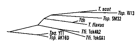
Figures lA-C show a sequence comparison of Thermus DNA ligases.
Figure lA illustrates the evolutionary tree for Thermus DNA ligases. Figure 1B
is a
regional sequence alignment of nine Thermus ligases. The aa (i.e. amino acid)
sequence of T. scot is retrieved from Genebank by accession number 1085749.
The
adenylation motif KXDG is underlined and the adenylation site is marked by *.
The
numbering of aa is based on Tsp. AK 16D ligase. Figure 1 C is a complete amino
acid
sequence of Tsp. AK16D ligase. The adenylation motif KXDG is underlined and
the
adenylation site 218K is shown with a (*) above the residue. The complete
sequence of
Tsp. AK16D ligase gene and partial sequences of six other Thermus ligase genes
have
been deposited with GenBank under accession No. AF092862 for Tsp. AK16D,
AF092863 for Thermus aquaticus YT-1, AF092864 for Thermus flavus, AF092865
for Thermus filiformis Tok4A2, AF092866 for Thermus filiformis Tok6A1,
AF092867 for Tsp. Vil3, and AF092868 for Tsp. SM32.
Figure 2 shows an SDS-PAGE analysis of Tsp. AK16D ligase protein.
Lane 1, molecular weight markers; Lane 2, uninduced cell lysate; Lane 3,
induced cell
lysate; Lane 4, supematant after heating at 70 C; Lane 5, fraction eluted from
Hitrap
blue column. The SDS-polyacrylamide gel was 0.1% SDS-7.5% polyacrylamide and
was stained with Coomassie brilliant blue after electrophoresis. The arrow
points to
the location of Tsp. AK16D ligase.
Figures 3A-C show the effects of salt, pH, and NAD+ on ligation
CA 02348776 2001-04-27
WO 00/26381 PCT/US99/25437
-8-
activity. Tsp. AK16D ligase: closed squares; Tth ligase: open squares. Figure
3A
reveals the pH effect. Reactions were performed in 20 l mixture containing
200 nM
nicked duplex substrate, 12.5 pM Tth ligase or Tsp. AK16D ligase, 20 mM Tris-
HCl
(pH values were determined at room temperature), 10 mM MgC12,100 mM KCI, 10
mM DTT, 1 mM NAD+ and 20 mg/ml BSA at 65 C for 10 min. Figure 3B shows the
salt effect. Reactions were performed in 20 l mixture containing 200 nM
nicked
duplex substrate, 12.5 pM Tth ligase or Tsp. AK16D ligase, 20 mM Tris-HCI, pH
8.5
(at room temperature) for Tth ligase, pH 8.0 for Tsp. AK16D ligase, 10 mM
MgC12,
indicated amount of KCI, 10 mM DTT, 1 mM NAD+ and 20 mg/ml BSA at 65 C for
10 min. Figure 3C shows the NAD+ effect. Tth ligation reactions were performed
in
l mixture containing 200 nM nicked duplex substrate, 12.5 pM Tth ligase and
indicated concentration of NAD+, 20 mM Tris-HCI, pH 8.5, 5 mM MgC12, 100 mM
KC15 10 mM DTT, 1 mM NAD+ and 20 mg/ml BSA at 65 C for 10 min. Tsp.
AK16D ligation reaction were performed in 20 l mixture containing 200 nM
nicked
15 duplex substrate, 12.5 pM Tth ligase and indicated concentration of NAD+,
20 mM
Tris-HCI, pH 8.5, 5 mM MgC12, 50 mM KCI, 10 mM DTT, 1 mM NAD+ and 20
mg/ml BSA at 65 C for 10 min.
Figures 4A-B show the divalent cation dependence of Tsp. AK16D
(stripped bars) and Tth (filled bars) ligase activity. Reaction mixtures
containing (20
20 l ) 20 nM nicked duplex substrate, 0.5 nM Tth ligase or 1 nM Tsp. AK16D
ligase
and 5 mM of indicated divalent cation in the reaction buffers as specified in
Figure 3C
were incubated at 65 C for 10 min. Figure 4A shows the ligation reactions with
different divalent ions as the metal cofactor. Figure 4B shows the
chromatogram of a
representative GeneScan gel illustrating ligation product and DNA adenylate
intermediate. (-): negative control reactions in which ligase was omitted.
Co2+ may
have caused precipitation of DNA substrate which resulted in disappearance of
the
unreacted substrate.
Figures 5A-B shows the time course of Tth (Figure 5A) and Tsp.
AK16D (Figure 5B) ligase activity in the presence of Mg2+ (open squares) or
Mn2+
(closed squares). Reactions were performed in 100 l mixture containing 20 nM
nicked duplex substrate, 0.5 nM Tth ligase or 1 nM Tsp. AK16D ligase and 5 mM
CA 02348776 2001-04-27
WO 00/26381 PCT/US99/25437
-9-
Mg2+ or Mn2+ in the reaction buffers as specified in Figure 3C at 65 C.
Aliquots (5
I ) were removed at the indicated time and reactions stopped by adding equal
volumes of stop solution.
Figures 6A-B show the divalent cation concentration dependence of
Tth (Figure 6A) and Tsp. AK16D (Figure 6B) ligase activity. Mg2+(open
squares);
Mn2+ (closed squares). Reactions were performed in 20 l mixture containing 20
nM
nicked duplex substrate, 0.5 nM Tth ligase or 1 nM Tsp. AK16D ligase and
indicated
concentration of Mg2+ or Mn2+ in the reaction buffers as specified in Figure
4C at
65 C for 2 min.
Figures 7A-B show the ligation of gapped and inserted substrates.
Figure 7A shows the formation of ligated product with gapped and inserted
substrates.
Reactions were performed in a 20 1 mixture containing 12.5 nM nicked duplex
substrate, 1.25 pM Tth ligase or 12.5 nM Tsp. AK16D ligase in the reaction
buffer at
65 C for 4 hours. Figure 7B shows the proposed reaction path leads to ligation
of 1 nt
(i.e. nucleotides) inserted substrate.
DETAILED DESCRIPTION OF THE INVENTION
The present invention relates to a high fidelity thermostable ligase
enzyme. This enzyme has the amino acid sequence of SEQ. ID. No. 1 as follows:
MTLEEARRRVNELRDLIRYHNYLYYVLDAPEISDAEYDRLLRELKELEERFPELKSP
DSPTEQVGARPLEATFRPVRHPTRMYSLDNAFSLDEVRAFEERIERALGRKGPFLYT
VERKVDGLSVNLYYEEGILVFGATRGDGETGEEVTQNLLTIPTIPRRLTGVPDRLEV
RGEVYMPIEAFLRLNQELEEAGERIFKNPRNAAAGSLRQKDPRVTARRGLRATFYAL
GLGLEETGLKSQHDLLLWLRERGFPVEHGFTRALGAEGVEEVYQAWLKERRKLPFEA
DGVWKLDDLALWRELGYTARTPRFALAYKFPAEEKETRLLSVAFQVGRTGRITPVG
VLEPVFIEGSEVSRVTLHNESFIEELDVRIGDWVLVHKAGGVIPEVLRVLKERRTGE
EKPIIWPENCPECGHALIKEGKVHRCPNPLCPAKRFEAIRHYASRKAMDIQGLGEKL
IEKLLEKGLVRDVADLYRLKKEDLVNLERMGEKSAENLLRQIEESKGRGLERLLYAL
GLPGVGEVLARNLALRFGHMDRLLEAGLEDLLEVEGVGELTARAILNTLKDPEFRDL
VRRLKEAGVEMEAKEREGEALKGLTFVITGELSRPREEVKALLRRLGAKVTDSVSRK
TSFLVVGENPGSKLEKAR.ALGVPTLSEEELYRLIEERTGKDPRALTA
CA 02348776 2001-04-27
WO 00/26381 PCTIUS99/25437
-10-
This protein has a molecular weight of 78 to 81 kDa, as measured by
SDS-PAGE. For purposes of the present application, the term "thermostable"
refers
to a DNA ligase which is resistant to inactivation by heat.
The thermostable ligase of the present invention has a 100 fold higher
fidelity than T4 ligase and 6 fold higher fidelity than wild-type Thermus
thermophilus
ligase, when sealing a ligation junction between a pair of oligonucleotide
probes
hybridized to a target sequence where there is a mismatch with the
oligonucleotide
probe having its 3' end abutting the ligation junction at the base immediately
adjacent
the ligation junction. This ligase also has a 50 fold higher fidelity than
T4ligase and
5 fold higher fidelity than wild-type Thermus thermophilus ligase, when
sealing a
ligation junction between a pair of oligonucleotide probes hybridized to a
target
sequence where there is a mismatch with the oligonucleotide probe having its
3' end
abutting the ligation junction at the base penultimate to the ligation
junction. Finally,
the thermostable ligase of the present invention, in the presence of a Mn2+
cofactor,
has a 12 fold higher fidelity than wild-type Thermus thermophilus ligase, when
sealing a ligation junction between a pair of oligonucleotide probes
hybridized to a
target sequence where there is a mismatch with the oligonucleotide probe
having its
3' end abutting the ligation junction at the base immediately adjacent to the
ligation
junction. For purposes of the present invention, "fidelity" is defined to mean
the ratio
of the initial rate of ligating two adjacent probes hybridized to a
complementary
template with a C-G match at the base of the probe with its 3' end at the
ligation
junction to the initial rate of ligating two adjacent probes hybridized to a
complementary template with a G-T mismatch at the base of the probe with its
3' end
at the ligation junction.
The thermostable ligase of the present invention is also characterized
by having an arginine adjacent to the active site lysine (i.e. K) in the KXDG
motif
(where X is any amino acid).
This protein is encoded by a DNA molecule having a nucleotide
sequence of SEQ. ID. No. 2 as follows:
ATGACCCTAGAGGAGGCCCGCAGGCGCGTCAACGAACTCAGGGACCTGATCCGTTAC
CACAACTACCTCTATTACGTCTTGGACGCCCCCGAGATCTCCGACGCCGAGTACGAC
CGGCTCCTTAGGGAGCTTAAGGAGCTGGAGGAGCGCTTTCCCGAGCTCAAAAGCCCC
CA 02348776 2001-04-27
WO 00/26381 PCT/US99/25437
-11-
GACTCCCCCACGGAACAGGTGGGGGCGAGGCCTCTGGAGGCCACCTTCCGCCCGGTG
CGCCACCCCACCCGCATGTACTCCCTGGACAACGCCTTTTCCTTGGACGAGGTGAGG
GCCTTTGAGGAGCGCATAGAGCGGGCCCTGGGGCGGAAGGGGCCCTTCCTCTACACC
GTGGAGCGCAAGGTGGACGGTCTTTCCGTGAACCTCTACTACGAGGAGGGCATCCTC
GTCTTTGGGGCCACCCGGGGCGACGGGGAGACCGGGGAGGAGGTGACCCAGAACCTC
CTCACCATCCCCACCATTCCCCGCCGCCTCACGGGCGTTCCGGACCGCCTCGAGGTC
CGGGGCGAGGTCTACATGCCCATAGAGGCCTTCCTCAGGCTCAACCAGGAGCTGGAG
GAGGCGGGGGAGCGCATCTTCAAAAACCCCAGGAACGCCGCCGCCGGGTCCTTGCGG
CAGAAAGACCCCAGGGTCACGGCCAGGCGGGGCCTGAGGGCCACCTTTTACGCCCTG
GGGCTGGGCCTGGAGGAAACCGGGTTAAAAAGCCAGCACGACCTTCTCCTATGGCTA
AGAGAGCGGGGCTTTCCCGTGGAGCACGGCTTTACCCGGGCCCTGGGGGCGGAGGGG
GTGGAGGAGGTCTACCAGGCCTGGCTCAAGGAGAGGCGGAAGCTTCCCTTTGAGGCC
GACGGGGTGGTGGTCAAGCTGGACGACCTCGCCCTCTGGCGGGAGCTGGGGTACACC
GCCCGCACCCCCCGCTTCGCCCTCGCCTACAAGTTCCCGGCCGAGGAGAAGGAGACC
CGCCTCCTCTCCGTGGCCTTCCAGGTGGGGCGGACCGGGCGCATCACCCCCGTGGGC
GTTCTGGAGCCCGTCTTCATAGAGGGCAGCGAGGTGAGCCGGGTCACCCTCCACAAC
GAGAGCTTCATTGAGGAGCTGGACGTGCGCATCGGCGACTGGGTGCTGGTCCACAAG
GCGGGCGGGGTGATTCCCGAGGTGCTGAGGGTCCTGAAAGAGCGCCGCACCGGGGAG
GAGAAGCCCATCATCTGGCCCGAGAACTGCCCCGAGTGCGGCCACGCCCTCATCAAG
GAGGGGAAGGTCCACCGCTGCCCCAACCCCTTGTGCCCCGCCAAGCGCTTTGAGGCC
ATCCGCCACTACGCCTCCCGCAAGGCCATGGACATCCAGGGCCTGGGGGAGAAGCTC
ATAGAAAAGCTTCTGGAAAAGGGCCTGGTCCGGGACGTGGCCGACCTCTACCGCCTG
AAGAAGGAGGACCTGGTGAACCTGGAGCGCATGGGGGAGAAGAGCGCAGAGAACCTC
CTCCGCCAGATAGAGGAGAGCAAGGGCCGCGGCCTGGAGCGCCTCCTTTACGCCCTG
GGCCTTCCCGGGGTGGGGGAGGTGCTGGCCCGGAACCTGGCCCTCCGCTTCGGCCAC
ATGGACCGCCTTCTGGAGGCGGGCCTCGAGGACCTCCTGGAGGTGGAGGGGGTGGGC
GAGCTCACCGCCCGGGCCATCCTGAATACCCTAAAGGACCCGGAGTTCCGGGACCTG
GTGCGCCGCCTGAAGGAGGCCGGGGTGGAGATGGAGGCCAAAGAGCGGGAGGGCGAG
GCCTTGAAGGGGCTCACCTTCGTCATCACCGGGGAGCTTTCCCGGCCCCGGGAGGAG
GTGAAGGCCCTCCTTAGGCGGCTTGGGGCCAAGGTGACGGACTCGGTGAGCCGCAAG
ACGAGCTTCCTGGTGGTGGGGGAGAACCCGGGGAGCAAGCTGGAAAAGGCCCGCGCC
TTGGGGGTCCCCACCCTGAGCGAGGAGGAGCTCTACCGCCTCATTGAGGAGAGGACG
GGCAAGGACCCAAGGGCCCTCACGGCCTAG
Fragments of the above polypeptide or protein are also encompassed
by the present invention.
Suitable fragments can be produced by several means. In the first,
subclones of the gene encoding the protein of the present invention are
produced by
conventional molecular genetic manipulation by subcloning gene fragments. The
subclones then are expressed in vitro or in vivo in bacterial cells to yield a
smaller
protein or peptide that can be tested for ligase activity according to the
procedure
described below.
As an alternative, fragments of the ligase of the present invention can
be produced by digestion of the full-length ligase with proteolytic enzymes
like
CA 02348776 2001-04-27
WO 00/26381 PCT/US99/25437
-12-
chymotrypsin or Staphylococcus proteinase A, or trypsin. Different proteolytic
enzymes are likely to cleave ligase proteins at different sites based on the
amino acid
sequence of the ligase. Some of the fragments that result from proteolysis may
be
active ligases.
In another approach, based on knowledge of the primary structure of
the protein, fragments of the ligase encoding gene may be synthesized by using
the
PCR technique together with specific sets of primers chosen to represent
particular
portions of the protein. These then.would be cloned into an appropriate vector
for
increased expression of a truncated peptide or protein.
Chemical synthesis can also be used to make suitable fragments. Such
a synthesis is carried out using known amino acid sequences for the ligase
being
produced. Alternatively, subjecting the full length ligase to high
temperatures and
pressures will produce fragments. These fragments can then be separated by
conventional procedures (e.g., chromatography, SDS-PAGE).
Variants may also (or alternatively) be modified by, for example, the
deletion or addition of amino acids that have minimal influence on the
properties,
secondary structure and hydropathic nature of the polypeptide. For example, a
polypeptide may be conjugated to a signal (or leader) sequence at the N-
terminal end
of the protein which co-translationally or post-translationally directs
transfer of the
protein. The polypeptide may also be conjugated to a linker or other sequence
for
ease of synthesis, purification, or identification of the polypeptide.
Suitable DNA molecules are those that hybridize to a DNA molecule
comprising a nucleotide sequence of 50 continuous bases of SEQ. ID. No. 2
under
stringent conditions characterized by a hybridization buffer comprising 0.9M
sodium
citrate ("SSC") buffer at a temperature of 37 C and remaining bound when
subject to
washing with the SSC buffer at 37 C; and preferably in a hybridization buffer
comprising 20% formamide in 0.9M saline/0.09M SSC buffer at a temperature of
42 C and remaining bound when subject to washing at 42 C with 0.2x SSC buffer
at
42 C.
The protein or polypeptide of the present invention is preferably
produced in purified form (preferably at least about 80%, more preferably 90%,
pure)
by conventional techniques. Typically, the protein or polypeptide of the
present
CA 02348776 2007-03-05
WO 00/26381 PCT/US99/25437
-13-
invention is secreted into the growth medium of recombinant host cells.
Alternatively, the protein or polypeptide of the present invention is produced
but not
secreted into growth medium. In such cases, to isolate the protein, the host
cell (e.g.,
E. coli) carrying a recombinant plasmid is propagated, lysed by sonication,
heat, or
chemical treatment, and the homogenate is centrifuged to remove bacterial
debris.
The supematant is then subjected to sequential ammonium sulfate precipitation.
The
fraction containing the polypeptide or protein of the present invention is
subjected to
gel filtration in an appropriately sized dextran or polyacrylamide column to
separate
the proteins. If necessary, the protein fraction may be further purified by
HPLC.
The DNA molecule encoding the ligase of the present invention can be
incorporated in cells using conventional recombinant DNA technology.
Generally,
this involves inserting the DNA molecule into an expression system to which
the
DNA molecule is heterologous (i.e. not normally present). The heterologous DNA
molecule is inserted into the expression system or vector in proper sense
orientation
and correct reading frame. The vector contains the necessary elements for the
transcription and translation of the inserted protein-coding sequences.
U.S. Patent No. 4,237,224 to Cohen and Boyer,
describes the production of expression systems in the form
of recombinant plasmids using restriction enzyme cleavage and ligation with
DNA
ligase. These recombinant plasmids are then introduced by means of
transformation
and replicated in unicellular cultures including procaryotic organisms and
eucaryotic
cells grown in tissue culture.
Recombinant genes may also be introduced into viruses, such as
vaccina virus. Recombinant viruses can be generated by transfection of
plasmids into
cells infected with virus.
Suitable vectors include, but are not limited to, the following viral
vectors such as lambda vector system gtl 1, gt WES.tB, Charon 4, and plasmid
vectors
such as pBR322, pBR325, pACYC 177, pACYC 1084, pUC8, pUC9, pUC 18, pUC 19,
pLG339, pR290, pKC37, pKC101, SV 40, pBluescript II SK +/- or KS +/- (see
"Stratagene Cloning Systems" Catalog (1993) from Stratagene, La Jolla, Calif
),
pQE, pIH821, pGEX, pET series (see F.W.
Studier et. al., "Use of T7 RNA Polymerase to Direct Expression of Cloned
Genes,"
CA 02348776 2007-03-05
WO 00/26381 PCT/US99/25437
-14-
Gene Expression Technologv vol. 185 (1990)),
and any derivatives thereof. Recombinant molecules can be introduced
into cells via transformation, particularly transduction, conjugation,
mobilization, or
electroporation. The DNA sequences are cloned into the vector using standard
cloning procedures in the art, as described by Sambrook et al., Molecular
Cloning: A
Laboratorv Manual, Cold Springs Laboratory, Cold Springs Harbor, New York
(1989).
A variety of host-vector systems may be utilized to express the protein-
encoding sequence(s). Primarily, the vector system must be compatible with the
host
cell used. Host-vector systems include but are not limited to the following:
bacteria
transformed with bacteriophage DNA, plasmid DNA, or cosmid DNA;
microorganisms such as yeast containing yeast vectors; mammalian cell systems
infected with virus (e.g., vaccinia virus, adenovirus, etc.); insect cell
systems infected
with virus (e.g., baculovirus); and plant cells infected by bacteria. The
expression
elements of these vectors vary in their strength and specificities. Depending
upon the
host-vector system utilized, any one of a number of suitable transcription and
translation elements can be used.
Different genetic signals and processing events control many levels of
gene expression (e.g., DNA transcription and messenger RNA (mRNA)
translation).
Transcription of DNA is dependent upon the presence of a promotor
which is a DNA sequence that directs the binding of RNA polymerase and thereby
promotes mRNA synthesis. The DNA sequences of eucaryotic promotors differ from
those of procaryotic promotors. Furthermore, eucaryotic promotors and
accompanying genetic signals may not be recognized in or may not function in a
procaryotic system, and, further, procaryotic promotors are not recognized and
do not
function in eucaryotic cells.
Similarly, translation of mRNA in procaryotes depends upon the
presence of the proper procaryotic signals which differ from those of
eucaryotes.
Efficient translation of mRNA in procaryotes requires a ribosome binding site
called
the Shine-Dalgamo ("SD") sequence on the mRNA. This sequence is a short
nucleotide sequence of mRNA that is located before the start codon, usually
AUG,
which encodes the amino-terminal methionine of the protein. The SD sequences
are
CA 02348776 2007-03-05
WO 00/26381 PCT/US99/25437
-15-
complementary to the 3'-end of the 16S rRNA (ribosomal RNA) and probably
promote binding of mRNA to ribosomes by duplexing with the rRNA to allow
correct
positioning of the ribosome. For a review on maximizing gene expression, see
Roberts and Lauer, Methods in Enzymoloay, 68:473 (1979).
Promotors vary in their "strength" (i.e. their ability to promote
transcription). For the purposes of expressing a cloned gene, it is desirable
to use
strong promotors in order to obtain a high level of transcription and, hence,
expression of the gene. Depending upon the host cell system utilized, any one
of a
number of suitable promotors may be used. For instance, when cloning in E.
coli, its
bacteriophages, or plasmids, promotors such as the T7 phage promoter, lac
promotor,
trp promotor, recA promotor, ribosomal RNA promotor, the PR and PL promotors
of
coliphage lambda and others, including but not limited, to lacUV 5, ompF, bla,
lpp,
and the like, may be used to direct high levels of transcription of adjacent
DNA
segments. Additionally, a hybrid trp-IacUV5 (tac) promotor or other E. coli
promotors produced by recombinant DNA or other synthetic DNA techniques may be
used to provide for transcription of the inserted gene.
Bacterial host cell strains and expression vectors may be chosen which
inhibit the action of the promotor unless specifically induced. In certain
operations,
the addition of specific inducers is necessary for efficient transcription of
the inserted
DNA. For example, the lac operon is induced by the addition of lactose or IPTG
(isopropylthio-beta-D-galactoside). A variety of other operons, such as trp,
pro, etc.,
are under different controls.
Specific initiation signals are also required for efficient gene
transcription and translation in procaryotic cells. These transcription and
translation
initiation signals may vary in "strength" as measured by the quantity of gene
specific
messenger RNA and protein synthesized, respectively. The DNA expression
vector,
which contains a promotor, may also contain any combination of various
"strong"
transcription and/or translation initiation signals. For instance, efficient
translation in
E. coli requires an SD sequence about 7-9 bases 5' to the initiation codon
("ATG") to
provide a ribosome binding site. Thus, any SD-ATG combination that can be
utilized
by host cell ribosomes may be employed. Such combinations include but are not
CA 02348776 2007-03-05
WO 00/26381 PCT/US99/25437
-16-
limited to the SD-ATG combination from the cro gene or the N gene of coliphage
lambda, or from the E. coli tryptophan E, D, C, B or A genes. Additionally,
any SD-
ATG combination produced by recombinant DNA or other techniques involving
incorporation of synthetic nucleotides may be used.
Once the isolated DNA molecule encoding the ligase of the present
invention has been cloned into an expression system, it is ready to be
incorporated
into a host cell. Such incorporation can be carried out by the various forms
of
transformation noted above, depending upon the vector/host cell system.
Suitable
host cells include, but are not limited to, bacteria, virus, yeast, mammalian
cells,
insect, plant, and the like.
The present invention is useful in a number of processes where a ligase
enzyme is conventionally utilized at high temperatures. Generally, these
procedures
include the ligase detection reaction and the ligase chain reaction.
Both of the ligase detection reaction and ligase chain reaction involve
detection of a target sequence and amplification of that sequence at elevated
temperatures. In carrying out these procedures, the enzyme is subjected to
elevated
temperatures but is not degraded due to its thermostable character. The ligase
detection reaction and ligase chain reaction procedures are generally
described in WO
90/17239 to Barany et. al., F. Barany, et. al., "Cloning, Overexpression, and
Nucleotide Sequence of a Thennostable DNA Ligase-Encoding Gene," Gene 109: 1-
11 (1991), and F. Barany, et. al., "Genetic Disease Detection and DNA
Amplification
Using Cloned Thermostable Ligase," Proc. Nat'1 Acad. Sci. USA 88: 189-93.
The ligase detection reaction process is useful in detecting in a sample
a target nucleotide sequence as described more fully below.
One or more oligonucleotide probe sets are provided for use in
conjunction with this method. Each set includes (a) a first oligonucleotide
probe
having a target-specific portion and (b) a second oligonucleotide probe having
a
target-specific portion. The oligonucleotide probes in a particular set are
suitable for
ligation together when hybridized adjacent to one another on a corresponding
target
nucleotide sequence, but have a mismatch which interferes with such ligation
when
hybridized to any other nucleotide sequence present in the sample.
CA 02348776 2001-04-27
WO 00/26381 PCT/US99/25437
-17-
The sample, the one or more oligonucleotide probe sets, and the ligase
are blended to form a ligase detection reaction mixture. The ligase detection
reaction
mixture is subjected to one or more ligase detection reaction cycles
comprising a
denaturation treatment and a hybridization treatment. In the denaturation
treatment,
any hybridized oligonucleotides are separated from the target nucleotide
sequences.
The hybridization treatment involves hybridizing the oligonucleotide probe
sets at
adjacent positions in a base-specific manner to the respective target
nucleotide
sequences, if present in the sample. The hybridized oligonucleotide probes
from each
set ligate to one another to form a ligation product sequence containing the
target-
specific portions connected together. The ligation product sequence for each
set is
distinguishable from other nucleic acids in the ligase detection reaction
mixture. The
oligonucleotide probe sets may hybridize to adjacent sequences in the sample
other
than the respective target nucleotide sequences but do not ligate together due
to the
presence of one or more mismatches. When hydridized oligonucleotide probes do
not
ligate, they individually separate during the denaturation treatment.
During the ligase detection reaction phase, the denaturation treatment
is carried out at a temperature of 80-105 C, while hybridization takes place
at 50-
85 C. Each cycle comprises a denaturation treatment and a thermal
hybridization
treatment which in total is from about one to five minutes long. Typically,
the
ligation detection reaction involves repeatedly denaturing and hybridizing for
2 to 50
cycles. The total time for the ligase detection reaction process is I to 250
minutes.
The oligonucleotide probe sets can be in the form of ribonucleotides,
deoxynucleotides, modified ribonucleotides, modified deoxyribonucleotides,
modified
phosphate-sugar-backbone oligonucleotides, nucleotide analogs, and mixtures
thereof.
In one variation, the oligonucleotides of the oligonucleotide probe sets
each have a hybridization or melting temperature (i.e. Tm) of 66-70 C. These
oligonucleotides are 20-28 nucleotides long.
The oligonucleotide probe sets, as noted above, have a reporter label
suitable for detection. Useful labels include chromophores, fluorescent
moieties,
enzymes, antigens, heavy metals, magnetic probes, dyes, phosphorescent groups,
radioactive materials, chemiluminescent moieties, and electrochemical
detecting
moieties.
CA 02348776 2001-04-27
WO 00/26381 PCT/US99/25437
-18-
The product of the ligase detection reaction can be detected in either of
two formats. These are fully described in WO 98/03673, to Barany et al., which
is
hereby incorporated by reference. In one of these formats, ligase detection
reaction
products are detected by capillary or gel electrophoresis. Alternatively,
ligation
products can be detected on an array by specific hybridization to a
complementary
sequence on the array.
The ligation detection reaction mixture may include a carrier DNA,
such as salmon sperm DNA.
The hybridization step in the ligase detection reaction, which is
preferably a thermal hybridization treatment discriminates between nucleotide
sequences based on a distinguishing nucleotide at the ligation junctions. The
difference between the target nucleotide sequences can be, for example, a
single
nucleic acid base difference, a nucleic acid deletion, a nucleic acid
insertion, or
rearrangement. Such sequence differences involving more than one base can also
be
detected. Preferably, the oligonucleotide probe sets have substantially the
same
length so that they hybridize to target nucleotide sequences at substantially
similar
hybridization conditions. As a result, the process of the present invention is
able to
detect infectious diseases, genetic diseases, and cancer. It is also useful in
environmental monitoring, forensics, and food science.
A wide variety of infectious diseases can be detected by the process of
the present invention. Typically, these are caused by bacterial, viral,
parasite, and
ftingal infectious agents. The resistance of various infectious agents to
drugs can also
be determined using the present invention.
Bacterial infectious agents which can be detected by the present
invention include Escherichia coli, Salmonella, Shigella, Klebsiella,
Pseudomonas,
Listeria monocytogenes, Mycobacterium tuberculosis, Mycobacterium avium-
intracellulare, Yersinia, Francisella, Pasteurella, Brucella, Clostridia,
Bordetella
pertussis, Bacteroides, Staphylococcus aureus, Streptococcus pneumonia, B-
Hemolytic strep., Corynebacteria, Legionella, Mycoplasma, Ureaplasma,
Chlamydia,
Neisseria gonorrhea, Neisseria meningitides, Hemophilus influenza,
Enterococcus
faecalis, Proteus vulgaris, Proteus mirabilis, Helicobacter pylorf, Treponema
CA 02348776 2001-04-27
WO 00/26381 PCT/US99/25437
-19-
palladium, Borrelia burgdorferi, Borrelia recurrentis, Rickettsial pathogens,
Nocardia, and Acitnomycetes.
Fungal infectious agents which can be detected by the present
invention include Cryptococcus neoformans, Blastomyces dermatitidis,
Histoplasma
capsulatum, Coccidioides immitis, Paracoccidioides brasiliensis, Candida
albicans,
Aspergillusfumigautus, Phycomycetes (Rhizopus), Sporothrix schenckii,
Chromomycosis, and Maduromycosis.
Viral infectious agents which can be detected by the present invention
include human immunodeficiency virus, human T-cell lymphocytotrophic virus,
hepatitis viruses (e.g., Hepatitis B Virus and Hepatitis C Virus), Epstein-
Barr Virus,
cytomegalovirus, human papillomaviruses, orthomyxo viruses, paramyxo viruses,
adenoviruses, corona viruses, rhabdo viruses, polio viruses, toga viruses,
bunya
viruses, arena viruses, rubella viruses, and reo viruses.
Parasitic agents which can be detected by the present invention include
Plasmodiumfalciparum, Plasmodium malaria, Plasmodium vivax, Plasmodium ovale,
Onchoverva volvulus, Leishmania, Trypanosoma spp., Schistosoma spp., Entamoeba
histolytica, Cryptosporidum, Giardia spp., Trichimonas spp., Balatidium coli,
Wuchereria bancrofti, Toxoplasma spp., Enterobius vermicularis, Ascaris
lumbricoides, Trichuris trichiura, Dracunculus medinesis, trematodes,
Diphyllobothrium latum, Taenia spp., Pneumocystis carinii, and Necator
americanis.
The present invention is also useful for detection of drug resistance by
infectious agents. For example, vancomycin-resistant Enterococcusfaecium,
methicillin-resistant Staphylococcus aureus, penicillin-resistant
Streptococcus
pneumoniae, multi-drug resistant Mycobacterium tuberculosis, and AZT-resistant
human immunodeficiency virus can all be identified with the present invention.
Genetic diseases can also be detected by the process of the present
invention. This can be carried out by prenatal or post-natal screening for
chromosomal and genetic aberrations or for genetic diseases. Examples of
detectable
genetic diseases include: 21 hydroxylase deficiency, cystic fibrosis, Fragile
X
Syndrome, Turner Syndrome, Duchenne Muscular Dystrophy, Down Syndrome or
other trisomies, heart disease, single gene diseases, HLA typing,
phenylketonuria,
sickle cell anemia, Tay-Sachs Disease, thalassemia, Klinefelter Syndrome,
CA 02348776 2001-04-27
WO 00/26381 PCT/US99/25437
-20-
Huntington Disease, autoimmune diseases, lipidosis, obesity defects,
hemophilia,
inborn errors of metabolism, and diabetes.
Cancers which can be detected by the process of the present invention
generally involve oncogenes, tumor suppressor genes, or genes involved in DNA
amplification, replication, recombination, or repair. Examples of these
include:
BRCA1 gene, p53 gene, APC gene, Her2/Neu amplification, Bcr/Abl, K-ras gene,
and human papillomavirus Types 16 and 18. Various aspects of the present
invention
can be used to identify amplifications, large deletions as well as point
mutations and
small deletions/insertions of the above genes in the following common human
cancers: leukemia, colon cancer, breast cancer, lung cancer, prostate cancer,
brain
tumors, central nervous system tumors, bladder tumors, melanomas, liver
cancer,
osteosarcoma and other bone cancers, testicular and ovarian carcinomas, head
and
neck tumors, and cervical neoplasms.
In the area of environmental monitoring, the present invention can be
used for detection, identification, and monitoring of pathogenic and
indigenous
microorganisms in natural and engineered ecosystems and microcosms such as in
municipal waste water purification systems and water reservoirs or in polluted
areas
undergoing bioremediation. It is also possible to detect plasmids containing
genes
that can metabolize xenobiotics, to monitor specific target microorganisms in
population dynamic studies, or either to detect, identify, or monitor
genetically
modified microorganisms in the environment and in industrial plants.
The present invention can also be used in a variety of forensic areas,
including for human identification for military personnel and criminal
investigation,
patemity testing and family relation analysis, HLA compatibility typing, and
screening blood, sperm, or transplantation organs for contamination.
In the food and feed industry, the present invention has a wide variety
of applications. For example, it can be used for identification and
characterization of
production organisms such as yeast for production of beer, wine, cheese,
yogurt,
bread, etc. Another area of use is with regard to quality control and
certification of
products and processes (e.g., livestock, pasteurization, and meat processing)
for
contaminants. Other uses include the characterization of plants, bulbs, and
seeds for
CA 02348776 2007-03-05
WO 00/26381 PCT/US99/25437
-21 -
breeding purposes, identification of the presence of plant-specific pathogens,
and
detection and identification of veterinary infections.
Desirably, the oligonucleotide probes are suitable for ligation together
at a ligation junction when hybridized adjacent to one another on a
corresponding
target nucleotide sequence due to perfect complementarity at the ligation
junction.
However, when the oligonucleotide probes in the set are hybridized to any
other
nucleotide sequence present in the sample, there is a mismatch at a base at
the ligation
junction which interferes with ligation. Most preferably, the mismatch is at
the base
at the 3' base at the ligation junction. Alternatively, the mismatch can be at
the bases
adjacent to bases at the ligation junction.
Before carrying out the ligase detection reaction, in accordance with
the present invention, target nucleotide sequences in the sample can be
preliminarily
amplified. This preferably carried out with polymerase chain reaction. The
polymerase chain reaction process is fully described in H. Erlich, et. al,
"Recent
Advances in the Polymerase Chain Reaction," Science 252: 1643-50 (1991); M.
Innis,
et. al., PCR Protocols: A Guide to Methods and Applications, Academic Press:
New
York (1990) and R. Saiki, et. al., "Primer-directed Enzymatic Amplification of
DNA
with a Thermostable DNA Polymerase," Science 239: 487-91 (1988).
The ligase detection reaction process achieves linear amplification of a
target sequence. The ligase chain reaction utilizes essentially the same steps
and
conditions as the ligase detection reaction to achieve exponential
amplification. This
greater level of amplification is achieved by utilizing 2 sets of
complementary
oligonucleotides with each set hybridizing to complementary strands of a
target
nucleic acid sequence.
In the ligase chain reaction process of the present invention, the
presence of a target double stranded nucleic acid formed from first and second
complementary target nucleotide sequences is detected in a sample. The target
double
stranded nucleic acid differs from other nucleotide sequences by one or more
single
base changes, insertions, deletions, or translocations.
This method involves providing a sample potentially containing a
target double stranded nucleic acid formed from first and second complementary
CA 02348776 2001-04-27
WO 00/26381 PCT/US99/25437
-22-
nucleotide sequence. This nucleic acid differs from other nucleotide sequences
in the
sample by one or more single base changes, insertions, deletions, or
translocations.
The method further includes providing a first oligonucleotide probe
set, characterized by (a) a first oligonucleotide probe having a target
specific portion
and (b) a second oligonucleotide probe having a target-specific portion. The
oligonucleotide probes in the first set are complementary to the first target
nucleotide
sequence which differs from other nucleotide sequences in the sample by one or
more
single base changes, insertions, dele.tions, or translocations. The probes are
also
suitable for ligation together when hybridized adjacent to one another on the
first
target nucleotide sequence, but have a mismatch which interferes with such
ligation
when hybridized to any other nucleotide sequence present in the sample. The
method
of the present invention also requires providing a second oligonucleotide
probe set,
characterized by (a) a third oligonucleotide probe having a target specific
portion and
(b) a fourth oligonucleotide probe having a target-specific portion. The
oligonucleotide probes in the second set are complementary to the second
target
nucleotide sequence which differs from other nucleotide sequences in the
sample by
one or more single base changes, insertions, deletions, or translocations. The
probes
of the second set are suitable for ligation together when hybridized adjacent
to one
another on the second target nucleotide sequence, but have a mismatch which
interferes with such ligation when hybridized to any other nucleotide sequence
present in the sample.
The sample, the first and second oligonucleotide probe sets, and the
thermostable ligase are blended together to form a ligase chain reaction
mixture. The
ligase chain reaction mixture is subjected to one or more ligase chain
reaction cycles
comprising a denaturation treatment and a hybridization treatment. During the
denaturation treatment, any hybridized oligonucleotides are separated from the
target
nucleotide sequences. In the hybridization treatment, the oligonucleotide
probe sets
hybridize at adjacent positions in a base specific manner to their respective
target
nucleotide sequences, if present in the sample. The probes also ligate to one
another
to form a ligation product sequence containing the target specific portions
connected
together with the ligation product sequences for each set being
distinguishable from
other nucleic acids in the ligase chain reaction mixture. The oligonucleotide
probe
CA 02348776 2007-03-05
WO 00/26381 PCT/US99/25437
-23-
sets may hybridize to nucleotide sequences in the sample other than their
respective
target nucleotide sequences but do not ligate together due to a presence of
one or
more mismatches and individually separate during the denaturation treatment.
The
presence of ligation product sequences produced as a result of the target
nucleotide
sequence being present in the sample are then detected.
EXAMPLES
Example 1- Reagents, Media, and Strains
All routine chemical reagents were purchased from Sigma Chemicals
(St. Louis, MO) or Fisher Scientific (Fair Lawn, NJ). Restriction enzymes and
T4
DNA ligase were obtained from New England Biolabs (Beverly, MA).
Oligonucleotide synthesis reagents, DNA sequencing kits, and PCR kits were
obtained from Applied Biosystems Division of Perkin-Elmer Corporation (Foster
City, CA). dNTPs, BSA (i.e. bovine serum albumin), ATP were purchased from
Boehringer-Mannheim (Indianapolis, IN). Pfu DNA polymerase was purchased from
Stratagene (La Jolla, CA). E. coli strain NovaBlue(DE3)pLysS, and plasmid pETI
l c
were purchased from Novagen, Inc. (Madison, WI). Protein assay kit was from
Bio-
Rad (Hercules, CA). HiTrap Blue affinity column was from Pharmacia
(Piscataway,
NJ). LB medium was prepared according to standard formula (Sambrook, et al.,
(1989) Molecular Cloning-A Laboratory Manual, 2nd Ed., Cold Spring Harbor
Laboratory Press, Cold Spring Harbor, New York (1994)).
Sonication buffer consisted of 50 mM Tris-HCI, pH 8.0
and 1 mM EDTA. TE buffer consisted of 10 mM Tris-HCI, pH 8.0 and 1 mM EDTA.
Tth DNA ligase and its mutant K294R were purified as previously described
(Luo, et
al., Nucleic Acids Res, 24(15):3071-3078 (1996)).
xam le 2 - Oligonucleotide Synthesis
Oligonucleotides were synthesized by using a 394 automated DNA
synthesizer from Applied Biosystems Division of Perkin-Elmer Corp. PCR and
sequencing primers were purified by ethanol precipitation according to
instruction
CA 02348776 2007-03-05
WO 00/26381 PCT/US99/25437
-24-
manual. The degenerate sense primer 5'-ATC(T/A)(C/G)CGACGC(C/G)-
GA(G/A)TA(T/C)GA-3' (SEQ. ID. No. 3) corresponding to amino acids 32-38
(ISDAEYD) (SEQ. ID. No. 4) in the T. thermophilus HB8 DNA ligase gene, and
antisense primers 5'-CC(C/G)GT(C/G)C(G/T)-(G/C)CC(G/C)AC(C/T)TG(A/G)AA-
3' (SEQ. ID. No. 5) and 5'-GCCTTCTC(C/G/A)A(A/G)(T/C)TTG-
(C/G)(A/T)(G/C)CC-3' (SEQ. ID. No. 6) corresponding to amino acids 333-339
(FQVGRTG) (SEQ. ID. No. 7) and 641-647 (GSKLEKA) (SEQ. ID. No. 8) were
used to amplify DNA ligase gene fragments from Thermus strains. Additional PCR
and sequencing primers were synthesized as required. PCR amplification primers
for
cloning Tsp. AK16D DNA ligase gene into pETl ic vector were 5'-
GCGATTTCATATGACCCTAGAGGAGGCCCG-3' (SEQ. ID. No. 9) and 5'-
GCGGGATCCGAGGC CTTGGAGAAGCTCTT-3', (SEQ. ID. No. 10) where the
Ndel and BamHI sites are underlined and the initiation codon in the forward
primer is
shown in bold. Oligonucleotide substrates for ligation assay were purified on
a
denaturing sequencing gel (7 M urea/10% polyacrylamide) (Applied Biosystems
Inc.,
The complete guide to evaluating and isolating synthetic oligonucleotides,
Applied
Biosystems Inc., Foster City, CA (1992)). 5'-phosphorylation of
oligonucleotides was
achieved during synthesis by using Chemical Phosphorylation Reagent (Glen
Research, Sterling, VA). Fluorescent group was attached to a 3'-terminus using
Fluorescein CPG column (Glen Research).
Example 3 - DNA Amplification, Cloning And Sequence Analysis
Genomic DNAs from Thermus strains were isolated as previously
described (Cao, et al., Gene, 197:205-214 (1997)).
PCR amplifications with degenerate and unique primers and inverse PCR
on circularized templates were carried out in a GeneAmp PCR System 9700
thermocycler (Applied Biosystems Division of Perkin Elmer) as described
(Wetmur,
et al., J Biol Chem, 269(41):25928-25935 (1994)).
The nucleotide sequences of amplified ligase fragments were directly
determined on an ABI 373 sequencer using ABI PRISMTM Dye Terminator Cycle
Sequencing Ready Reaction Kit (Perkin-Elmer). Full length Tsp. AK16D DNA
ligase gene was amplified using PCR amplification primers as described above,
CA 02348776 2007-03-05
WO 00/26381 PCT/US99125437
-25-
digested with Ndel and BamHI, ligated into the cloning vector pET 11 c treated
with
the same pair of restriction enzymes, and transformed into E. coli strain
NovaBlue(DE3)pLysS. Inserts in pET expression vectors were sequenced in both
orientations to ensure that the plasmid constructs were free of PCR or
ligation error.
Nucleic acid and protein sequence analyses were carried out by Clustal method
(Higgins, et al., Comput Appl Biosci, 5(2):151-153 (1989))
using MegAlign program of DNASTAR (Madison, WI).
Example 4 - Expression and Purification of Tsp. AK16D DNA Ligase
E. coli NovaBlue(DE3)pLysS cells containing plasmid pTAK
encoding the Tsp. AK16D DNA ligase gene from a pETl lc construct was
propagated
overnight at 37 C in LB medium containing 50 g/ml ampicillin, 25 g/ml
chloramphenicol, and 0.2% glucose. Overnight cultures were diluted 100-fold
into
the same medium, grown until the optical density of the culture reached 0.5 at
600
nm, then induced by the addition of IPTG to a final concentration of 1 mM, and
grown for an additional 4 hrs under the same conditions. Cells were collected
by
centrifugation, frozen/thawed at-20 C/23 C, disrupted by sonication, and
clarified by
centrifugation as previously described (Wetmur, et al., J Biol Chem,
269(41):25928-
25935 (1994)). The resulting supernatants
were heated at 70 C for 15 min to denature thermolabile E. coli proteins,
placed on
ice for 30 min to aggregate the denatured proteins, and cleared of denatured
proteins
by microcentrifugation for 15 min at 4 C. The partially pure DNA ligase was
further
purified by chromatography using I ml HiTrap Blue affinity column. Briefly,
the
column containing Tsp. AK16D DNA ligase was washed extensively with TE buffer
(pH 7.8) containing 0.1 M NaOAc, and the ligase was eluted with TE buffer (pH
7.8)
containing 2 M NaCI. After dialysis against TE buffer (pH 8.0) containing 0.2
M KC]
and concentration using Centricon-30 (Amicon), protein concentration was
assayed
by the Bradford method with reagents supplied by Bio-Rad protein assay kit.
The
amount of protein was determined using BSA as the standard. The purity of the
ligase
was verified through 7.5% SDS (i.e. sodium dodecyl sulfate)-PAGE (i.e.
polyarcylamide gel electrophoresis) analysis followed by visualizing the
overloaded
gel with routine Coomassie Brilliant Blue R staining.
CA 02348776 2007-03-05
WO 00/26381 PCT/US99/25437
-26-
Exa e 5- Substrates And Ligation Assay
The oligonucleotide perfect match substrate was formed by annealing
two short oligonucleotides (33-mer for LP3'C (SEQ. ID. No. 11) and 30-mer for
Com3F (SEQ. ID. No. 12)) with a 59-mer complementary oligonucleotide (Glg).
Oligonucleotides LP3'C and Glg (SEQ. ID. No. 14) were in 1.5-fold excess so
that the
all the 3' Fam labeled Com3F represented nicked substrates (see Luo, et al.,
Nucleic
Acids Res, 24(15):3071-3078 (1996)).
The T/G mismatch substrate was formed by annealing LP3'T (SEQ. ID. No. 13),
which introduced a single base-pair mismatch at the 3'-end of the nick
junction, along
with Com 3'F to the complementary strand (Glg). The nicked DNA duplex
substrates
were formed by denaturing DNA probes at 94 C for 2 min followed by re-
annealing
at 65 C for 2 min in ligation buffer. The sequences of the oligonucleotides
were
listed below (p represents 5' phosphate group):
pAGTTGTCATAGTTTGATCCTCTAGTCTGGG-Fam-3' Com3
LP3'T 5'-CCCTGTTCCAGCGTCTGCGGTGTTGCGTT
LP3'C 5'-AAAACCCTGTTCCAGCGTCTGCGGTGTTGCGTC
Glg 3'-GGGACAAGGTCGCAGACGCCACAACGCAGTCAACAGTATCAAACTAGGAGATCAGACCC-5'
Ligation mixtures (20 l) containing indicated amount of DNA ligase
and match or mismatch substrate in the ligase buffer (20 mM Tris-HCI, pH 7.6
at
room temperature; 10 mM MgCI) 100 mM KCI; 10 mM DTT (i.e. dithiothreitol); 1
mM NAD+; and 20 mg/ml BSA) were incubated at 65 C for a predetermined time.
Reactions were terminated by the addition of an equal volume of stop solution
(i.e. 50
mM EDTA, 80% formamide, and 1% Blue Dextran). Samples (5 l) were
electrophoresed through an 8 M urea-10% polyacrylamide GeneScan gel according
to
instructional manual (Perkin Elmer). The unreacted substrates were represented
by
the 30-mer com3F and products were represented by a ligated 63-mer in the case
of
the match substrate. Both the remaining substrates and ligated products were
quantified using GeneScan analysis software 672 (version 2.0, Perkin Elmer).
CA 02348776 2007-03-05
WO 00/26381 PCT/US99/25437
-27-
Example 6 - Steady State Kinetics
Steady state kinetic constants were determined by measuring initial
rates of the ligation reaction at a given substrate concentration (nicked DNA
duplex
substrate concentration ranging from 25-400 nM) and a given ligase
concentration
(12.5 pM for both Tih and Tsp. AK16D) in 100 l reaction volume at 65 C. A 5
I
aliquot was removed at 0, 2, 4, 6, 8, 10 min, and mixed with 5 1 of stop
solution. The
remaining substrate was separated from ligated product by GeneScan gel as
described
above. Initial rates of the ligation reactions were calculated from the
generation of
ligated product over time. The Km and kcat values were determined using
computer
software Ultrafit (Biosoft, Ferguson, MO).
Example 7 - Sequence Analysis Of Seven Tlterntus Ligase Genes
Amino acid sequence alignment of five Gram negative bacterial
NAD+-dependent DNA ligases indicates that Tth ligase is 93% identical to
Thermus
scotoductus ligase, 49% to Rhodothermus marinus ligase, 48% to E. coli ligase,
and
38% to Zymomonas mobilis based on sequence data retrieved from GeneBank.
Degenerate primers corresponding to highly conserved regions of these ligases
were
used to amplify fragments of ligase genes from seven Thermus strains which
represent
a worldwide collection: Thermusflavus from Japan (SEQ. ID. No. 16), Thermus
aquaticus YT-1 (SEQ. ID. No. 15) and Thermus sp. AK16D from Yellowstone
National Park in the United States, Thermus frliforniis Tok4A2 (SEQ. ID. No.
17) and
Thermus filiformis Tok6A1 (SEQ. ID. No. 18) from New Zealand, Thermus sp. SM32
(SEQ. ID. No. 19) from Azores, and Thermus sp. Vi13 (SEQ. ID. No. 20) from
Portugal. The sequences of amplified ligase fragments ranging from 1.4 to 1.6
kb
were determined by directly sequencing the PCR products using an ABI 373
automated sequencer. Thermus ligases, in general, were highly conserved during
evolution as demonstrated by 85%-98% sequence identity. In contrast, the amino
acid
sequences of the restriction endonuclease Taql and its isoschizomers from the
identical strains show only 50-70% aa identities (Cao, et al., Gene, 197:205-
214
(1997)). Thermus ligases in general show
30-40% sequence identities as compared with DNA ligases from other bacteria.
The
CA 02348776 2001-04-27
WO 00/26381 PCT/US99/25437
-28-
sequence divergence is slightly higher among the different geographic groups
than
within the same group, which may reflect random drift or adaptation to their
respective local environments (Figure 1). Thermus flavus, Thermus filiformis
Tok4A2, Thermus filiformis Tok6A1, Thermus sp. SM32, Thermus sp. Vil3, Thermus
aquaticus YT-1, and Thermus sp. AK16D (SEQ. ID. No. 14) ligases shared 98.2%,
89.9%, 89.5%, 89.8%, 88.3%, 88.2%, 88.1% with Thermus thermophilus HB8 DNA
ligase, respectively. The adenylation site of the enzymes (118 KXDG where X is
in
general a hydrophobic residue), as identified by site-directed mutagenesis of
Tth DNA
ligase, is completely identical among all Thermus ligases, furthermore, the
flanking
sequences of the adenylation motif are also identical except Tsp. AK16D in
which the
aa residues 11 TH before the 118K is substituted by an 118R (Figure 1 B). In
non-Thermus
NAD+-dependent ligases discovered to date, the corresponding position is
either a Pro
or a Leu. The two isolates from Japan can be distinguished from the other
Thermus
strains by a 3-aa-insertion at position 234.
Example 8- Cloning, Expression And Purification Of DNA Ligase From Tsp.
AK16D
To maximize the chance of finding a Thermus ligase with novel
properties, Tsp. AK16D ligase was chosen which showed the least sequence
identity
as compared with T. thermophilus ligase. To obtain the complete sequence of
the
ORF (i.e. open reading frame), the fragments of the N- and C-terminus of the
gene
were amplified by inverse PCR and were subject to direct sequencing. The
complete
ORF of the Thermus sp. AK16D ligase gene consists of 674 amino acids, as
compared
to 676 aa for Tth ligase and 674 aa for T. scot ligase (Figure 1 C). The full-
length
Thermus sp. AK16D ligase gene was PCR amplified using Pfu polymerase and
cloned
into expression plasmid pET 11 c(Novagen). The integrity of the insert
containing the
ligase gene was verified by DNA sequencing. The pET11c plasmid expressing Tsp.
AK16D ligase was transformed into competent E. coli cells NovaBlue(DE3)pLysS.
Production of ligases was induced by adding IPTG to 1 mM final concentration.
Tsp.
AK16D ligase protein was expressed to approximately 10% of total cellular
proteins
(Figure 2, lane 3). Heating at 70 C for 15 minutes denatured most of E. coli
proteins
while leaving the thermostable ligases as the dominant band (Figure 2, lane
4). A
CA 02348776 2001-04-27
WO 00/26381 PCT/US99/25437
-29-
cibacron blue based affinity chromatography (Pharmacia) further removed
residual E.
coli proteins and nucleic acids, yielding apparently homogenous Tsp. AK16D
ligase
protein as judged by Coomassie staining (Figure 2, lane 5).
Examlile 2- Salt, pH, and NAD+ Dependence Of The Ligation Reaction
Figure 3A depicts the pH dependence of ligase activity of Tth and Tsp.
AK16D ligase proteins. The shape of the pH dependence curves of Tth ligase and
Tsp. AK16D ligase is essentially superimposable. The optimal pH is 8.5 for
both Tth
ligase and Tsp. AK16D ligase with greater than 80% activity observed between
pH
7.8 and 9.5. The identity of pH effect suggests that both of the ligases
possess similar
local environment at their catalytic center, which is in agreement with the
degree of
sequence conservation between the two ligases. Figure 3B depicts the salt
concentration dependence of ligase activity of Tth and Tsp. AK16D ligase
proteins.
The optimum KCI concentration for Tth ligase and Tsp. AK16D ligase are 100 and
50
mM, respectively. Figure 3C depicts the NAD+ concentration dependence of
ligase
activity of Tth and Tsp. AK16D proteins. The optimum NAD+ concentration is 1
mM
for both Tth ligase and Tsp. AK16D ligase. The similarity of the NAD profiles
is in
keeping with the highly conserved nature of the N-terminal domain of the
ligases
which is involved in NAD+ binding.
Example 10 - Effects Of Divalent Metals On The Ligation Reaction
Divalent metal ion is indispensable for each of the three steps in a
ligation reaction: (i) adenylation of a lysine residue in the adenylation
motif KXDG;
(ii) transfer of the adenylate to the 5' phosphate to form a DNA-adenylate
intermediate; and (iii) formation of the phosphodiester bond with the release
of
adenosine monophosphate (AMP). In general, Mg2+ is the preferred metal ion for
both ATP-dependent and NAD+-dependent ligases. MgZ+ was substituted with
alkaline earth metal ion Caz+ and commonly studied period 4 transition metal
ions.
Tth and Tsp. AK16D ligases could use Mn2+ as an alternative metal cofactor to
support ligation activity (Figure 4). Both enzymes were less active with Caz+,
while
Co2+, Ni2+, Cu2+, and Zn2+ failed to support ligation. In comparison, ATP-
dependent
CA 02348776 2007-03-05
WO 00/26381 PCT/US99/25437
-30-
ligase from Hin (i.e. Haemophilus influerzzae) uses only Mg2+ and Mn'+ as the
metal
cofactor for nick closure but not Ca2+, Co2+, Cuz+, and Zn2+ (Cheng, et al.,
Nucleic
Acids Res, 25(7):1369-1374 (1997));
ATP-dependent ligase from Chlorella virus PBCV-1 can use Mg2+, Mn2+, and Co2
but not Ca''+, Cu2+, and Zn2+ (Ho, et al., J Virol, 71(3):1931-1937 (1997))=
Using Ca2+ as the metal cofactor, Thermus
enzymes were able to convert most of the substrate into the DNA-adenylate
intermediate. However, the rates of nick closure were reduced which led to the
accumulation of the DNA-adenylate intermediate (Figure 4B). A small amount of
the
intermediate was observed with Ni2+; however, ligation product was not
observed at
the current detection level, suggesting that Ni'j could not support the nick
closure step
(Figure 4B). To further compare the relative activity of the two Therrnus
ligases with
Mg2+ and Mn''+, the generation of ligation product was first monitored over a
20-min
time period. As shown in Figure 5, the Thermus enzymes were consistently more
active with Mg2+ than with Mn2+. Second, ligation activity up to 40 mM Mg2+ or
Mn'+ concentrations (Figure 6) was assayed. Both of the enzymes responded
sensitively to the change of the metal ion concentration in the reaction
mixture. At
high M2+ concentrations, the high ionic strength may inhibit the enzyme
activity,
consistent with KC1 dependence profile (Figure 4). Similar to the time-course
results,
the Thermus enzymes were more active with Mgz+ than with Mn2+ (Figure 6). The
discrepancy on the relative activity of Thermus ligases between this study and
an
earlier report may be due to use here of cloned enzymes while the earlier work
used
purified native enzyme (Takahashi, et al., J Bio) Chem, 259(16):10041-10047
(1984)).
Example 11 - Steady State Kinetics
The steady state kinetic constants were measured by monitoring the
formation of fluorescently labeled ligation product over time using substrate
concentrations spanning estimated Km values (Table 1).
CA 02348776 2007-03-05
WO 00126381 PCT/US99/25437
-31 -
Table 1. Steady state kinetics of Tth and Tsp. AK16D ligase a
Ligase Km (nM) kcat (min-') kcat/Km (M-1 s"1)
Tth 87 56 1.1 x 10'
Tsp.AK16D 104 38 0.62 x 10'
a: Results represent the average of at least three experiments.
The steady state properties of Tsp. AK16D ligase were similar to Tih
ligase, indicating that the catalytic channels are highly conserved in Thermus
ligases.
The average Km value of about 90 nM for Thermus ligases is similar to the Km
value
of 50 nM for E. coli ligase (Modrich, et al., J Biol Chem, 248(21):7495-7501
(1973))
and about 10-fold higher than vaccinia
virus ATP-dependent ligase (Sekiguchi, et al., Nucleic Acids Res, 25(4):727-
734
(1997)). The average kcat value of about
45 turnovers per min for Thermus ligases is higher than the kcat value of 28
turnovers
per min for E. coli ligase (Modrich, et al., J Biol Chem, 248(21):7495-7501
(1973)).
Example 12 - Ligation Of Gapped Or Inserted DNA Duplex Substrates
Gapped substrates were formed by deleting one or two nt from the 3'
hydroxyl site of oligonucleotide LP3'C, and inserted substrates were formed by
adding one or two nt at the 3' hydroxyl site of oligonucleotide LP3'C. Gapped
or
inserted duplexed DNA sequences are distinctively different from normal nicked
substrate. Under our experimental conditions, no ligation was detectable with
1-nt
(i.e. nucleotide) or 2-nt gapped or 2-nt insertion substrates for either Tth
or Tsp.
AK16D ligase (Figure 7A). As for 1-nt insertion substrates, only A insertion
gave a
trace amount of ligated products for both ligases (Figure 7A). All other 1-nt
insertions at the ligation junction could not be ligated. In contrast, Hin
ligase and
Chlorella ligase demonstrate observable ligation with 1-nt gap (Ho, et al., J
Virol,
71(3):1931-1937 (1997) and Cheng, et al., Nucleic Acids Res, 25(7):1369-1374
(1997)). In the case of vaccinia ligase,
the ligation of 1-nt gap is negligible but the formation of DNA-adenylate
intermediate
is significant, suggesting the major impact of using 1-nt gapped substrate is
on nick
CA 02348776 2007-03-05
WO 00/26381 PCT/US99/25437
-32-
closure (Shuman, S., Biochemistry, 34(49):16138-16147 (1995)).
The formation of DNA-adenylate intermediate -.vith the
Thermus enzymes was not observed, suggesting that most of the gapped or
inserted
substrates may have abolished the possibility of completing the second step in
the
ligation cycle - adenylation of DNA substrate at the 5' phosphate. The 1-nt A
insertion mis-ligation could be due to slippage (Figure 7B). Although Thermus
ligase
slippage is far less than Thernzus DNA polymerase, it does occur at a low
frequency.
Given the fact that the adjacent nt is a T, the slippage could have occurred
at 5'
phosphate side where a 5'A/C mismatch is ligated (Luo, et al., Nucleic Acids
Res,
24(15):3071-3078 (1996)). It is unlikely
that the enzyme tolerates slippage on the 3' side, because a I nt C insertion
did not
vield detectable ligation product (Figure 7).
Example 13 - Tfiern:us DNA Ligase Fidelity
Tth DNA ligase is more discriminative when the mismatch is located at
the 3' side of the nick junction. 3'G/T or 3'T/G is the only mismatch that
shows
observable mismatch ligation (Luo, et al., Nucleic Acids Res, 24(15):3071-3078
(1996)). To evaluate the fidelity of the
cloned Tsp. AK16D ligase, the rate ratio of match over 3'T/G mismatch ligation
was
compared with wild-type and K294R mutant Tth DNA ligases along with T4 ligase
from a commercial source (Table 2).
CA 02348776 2001-04-27
WO 00/26381 PCT/US99/25437
-33-
Table 2. DNA ligase fidelity a
Ligase Enzyme Initial rates of Initial natas ofT- Initial rates of T-G
Ligation Ligation
Concentration C-G match G mismatc6 at mismatch at fidelity 1b fidelity 2c
(nM) (finol/min) 3'-ead penultimate 3'-end
(fmoVmin) (6noVmin)
T4 0.5 1.4x10² 2.8 7=1 5.Ox 101 1.9x101
Tth-wt 1.25 5.5x101 6.5x10⁻² 2.9x10'1 8.4x10² 1.9x10²
Tth4(294R 12.5 1.5 x 10² 2.3x 10⁻² 4.3 x 10'1 6.3 x 10³ 3.4 x 10²
Tsp. AK16D 12.5 1.3 x 10² 2.5 x 10⁻² 1.2 x 10'1 5.lx 103 1.1 x 10³
a The reaction mixture consisted of 12.5 nM nicked DNA duplex substrates,
indicated the amount
of DNA ligases in ligation reaction buffer. T4 DNA ligase fidelity was assayed
at 37 C,
thermophilic ligase fidelity was assayed at 65 C. Five l Aliquots from a 160
l reaction
mixture were removed at 0, 10, 20, 30, 40, 50, 60 s for reactions containing
matched substrates
and at 0, 1, 2, 3, 4, 5, 6 h for reactions containing mismatched substrates,
and mixed with 5 l of
stop solution. Samples (5 l) were electophoresed through an 8 M urea-10%
polyacrylamide gel
as described. Fluorescently labeled ligation products were analyzed and
quantified using
Genescan 672 version 2.0 software (Applied Biosystems, Foster City, CA). The
results were
plotted using DeltaGraph Pro3 software (DeltaPoint Inc., Monterey, CA). The
initial rates were
determined as the slope of linear range of the gragh with the x-axis as the
time and the y-axis as
the amount of the ligation product generated. A schematic illustration of
matched and
mismatched substrates are as follows:
C-G match at 3'-end T-G mismatch at 3'-end T-G mismatch at penultimate 3'-end
GTC P F GTT P F GTC P F
CAG CAG CGG
b Ligation fidelity 1= Initial Rate of C-G match / Initial Rate of T-G
mismatch at 3'-end.
c Ligation fidelity 2= Initial Rate of C-G match / Initial Rate of T-G
mismatch at penultimate 3'-
end. The concentrations of DNA ligases used in each experiment are as
indicated. Results
were calculated as the average of at least two experiments.
T4 ligase demonstrated high catalytic efficiency toward both match and 3'T/G
mismatch substrate such that a ligation fidelity of 50 was obtained. Thermus
ligases
appeared to be less efficient in match ligation as evidenced by the
requirement of
higher enzyme concentration to achieve comparable match ligation rate.
However,
under the same assay conditions, Thermus enzymes were far less prone to ligate
a
3'T/G mismatch. As a result, the fidelity of Thermus enzymes was 17- to 126-
fold
higher than T4 ligase (Table 2, Ligation fidelity 1). The fidelity of the
newly cloned
Tsp. AK16D ligase was similar to K294R Tth mutant but 6-fold higher than wild-
type
Tth enzyme. A DNA-adenylate intermediate was observed with 3'T/G mismatch
StlBSTTTM SHEET (RULE 26)
CA 02348776 2001-04-27
WO 00/26381 PCT/US99/25437
-34-
ligation, suggesting that a mismatch at the 3' ligation junction imposes
substantial
constraints on the ability of Thermus ligases to close the nick, thereby
limiting the
turnover of DNA-adenylate intermediate into ligated product and free AMP (the
third
step of ligation cycle). The effects of moving the T/G mismatch one base-pair
away
from the ligation junction was further examined. The rates of ligation with a
T/G
mismatch at the penultimate 3' end in general improved several-fold as
compared with
the T/G mismatch at the 3' end of the ligation junction. However, the ligation
rates
were still much slower than those of.match ligation, emphasizing the
importance of
nucleotide complementarity near the ligation junction as well as the ultimate
critical
role of the perfect base-pair at the 3' end in controlling ligation reaction.
Consequently, the ligation fidelity when the mismatch was at the second
position
from the 3' side (ligation fidelity 2) was lower than that when the mismatch
was
located immediately at the ligation junction. It is noteworthy that the Tsp.
AK16D
enzyme maintains extremely high fidelity (1.1 x 10³) even when the mismatch is
at
the penultimate position, further underscoring the discriminative power of
this new
Thermus ligase.
Exam lu e 14 - Thermostable DNA Ligase Fidelity In The Presence Of Mn2+
Many enzymes such as DNA polymerase and restriction endonucleases
demonstrate relaxed specificity when Mn2+ is used as the metal cofactor. The
influence of metal ion substitution on ligase fidelity has not been fully
investigated
although it is known that Mn2+ can be used as an alternative metal cofactor
for a
ligation reaction ((Ho, et al., J Virol, 71(3):1931-1937 (1997) and Cheng, et
al.,
Nucleic Acids Res, 25(7):1369-1374 (1997), which are hereby incorporated by
reference). The reaction rates of the match and mismatch ligation for Tsp.
AK16D
ligase and Tth ligase were determined. As shown in Table 3, the match ligation
rates
were higher with Mg2+ than with Mn2+ (Table 3), in agreement with the
consistent
high ligation rate under various Mg2+ conditions (Figure 4-6).
CA 02348776 2007-03-05
WO 00/26381 PCT/US99/25437
-35-
Table 3. DNA ligase fidelity with Mn'-+a
Ligase Concentration Initial rate of C-G Initial rate of T-G Ligation
(nM) match (fmol/min) mismatch (fmoUmin) fidelity
Tth-wt 1.25 2.6 X 101 3.7 X 10⁻¹ 7.0 X 101
Tsp. AK16D 12.5 9.5 X 101 1.1 X 10-I 8.6 X 10²
a Reaction conditions were identical to those in Table 2, except that 10 mM
Mn'-+ was used in
place of Mg2+ Ligation fidelity was defined as the ratio of Initial Rate of C-
G match divided
by Initial Rate of T-G mismatch at 3'-end. Results were calculated as the
average of at least
two experiments.
The mismatch ligation rate of Tth ligase was about six-fold higher with Mn2+
than
with Mg2+ while that of Tsp. AKI 6D ligase was about 4-fold higher. Thus, as
with
other previously studied DNA enzymes, DNA ligases also demonstrate relaxed
specificity when Mg2+ is substituted with Mn2+. As a result, the fidelity
factors of Tth
ligase and Tsp. AK16D ligase were reduced 12- and 6-fold, respectively (Tables
2-3).
Remarkably, the Tsp. AK16D enzyme retains 12-fold higher fidelity against
mismatch
ligation than the Tth enzyme. In contrast to using Mg2+ as the metal cofactor,
Tth
ligase did not generate DNA-adenylate intemiediate during 3'T/G mismatch
ligation
with Mn2+. This observation suggests that the nick closure of a 3'T/G mismatch
by
the Tth enzyme is accelerated with Mn'+. On the other hand, the Tsp. AK16D
enzyme
accumulated DNA-adenylate intermediate during 3'T/G mismatch ligation with
either
Mg+ or Mn'+. These results indicate that the nick closure of a 3'T/G mismatch
with
'
Mn2+ by Tsp. AK16D DNA ligase remains as the rate-limiting step, which
accounts
for the higher fidelity of this enzyme.
Studies on Tth DNA ligase has deepened understanding of
thermostable ligases and has reaffirmed the common theme of ligation -
adenylation
of ligase at the KXDG motif (Luo, et al., Nucleic Acids Res, 24(15):3079-3085
(1996)). This study reveals that Thermus
ligases may differ from each other as to substrate specificity despite their
highly
identical primary protein sequences. A highly homologous structure can be
anticipated from various Thermus ligases, but subtle local environments may
dictate
the probability of accepting a particular mismatch as the substrate. The
fidelity of the
CA 02348776 2007-03-05
WO 00/26381 PCT/US99/25437
-36-
Thermus ligases may be determined by multiple domains, multiple motifs and/or
multiple sequence elements. In comparison of Tth and Tsp. AK16D ligases, one
can
find that although K294R (in an identical local environment, see Figure 1 B)
enhances
the fidelity of Tth ligase (Luo, et al., Nucleic Acids Res, 24(15):3071-3078
(1996))
Tsp. AK16D ligase with a K in this
position can still demonstrate superior mismatch discrimination. Additional
sequence
elements remain to be uncovered. The R substitution at the adjacent position
to the
KXDG motif may have an effect on the Tsp. AK16D ligase's specificity, because
studies on Chlorella ligase has emphasized the importance of occupying AMP
binding pocket for nick recognition (Sriskanda, et al., Nucleic Acids Res,
26(2):525-
531 (1998)). The accumulation of DNA-adenylate intermediate with some divalent
metal ions by Tsp. AK16D ligase asserts that the nick closure step of a
ligation
reaction can be sensitive to the selection of metal ions, gapped substrates
and
mismatch substrates. More structural and functional studies on Tsp. AK16D
ligase
could reveal how this enzyme achieves high fidelity with different substrates
and
different metal ions.
Although the invention has been described in detail for the purpose of
illustration, it is understood that such details are solely for that purpose
and that
variations can be made therein by those skilled in the art without departing
from the
spirit of the scope of the invention which is defined by the following claims.
CA 02348776 2001-08-23
-36- 1
SEQUENCE LISTING
<110> Cornell Research Foundation, Inc.
<120> HIGH FIDELITY THERMOSTABLE LIGASE AND USES THEREOF
<130> 08-891193CA
<140> 2,348,776
<141> 1999-10-29
<150> 60/106,461
<151> 1998-10-30
<160> 20
<170> PatentIn Ver. 2.1
<210> 1
<211> 674
<212> PRT
<213> Thermus sp.
<400> 1
Met Thr Leu Glu Glu Ala Arg Arg Arg Val Asn Glu Leu Arg Asp Leu
1 5 10 15
Ile Arg Tyr His Asn Tyr Leu Tyr Tyr Val Leu Asp Ala Pro Glu Ile
20 25 30
Ser Asp Ala Glu Tyr Asp Arg Leu Leu Arg Glu Leu Lys Glu Leu Glu
35 40 45
Glu Arg Phe Pro Glu Leu Lys Ser Pro Asp Ser Pro Thr Glu Gln Val
50 55 60
Gly Ala Arg Pro Leu Glu Ala Thr Phe Arg Pro Val Arg His Pro Thr
65 70 75 80
Arg Met Tyr Ser Leu Asp Asn Ala Phe Ser Leu Asp Glu Val Arg Ala
85 90 95
Phe Glu Glu Arg Ile Glu Arg Ala Leu Gly Arg Lys Gly Pro Phe Leu
100 105 110
Tyr Thr Val Glu Arg Lys Val Asp Gly Leu Ser Val Asn Leu Tyr Tyr
115 120 125
Glu Glu Gly Ile Leu Val Phe Gly Ala Thr Arg Gly Asp Gly Glu Thr
130 135 140
Gly Glu Glu Val Thr Gln Asn Leu Leu Thr Ile Pro Thr Ile Pro Arg
145 150 155 160
CA 02348776 2001-08-23
-36- 2
Arg Leu Thr Gly Val Pro Asp Arg Leu Glu Val Arg Gly Glu Val Tyr
165 170 175
Met Pro Ile Glu Ala Phe Leu Arg Leu Asn Gln Glu Leu Glu Glu Ala
180 185 190
Gly Glu Arg Ile Phe Lys Asn Pro Arg Asn Ala Ala Ala Gly Ser Leu
195 200 205
Arg Gln Lys Asp Pro Arg Val Thr Ala Arg Arg Gly Leu Arg Ala Thr
210 215 220
Phe Tyr Ala Leu Gly Leu Gly Leu Glu Glu Thr Gly Leu Lys Ser Gln
225 230 235 240
His Asp Leu Leu Leu Trp Leu Arg Glu Arg Gly Phe Pro Val Glu His
245 250 255
Gly Phe Thr Arg Ala Leu Gly Ala Glu Gly Val Glu Glu Val Tyr Gln
260 265 270
Ala Trp Leu Lys Glu Arg Arg Lys Leu Pro Phe Glu Ala Asp Gly Val
275 280 285
Val Val Lys Leu Asp Asp Leu Ala Leu Trp Arg Glu Leu Gly Tyr Thr
290 295 300
Ala Arg Thr Pro Arg Phe Ala Leu Ala Tyr Lys Phe Pro Ala Glu Glu
305 310 315 320
Lys Glu Thr Arg Leu Leu Ser Val Ala Phe Gln Val Gly Arg Thr Gly
325 330 335
Arg Ile Thr Pro Val Gly Val Leu Glu Pro Val Phe Ile Glu Gly Ser
340 345 350
Glu Val Ser Arg Val Thr Leu His Asn Glu Ser Phe Ile Glu Glu Leu
355 360 365
Asp Val Arg Ile Gly Asp Trp Val Leu Val His Lys Ala Gly Gly Val
370 375 380
Ile Pro Glu Val Leu Arg Val Leu Lys Glu Arg Arg Thr Gly Glu Glu
385 390 395 400
Lys Pro Ile Ile Trp Pro Glu Asn Cys Pro Glu Cys Gly His Ala Leu
405 410 415
Ile Lys Glu Gly Lys Val His Arg Cys Pro Asn Pro Leu Cys Pro Ala
420 425 430
Lys Arg Phe Glu Ala Ile Arg His Tyr Ala Ser Arg Lys Ala Met Asp
435 440 445
CA 02348776 2001-08-23
-36- 3
Ile Gln Gly Leu Gly Glu Lys Leu Ile Glu Lys Leu Leu Glu Lys Gly
450 455 460
Leu Val Arg Asp Val Ala Asp Leu Tyr Arg Leu Lys Lys Glu Asp Leu
465 470 475 480
Val Asn Leu Glu Arg Met Gly Glu Lys Ser Ala Glu Asn Leu Leu Arg
485 490 495
Gln Ile Glu Glu Ser Lys Gly Arg Gly Leu Glu Arg Leu Leu Tyr Ala
500 505 510
Leu Gly Leu Pro Gly Val Gly Glu Val Leu Ala Arg Asn Leu Ala Leu
515 520 525
Arg Phe Gly His Met Asp Arg Leu Leu Glu Ala Gly Leu Glu Asp Leu
530 535 540
Leu Glu Val Glu Gly Val Gly Glu Leu Thr Ala Arg Ala Ile Leu Asn
545 550 555 560
Thr Leu Lys Asp Pro Glu Phe Arg Asp Leu Val Arg Arg Leu Lys Glu
565 570 575
Ala Gly Val Glu Met Glu Ala Lys Glu Arg Glu Gly Glu Ala Leu Lys
580 585 590
Gly Leu Thr Phe Val Ile Thr Gly Glu Leu Ser Arg Pro Arg Glu Glu
595 600 605
Val Lys Ala Leu Leu Arg Arg Leu Gly Ala Lys Val Thr Asp Ser Val
610 615 620
Ser Arg Lys Thr Ser Phe Leu Val Val Gly Glu Asn Pro Gly Ser Lys
625 630 635 640
Leu Glu Lys Ala Arg Ala Leu Gly Val Pro Thr Leu Ser Glu Glu Glu
645 650 655
Leu Tyr Arg Leu Ile Glu Glu Arg Thr Gly Lys Asp Pro Arg Ala Leu
660 665 670
Thr Ala
<210> 2
<211> 2025
<212> DNA
<213> Thermus sp.
<400> 2
atgaccctag aggaggcccg caggcgcgtc aacgaactca gggacctgat ccgttaccac 60
aactacctct attacgtctt ggacgccccc gagatctccg acgccgagta cgaccggctc 120
cttagggagc ttaaggagct ggaggagcgc tttcccgagc tcaaaagccc cgactccccc 180
CA 02348776 2001-08-23
-36- 4
acggaacagg tgggggcgag gcctctggag gccaccttcc gcccggtgcg ccaccccacc 240
cgcatgtact ccctggacaa cgccttttcc ttggacgagg tgagggcctt tgaggagcgc 300
atagagcggg ccctggggcg gaaggggccc ttcctctaca ccgtggagcg caaggtggac 360
ggtctttccg tgaacctcta ctacgaggag ggcatcctcg tctttggggc cacccggggc 420
gacggggaga ccggggagga ggtgacccag aacctcctca ccatccccac cattccccgc 480
cgcctcacgg gcgttccgga ccgcctcgag gtccggggcg aggtctacat gcccatagag 540
gccttcctca ggctcaacca ggagctggag gaggcggggg agcgcatctt caaaaacccc 600
aggaacgccg ccgccgggtc cttgcggcag aaagacccca gggtcacggc caggcggggc 660
ctgagggcca ccttttacgc cctggggctg ggcctggagg aaaccgggtt aaaaagccag 720
cacgaccttc tcctatggct aagagagcgg ggctttcccg tggagcacgg ctttacccgg 780
gccctggggg cggagggggt ggaggaggtc taccaggcct ggctcaagga gaggcggaag 840
cttccctttg aggccgacgg ggtggtggtc aagctggacg acctcgccct ctggcgggag 900
ctggggtaca ccgcccgcac cccccgcttc gccctcgcct acaagttccc ggccgaggag 960
aaggagaccc gcctcctctc cgtggccttc caggtggggc ggaccgggcg catcaccccc 1020
gtgggcgttc tggagcccgt cttcatagag ggcagcgagg tgagccgggt caccctccac 1080
aacgagagct tcattgagga gctggacgtg cgcatcggcg actgggtgct ggtccacaag 1140
gcgggcgggg tgattcccga ggtgctgagg gtcctgaaag agcgccgcac cggggaggag 1200
aagcccatca tctggcccga gaactgcccc gagtgcggcc acgccctcat caaggagggg 1260
aaggtccacc gctgccccaa ccccttgtgc cccgccaagc gctttgaggc catccgccac 1320
tacgcctccc gcaaggccat ggacatccag ggcctggggg agaagctcat agaaaagctt 1380
ctggaaaagg gcctggtccg ggacgtggcc gacctctacc gcctgaagaa ggaggacctg 1440
gtgaacctgg agcgcatggg ggagaagagc gcagagaacc tcctccgcca gatagaggag 1500
agcaagggcc gcggcctgga gcgcctcctt tacgccctgg gccttcccgg ggtgggggag 1560
gtgctggccc ggaacctggc cctccgcttc ggccacatgg accgccttct ggaggcgggc 1620
ctcgaggacc tcctggaggt ggagggggtg ggcgagctca ccgcccgggc catcctgaat 1680
accctaaagg acccggagtt ccgggacctg gtgcgccgcc tgaaggaggc cggggtggag 1740
atggaggcca aagagcggga gggcgaggcc ttgaaggggc tcaccttcgt catcaccggg 1800
gagctttccc ggccccggga ggaggtgaag gccctcctta ggcggcttgg ggccaaggtg 1860
acggactcgg tgagccgcaa gacgagcttc ctggtggtgg gggagaaccc ggggagcaag 1920
ctggaaaagg cccgcgcctt gggggtcccc accctgagcg aggaggagct ctaccgcctc 1980
attgaggaga ggacgggcaa ggacccaagg gccctcacgg cctag 2025
<210> 3
<211> 20
<212> DNA
<213> Artificial Sequence
<220>
<223> Description of Artificial Sequence: probe or
primer
<400> 3
atcwscgacg csgartayga 20
<210> 4
<211> 7
<212> PRT
<213> Artificial Sequence
<220>
<223> Description of Artificial Sequence: probe or
primer
CA 02348776 2001-08-23
-36- 5
<400> 4
Ile Ser Asp Ala Glu Tyr Asp
1 5
<210> 5
<211> 20
<212> DNA
<213> Artificial Sequence
<220>
<223> Description of Artificial Sequence: probe or
primer
<400> 5
ccsgtscksc csacytgraa 20
<210> 6
<211> 20
<212> DNA
<213> Artificial Sequence
<220>
<223> Description of Artificial Sequence: probe or
primer
<400> 6
gccttctcva ryttgswscc 20
<210> 7
<211> 7
<212> PRT
<213> Artificial Sequence
<220>
<223> Description of Artificial Sequence: probe or
primer
<400> 7
Phe Gln Val Gly Arg Thr Gly
1 5
<210> 8
<211> 7
<212> PRT
<213> Artificial Sequence
<220>
<223> Description of Artificial Sequence: probe or
primer
<400> 8
CA 02348776 2001-08-23
-36- 6
Gly Ser Lys Leu Glu Lys Ala
1 5
<210> 9
<211> 30
<212> DNA
<213> Artificial Sequence
<220>
<223> Description of Artificial Sequence: probe or
primer
<400> 9
gcgatttcat atgaccctag aggaggcccg 30
<210> 10
<211> 29
<212> DNA
<213> Artificial Sequence
<220>
<223> Description of Artificial Sequence: probe or
primer
<400> 10
gcgggatccg aggccttgga gaagctctt 29
<210> 11
<211> 33
<212> DNA
<213> Artificial Sequence
<220>
<223> Description of Artificial Sequence: probe or
primer
<400> 11
aaaaccctgt tccagcgtct gcggtgttgc gtc 33
<210> 12
<211> 30
<212> DNA
<213> Artificial Sequence
<220>
<223> Description of Artificial Sequence: probe or
primer
<400> 12
agttgtcata gtttgatcct ctagtctggg 30
CA 02348776 2001-08-23
-36- 7
<210> 13
<211> 29
<212> DNA
<213> Artificial Sequence
<220>
<223> Description of Artificial Sequence: probe or
primer
<400> 13
ccctgttcca gcgtctgcgg tgttgcgtt 29
<210> 14
<211> 59
<212> DNA
<213> Artificial Sequence
<220>
<223> Description of Artificial Sequence: probe or
primer
<400> 14
gggacaaggt cgcagacgcc acaacgcagt caacagtatc aaactaggag atcagaccc 59
<210> 15
<211> 184
<212> PRT
<213> Thermus aquaticus
<400> 15
Tyr Thr Val Glu His Lys Val Asp Gly Leu Ser Val Asn Leu Tyr Tyr
1 5 10 15
Glu Xaa Xaa Xaa Xaa Xaa Xaa Xaa Xaa Xaa Xaa Xaa Xaa Xaa Xaa Xaa
20 25 30
Xaa Xaa Xaa Xaa Xaa Xaa Xaa Xaa Xaa Xaa Xaa Xaa Xaa Xaa Xaa Xaa
35 40 45
Xaa Xaa Xaa Xaa Xaa Xaa Xaa Xaa Xaa Xaa Xaa Xaa Xaa Xaa Xaa Xaa
50 55 60
Xaa Xaa Xaa Xaa Xaa Xaa Xaa Xaa Xaa Xaa Xaa Xaa Xaa Xaa Xaa Xaa
65 70 75 80
Xaa Xaa Xaa Xaa Xaa Xaa Xaa Xaa Xaa Xaa Xaa Xaa Xaa Xaa Xaa Xaa
85 90 95
Xaa Xaa Xaa Xaa Xaa Xaa Xaa Xaa Xaa Xaa Xaa Xaa Xaa Xaa Xaa Xaa
100 105 110
Xaa Xaa Xaa Xaa Xaa Xaa Xaa Xaa Leu Glu Glu Thr Gly Xaa Xaa Xaa
115 120 125
CA 02348776 2001-08-23
-36- 8
Xaa Xaa Xaa Xaa Xaa Xaa Xaa Xaa Xaa Xaa Xaa Xaa Xaa Xaa Xaa Xaa
130 135 140
Xaa Xaa Xaa Xaa Xaa Xaa Xaa Xaa Xaa Xaa Xaa Xaa Xaa Xaa Xaa Xaa
145 150 155 160
Xaa Xaa Xaa Xaa Xaa Xaa Xaa Xaa Xaa Xaa Xaa Xaa Pro Phe Glu Ala
165 170 175
Asp Gly Val Val Val Lys Leu Asp
180
<210> 16
<211> 187
<212> PRT
<213> Thermus flavus
<400> 16
Tyr Thr Val Glu His Lys Val Asp Gly Leu Ser Val Asn Leu Tyr Tyr
1 5 10 15
Glu Xaa Xaa Xaa Xaa Xaa Xaa Xaa Xaa Xaa Xaa Xaa Xaa Xaa Xaa Xaa
20 25 30
Xaa Xaa Xaa Xaa Xaa Xaa Xaa Xaa Xaa Xaa Xaa Xaa Xaa Xaa Xaa Xaa
35 40 45
Xaa Xaa Xaa Xaa Xaa Xaa Xaa Xaa Xaa Xaa Xaa Xaa Xaa Xaa Xaa Xaa
50 55 60
Xaa Xaa Xaa Xaa Xaa Xaa Xaa Xaa Xaa Xaa Xaa Xaa Xaa Xaa Xaa Xaa
65 70 75 80
Xaa Xaa Xaa Xaa Xaa Xaa Xaa Xaa Xaa Xaa Xaa Xaa Xaa Xaa Xaa Xaa
85 90 95
Xaa Xaa Xaa Xaa Xaa Xaa Xaa Xaa Xaa Xaa Xaa Xaa Xaa Xaa Xaa Xaa
100 105 110
Xaa Xaa Xaa Xaa Xaa Xaa Xaa Xaa Leu Glu Glu Val Glu Arg Glu Gly
115 120 125
Xaa Xaa Xaa Xaa Xaa Xaa Xaa Xaa Xaa Xaa Xaa Xaa Xaa Xaa Xaa Xaa
130 135 140
Xaa Xaa Xaa Xaa Xaa Xaa Xaa Xaa Xaa Xaa Xaa Xaa Xaa Xaa Xaa Xaa
145 150 155 160
Xaa Xaa Xaa Xaa Xaa Xaa Xaa Xaa Xaa Xaa Xaa Xaa Xaa Xaa Xaa Pro
165 170 175
Phe Glu Ala Asp Gly Val Val Val Lys Leu Asp
180 185
CA 02348776 2001-08-23
-36- 9
<210> 17
<211> 184
<212> PRT
<213> Thermus filiformis
<400> 17
Tyr Thr Val Glu His Lys Val Asp Gly Leu Ser Val Asn Leu Tyr Tyr
1 5 10 15
Glu Xaa Xaa Xaa Xaa Xaa Xaa Xaa Xaa Xaa Xaa Xaa Xaa Xaa Xaa Xaa
20 25 30
Xaa Xaa Xaa Xaa Xaa Xaa Xaa Xaa Xaa Xaa Xaa Xaa Xaa Xaa Xaa Xaa
35 40 45
Xaa Xaa Xaa Xaa Xaa Xaa Xaa Xaa Xaa Xaa Xaa Xaa Xaa Xaa Xaa Xaa
50 55 60
Xaa Xaa Xaa Xaa Xaa Xaa Xaa Xaa Xaa Xaa Xaa Xaa Xaa Xaa Xaa Xaa
65 70 75 80
Xaa Xaa Xaa Xaa Xaa Xaa Xaa Xaa Xaa Xaa Xaa Xaa Xaa Xaa Xaa Xaa
85 90 95
Xaa Xaa Xaa Xaa Xaa Xaa Xaa Xaa Xaa Xaa Xaa Xaa Xaa Xaa Xaa Xaa
100 105 110
Xaa Xaa Xaa Xaa Xaa Xaa Xaa Xaa Leu Glu Glu Ser Gly Xaa Xaa Xaa
115 120 125
Xaa Xaa Xaa Xaa Xaa Xaa Xaa Xaa Xaa Xaa Xaa Xaa Xaa Xaa Xaa Xaa
130 135 140
Xaa Xaa Xaa Xaa Xaa Xaa Xaa Xaa Xaa Xaa Xaa Xaa Xaa Xaa Xaa Xaa
145 150 155 160
Xaa Xaa Xaa Xaa Xaa Xaa Xaa Xaa Xaa Xaa Xaa Xaa Pro Phe Glu Ala
165 170 175
Asp Gly Val Val Val Lys Met Asp
180
<210> 18
<211> 184
<212> PRT
<213> Thermus filiformis
<400> 18
Tyr Thr Val Glu His Lys Val Asp Gly Leu Ser Val Asn Leu Tyr Tyr
1 5 10 15
Glu Xaa Xaa Xaa Xaa Xaa Xaa Xaa Xaa Xaa Xaa Xaa Xaa Xaa Xaa Xaa
20 25 30
CA 02348776 2001-08-23
-36- 10
Xaa Xaa Xaa Xaa Xaa Xaa Xaa Xaa Xaa Xaa Xaa Xaa Xaa Xaa Xaa Xaa
35 40 45
Xaa Xaa Xaa Xaa Xaa Xaa Xaa Xaa Xaa Xaa Xaa Xaa Xaa Xaa Xaa Xaa
50 55 60
Xaa Xaa Xaa Xaa Xaa Xaa Xaa Xaa Xaa Xaa Xaa Xaa Xaa Xaa Xaa Xaa
65 70 75 80
Xaa Xaa Xaa Xaa Xaa Xaa Xaa Xaa Xaa Xaa Xaa Xaa Xaa Xaa Xaa Xaa
85 90 95
Xaa Xaa Xaa Xaa Xaa Xaa Xaa Xaa Xaa Xaa Xaa Xaa Xaa Xaa Xaa Xaa
100 105 110
Xaa Xaa Xaa Xaa Xaa Xaa Xaa Xaa Leu Glu Glu Ser Gly Xaa Xaa Xaa
115 120 125
Xaa Xaa Xaa Xaa Xaa Xaa Xaa Xaa Xaa Xaa Xaa Xaa Xaa Xaa Xaa Xaa
130 135 140
Xaa Xaa Xaa Xaa Xaa Xaa Xaa Xaa Xaa Xaa Xaa Xaa Xaa Xaa Xaa Xaa
145 150 155 160
Xaa Xaa Xaa Xaa Xaa Xaa Xaa Xaa Xaa Xaa Xaa Xaa Pro Phe Glu Ala
165 170 175
Asp Gly Val Val Val Lys Leu Asp
180
<210> 19
<211> 184
<212> PRT
<213> Thermus sp.
<400> 19
Tyr Thr Val Glu His Lys Val Asp Gly Leu Ser Val Asn Leu Tyr Tyr
1 5 10 15
Glu Xaa Xaa Xaa Xaa Xaa Xaa Xaa Xaa Xaa Xaa Xaa Xaa Xaa Xaa Xaa
20 25 30
Xaa Xaa Xaa Xaa Xaa Xaa Xaa Xaa Xaa Xaa Xaa Xaa Xaa Xaa Xaa Xaa
35 40 45
Xaa Xaa Xaa Xaa Xaa Xaa Xaa Xaa Xaa Xaa Xaa Xaa Xaa Xaa Xaa Xaa
50 55 60
Xaa Xaa Xaa Xaa Xaa Xaa Xaa Xaa Xaa Xaa Xaa Xaa Xaa Xaa Xaa Xaa
65 70 75 80
Xaa Xaa Xaa Xaa Xaa Xaa Xaa Xaa Xaa Xaa Xaa Xaa Xaa Xaa Xaa Xaa
85 90 95
CA 02348776 2001-08-23
-36- 11
Xaa Xaa Xaa Xaa Xaa Xaa Xaa Xaa Xaa Xaa Xaa Xaa Xaa Xaa Xaa Xaa
100 105 110
Xaa Xaa Xaa Xaa Xaa Xaa Xaa Xaa Leu Glu Glu Ser Gly Xaa Xaa Xaa
115 120 125
Xaa Xaa Xaa Xaa Xaa Xaa Xaa Xaa Xaa Xaa Xaa Xaa Xaa Xaa Xaa Xaa
130 135 140
Xaa Xaa Xaa Xaa Xaa Xaa Xaa Xaa Xaa Xaa Xaa Xaa Xaa Xaa Xaa Xaa
145 150 155 160
Xaa Xaa Xaa Xaa Xaa Xaa Xaa Xaa Xaa Xaa Xaa Xaa Pro Phe Glu Ala
165 170 175
Asp Gly Val Val Val Lys Leu Asp
180
<210> 20
<211> 184
<212> PRT
<213> Thermus sp.
<400> 20
Tyr Thr Val Glu His Lys Val Asp Gly Leu Ser Val Asn Leu Tyr Tyr
1 5 10 15
Glu Xaa Xaa Xaa Xaa Xaa Xaa Xaa Xaa Xaa Xaa Xaa Xaa Xaa Xaa Xaa
20 25 30
Xaa Xaa Xaa Xaa Xaa Xaa Xaa Xaa Xaa Xaa Xaa Xaa Xaa Xaa Xaa Xaa
35 40 45
Xaa Xaa Xaa Xaa Xaa Xaa Xaa Xaa Xaa Xaa Xaa Xaa Xaa Xaa Xaa Xaa
50 55 60
Xaa Xaa Xaa Xaa Xaa Xaa Xaa Xaa Xaa Xaa Xaa Xaa Xaa Xaa Xaa Xaa
65 70 75 80
Xaa Xaa Xaa Xaa Xaa Xaa Xaa Xaa Xaa Xaa Xaa Xaa Xaa Xaa Xaa Xaa
85 90 95
Xaa Xaa Xaa Xaa Xaa Xaa Xaa Xaa Xaa Xaa Xaa Xaa Xaa Xaa Xaa Xaa
100 105 110
Xaa Xaa Xaa Xaa Xaa Xaa Xaa Xaa Leu Glu Glu Ser Gly Xaa Xaa Xaa
115 120 125
Xaa Xaa Xaa Xaa Xaa Xaa Xaa Xaa Xaa Xaa Xaa Xaa Xaa Xaa Xaa Xaa
130 135 140
Xaa Xaa Xaa Xaa Xaa Xaa Xaa Xaa Xaa Xaa Xaa Xaa Xaa Xaa Xaa Xaa
145 150 155 160
CA 02348776 2001-08-23
-36- 12
Xaa Xaa Xaa Xaa Xaa Xaa Xaa Xaa Xaa Xaa Xaa Xaa Pro Phe Glu Ala
165 170 175
Asp Gly Val Val Val Lys Leu Asp
180