Note: Descriptions are shown in the official language in which they were submitted.
CA 02350028 2012-06-07
WO 00127293 PCTIUS99/26163
A METHOD TO IMPROVE CIRCULATION TO ISCHEMIC TISSUE
FIELD OF THE INVENTION
The expanding use of fibrinolytic therapy has resulted in improved
outcomes in patients with myocardial infarction and peripheral vascular
disease
and promise of reduced disability following stroke. These advances also
focused
attention on the limitations of therapy and stimulated efforts to improve
effectiveness and decrease adverse effects. Thus, in patients with acute
myocardial infarction, up to 20% do not achieve reperfusion, and the benefit
decreases with longer periods of ischemia, emphasizing the need for rapidly
acting
therapy. For stroke, the need for very early treatment and serious
consequences of
intracranial bleeding limit application. Problems with treatment of peripheral
arterial occlusion include the need for proper catheter replacement, a longer
duration of treatment, and a requirement for subsequent endovascular or
surgical
reconstruction in most patients. Its limited use for venous thromboembolic
disease reflects the high incidence of therapeutic failure and lower benefit-
to-risk
ratio. Efforts to overcome these obstacles have focused on development of new
plasminogen activators, more effective dosing regimens, and the use of
adjunctive
aaitiplatelet and anticoagulant therapy.
The use of ultrasound represents a completely different, nonpharmacologic
approach to improving fibrinolytic therapy and offers unique potential to
increase
reperfusion and limit bleeding complications. Several reports have shown
marked
acceleration of fibrinolysis using low intensity ultrasound in vitro (1-5) and
in
animal models (6-10). Miniaturized transducers have also been attached to
catheters for endovascular use (11-13), and this offers the potential to
deliver
localized ultrasound at the site of thrombosis while limiting insonification
of
CA 02350028 2002-01-21
WO 00/27293 PCT/US"/26163
-2-
normal tissue. The choice of ultrasound frequency is critical for successful
clinical application as it influences both efficacy and safety. Early studies
employed frequencies of 500 kHz or greater but poor tissue penetration and
unacceptable heating were limiting. These problems are less at lower
frequencies,
and the enhancement of thrombolysis in vitro is greater at 40 kHz than at 1
MHz
(5). Even though ultrasound has some benefit, a need exists for improved
methods of treating tissue with ultrasound.
The emergence of nitric oxide (NO), a reactive, inorganic radical gas as a
molecule contributing to important physiological and pathological processes is
one of the major biological revelations of recent times. This molecule is
produced
under a variety of physiological and pathological conditions by cells
mediating
vital biological functions. Examples include endothelial cells lining the
blood
vessels; nitric oxide derived from these cells relaxes smooth muscle and
regulates blood pressure and has significant effects on the function of
circulating
blood cells such as platelets and neutrophils as well as on smooth muscle,
both of
the blood vessels and also of other organs such as the airways. In the brain
and
elsewhere nitric oxide serves as a neurotransmitter in non-adrenergic non-
cholinergic neurons. In these instances nitric oxide appears to be produced in
small amounts on an intermittent basis in response to various endogenous
molecular signals. In the immune system nitric oxide can be synthesized in
much
larger amounts on a protracted basis. Its production is induced by exogenous
or
endogenous inflammatory stimuli, notably endotoxin and cytokinos elaborated by
cells of the host defense system in response to infectious and inflammatory
stimuli. This induced production results in prolonged nitric oxide release
which
contributes both to host defense processes such as the killing of bacteria and
viruses as well as pathology associated with acute and chronic inflammation in
a
wide variety of diseases. The discovery that nitric oxide production is
mediated
by a unique series of three closely related enzymes, named nitric oxide
syntheses,
which utilize the amino acid arginine and molecular oxygen as co-substrates
has
provided an understanding of the biochemistry of this molecule and provides
distinct pharmacological targets for the inhibition of the synthesis of this
mediator,
which should provide significant beneficial effects in a wide variety of
diseases.
Thus it is desirable to have methods for controlling NO production.
CA 02350028 2002-01-21
WO 00127293 PMUS99126163
-3-
The present invention overcomes the prior limitations of ultrasound
fibrinolytic therapy. In addition, the present invention provides a safe and
easy
method for controlling the production of NO. These methods will greatly expand
the utility of ultrasound in the treatment of disease.
SUMMARY OF THE INVENTION
The present invention provides a method for improving vasodilation in
ischemic tissue by applying ultrasound to the ischemic tissue under conditions
effective to increase blood flow to said ischemic tissue.
In another embodiment the invention provides a method of treating tissue
damage by applying ultrasound to said tissue damage under conditions effective
to
treat said tissue damage
The present invention may also be used to accelerate wound healing in a
patient. Again, ultrasound is applied to the wound under conditions effective
to
accelerate healing. This approach can be utilized to accelerate healing of
various
types of wounds, including ulcers and bone fractures.
The invention also provides a method for increasing nitric oxide
production in tissue by applying ultrasound to the tissue.
Also within the scope of the invention is a method of treating
neurodegenerative and muscle diseases. Ultrasound is applied to diseased
tissue
under conditions effective to increase nitric oxide production in, the
diseased.
tissue.
BRIEF DESCRIPTION OF THE DRAWINGS
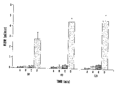
Figure 1 demonstrates the effects of ultrasound on blood flow after
femoral artery thrombolysis. Thrombi were formed by constriction of the
femoral
artery and external application of ferric chloride. Controls (A) received no
treatment. Treatment was administered with 40 kHz ultrasound at 0.75 W/cm2
(B), streptokinase administered as an intravenous bolus of 15,000 U/kg
followed
by an infusion of 15,000 U/kg/hr (C) or the combination of both ultrasound and
CA 02350028 2002-01-21
WO 00/27293 PCT/US99/26163
-4-
streptokinase (D). Data are shown as mean SD. There were 9, 7, 6 and 6
rabbits in groups A, B, C and D, respectively.
Figure 2 shows histologic changes in vessels exposed to US. Figure 2A
shows vessel walls with endothelial cells showing occasional cytoplasmic
vacuoles (arrowheads) and lifting of a single endothelial cell (arrow). Figure
2B
shows a Segment of treated artery with all endothelial cells showing
cytoplasmic
vacuoles (arrowheads), and some rounding up with lifting (arrow). Figure 2C
shows a segment of artery with vacuolization (arrowheads) and complete lifting
of
an endothelial cell (arrow) with erythrocytes under the lifted endothelial
cell in
contact with basement membrane. Bar - 20 micrometers.
Figure 3 is a schematic drawing of the experimental site.
Figure 4 shows the effects of ultrasound on tissue perfusion and pH. For
Figure 4A, perfusion was measured using a laser-Doppler probe placed over the
gracilus muscle near the center of the transducer. In control animals (dotted
line)
perfusion was reduced after clot formation and declined further over the
period of
observation. The nine animals receiving 40 kHz ultrasound at 0.75 W/cm2 (solid
line) demonstrated increased perfusion until 60 min when the ultrasound
transducer was turned off. Ultrasound was applied again between 120 min and
180 min and then turned off again. For Figure 4B, the same protocol was
followed using an electrode to measure muscle pH.
Figure 5 shows the regional distribution of tissue perfusion and pH in
relation to ihe,,l n of the ultrasound transducer. In Figure 5A tissue
perfusion
was measured in five rabbits by placing the probe near the center of the
transducer
(0 cm) or 1, 2, 3, or 4 an distally. In control limbs (no clot), there was no
external
constriction or thrombosis. Following formation of a clot, tissue perfusion
was
reduced. 40 kHz ultrasound at 0.75 Wlem2 was then applied for 30 or 60 min.
The diameter of the insonified area was I cm (Figure 3) The same protocol was
used with measurement of tissue pH in five additional rabbits (Figure 5B). No
thrombolytic therapy was given, and there was no flow in the femoral artery
throughout the experiment. Data are mean SD.
Figures 6A and 6B the effect on tissue perfusion and pH after the right
femoral artery was ligated causing total occlusion (occl). TPU was measured
with
a laser Doppler probe and pH with a sensitive surface electrode. Over the
CA 02350028 2002-01-21
WO 00/27293 PCT/US99126163
-5-
duration of the two hour experiment the tissue perfusion and pH remained
stable
or declined slightly.
Figures 7A and 7B show the effects of ultrasound on tissue perfusion and
pH in the ischemic rabbit leg. Following ligation and occlusion of the femoral
artery, tissue perfusion and pH declined. Application of 40 kHz ultrasound at
0.75
W/cm2 was then commenced and continued for 60 minutes. Over this time, tissue
perfusion improved (maximum effect at 60 minutes) and pH increased during the
duration of ultrasound exposure from 30 minutes to 90 minutes to over 7.3.
After
discontinuation of US, both tissue perfusion and pH declined.
Figures 8A and 8B show the effect of the nitric oxide inhibitor L-NAMA
on ultrasound induced changes in pH and tissue perfusion. The experiments in
Figures 3 and 4 were repeated, but in animals that had been pretreated with L-
NAMA. Ultrasound was applied for one hour following vessel occlusion. No
change in pH or TPU was observed.
DETAILED DESCRIPTION OF THE INVENTION
The present invention provides broadly applicable methods for enhancing
tissue perfusion or growth and accelerating the recovery of damaged tissues
using
ultrasound.
Vasodilation in ischemic tissue may be improved by applying ultrasound
to said ische iF~issue: under con4i4aus. effective to increase blood flow to
said
ischemic tissue. Ultrasound has been used previously to disrupt blood clots to
treat ischemia, however, the present invention provides a method for
increasing,
vasodilation by treating the surrounding ischemic tissue with ultrasound. Such
treatment results in increased vasodilation and blood flow in the tissue. This
approach provides substantial benefit even when the obstruction can not be
disrupted, such as a surgical severing of the blood vessel.
In a preferred embodiment of the invention ultrasound is used to treat
arterial obstruction. Arterial obstruction may be acute vascular obstruction
or
chronic vascular obstruction. Obstruction may results from any number of
internal or external factors. Examples include obstructions resulting from
clots,
CA 02350028 2002-01-21
WO 00127293 PCT1US99126163
-6-
sutures (i.e. vessels being tied oaf), tumors, emboli, hematomas, trauma,
vasospasm, and artherosclorosis.
In one particular embodiment, the present invention may be used to treat
patients after subarachnoid hemmorages to prevent or control vasospasm.
Ultrasound may be applied to the tissue in a variety of manners. It may be
applied to surgically exposed muscle. Alternatively it may be applied to skin
overlying the tissue to be treated. Another approach is to place the
ultrasound
transmitter on a catheter which can be introduced into the area without full
scale
surgery.
In a preferred embodiment, the ultrasound is applied to the tissue by
placing an ultrasound transducer adjacent to the tissue and then subjecting
the
tissue to ultrasound treatment with said ultrasound transducer. In a preferred
embodiment, the ultrasound transducer operates at a frequency of from about 10
to 300 kHz. Preferably, the ultrasound transducer operates at an intensity of
from
about 0.25 to 2 W/cm2. In a more preferred embodiment, the ultrasound
transducer operates at a frequency of about 40 kHz and an intensity of about
0.75
W/cm2.
The present invention also provides a method of treating tissue damage by
applying ultrasound to said tissue damage under conditions effective to treat
said
tissue damage. Although the method is broadly applicable to damaged tissues,
it
is particularly suited to treating damage which is the result of a stroke,
coronary
artery occlusion, or periphcralA{rterial occlusion.
Another embodiment of the invention provides a method of accelerating
wound healing in a patient. Wound healing can be accelerated by applying
ultrasound to said wound under conditions effective to increase blood flow to
said
wound or to increase the production of nitric oxide. Although the method of
the
invention may be used with a wide variety of different wounds, for example,
the
method may be used to accelerate the healing of a bone fracture or an ischemic
ulcer.
The invention also provides a method for increasing the production of
nitric oxide by applying ultrasound to tissue under conditions effective to
increase
nitric oxide concentrations. Such an approach may be used to increase nitric
oxide production to regulate vascular tone. More specifically, the method may
be
CA 02350028 2002-01-21
WO 0087293 PCTNS99/26163
-7-
used to increase nitric oxide production to increase vasodilation. In a
preferred
embodiment, the tissue being treated is ischemic tissue.
Nitric Oxide ("NO") has been demonstrated to play a key role in a wide
variety of physiological pathways (34-36). NO is a uniquely diffusible and
reactive molecular messenger in the vascular and immune systems. In the
peripheral nervous system, NO acts as a classical neurotransmitter in
regulating
gastrointestinal motility, regional blood flow, and neuroendocrine function.
In the
brain, NO acts as a neuromodulator to control behavioral activity, influence
memory function, and intensify responses to painful stimuli. NO is not
restricted
to neurons. Skeletal muscle is also a major source of NO where NO regulates
both metabolism and muscle contractility.
Increased NO production can modulate tissue injury. For example large
amounts of NO produced during ischemia mediate neuronal injury resulting from
stroke (39). NO-mediated damage may account for neurodegeration in a number
of other conditions as well, including Parkinson's disease, amyotrophic
lateral
sclerosis, and Huntington's disease. NO signaling is also perturbed in various
muscle diseases, particularly in Duchenne muscular dystrophy, and that
perturbation may contribute to the disease process.
Physiological functions for neuron-derived NO were first demonstrated in
the gastrointestinal tract. Molecular biological studies have helped detail
the
mechanisms for NO-mediated neurotransmission. In the intestine, neuronal NOS
(nNOS) is selectively concert r ted in axon varicosities of myenteric neurons.
Adjacent intestinal smooth muscle cells contain an "NO receptor," the soluble
guanylyl cyclase. During intestinal peristalsis, myenteric neurons fire action
potentials, and the resulting calcium influx activates calmodulin, which in
turn
stimulates nNOS. The NO then diffuses into adjacent smooth muscle cells and
augments accumulation of cOMP, which mediates intestinal relaxation. Knockout
mice lacking nNOS display a grossly enlarged stomach that histologically
resembles the human disease hypertrophic pyloric stenosis (40). Alterations in
NOS may also play a causal role in some newborns with this disorder, as recent
genetic studies indicate that nNOS is a suseptibility locus for infantile
pyloric
stenosis (41).
CA 02350028 2002-01-21
WO 00127293 PCT/US99126163
-8-
Neuron-derived NO also plays a major role in regulation of blood flow. In
.brain, neuronal activity is associated with an increase in local blood flow,
and this
response is prevented by NOS inhibitors (42). Particularly high levels of nNOS
occur in vasodilator nerves that innervate the large cerebral blood vessels
(43).
Abnormal reactivity of these vessels appears to mediate migraine headache, as
sumatriptan constricts these large vessels and controls headache (44).
Sumatriptan is also effective in treatment of nitroglycerininduced headache,
suggesting a role for endogenous NO in migraine. Therefore, pharmacological
manipulations of nNOS may offer an avenue for migraine therapy.
Neuron-derived NO also mediates penile erection through regulation of
blood flow. nNOS is enriched in neurons of the pelvic plexus and NOS
inhibitors
block penile erection in animal models in vivo (45) and in strips of human
cavernosal tissue in vitro (46). However, nNOS mutant mice display normal
erectile function (47). Apparently NO derived from other NOS isoforms
compensates for the loss of nNOS, as NOS inhibitors block penile erection in
nNOS mutant mice. Recent studies demonstrate that abnormalities in NO
biosynthesis may also underlie erectile dysfunction. Diabetes mellitus is
associated with impaired NOS-dependent erectile function (48). NOS levels in
penis are also decreased in aging rats, and this age-related decrease
correlates with
impaired erectile responses (49). Androgens are essential for penile reflexes
in
the rat and are also essential for normal libido. Similarly, nNOS expression
in
penis is dependent upon active androgens as nNOS levels decrease,by 60% l wk
after castration but are restored to normal levels with testosterone
replacement
(50). Therefore, pharmacological manipulation of NO or NOS expression may
offer a viable strategy for treatment for some causes of erectile dysfunction.
Because NO is a uniquely diffusible mediator, it was proposed on
theoretical grounds that NO may mediate neuronal plasticity, which underlies
aspects of both development and information storage in brain. Evidence for NO
involvement in synaptic plasticity has accumulated steadily. At the cellular
level,
NO signaling appears to be essential for two forms of neuronal plasticity:
long-
term potentiation (LTP) in the hippocampus (51) and long-term depression in
the
cerebellum (52). In these cellular models, repeated neuronal stimulation
yields
long-lasting changes in synaptic strength. NOS inhibitors prevent these
changes.
CA 02350028 2002-01-21
WO 00/27293 PCTNS99t26163
-9-
Studies with NOS inhibitors have been controversial because these arginine
analogues often have nonspecific effects. This controversy may now be resolved
by studies of NOS knockout mice. Both endothelial NOS (eNOS) and nNOS
activities are found in hippocampus. Mice that either lack eNOS or nNOS have
essentially normal LTP, whereas mutant mice deficient in both eNOS and nNOS
have substantially decreased LTP (53).
Although endogenous NO was originally appreciated as a mediator of
smooth muscle relaxation, more 'recent studies indicate a role for NO in
skeletal
muscle as well. nNOS mRNA is expressed at high levels in human skeletal
muscle (37), where it is alternatively spliced, yielding a muscle-specific
isoform
(nNOSmiero) (54). Understanding functions for nNOS in skeletal muscle has
been facilitated by the discrete localization of nNOS in myofibers. In rodent
muscle, nNOS is specifically enriched beneath the sarcolemma of fast twitch
muscle fibers (38). NOS activity stimulated during muscle membrane
depolarization inhibits contractile force in fast twitch fibers.
In addition to modulating contractile force, NO derived from sarcolemmal
nNOS regulates physiologic functions at the muscle membrane. During muscle
development, myocytes fuse to form muscle myotubes, and this membrane fusion
is blunted by NOS inhibitors (55). In myocyte/motor neuron co-cultures, NOS
produced at the postsynaptic muscle membrane functions as a retrograde
messenger to regulate myotube innervation (56). In mature muscle fibers, NOS
regulates glucose uptake across thess p rcolemma. Glucose uptake in skeletal
muscle is regulated by both acute exercise and by insulin. NOS inhibitors
selectively blunt exercise-induced uptake but have no effect on insulin-
stimulated
glucose transport (57). Interestingly, chronic exercise increases nNOS protein
expression in muscle and this has long-lasting enhancing effects on glucose
transport in heavily used muscle (57).'
Since NO plays an important role in neurodegenerative and muscle
diseases, the present invention can be used to treat neurodegenerative and
muscle
diseases by applying ultrasound to diseased tissue under conditions effective
to
increase nitric oxide production in the diseased tissue.
CA 02350028 2002-01-21
WO 00121293 PCTIUS"/26163
-10-
EXAMPLES
Exgmnle 1 - Materials and Methods
Animal Pregaration: Rabbits were anesthetized with ketamine (60 mg/kg),
xylazine (6 mg/kg) and chlorpromazine (25 mg/kg), and sedation was maintained
with sodium pentobarbital as needed. The femoral arteries were dissected 5 cm
distal to the origin of the superficial branch, and the profunda femorus and
superficial arteries were ligated close to their origin. A Doppler flow probe
was
placed distally around the isolated segment and two parallel ligatures were
placed around the femoral artery 1 cm distal to the profunda branch. These
reduced flow by approximately 50% remained in place for the duration of the
experiment. Following this, of filter paper saturated with 20% ferric chloride
was
placed on the femoral artery and thrombosis was assessed by monitoring flow
which approached 0 after occlusion. In some animals a completely occlusive
suture was placed around the artery.
Experimental Protocol: Rabbits were assigned to receive: 1) ultrasound
alone, 2) streptokinase alone, 3) both ultrasound and streptokinase, or 4) no
treatment. There were 7, 6, 6 and 9 rabbits in groups 1, 2, 3 and 4,
respectively.
The source of ultrasound was a 3.5 cm diameter probe with a 1 cm diameter
transducer driven in continuous mode at 0.75 W/cm2, and acoustic pressures
were
geaspted.llefore and after each experiment with a hydrophone. A ba loon filled
with water at 37 C was placed over the thrombosed segment for temperature
control and ultrasound transduction. The interface between the balloon and the
artery was covered by a layer of ultrasound transmission gel. Streptokinase
was
administered as an intravenous bolus of 15,000 U/kg followed by an infusion of
15,000 U/kg/hr for two hours. This dose was selected because our prior
experience with this model indicated that it was relatively ineffective alone,
that I
MHz ultrasound enhanced its effects (18) and data in vitro indicated that 40
kHz
had a greater effect on thrombolysis-than 1 MHz ultrasound (14). The pH of the
muscle was also monitored using a pH meter (Model HI 9023C, Hanna
Instruments, Woonsocket, RI). Perfusion in the gracilis muscle was measured
using a laser-Doppler flow meter (BFL 21, Transonic Systems)with an output in
CA 02350028 2002-01-21
WO 00/27293 PCT/US99126163
-Il-
units (TPU) that is linearly related to the number of red cells times their
velocity
in the hemispheric measuring volume (26-28). The measuring surface was 1 mm2,
and the light penetration depth was approximately 1 mm.
Temperature monitoring in four rabbits was performed with a copper-
constantin fine wire thermocouple placed under the femoral artery or on the
exposed surface of the femur and connected to a temperature gauge. To assess
the
effects of heating on tissue perfusion, a balloon containing water at 32 C to
42 C
was laid over the gracilis muscle. At the completion of each experiment,
animals
were euthanized and samples for histology were excised and fixed in 10%
buffered formalin. Specimens were processed in paraffin, sectioned at 4
microns,
and stained with hematoxylin and eosin. The stained sections were encoded to
obscure treatment, and examined by an observer (RBB) blinded as to code and
particular attention was paid to the endothelial cells.
Statistical Methods: The three primary outcome measures that were used
to assess the effect of ultrasound were flow intensity, TPU and pH. The mixed
linear model was used for statistical analysis of each of the primary
measures.
The responses were grouped into clusters by the individual animal (random
effect)
and were treated as repeated measurements taken over time and/or at different
distances. Based on these models, the least square means, their standard
errors
and covariances were calculated, and the adjusted differences between
treatment
means at different time points were obtained. They were used for testing for
tfe4tmcttt c fect as well as for effect sizes for each level of grouping
variables.
Example 2 - Treatment of Surgically Induced Arterial Occlusion with
Ultrasound
Occlusive thrombi formed in all femoral arteries within 20-30 min of
placement of the constriction and application of 20% ferric chloride. Arterial
flow
was 12.0 0.7 ml/min at baseline, declined to 5.8 0.4 after placement of
the
constriction and was 0.1 0.1 after thrombosis. Three different treatment
regimens were administered following thrombosis: ultrasound alone,
streptokinase alone or the combination of streptokinase and US. Flow in
control
vessels receiving no treatment remained at near 0 after 30, 60, and 120 min
(Fig.
CA 02350028 2002-01-21
WO 00/27293 PCTNS99126163
-12-
1). Treatment with ultrasound alone resulted in no significant increase in
flow,
whereas treatment with streptokinase alone resulted in a small but
significantly
increased flow at 120 min to 0.4:t0.1 ml/min (p < 0.001). The combination of
streptokinase and ultrasound resulted in greater reperfusion, with flow of 2.6
0.7
ml/min at 30 min, 4.6 0.4 ml/min at 60 min and 4.8 0.6 ml/min at 120 min.
The 120 minute flow represented 83% of the flow of 5.8 0.4 ml/min after
placement of the external constrictor but prior to thrombosis. These results
indicate that the application of ultrasound markedly accelerates arterial
reperfusion as compared with streptokinase alone and that ultrasound by itself
had
no appreciable effect.
Exgmpe -Monitoring of Heating by Ultrasound
US application can cause heating, and temperature was monitored using a
thermocouple placed adjacent to the thrombosed vessel or at the surface of the
femur. With application of ultrasound the average initial temperature increase
at
the femoral artery was 0.02 C/min, and it was 0.04 C/min at the surface of the
femur. The maximum temperature increase after 60 min was 1.6 t 1.3 C at the
femoral artery and L I t 0.7 C at the femur. Histologically, examination
showed
that vessels exposed to US, regardless of other treatment components
(streptokinase, ligation, clot), had a pronounced tendency for endothelial
cell
vacuolization, and some cells lifted up off the underlying basement membrane
(Fig. 2). Occasionally, erythrocytes were seen in direct contact with the
basement
membrane.
Example 4 -Capillary Perfusion after Ultrasound Treatment
During these experiments, it was observed that muscle adjacent to the
femoral artery lost its normal pink, vital color soon after thrombosis and
became a
brownish-purple. Application of ultrasound restored the normal pink color even
when no thrombolysis occurred and femoral artery flow remained near zero.
Therefore, perfusion in the gracilis muscle was characterized using a probe
which
CA 02350028 2002-01-21
WO 00127293 PCTNS99126163
-13-
is sensitive to capillary blood flow (Fig. 3). In control experiments, tissue
perfusion was stable for periods up to 60 min indicating that application of
the
unactivated probe by itself did not tissue perfusion. At baseline, prior to
vessel
constriction or thrombosis, capillary perfusion was 13.7 t 0.4 U (Fig. 4A).
This
declined to 6.8:t 0.4 U immediately after thrombosis and then declined
progressively to 4.5 t 0.4 U after 240 min in animals receiving no treatment.
The
application of ultrasound resulted in a significant increase in perfusion to
10.0 t
0.5 U at 30 min and a further increase to 12.1 0.5 U at 60 min (p < 0.001
for
both). To determine if the effect of ultrasound was reversible, the transducer
was
switched off at 60 min, and perfusion then declined progressively to 9.7 0.2
U at
90 min and to 8.5;t 0.2 U at 120 min (p < 0.001 for both in comparison with 60
min). At 120 min the transducer was reactivated, and this resulted again in
improved perfusion to 11.8 0.8 U at 1 50 min and 12.7 0.4 U at 180 min (p
<
0.001 for both in comparison with 120 min). When the transducer was switched
off at 180 min, perfusion again declined and reached 8.2 0.8 U at 240 min.
The
Doppler flow probe placed distally on the artery showed no flow for the
duration
of the experiment. The same changes were observed when the vessel was ligated
completely with sutures to preclude any change in femoral artery flow. Because
tissue perfusion is sensitive to temperature, experiments were carried out to
determine whether US-induced heating could explain the changes. Muscle was
heated from 32 C to 42 C using warm water in a balloon and TPU increased from
6.2 to 7.2 U over this temperature range. Since the maximum temperature
increase with ultrasound was less than 2 C, heating alone could not account
for
the US-induced increase in perfusion in the ischemic area.
Example 5 - Metabolism and Acidosis in Ultrasound Treated Ischemic
Muscle
The increased tissue perfusion following application of ultrasound
appeared to improve the metabolism of ischemic muscle and ameliorate acidosis,
and this was investigated with a similar experimental design using a pH probe
(Fig. 4B). Following surgical exposure but prior to thrombosis, the baseline
muscle pH was 7.41 0.02, but this declined to 7.05 0.02 after thrombotic
CA 02350028 2002-01-21
WO 00/27293 PCT/US99R6163
-14-
occlusion. In the absence of treatment, pH declined slowly but progressively
to
reach 6.86 0.02 at 240 min. The application of ultrasound reversed the
acidosis,
and muscle pH increased significantly to 7.31 0.02 after 30 min and to 7.34
'
0.03 following 60 min (p < 0.001 for both). At 60 min the ultrasound was
turned
off, and muscle pH declined to 7.17 0.01 at 90 min and then showed little
change to 7.16 0.02 at 120 min. The transducer was then turned on, and pH
again improved to 7.32 0.02 at 150 min and 7.30 0.03 at 180 min. At this
time
the transducer was again turned off, and pH declined to 7.13 0.04 at 210
min.
The differences between means were all significant (p < 0.001).
To determine whether the effect of ultrasound on tissue perfusion and pH
was limited to the insonified tissue, measurements were made at multiple
locations laterally and distally during insonification (Fig. 5A). Perfusion
measured from the center of the transducer to 4 cm distally at baseline was
between 12.4 and 13.6 U, and after thrombosis it decreased to between 6.3 and
6.5
U. Perfusion measurements were then made after application of ultrasound for
30
and 60 min. At 0 cm (Fig 5A), perfusion increased to 11.1 0.5 U at 30 min
and
further to 14.0 0.6 U at 60 min. The effect declined at sites distal from
the
center of the transducer, This was most evident at 60 min with values of 12.1
t
0.4, 8.3 0.3, 6.0 0.3 and 5.3 0.3 at 1, 2, 3, and 4 cm, respectively.
The
readings at 3 and 4 cm were the same as those in control animals not exposed
to
US. Since the diameter of the transducer was 1 cm, these findings suggest that
the
ultrasound effect is limited to the insonified tissue. In other experiments,
the
transducer was applied at sites 1, 2, 3 and 4 can distally. Insonification at
these
sites resulted in normalization of TPU, indicating that the effect could be
induced
at these sites, but required direct ultrasound exposure.
Similar experiments were performed measuring muscle pH (Fig. 5B). At
baseline and before thrombosis, muscle pH was between 7.36 and 7.42 within the
4 cm area. This declined to between 7.03 and 7.08 after thrombus formation.
Ultrasound application improved tissue pH at the center of the transducer to
7.34
0.04 at 30 min and to 7.39 0.07 units at 60 min. As with perfusion (Fig.
5A),
the effect was limited to the insonified area, and muscle pH at 3 and 4 cm was
not
improved during insonification.
CA 02350028 2002-01-21
WO 00/27293 PCT/US99/26163
-15-
Exa Rk ¾ - Investigation of the Role of Nitric Oxide
Nitric oxide is an important regulator of vascular tone, and its synthesis
can be affected by mechanical stresses such as alterations in flow. Therefore,
it
was hypothesized that ultrasound may improve tissue perfusion and pH in
ischemic tissue by effecting nitric oxide production. To test this hypothesis,
experiments were repeated in the'rabbit femoral artery thrombosis model to
document the effect of ultrasound on acidosis and tissue perfusion. An
inhibitor
of nitric oxide production, N-Nitro-L-arginine methyl esther hydrochloride
(MAMA) was incorporated into the experiments.
Methods: The rabbit femoral artery thrombosistligation method as
described above was used. 40 kHz ultrasound was applied at 0.75 W/cm2 in
selected limbs and omitted in controls. In some animals L-NAMA was injected in
a bolus of 50 mg/kg 30 minutes before occlusion of the femoral artery.
Controls
received no NAMA. Measurements of tissue perfusion using a laser Doppler
probe and of muscle pH were performed as described previously.
In control animals receiving no ultrasound tissue perfusion (Figure 6A)
and pH (Figure 6B) declined after occlusion of the femoral artery and remained
low for the duration of the experiment. Application of 40 kHz ultrasound at
0.75
W/cm2 resulted in improvement in tissue perfusion (Figure 7A) and pH (Figure
7B) as noted previously. The effect was reversible after ultrasound was
discontinued in those experiments. After administration of NAMA the effect of
ultrasound on TPU (Figure 8A) and pH (Figure 8B) was eliminated and perfusion
and pH remained low or declined for the duration of the experiment, similar to
the
findings in the control animals (Figures 6A and 6B). The administration of L-
NAMA alone resulted in no change in control experiments in the absence of US.
The results confirm and extend the previous findings examining the effects
of 40 kHz ultrasound on tissue perfusion and pH in the rabbit ischemic leg
model.
Application of 40 kHz ultrasound at 0.75 W/cm2 improved tissue perfusion and
pH in the grasilus muscle. Pretreatment of the animals with L-NAMA, a
selective
nitric oxide inhibitor, completely aggregated the effects of US, and the
animals
behaved as controls in the absence of US. These findings indicate that the
CA 02350028 2002-01-21
WO 00/27293 PCT/11S99/26163
-16-
beneficial effect of ultrasound on tissue perfusion and pH requires an intact
nitric
oxide system.
Femoral artery thrombosis results in distal muscle ischemia and metabolic
changes including acidosis. In the experiments reported, muscle perfusion was
measured using a probe sensitive to movement of red blood cells to a depth of
approximately I mm in the regional microcirculation (31). Unexpectedly, the
application of ultrasound improved tissue perfusion, and this resulted in
reversal
of acidosis. Although the laser-Doppler measurement is limited to 1 mm in
depth, the prolonged duration of the ultrasound effect accompanied by
normalization of tissue pH and muscle color suggest that it was more general.
This occurred without clot lysis and was observed even when the vessel was
completely ligated, indicating that reperfusion with flow through the femoral
artery was not the explanation.. The improved perfusion was reversible, and
the
effect was limited to the insonified area with no discernable increase in
perfusion
either distally or laterally. The proximal leg muscles receive their primary
blood
supply from the femoral artery, and occlusion reduced perfusion to
approximately
50'/ of baseline, but the residual perfusion following occlusion indicates
that an
alternate arterial supply provided collateral flow. The US-induced increase in
perfusion in the absence of femoral artery flow suggests that arterial supply
through these collateral vessels increased.
The mechanism of improved perfusion is unclear, but redistribution of
collateral flow into the ischemic area may be modulated by neural or hormonal
influences. Humoral mediators of vasomotor tone include endothelin,
prostacyclin, and nitric oxide. The local secretion of nitric oxide is
regulated by
nitric oxide synthase, an enzyme which can be induced by mechanical stress on
endothelial cells (32,33) that could result from US. Thus, we hypothesize that
nitric oxide-induced redistribution of flow may account for the effects of US,
but
further studies will be required to elucidate the physiologic mechanisms.
Therapeutic application will require careful attention to limiting application
of
ultrasound only to ischemic tissue, as inappropriate insonification to
adjacent non-
ischemic tissues could result in redistribution of flow away from ischemic
tissue,
thereby extending ischemia.
CA 02350028 2012-06-07
WO 00/27293 PCTIUS99/26163
- 17-
The increased tissue perfusion resulting from ultrasound has the potential
to improve clinical outcomes. Rapid reversal of tissue ischemia is essential
in
preventing myocardial necrosis and particularly neuronal loss with stroke.
This
could be achieved through either removal of the arterial obstruction or by an
increase flow to the ischemic area through collateral vessels. The
augmentation of
collateral flow offers a new approach to increasing perfusion of ischemic
tissue
and limiting dysfunction and necrosis.
Although preferred embodiments have been depicted and described in
detail herein, it will be apparent to those skilled in the relevant art that
various
modifications, additions, substitutions, and the like can be made. The claims
are to
be given a purposive construction, in view of the application as a whole.
CA 02350028 2008-04-04
= WO 00/27293 PCT/US99/26163
-18-
References Cited
The following references were cited herein.
1. Lauer CG, Burge R, Tang DB, Bass BG, Gomez ER, Alving BM. Effect of
US on tissue-type plasminogen activator-induced thrombolysis. Circulation.
1992;86:1257-1264.
2. Francis CW, Onundarson PT, Carstensen EL, Blinc A, Meltzer RS..
Enhancement of fibrinolysis in vitro by US. JClin Invest. 1992;90:2063-
2068.
3. Luo H, Steffen W, Cercek B, Arunasalam S, Maurer G, Siegel RJ.
Enhancement of thrombolysis by external US. Am Heart J. 1993; 1125:1564-
1569.
4. Tachibana K. Enhancement of fibrinolysis with US energy. J Vasc Interv
Radiol. 1992;3:299-303.
5. Suchkova V, Siddiqi FN, Carstensen EL, Dalecki D, Child S, Francis CW.
Enhancement of fibrinolysis with 40-kHz US. Circulation. 1998;98:1030-
1035.
6. Hamano K. Thrombolysis enhanced by transcutaneous ultrasonic irradiation.
Tokyo Jikekai MedJ. 1991;106:533-542.
7. Kornowski R, Meltzer RS, Chernine A, Vered Z, Battler A. Does external US
accelerate thrombolysis? Results from a rabbit model. Circulation.
1994;89:339-344.
8. Riggs PN, Francis CW, Bartos RS, Penney DP. US enhancement of rabbit
femoral artery thrombolysis. Cardiovasc Surg. 1997;5:201-207.
9. Luo H, Nishioka T, Fishbein MC, Cercek BM Forrester JS, Kim C-J,
Berglund H. Siegel RJ. Myocardial ischemia/infarction/thrombolysis:
transcutaneous US augments lysis of arterial thrombi in vivo. Circulation.
1996;94:775-778.
10. Kashyap A, Blinc A, Marder VJ, Penney DP, Francis CW. Acceleration of
fibrinolysis by US in a rabbit ear model of small vessel injury. Thromb Res.
1994;76:475-485.
CA 02350028 2002-01-21
WO 00/27293 PCT/US99/16163
-19-
11. Birnbaum Y, Luo H, Nagai T, Fishbein MC, Peterson TM, Li S, Kricsfeld D,
Porter TR, Siegel RJ. Noninvasive in vivo clot dissolution without a
thrombolytic drug: recanalization of thrombosed iliofemoral arteries by
transcutaneous US combined with intravenous infusion of microbubbles.
Circulation. 1998; 97:130-134.
12. Shlansky-Goldberg RD, Cinea DB, Sehgal CM. Catheter-delivered US
potentiates in vitro thrombolysis..1 Vase Interv Radio[. 1996;7:313-320.
13. Sehgal CM, Leven RF, Shlansky-Goldberg RD. US-assisted thrombolysis.
Invest Radial. 1993;28:939-943.
14. Haberl RL, Heizer ML, Marmarou A, Ellis EF. Laser-Doppler assessment of
brain microcirculation: effect of systemic alterations. Am J Physiol.
1989;256:1247-1254.
15. Summaria L, Arzadon L, Bernabe P, Robbins KC. The interaction of
streptokinase with human cat, dog, and rabbit plasminogens. J Biol Chem.
1974;249:4760-4769.
16. Blinc A, Francis CW. Transport processes in fibrinolysis and fibrinolytic
therapy. Thromb Haemost. 1996;76:481-491.
17. Francis CW, Blinc A, Lee S, Cox C. US accelerates transport of recombinant
tissue plasminogen activator into clots. US Med Biol. 1995;21:419-424.
18. Siddiqi F, Blinc A, Braaten J, Francis CW. US increases flow through
fibrin
gels. Thromb Haemost. 1995;73:495-498.
19. Braaten JV, Goss RA, Francis CW. US reversibly disaggregates fibrin
fibers.
Thromb Haemost 1997;78:1063-1068.
20. Siddiqi F, Odrljin TM, Fay PJ, Cox C, Francis CW. Binding of tissue-
plasminogen activator to fibrin: Effect of US. Blood 1998;91:2019-2025.
21. Nishioka T, Luo H. Fishbein MC, Cercek B, Forrester JS, Kim C-J, Bergima
J,
Siegel RL. Dissolution of thrombotic arterial occlusion by high intensity, low
frequency US and dodecafluoropentane emulsion: An in vitro and in vivo
study. JAm Coll Cardiol. 1997;30:561-568.
22. Rosenschein U, Bernstein JJ, DdiSegni E, Kaplinsky E, Bernheim J,
Rozenzsajn LA. Experimental ultrasonic angioplasty: Disruption of
atherosclerotic plaque and thrombi in vitro and arterial recanalization in
vivo.
JAm Coll Cardiol. 1990;15:711-717.
CA 02350028 2002-01-21
WO 00/27293 PCT/1JS99/26163
- 20 -
23. Arian M, Fishbein MC, Chae JS, Sadeghu H, DonMichael A, Dubin SB,
Siegel RJ. Dissolution of peripheral arterial thrombi by US. Circulation.
1991;84:1680-1688.
24. Trubstein G, Engel C, Etzerl F, Sobbe H, Cremer H, Stumpff U.
Thrombolysis by US. Clin Sci Mol Med. I976;51:697s-698s.
25. Eccleston DS, Cumpston GN, Hodge AJ, Peame-Rowe D, DonMichael TA.
Ultrasonic coronary angioplasty during coronary artery bypass grafting. JAm
Coll Cardfol. 19%-,78-1172-1175.
26. Rosenschein U, Rozenszajn LA, Kraus L, Marboe CC, Watkins JF, Rose EA,
David D, Cannon PJ, Weinstein IS. Ultrasonic angioplasty in totally occluded
peripheral arteries: initial clinical, histological, and angiographic results.
Circulation. 1991;83:1976-1986.
27. Siegel RJ, Gaines P, Crew JR, Cumberland DC. Clinical trial of
percutaneous
peripheral US angioplasty. JAm Coll Cardfol. 1993;22:480-488.
28. Rosenschein U, Gaul G, Erbel R, Amann F, Velasguez D, Stoerger H, Simon
R, Gomez G, Troster J, Bartorelli A, Peiper M, Kyriakides Z, Laniado S,
Miller HI, Cribier A, Fajedet J. Percutaneous transluminal therapy of
occluded saphenous vein grafts. Can the challenge be met with US
thrombolysis? Circulation 1999;99:26-29.
29. Behrens S, Daffertshofer M, Spiegel D, Hennerici M. Low frequency, low-
intensity US accelerates thrombolysis through the skull. US Med Biol.
1999;25:269-273.
30. Akiyama M. Ishibashi T, Yamada T, Furuhata H. Low-frequency US
penetrates the cranium and enhances thrombolysis in vitro. Neurosurgery
1998;43:828-833.
31. Stern MD, Lappe DL, Bowen PD, himosky JE, Holloway GA Jr, Keiser HR,
Bowman RL. Continuous measurement of tissue blood flow by laser-Doppler
spectroscopy. Am J Physiol. 1977;232:441-448.
32. Corson MA, James NL, Latta SE, Nerem RM, Berk BC, Harrison DO.
Phosphorylation of endothelial nitric oxide synthase in response to fluid
shear
stress. Circ Res. 1996;9:984-991.
CA 02350028 2002-01-21
WO 00/27293 PCT/US99/26163
-21-
33. Uematsu M. Ohara Y. Navas JP, Nishida K, Murphy TJ, Alexander RW,
Nerem RM, Harrison DG. Regulation of endothelial cell nitric oxide synthase
mRNA expression by shear stress. Am JPhysiol. 1995;269:1371-1378.
34. Christopherson KS. and Bredt DS. Nitric Oxide in Excitable Tissues:
Physiological Roles and Disease. J. Clin. Investig. 1997;100:2424-2429.
35. Bredt DS and Snyder SH. Nitric Oxide: A Physiologic Messenger Molecule.
Annu. Rev. Biochem. 1994;63:175-195.
36. Cooke J.P. and Dzau, VJ. Derangements of the Nitric Oxide Synthase
Pathway, L-arginine and Cardiovascular Diseases. Circulation 1997;96:379-
382.
37..Nakane M. Schmidt HH; Pollock JS, Forstennann U and Murad F. Cloned
human brain nitric oxide synthase is highly expressed in skeletal muscle.
FEBS Lett. 1993;316:175-180.
38. Kobzik L, Reid MB, Bredt DS and Stamler JS. Nitric oxide in skeletal
muscle. Nature 1994;372:546-548.
39. Samdani. AF, Dawson TM, and Dawson VL. Nitric Oxide Synthase in
Models of Focal Ischemia. Stroke 1997;28:1283-1288.
40. Huang PL, Dawson TM, Bredt DS, Snyder SH, Fishman MC. Targeted
Disruption of the Neuronal Nitric Oxide Synthase Gene. Cell 1993;75:1273-
1286.
41. Chung ED, Curtis G, Chen PA, Marsden R, Twells R, Xu W, and Gardiner M.
Genetic Evidence for the Neuronal Nitric Oxide Synthase Gene (NOS1) as a
Susceptibility Locus for Infantile Pyloric Stenosis. Am. J. Hum. Genet.
1996;58:363-370.
42. Iadecola C, Zhang F, and Xu X. Role of nitric oxide synthase-containing
vascular nerves in cerebrovasodilation elicited from cerebellum. An J.
PhysioL 1993;264:R738-R746.
43. Bredt DS, Hwang PM, and Snyder, SH. Localization of nitric oxide synthase
indicating a neural role for nitric oxide. Nature 1990;347:768-770.
44. Welch KM. Drug therapy of migraine [see comments). N. Engl. J. Med.
1993;329:1476-1483.
45. Burnett AL, Tillman SL, Chang TS, Epstein JI, Lowenstein CJ, Bredt DS,
Snyder S11, Walsh PC. Immunohistochemical localization of nitric oxide
CA 02350028 2002-01-21
WO 00/27293 PCT/US"/26163
-22
synthase in the autonomic innervation of the human penis. J Urol.
1993;150:73-76.
46. Raijfer J, Aronson WJ, Bush PA, Dorey FJ, and Ignarro U. Nitric oxide as a
mediator of relaxation of the corpus cavernosum in response to nonadrenergic,
noncholinergic neurotransmission. N. Engl. J. Med. 1992;326:90-94.
47. Burnett AL, Nelson RI, Calvin DC, Liu JX, Demas GE, Klein SL, Kriegsfeld
LJ, Dawson VL, and Snyder SH. Nitric oxide-dependent penile erection in
mice lacking neuronal nitric oxide synthase. Mol. Med. 1996;2:228-296.
48. Vernet D, Cai L, Garban H, Babbitt ML, Murray FT, Raijfer J, and Gonzalez-
Cadavid NF. Reduction of penile nitric oxide synthase in diabetic
BB/WORdp (type 1) and BBZ/WORdp (type 11) rats with erectile dysfunction.
Endocrinology 1995;136:5709-5717.
49. Carrier S, Nagaraju P, Morgan DM, Baba K, Nunes L, and Lue TF. Age
decreases nitric oxide synthase-containing nerve fibers in the rat penis. J.
Urol. 1997;157:1088-1092.
50. Penson DF, Ng C, Cai L, Raijfer J, and Gonzalez-Cadavid NF. Androgen and
pituitary control of penile nitric oxide synthase and erectile function in the
rat.
Biol. Reprod 1996;55:567-574.
51. Schuman EM, and Madison DV. Nitric oxide and synaptic function. Annu.
Rev. Neurosci. 1"4;17:153-183.
52. Shibuki K, and Okada D. Endogenous nitric oxide release required for long-
term synaptic depression in the cerebellum. Nature 1991;349:326-328.
53. Son H, Hawkins RD, Martin K, Kiebler M, Huang PL, Fishman MC, and
Kandel ER. Long-term potentiation is reduced in mice that are doubly mutant
in endothelial and neuronal nitric oxide synthase. Cell. 1996;87:1015-1023.
54. Silvagno F, Xia H, and Bredt DS. Neuronal nitric oxide synthase-micro, an
alternatively spliced isoform expressed in differentiated skeletal muscle. J.
Biol. Chem. 1996;271:11204-11208.
55. Lee KH, Back MY, Moon KY, Song WK, Chung CH, Ha DB, and Kang MS.
Nitric oxide as a messenger molecule for myoblast fusion. J. Biol. Chem.
1994;269:14371-14374.
CA 02350028 2002-01-21
WO 00/27293 PGT/US99/26163
- 23 -
56. Wang T, Xie Z, and Lu B. Nitric oxide mediates activity-dependent synaptic
suppression at developing neuromuscular synapses. Nature 1995;374:262-
266.
57. Roberts CK, Barnard RJ, Scheck SH, and Balon TW. Exercise-stimulated
glucose transport in skeletal muscle is nitric oxide dependent. Am. J.
Physiol.
1997;273:E220-E225.