Note: Descriptions are shown in the official language in which they were submitted.
CA 02351216 2009-01-07
1
METHOD FOR TISSUE FIXATION USING EPOXIDES
BACKGROUND OF THE INVENTION
1. Field of the Invention
The present invention relates generally to tissue
fixation, and in particular, to an epoxy compound
and method for use in tissue fixation.
2. Description of the Prior Art
Biological tissues such as autologous pericardium
and homologous aortic valves have been used in
various surgical applications because of their good
mechanical properties and biocompatibility.
Biological tissue-derived, chemically-modified
heterologous tissues have been provided as conduits
for peripheral or coronary revascularization,
patches, ligament substitutes, and prosthetic heart
valves. It is well-known that collagen fibers
constitute the fundamental structural framework of
biological tissues.
The physiochemical and biomechanical properties of
collagen matrices are directly related to the
structure of the collagen fibrils. The collagen
molecules are stabilized in the fibrils by covalent
intermolecular crosslinks, which provide the
fibrillate matrices with an adequate degree of
tensile strength and biostability.
After a prosthesis having heterologous tissue has
been implanted in a living host environment, the
biological tissue will be subject to a host
response, which includes both cellular and enzymatic
attack. Previous studies have shown that implanted
heterologous collagenous tissues provoke a cellular
response which leads to physical invasion of the
implanted prosthesis by phacocytes
(polymorphonuclear leukocytes, macrophages) and
fibroblasts. Phagocytes are known to be able to
CA 02351216 2001-05-09
WO 00/35374 PCT/US99/29617
2
secrete collagenase and other proteases and oxygen
free radicals. Heterologous biological tissues can
be readily degraded by such proteolytic enzymes,
and/or through an oxidation process, significantly
reducing the strength and life span of the collagen
fibrils. To achieve long-term stability,
bioprostheses derived from heterologous tissues have
to be chemically modified to increase their
resistance to enzymatic degradation before they can
be implanted into a human being for long term use.
These chemical modifications include:
(1) Crosslinking to stabilize the collagen
matrix, such as enhancing the molecular interaction
between collagen fibrils, elastin and other
proteins; increasing tissue fatigue limit under
stress; and maintaining the tissue integrity and
preventing inflammatory cell infiltration;
(2) Modification of collagenous tissue to
minimize the immunogenicity: heterologous tissue
needs to be modified to reduce the immunogenicity so
that systemic and local adverse effects (e.g.,
chronic inflammation or rejection) will be
minimized;
(3) Modification to minimize enzymatic attack:
chemically modified tissue might be less
recognizable by proteolytic enzymes; and
The crosslinking and modification are preferably
stable to achieve optimum long-term results.
The extent of enzymatically catalyzed breakdown of
fibrous collagen may be influenced by two factors:
the availability to the enzyme of recognizable
cleavage sites, and the extent of the helical
integrity of the collagen. Previous works have
suggested that tissue subjected to fixation and
CA 02351216 2001-05-09
WO 00/35374 PCT/US99/29617
3
having greater crosslinking density will have a
greater resistance to degradation.
Fixation refers to the deactivation of the amino
acid of a collagen by reaction with a chemical to
minimize the antigenicity of the heterologous
biological material and the possibility of enzymatic
degradation by collagenase and other proteases.
Thus, fixation would enhance the durability of the
collagen.
Two types of fixation treatment can be
differentiated. The first type is crosslinking, in
which one molecule of a fixation agent having
multiple functional groups reacts with two or more
groups in a collagen. After crosslinking, the
mechanical properties of the tissue change. The
second type of fixation treatment can be referred to
as branching, in which the fixative reacts with a
single group only, resulting in a branch produced by
the reacted amino acid. In branching, the
mechanical properties (e.g., flexibility) of the
tissue will normally experience little change.
Both cross-linking and branching will alter the
antigenicity of the collagenous tissue if there is
modification of a sufficient amount of amino acids,
and if the grafting structure (i.e., branching) is
large enough to change the local molecular
conformation (i.e., both sequential and conformatial
antigen determination sites/epitopes). A higher
degree of fixation of the fixed. biomaterial (tissue)
will generally result in lower antigenicity.
Since the host cellular and enzymatic activity is
highly associated with inflammation, and the
toxicity of the residual fixative may contribute to
the local chronic inflamation, a minimal residual
toxicity of the prosthesis is desirable.
CA 02351216 2009-01-07
4
Collagenous tissue for blood-contacting
applications, such as for heart valves and conduits,
should also have excellent hemocompatibility.
Hydrophilicity, charge, surface texture and other
surface characteristics on the blood-contacting
surface can significantly impact the performance and
durability of the tissue when used in these
applications. Some trends can be observed in
relation to surface tension and hemocompatibility/
bioadhesion. R.R. Baier and V.A. DePalma, "The
Relation of the Internal Surface of Grafts to
Thrombosis", Management of Arterial Occlusive
Disease, Year Book Medical Publisher, Chicago, IL,
147-163 (1971) has accumulated an extensive amount
of data over many years on the observed trend of
biological reactivity of materials as a function of
their relative critical surface tensions. An
empirically derived graph from their work is divided
into three zones: (1) A first zone, coincident to a
minimum in biological interaction, is the
"hypothetical zone of biocompatability:, which
surface tension ranges from 20 to 30 dynes/cm
(hydrophobic surface). This zone is the range of
surface tensions that most natural arteries possess
and is descriptive of relatively nonthrombogenic
surfaces. (2) A second zone which ranges from 33 to
38 dynes/cm and comprises the surface tensions of
most commonly available polymers, which
surprisingly, excludes the most commonly used
polymers for vascular grafts (i.e., ePTFE and
DacronTm). (3) A third zone which ranges from 40 to
72 dynes/cm and known as the zone of "good
bioadhesion". This "good bioadhesion" zone would be
favored by prostheses in which good ingrowth is
required, such as orthopedic and dental implants.
CA 02351216 2001-05-09
WO 00/35374 PCT/US99/29617
Critical surface tensions in the range of 20 to 30
dynes/cm, which correlate to surfaces dominant with
methyl (CH3) groups, do indicate inherent
thromboresistance for implanted specimens.
5 Biological tissues can be chemically modified or
fixed with formaldehyde (FA) or glutaraldehyde (GA).
Heterologous and homologous tissues have been fixed
and implanted as prostheses for over the past thirty
years. Clinically, GA has been the most common
fixative. GA modifies most lysyl i-amino groups,
forms cross-linkage between nearby structures, and
it polymerizes and gains stability through Schiff
base interaction. GA provides adequate modification
to minimize the antigenicity of the prosthesis while
making the prosthesis hydrophobic and negatively-
charged on the surface for good blood interaction.
However, the tendencies of GA to markedly alter
tissue stiffness and promote tissue calcification
are well-known drawbacks of this fixative. For
these reasons, GA has been linked to a number of
prosthesis failures.
Attempts have been made to reduce the potential
for calcification in prostheses that have been fixed
with GA. For example, U.S. Patent No. 5,080,670 to
Imamura et al. discloses a number of polyglycidl
ethers (sold under the trademark DENACOL by Nagasi
Chemicals, Osaka, Japan) for cross-linking tissue
heart valves. Imamura et al. believe that the
existence of the ether linkage (C-O) in the backbone
of the fixative will allow the oxygen arm to work as
a flexible joint in the cross-linking bridge, so
that the cross-linked tissue can be more flexible
and hydrophilic. Biological tissues cross-linked
with polyglycidl ethers have shown great flexibility
(pliability) and resistance to calcification when
CA 02351216 2001-05-09
WO 00/35374 PCT/US99/29617
6
compared with GA fixation as used with tissue heart
valves. Further, the epoxy compound is less
cytotoxic than GA solutions.
Unfortunately, hydrophilic material has a tendency
for water to attach thereto. In addition, more
protein and cellular activation has been observed on
such hydrophilic surfaces. These interactions may
affect or reduce hemocompatibility of the biological
tissue.
Another possible drawback with Imamura et al.'s
approach is that ether linkages may be highly
susceptible to oxidation and thereby lose their
cross-linkage within a matter of days after in vivo
implantation, especially under stress. See M.A.
Schubert, M.J. Wiggins, M.P. Schaefer, A. Hiltner,
and J.M. Anderson, "Oxidative Biodegradation
Mechanisms of Biaxially Strained Poly(etherurethane
urea) Elastomers", J. Biomed. Mater. Res., Vol. 29,
337-347 (1995) ("Schubert et al.").
After implantation of a foreign biological tissue
into a human host, macrophages adhere to the
implanted or foreign surface, become activated, and
can form foreign-body giant cells. These phagocytic
cells release superoxide anions, hydrogen peroxide,
hypochlorite and hydrolytic enzymes. Local
concentrations of these by-products can be quite
high. Further, the interfacial environment (i.e.,
surrounding the implant) changes into the acidic
(i.e., lower pH) range. Absorption of 0(2-
macroglobulin has also been observed to play an
important role in the biodegradation that results in
oxidation and the loss of cross-linkage.
The breakdown of the ether linkage can be clearly
observed. A possible explanation for this breakdown
will now be posited for this observed degradation
CA 02351216 2001-05-09
WO 00/35374 PCT/US99/29617
7
(i.e., breakdown). The appearance of new bands in
the infrared spectra of explanted hydrophobic
polyether tissue samples might be explained by
assuming a mechanism similar to that proposed by Wu
et al. for the in vivo degradation of poly(ether
urethane)s with poly(THF) as the soft segment and/or
by assuming a mechanism for the autoxidation of
polyethers. Y. Wu, C. Sellitti, J.M. Anderson, A.
Hiltner, G.A. Lodoen and C.R. Payet, "An FTIR-ATR
Investigation of In Vivo Poly(ether urethane)
Degradation", J. Appl. Polym. Sci., Vol. 46, 201-211
(1992). Wu et al. reported that superoxide anion
radicals combine rapidly with protons to form
hydroperoxide radicals HOO, which attack the polymer
backbone leading to the hydroperoxide groups POOH.
The hydroperoxide subsequently dehydrates to form an
ester, which will then hydrolyze due to esterases,
leading to chain scission and resulting in the
formation of carboxylic acid and alcohol groups.
This is illustrated on the left side of the chain in
FIG. 1.
Schubert et al. suggest that the radicals P' of
might be formed by hydrogen abstraction from the
polyether soft segment by thiyl radicals that formed
after the reaction of hydroxy radicals with free
thiol groups of (absorbed) 2-macroglobulin. This
is illustrated on the right side of the chain in
FIG. 1.
Another form of degradation (autoxidation) can
take place by way of a variety of reaction paths,
all involving radical mechanisms. In short, the
propagation reactions of this autoxidation consist
of the formation and decomposition of hydroperoxide
groups on the polymer backbone. Homolysis of the
hydroperoxide leads to hydroxyl and alcoxy radicals
CA 02351216 2001-05-09
WO 00/35374 PCT/US99/29617
8
(PO). The latter can form an ester by hydrogen
fragmentation or can lead to chain scission,
resulting in the formation of aldehyde and ester
groups. These reactions occur without the loss of
radical activity, and the remaining radicals can
continue the dehydration. Hydrolysis of the ester
bonds will lead to the formation of alcohol and acid
groups.
Once the cross-linkage formed by polyglycidl ether
is cleaved at its ester linkage, modification to the
tissue will be the same regardless of whether it is
a mono- or poly-epoxide, and regardless of the type
of polyglycidl ether used. The structure at the
modification site is always either:
-CH 2-CH-COOH or -CH2-CH-CH 2-OH
OH OH
From previous studies, it is observed that mono-
functional glycidyl ethers cannot block the
recognition of enzyme and possible antigenicity. As
observed with fresh tissue, methyl glycidyl ether
(DENACOL EX-131) fixed tissue disintegrated into
pieces with bacterial collagenase when the test tube
was shaken. See R. Tu, S.H. Shen, D. Lin, C. Hata,
K. Thyagarajan, Y. Noishiki and R.J. Quijano,
"Fixation of Bioprosthetic Tissues With
Monofunctional and Multifunctional Polyepoxy
Compounds", J. Biomed. Mater. Res., Vol. 28, 677-684
(1994). In addition, the increment in its free
amino group content due to the cleavage of peptide
bonds was comparable to that seen in the fresh
tissue. In other words, the glycidyl ether was not
effective in effecting cross-linkage.
The above strongly suggests that the glycidyl
ether is highly susceptible to oxidation at its
ether linkage. Distintegrated linkage failed to
CA 02351216 2001-05-09
WO 00/35374 PCT/US99/29617
9
protect the recognition of'collagenase. Thus, the
glycidyl ether did not provide the desired results.
Thus, there still remains a need for a tissue
fixation method and treatment which minimizes
calcification while avoiding the problems
experienced by the known methods and treatments
described above.
SUMMARY OF THE DISCLOSURE
It is an object of the present invention to
provide a stable epoxide tissue treatment agent for
collagenous tissue modification that reduces the
possibility of oxidation enzymatic attack and
antigenicity.
In order to accomplish the objects of the present
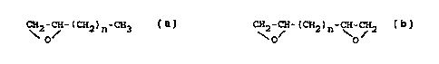
invention, the present invention provides an epoxy
compound that has a hydrocarbon backbone, that is
water-soluble, and which does not contain an ether
or ester linkage in its backbone. Examples of
suitable epoxide agents include mono- or diepoxides
that have the following basic formulas:
Monoepoxide: CH2-CH-(CH2)n-CH3
O/
Diepoxide: CH-CH-(CH2)n-CH-CH2
' O \0
where n = 1 to 10.
BRIEF DESCRIPTION OF THE DRAWINGS
FIG. 1 illustrates proposed mechanisms for the in
vivo degradation of polyethylene oxide.
FIG. 2 is a flowchart illustrating a method for
producing a bioprosthesis according to the present
invention.
CA 02351216 2001-05-09
WO 00/35374 PCTIUS99/29617
DETAILED DESCRIPTION OF THE PREFERRED EMBODIMENTS
The following detailed description is of the best
presently contemplated modes of carrying out the
invention. This description is not to be taken in a
5 limiting sense, but is made merely for the purpose
of illustrating general principles of embodiments of
the invention. The scope of the invention is best
defined by the appended claims. In certain
instances, detailed descriptions of well-known
10 devices, compositions, components, mechanisms and
methods are omitted so as to not obscure the
description of the present invention with
unnecessary detail.
For purposes of the present invention, the term
"collagenous tissue" refers to material which may be
derived from different animals, such as mammals.
Specific examples include, but are not limited to,
porcine heart valves; bovine pericardium; connective
tissue derived materials such as dura mater,
tendons, ligaments, skin patches; arteries; veins;
and the like.
The present invention provides a cross-linking
agent for use in tissue fixation of collagenous
material. The agent is an epoxy compound that has a
hydrocarbon backbone, that is water-soluble and
which does not contain an ether or ester linkage in
its backbone. Examples of suitable epoxide agents
include mono- or diepoxides that have the following
basic formulas:
Monoepoxide: CH2-~ -CH-CH3
O
Diepoxide: CH2-CH-(CH2-)n-CH-CH 2
\O/ "O/
where n = 1 to 10. For example, a monoepoxide where
n is equal to 3 is as follows:
CA 02351216 2009-01-07
11
H2-CH-CH 2-CH2-CH2-CH3
O Monoepoxides according to the present invention
are generally used to modify tissue where a greater
flexibility is important. Examples of such tissues
include venous valves, esophagus and ureters.
Polyepoxides (i.e., diepoxides and epoxides having
two or more reactive epoxide groups) according to
the present invention are generally used to modify
tissue which may be used in applications where
significant stress and load are experienced after
implantation. Examples of such tissues include
heart valves in arterial systems, ligaments and
tendons.
Bioprosthetic tissue may be cross-linked by immersing a
bioprosthetic tissue in a solution containing an epoxy com-
pound having a hydrocarbon backbone, the epoxy compound being
water soluble and having a backbone devoid of either an ether
or ester linkage. A bioprosthetic tissue may thus contain a
cross-linked epoxy compound having a hydrocarbon backbone, the
epoxy compound being water soluble and having a backbone
devoid of either an ether or ester linkage.
The cross-linking agent of the present invention can be used
to fix or modify a wide variety of bioprosthetic tissues,
including bovine pericardium and porcine aortic valves. The
method of treating and preparing the bioprosthetic tissue is
summarized in the flowchart of FIG. 2, and is set forth as
follows.
In step 10, a collagenous tissue is harvested and processed.
A suitable collagenous tissue, such as an artery or vein, is
harvested from a mammal, and excess muscle, fat and connective
tissues are trimmed according to known methods. The
collagenous tissue is cleaned and prepared in accordance with
known methods. The blood vessel is washed inside and out with
cold saline solution to remove any remaining blood.
CA 02351216 2009-01-07
11A
In step 20, bioburden levels are reduced by immersing each
tissue in 70% ethanol for about one hour. The tissues are then
stored in 30% ethanol for any desired period of time.
CA 02351216 2001-05-09
WO 00/35374 PCT/US99/29617
12
In step 30, the cellular component is inflated.
This can be done by injecting the lumen of each
tissue vessel with fresh filtered water and then
transferring them to a container of fresh filtered
water. The tissue is then kept refrigerated for at
least one hour while in the fresh filtered water
prior to sonication.
In step 40, the tissue is sonicated in filtered
water for a period of time sufficient to remove the
cellular component. It is desirable to remove the
cellular component because it has greater
antigenicity. The tissue is then thoroughly washed
with water.
In step 50, fixation is performed. The previously
prepared collagenous tissue is immersed in an
aqueous solution of the water-soluble epoxide cross-
linking agent of the present invention at a pH of
8.5 to 10.5 for a time (e.g., 1 to 30 days)
sufficient to permit irreversible cross-linking.
The concentration of the epoxide crosslinking agent
preferably ranges from 0.01 M to 1.0 M, and more
preferably, is between 0.05 M to 0.5 M. The
fixation solution is changed every two to three
days.
In step 60, the collagenous tissue is removed from
the fixation solution and is rinsed with a suitable
rinsing solution such as phosphate buffered saline,.
with or without amino acid. This rinsing removes
residual fixative reactivity.
In step 70, final trimming and branch ligation are
performed. Excess connective tissue is carefully
trimmed away without damaging the vessel branches.
Any tissue vessel having holes, avulsed branches,
blood stains or other visual structural defects will
CA 02351216 2001-05-09
WO 00/35374 PCT/US99/29617
13
not be used. All branches are suture-ligated using
4-0 or 5-0 Prolene suture.
In step 80, final sterilization is performed. The
collagenous tissue is sterilized with a non-aldehyde
sterilant, such as 0.1% iodine solution, and then
stored in 30% ethanol solution until the tissue is
to be implanted.
FIRST EXAMPLE -- CROSS-LINKING ARTERIAL GRAFT WITH
DIEPOXIDES
A fresh bioprosthetic tissue, such as a bovine
artery, is incubated in an aqueous solution of a
water-soluble polyepoxide cross-linking agent. More
specifically, a 1,2,7,8-diepoxyoctane at 0.2 M is
jr- buffered to a pH of 9.5 with carbonate-bicarbonate
buffer with 5% ethanol. The artery is exposed to
the solution for 14 days at room temperature (e.g.,
25 degrees Celcius) to permit irreversible cross-
linking. The fixation solution is changed every
three days.
SECOND EXAMPLE -- MODIFICATION OF VENOUS CONDUIT
WITH VALVE
A vein conduit having venous valves is incubated
in an aqueous solution of a water-soluble
polyepoxide cross-linking agent. More specifically,
a 1,2-epoxyoctane at 0.2 M is buffered to a pH of
9.5 with carbonate-bicarbonate buffer with 100
ethanol. The vein is exposed to the solution for 14
days at 25 degrees Celcius to permit complete
modification.