Note: Descriptions are shown in the official language in which they were submitted.
CA 02352699 2008-06-26
- 1 -
BIPOLAR MAPPING OF INTRACARDIAC POTENTIALS
FIELD OF THE INVENTION
The present invention is directed to a method for
measuring electrical activity in the heart and a catheter
useful for performing the method.
The present invention is related to other commonly
owned U.S. patents: U.S. Patent No. 6,872,762, entitled
Catheter with Tip Electrode Having a Recessed Ring Electrode
Mounted Thereon; U.S. Patent No. 6,477,396, entitled Mapping
and Ablation Catheter; U.S. Patent No. 6,546,270, entitled
Multi-Electrode Catheter, System and Method; and U.S. Patent
No. 6,569,160, entitled System and Method for Detecting
Electrode-Tissue Contact; all commonly owned by the assignee
of the present invention.
BACKGROUND OF THE INVENTION
Electrode catheters have been in common,use in medical
practice for many years. They are used to stimulate and map
electrical activity in the heart and to ablate sites of
aberrant electrical activity.
In use, the electrode catheter is inserted into a
major vein or artery, e.g., femoral artery, and then guided
into the chamber of the heart which is of concern. Within
the heart, the ability to control the exact position and
orientation of the catheter tip is critical and largely
determines how useful the catheter is.
CA 02352699 2001-07-05
2 -
In healthy humans the heartbeat is controlled by
the sinoatrial node ("S-A node") located in the wall of
the right atrium. The S-A node generates electrical
signal potentials that are transmitted through pathways
of conductive heart tissue in the atrium to the
atrioventricular node ("A-V node") which in turn
transmits the electrical signal potentials throughout
the ventricle by means of the His and Purkinje
conductive tissues. Improper growth of or damage to the
io conductive tissue in the heart can interfere with the
passage of regular electrical signals from the S-A and
A-V nodes. Electrical signal irregularities resulting
from such interference can disturb the normal rhythm of
the heart and cause an abnormal rhythmic condition
is referred to as cardiac arrhythmia.
Electrophysiological ablation is a procedure often
successful in terminating cardiac arrhythmia. This
procedure involves applying sufficient energy to the
interfering tissue to ablate that tissue thus removing
20 the irregular signal pathway. However, before the
ablation procedure can be carried out, the interfering
tissue must first be located.
One location technique involves an
electrophysiological mapping procedure whereby the
25 electrical signals emanating from the conductive
endocardial tissues are systematically monitored and a
map is created of those signals. By analyzing that map,
the interfering electrical pathway can be identified. A
conventional method for mapping the electrical signals
30 from conductive heart tissue is to percutaneously
introduce an electrophysiology catheter (electrode
catheter) having mapping electrodes mounted on its
CA 02352699 2001-07-05
- 3 -
distal extremity. The catheter is maneuvered to place
these electrodes in contact with or in close proximity
to the endocardium. By monitoring the electrical
signals at the endocardium, aberrant conductive tissue
sites responsible for the arrhythmia can be pinpointed.
Once the origination point for the arrhythmia has
been located in the tissue, the physician may use an
ablation procedure to destroy the tissue causing the
arrhythmia in an attempt to remove the electrical signal
io irregularities and restore normal heart beat or at least
an improved heart beat. Successful ablation of the
conductive tissue at the arrhythmia initiation site
usually terminates the arrhythmia or at least moderates
the heart rhythm to acceptable levels.
Conventional unipolar electrode catheters utilize a
primary tip or ring electrode that cooperates with a
reference electrode outside the patient's body. Such
catheters are known to map inaccurate electrical
readings due to the reference electrode being located
outside the patient's body.
Previous attempts have been made to design a
bipolar electrode catheter having two electrodes within
the patient's body. However, such catheters also have
limited accuracy. Specifically, both electrodes pick up
near field electrical signals emanating from the
conductive endocardial tissues due to their contact with
the heart tissue, and far-field electrical signals which
propagate from other regions of the heart due to their
contact with the blood. The far-field signals interfere
with the near-field signals, making accurate measurement
of the near-field signals difficult. Accordingly, a
CA 02352699 2001-07-05
- 4 -
need exists for a bipolar electrode catheter that more
accurately measures near-field signals.
U.S. Patent No. 5,749,914 to Janssen discloses a
catheter for removing obstructions from a tubular
passageway in a patient. In one embodiment, Janssen
describes a catheter having a distal end with a recessed
annular ridge that defines a groove in which a plurality
of electrodes are seated. The electrodes are sized so
that they are recessed within the annular ridge. A
return electrode is located on the catheter proximal to
the recessed electrodes. The electrodes are connected
to a radio-frequency energy source that generates and
supplies current to the electrodes to ablate
constructive material. Janssen nowhere teaches or
suggests, however, using this catheter to map electrical
activity in the heart.
U.S. Patent No. 4,966,597 to Cosman discloses a
cardiac ablation electrode catheter with a thermosensing
detector at a position in the distal end of the
catheter. In one embodiment, the ablation electrode has
an insulative exterior with openings that provide
exposed electrode surfaces. Each of the electrode
surfaces can be independently connected to different
contacts, which are then connected to a voltage source,
or the electrode surfaces can all be connected together.
A temperature-measuring conductor is attached to one or
more of the electrode surfaces. The object of the
invention described in Cosman is to provide a cardiac
catheter for tissue ablation with ultra-fast faithful
recording of temperature in the affected tissue. Cosman
nowhere discloses, however, obtaining electrical signals
CA 02352699 2001-07-05
- 5 -
with different electrodes and comparing the signals to
obtain near-field electrical activity information.
SUMMARY OF THE INVENTION
s The present invention is directed to a catheter
having two electrodes for bipolar mapping and a method
for using the catheter. In one embodiment, the
invention is directed to a method for measuring near-
field electrical activity at a location in a heart. The
method comprises introducing into the heart a catheter
comprising an elongated tubular body having a distal
region and a circumferential recess along the length of
the distal region. A first electrode is mounted on the
distal region in close proximity to the circumferential
recess. A second electrode is mounted within the
circumferential recess. The method further comprises
positioning the distal region at the location in the
heart so that the first electrode is in direct contact
with heart tissue and the second electrode is not in
direct contact with heart tissue but is in contact with
blood. A first signal is obtained with the first
electrode, and a second signal is obtained with the
second electrode. The first signal and the second
signal are compared to obtain the near-field electrical
activity at the location in the heart.
In another embodiment, the invention is directed to
a method for measuring near-field electrical activity at
a location in a heart comprising introducing into the
heart a catheter comprising an elongated body having an
outer diameter and a distal region, a first electrode
mounted on the distal region, and a second electrode
mounted on the distal region in close proximity to and
CA 02352699 2001-07-05
- 6 -
electrically isolated from the first electrode, the
second electrode having an outer diameter less than the
outer diameter of the portion of the distal region on
which it is mounted. The distal region is positioned at
the location in the heart so that the first electrode is
in direct contact with heart tissue and the second
electrode is not in direct contact with heart tissue but
is in contact with blood. A first signal is obtained
with the first electrode, and a second signal is
io obtained with the second electrode. The first signal
and the second signal are compared to obtain the near-
field electrical activity at the location in the heart.
In still another embodiment, the invention is
directed to a method for measuring near-field electrical
activity at a location in a heart comprising introducing
into the heart a catheter comprising an elongated body
having a distal region, a first electrode mounted on the
distal region, and a second electrode mounted on the
distal region in close proximity to and electrically
isolated from the first electrode. The second electrode
is covered by a blood-permeable membrane that prohibits
direct contact between the second electrode and
surrounding heart tissue. The distal region is
positioned at the location in the heart so that the
first electrode is in direct contact with heart tissue
and the second electrode is not in direct contact with
heart tissue but is in contact with blood. A first
signal is obtained with the first electrode, and a
second signal is obtained with the second electrode.
The first signal and the second signal are compared to
obtain the near-field electrical activity at the
location in the heart.
CA 02352699 2008-06-26
- 7 -
In yet another embodiment, the invention is directed
to a catheter comprising an elongated body having a
distal region. A first electrode is mounted on the
distal region. A second electrode is mounted on the
distal region in close proximity to and electrically
isolated from the first electrode. The second electrode
is covered by a blood-permeable membrane that, in use,
prohibits direct contact between the second electrode and
surrounding heart tissue.
In yet another embodiment, the present invention
provides a system for determining an arrhythmiogenic
focus in heart tissue. The system comprises:
a catheter comprising:
(i) an elongated body having a distal region;
(ii) a first electrode mounted on the distal region
for obtaining a first signal;
(iii) a second electrode mounted on the distal
region in close proximity to and electrically isolated
from the first electrode for obtaining a second signal,
the second electrode being covered by a blood-permeable
membrane that, in use, prohibits direct contact between
the second electrode and surrounding heart tissue; and
a signal processing unit operatively connected to
the catheter for comparing the first signal and the
second signal and subtracting far-field activity from the
first signal for obtaining near-field electrical activity
and identifying a location of an arrhythmiogenic focus
based on near-field electrical activity.
CA 02352699 2008-06-26
- 7a -
In yet another embodiment, the present invention
provides use of a system of the present invention for
determining an arrhythmiogenic focus at a location in a
heart.
In yet another embodiment, the present invention
provides use of a system of the present invention for
generating a map of the electrical activity of a heart.
DESCRIPTION OF THE DRAWINGS
These and other features and advantages of the
present invention will be better understood by reference
to the following detailed description when considered in
conjunction with the accompanying drawings wherein:
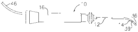
FIG. 1 is a side view of an embodiment of the
catheter of the invention.
FIG. 2 is a side cross-sectional view of a catheter
body according to the invention, including the junction
between the catheter body and tip section.
FIG. 3 is a side cross-sectional view of a catheter
tip section showing a tip electrode and a recessed ring
electrode.
FIG. 4 is a side cross-sectional view of an
alternative tip section according to the invention having
a ring electrode covered by a blood-permeable material.
FIG. 5A is a side cross-sectional view of another
alternative tip section according to the invention having
a first ring electrode and a second ring electrode that
is recessed.
CA 02352699 2001-07-05
- 8 -
FIG. 5B is a side cross-sectional view of another
alternative tip section according to the invention
having a first ring electrode and a second ring
electrode that is covered by a blood-permeable membrane.
FIG. 6 is a side cross-sectional view of another
alternative tip section according to the invention, the
tip section including an electromagnetic location
sensor.
FIG. 7 is an end cross-sectional view of the tip
lo section depicted in FIG. 6.
DETAILED DESCRIPTION
In a particularly preferred embodiment of the
invention, there is provided a steerable catheter having
is two electrodes for making bipolar measurements. As
shown in FIGs. 1 to 3, catheter 10 comprises an
elongated catheter body 12 having proximal and distal
ends, a tip section 14 at the distal end of the catheter
body 12, and a control handle 16 at the proximal end of
20 the catheter body 12.
With reference to FIG. 2, the catheter body 12
comprises an elongated tubular construction having a
single, axial or central lumen 18. The catheter body 12
is flexible, i.e., bendable, but substantially non-
25 compressible along its length. The catheter body 12 can
be of any suitable construction and made of any suitable
material. A presently preferred construction comprises
an outer wall 22 made of a polyurethane, or PEBAX. The
outer wall 22 comprises an imbedded braided mesh of
30 high-strength steel, stainless steel or the like to
increase torsional stiffness of the catheter body 12 so
that, when the control handle 16 is rotated, the tip
CA 02352699 2001-07-05
- 9 -
section 14 of the catheter 10 will rotate in a
corresponding manner. The outer diameter of the
catheter body 12 is not critical, but is preferably no
more than about 8 french (1 mm = 3 french), more
preferably about 7 french, still more preferably about 5
french. Likewise the thickness of the outer wall 22 is
not critical, but is thin enough so that the central
lumen 18 can accommodate an infusion tube, a puller
wire, lead wires, and any other wires, cables or tubes.
The inner surface of the outer wall 22 is lined with a
stiffening tube 20, which can be made of any suitable
material, such as polyimide or nylon. The stiffening
tube 20, along with the braided outer wall 22, provides
improved torsional stability while at the same time
minimizing the wall thickness of the catheter, thus
maximizing the diameter of the central lumen 18. The
outer diameter of the stiffening tube 20 is about the
same as or slightly smaller than the inner diameter of
the outer wall 22. Polyimide tubing is present.ly
preferred for the stiffening tube 20 because it may be
very thin walled while still providing very good
stiffness. This maximizes the diameter of the central
lumen 18 without sacrificing strength and stiffness. A
particularly preferred catheter has an outer wall 22
with an outer diameter of from about 0.090 inch to about
0.098 inch and an inner diameter of from about 0.061
inch to about 0.065 inch and a polyimide stiffening
tube 20 having an outer diameter of from about 0.060
inch to about 0.064 inch and an inner diameter of from
about 0.051 inch to about 0.056 inch. If desired, the
stiffening tube 20 can be eliminated. As would be
CA 02352699 2001-07-05
- 10 -
recognized by one skilled in the art, the catheter body
construction can be modified as desired.
As shown in FIG. 3, the tip section 14 comprises a
short section of tubing 19 having two lumens 30 and 32.
The tubing 19 is made of a suitable non-toxic material
that is preferably more flexible than the catheter
body 12. A presently preferred material for the tubing
19 is braided polyurethane, i.e., polyurethane with an
embedded mesh of braided high-strength steel, stainless
io steel or the like. The outer diameter of the tip
section 14, like that of the catheter body 12, is
preferably no greater than about 8 french, more
preferably 7 french, still more preferably about 5
french. The size of the lumens is not critical and can
vary depending on the specific application.
A preferred means for attaching the catheter
body 12 to the tip section 14 is illustrated in FIG. 2.
The proximal end of the tip section 14 comprises an
outer circumferential notch 24 that receives the inner
surface of the outer wall 22 of the catheter body 12.
The tip section 14 and catheter body 12 are attached by
adhesive (e.g., polyurethane glue) or the like. Before
the tip section 14 and catheter body 12 are attached,
however, the stiffening tube 20 is inserted into the
catheter body 12. The distal end of the stiffening
tube 20 is fixedly attached near the distal end of the
catheter body 12 by forming a glue joint (not
shown) with polyurethane glue or the like. Preferably a
small distance, e.g., about 3 mm, is provided between
the distal end of the catheter body 12 and the distal
end of the stiffening tube 20 to permit room for the
catheter body 12 to receive the- notch 24 of the tip
CA 02352699 2001-07-05
- 11 -
section 14. A force is applied to the proximal end of
the stiffening tube 20, and, while the stiffening
tube 20 is under compression, a first glue joint (not
shown) is made between the stiffening tube 20 and the
outer wall 22 by a fast drying glue, e.g. Super Glue .
Thereafter a second glue joint (not shown) is formed
between the proximal ends of the stiffening tube 20 and
outer wall 22 using a slower drying but stronger glue,
e.g., polyurethane.
At the distal end of the tip section 14 is a tip
electrode 36. Preferably the tip electrode 36 has a
diameter about the same as the outer diameter of the
tubing 19. The tip electrode 36 can be made from any
suitable material, such as platinum, gold, iridium or
stainless steel, and is preferably machined from
platinum-iridium bar (90% platinum/10% iridium).
A preferred tip electrode has a length ranging from
about 2.5 mm to about 8 mm, preferably about 3.5 mm.
Preferably the tip electrode 36 is attached to the
tubing 19 by polyurethane glue or the like. The wires
that extend into the tip electrode 36, described in more
detail below, help to keep the tip electrode in place on
the tubing 19 of the tip section 14.
In the embodiment shown in FIG. 3, there is a ring
electrode 39 mounted within a circumferential recess 26
in the tubing 19 of the tip section 14. The recess 26
is located near the distal end of the tip section 14 and
in close proximity to the tip electrode 36. As used
herein, "in close proximity" means a distance suitable
for conducting bipolar mapping. Preferably the
recess 26 is spaced apart from the tip electrode 36 a
distance no greater than about 4 mm, more preferably
CA 02352699 2001-07-05
- 12 -
from about 0.1 mm to about 2 mm, still more preferably
from about 0.5 mm to about 1.0 mm. The width and depth
of the recess 26 are designed such that, when the tip
section 14 is positioned on its side against the
adjacent heart tissue, the tissue des not come into
contact with the ring electrode 39. Preferably, the
width of the recess 26 ranges from about 0.5 mm to about
4 mm, more preferably from about 1 mm to about 3 mm,
with the depth of the recess 26 preferably ranging from
about 0.25 mm to about 1.5 mm, more preferably from
about 0.5 mm to about 1 mm.
In a preferred embodiment, the ring electrode 39
comprises a resilient ribbon-shaped conductive material
that is wrapped within the recess 26 and fixed in place
by glue or the like. The ring electrode 39 can be made
of any suitable conductive material, such as those
discussed above for the tip electrode. The width of and
thickness of the ring electrode 39 are suitable for
fitting within the recess 26 so that the outer surface
of the ring electrode 39 is recessed within the recess
26. In other words, the ring electrode 39 has an outer
diameter less than the outer diameter of the tubing 19
of the tip section 14. Preferably, the outer diameter
of the ring electrode 39 is at least about 10%, more
preferably from about 20% to about 50%, less than the
outer diameter of the portion of the tip section 14 on
which it is mounted. The ring electrode 39 has a width
preferably ranging from about 0.5 mm to about 4 mm, more
preferably from about 1 mm to about 3 mm. In an
alternative embodiment, the ring electrode 39 is in the
form of a snap ring, where the width and thickness of
CA 02352699 2001-07-05
- 13 -
the ring 39 are suitable for fitting within the recess
26, as described above.
The tip electrode 36 and ring electrode 39 are each
connected to a separate lead wire 44. The lead wires 44
s extend through the first lumen 30 of tip section 14, the
central lumen 18 of the catheter body 12, and the
control handle 16, and terminate at their proximal end
in an input jack (not shown) that may be plugged into an
appropriate signal processing unit (not shown). The
portion of the lead wires 44 extending through the
central lumen 18 of the catheter body 12, control
handle 16 and proximal end of the tip section 14 may be
enclosed within a protective sheath 49, which can be
made of any suitable material, preferably polyimide.
The protective sheath 49 is preferably anchored at its
distal end to the proximal end of the tip section 14 by
gluing it in the first lumen 30 with polyurethane glue
or the like.
The lead wires 44 are attached to the tip
electrode 36 and ring electrode 39 by any conventional
technique. Connection of a lead wire 44 to the tip
electrode 36 is accomplished, for example, by soldering
the lead wire 44 into a first blind hole 31 of the tip
electrode, as shown in FIG. 3.
Connection of a lead wire 44 to a ring electrode 39
is preferably accomplished by first making a small hole
through the tubing 19. Such a hole can be created, for
example, by inserting a needle through the tubing 19 and
heating the needle sufficiently to form a permanent
hole. A lead wire 44 is then drawn through the hole by
using a microhook or the like. The ends of the lead
wire 44 are then stripped of any coating and soldered or
CA 02352699 2001-07-05
- 14 -
welded to the underside of the ring electrode 39, which
is then slid into position over the hole and fixed in
place with polyurethane glue or the like.
A puller wire 50 extends through the catheter
body 12, is anchored at its proximal end to the control
handle 16, and is anchored at its distal end to the tip
section 14. The puller wire 50 is made of any suitable
metal, such as stainless steel or Nitinol, and is
preferably coated with Teflon(l) or the like. The coating
io imparts lubricity to the puller wire 50. The puller
wire 50 preferably has a diameter ranging from about
0.006 to about 0.010 inches.
A compression coil 52 is situated within the
catheter body 12 in surrounding relation to the puller
wire 50. The compression coil 52 extends from the
proximal end of the catheter body 12 to the proximal end
of the tip section 14. The compression coil 52 is made
of any suitable metal, preferably stainless steel. The
compression coil 52 is tightly wound on itself to
provide flexibility, i.e., bending, but to resist
compression. The inner diameter of the compression
coil 52 is preferably slightly larger than the diameter
of the puller wire 50. The Teflon coating on the
puller wire 50 allows it to slide freely within the
compression coil 52. If desired, particularly if the
lead wires 44 are not enclosed by a protective
sheath 49, the outer surface of the compression coil 52
can be covered by a flexible, non-conductive sheath 46,
e.g., made of polyimide tubing, to prevent contact
between the compression coil 52 and any other wires
within the catheter body 12.
CA 02352699 2008-06-26
- 15 -
The compression coil 52 is anchored at its proximal
end to the proximal end of the stiffening tube 20 in the
catheter body 12 by glue joint 51 and at its distal end
to the tip section 14 by glue joint 53. Both glue
s joints 51 and 53 preferably comprise polyurethane glue
or the like. The glue may be applied by means of a
syringe or the like through a hole made between the
outer surface of the catheter body 12 and the central
lumen 18. Such a hole may be formed, for example, by a
needle or the like that punctures the outer wall 22 of
the catheter body 12 and the stiffening tube 20 which is
heated sufficiently to form a permanent hole. The glue
is then introduced through the hole to the outer surface
of the compression coil 52 and wicks around the outer
is circumference to form a glue joint about the entire
circumference of the compression coil 52.
The puller wire 50 extends into the second lumen 32
of the tip section 14. The pu11=er wire 50 is anchored
at its distal end to the tip electrode 36 within a
second blind hole 33 by weld or the like. A preferred
method for anchoring the puller wire 50 within the tip
electrode 36 is by crimping metal tubing 54 to the
distal end of the puller wire 50 and soldering the metal
tubing 54 inside the second blind hole 33. Anchoring
the puller wire 50 within the tip electrode 36 provides
additional support for the tip electrode on the flexible
plastic tubing 19, reducing the likelihood that the tip
electrode will separate from the tubing. Alternatively,
the puller wire 50 can be attached to the side of the
tip section 14. Such a design is described in U.S.
Patent 6,123,699 (filed September 5, 1997.)
CA 02352699 2008-06-26
- 16 -
Within the second lumen 32 of the tip
section 14, the puller wire 50 extends through a
plastic, preferably Teflon , sheath 56, which prevents
the puller wire 50 from cutting into the wall of the
tubing 19 when the tip section is deflected.
Longitudinal movement of the puller wire 50
relative to the catheter body 12, which results in
deflection of the tip section 14, is accomplished by
suitable manipulation of the control handle 16. A
suitable control handle.design for use with the present
invention is described in U.S. Patent
6,120,476 filed December 1, 1997.
In operation, the present invention is ideal for
mapping the heart and ablating accessory signal pathways
causing arrhythmias. To perform this function, the
distal end of.the catheter 10 is inserted into a vein or
artery and advanced into the heart. To assist in
positioning the tip section 14 of the.catheter 10 at a
desired position within the heart, the puller wire 50
and control handle 16 are used to deflect the tip
section 14. Once the tip section 14 has been positioned
at or near the desired location of the heart tissue, the
electrical activity of the heart may be identified,
evaluated or mapped, and electrophysiological sources of
arrhythmia may be identified and/or treated.
Electrical activity within the heart is detected
using the tip electrode 36 and ring electrodes 39 of the
catheter 10. The catheter 10 of the present invention
is designed such that the tip electrode 36 is in direct
contact with the heart tissue. Thus, the tip electrode '-
36 senses both the local activation energy (near-field
CA 02352699 2001-07-05
- 17 -
signals) at the point of contact with the heart tissue
and far field activation energy (far-field signals)
received by the electrode through the blood.
As described above, the ring electrode 39 is
recessed relative to the tip section 14 to be protected
from direct contact with the heart tissue, but
permitting contact with surrounding blood. The close
proximity of the ring electrode 39 to the tip electrode
36 enables the ring electrode 36 to receive
approximately the same far-field signals as the tip
electrode 36. However, the ring electrode 39 does not
pick up the local activation potential (near-field
signals). The signals received by the tip electrode 36
and the ring electrode 39 are sent to a suitable signal
processing unit.
Within the signal processing unit, the signal
detected by the ring electrode 39, which is only far-
field signals, is subtracted from the signal detected by
the tip electrode 36, which includes both near-field and
far-field signals. Thus, the near-field signals can be
more accurately determined. This improved method of
detecting electrical activity allows the physician or
operator to determine the location of the
arrhythmiogenic focus more accurately for ablating and
other purposes.
Alternate bipolar electrode designs can also be
provided having one electrode in contact with blood but
not the heart tissue. For example, as shown in FIG. 4,
the ring electrode 39 is covered by a membrane 60 that
is permeable to the blood, but that prevents direct
physical contact between the ring electrode and the
heart tissue. In this embodiment the ring electrode 39
CA 02352699 2001-07-05
- 18 -
is mounted on the tubing 19 proximal to and in close
proximity to the tip electrode 39. The ring electrode
39 is slid over the tubing 19 and fixed in place by glue
or the like. The membrane 60 is wrapped around the ring
s electrode 39 and glued in place onto the tip section 14
by polyurethane or the like. The membrane 60 is
preferably in the form of a perforated film or a woven
or nonwoven fabric. The membrane 60 preferably
comprises a biocompatible polymer. Examples of suitable
biocompatible polymers for use in connection with the
invention include polyolefins such as polypropylene,
polyurethane, polyetheramide, polyetherimide, polyimide,
fluoropolymers such as polytetrafluoroethylene,
silicones and the like, and combinations thereof. The
blood-permeable membrane 60 thus allows the blood to
permeate the membrane 60 and contact the ring electrode
39, while protecting the ring electrode 39 from direct
contact with the heart tissue.
In alternative embodiments, as shown in FIGs. 5A
and 5B, ring electrode pairs may be provided instead of
the tip electrode/ring electrode combinations described
above. In these embodiments, the ring electrode pair 61
includes first and second ring electrodes 64 and 66
mounted in close proximity to each other. In one
alternative embodiment, the first ring electrode 64 is
mounted on the outer surface of the tubing 19 to make
direct contact with adjacent heart tissue. The second
ring electrode 66 is displaced within a recess 65
proximal to the first electrode 64 such that the second
electrode 66 is recessed from the outer surface of the
tubing 19 to prevent direct contact with adjacent heart
tissue, in a manner as described above.
CA 02352699 2008-06-26
- 19 -
In another alternative embodiment, the first ring
electrode 64 is mounted on the outer surface of the
tubing 19, to make direct contact with adjacent heart
tissue. The second ring electrode 66 is mounted on the
s tubing 19 proximal to the first electrode 64. A blood-
permeable membrane 60 is wrapped around the second
electrode 66, in a manner as described above, to protect
the second electrode 66 from direct contact with
adjacent heart tissue.
As would be recognized by one skilled in the art,
the relative locations of the ring electrodes can vary.
For example, in the embodiment of FIG. 5A, the second
electrode 66, which is recessed, can be di&tal to the
firs't electrode 64. Also, additional ring electrodes
is can be provided for any of the above-described
embodiments.
In an alternative embodiment, the catheter further
includes a location sensor, preferably an
electromagnetic location sensor. As shown in FIGs. 6
and 7, the tip section 14 includes a third lumen 34.
The electromagnetic sensor 72 is mounted in part in the
distal end of the tubing 19 and in part in a blind hole
in the tip electrode 36. Suitable electromagnetic
sensors for use in connection with the present invention
are described in U.S. Patent No. 6,201,387
(entitled "Miniaturized Position Sensor") and U.S.
Patent No.s. 5,558,091, 5,443,489, 5,480,422, 5.546.951,
5,568,809, and 5,391,199.
The electromagnetic
sensor 72 is connected to a electromagnetic sensor cable
74, which extends through the third lumen 34 of the-tip
section 14, through the central lumen 18 of the catheter
CA 02352699 2008-06-26
- 20 -
body 12, and into the control handle 16. The
electromagnetic sensor cable 74 then extends out the
proximal end of the control handle 16 within an
umbilical cord (not shown) to a sensor control module
s (not shown) that houses a circuit board (not shown).
Alternatively, the circuit, board can be housed within
the control handle 16, for example, as described in U.S.
Patent Serial No. 5,964,757, entitled
"Steerable Direct Myocardial Revascularization
Catheter".
The electromagnetic sensor cable 74
comprises multiple wires encased within a plastic
covered sheath. In the sensor control module, the wires
of the electromagnetic sensor cable are connected to the
i5 circuit board. The circuit board amplifies the signal
received from the electromagnetic sensor and transmits
it to a computer in a form understandable by the
computer by means of the sensor connector at the
proximal end of the sensor control module. Also,
because the catheter is designed for single use only,
the circuit board preferably contains an EPROM chip
which shuts down the circuit board approximately 24
hours after the catheter has been used. This prevents
the catheter, -or at least the electromagnetic sensor,
from being used twice. If desired, the sensor 72 can be
contained within a rigid plastic housing, e.g., made of
polyetheretherketone (PEEK), that is mounted between the
tip electrode 36 and the flexible tubing 19. Such a
design is described in U.S. Patent No. 5,938,603.
To use the electromagnetic sensor 72, the patient '
is placed in a magnetic field generated, for example, by
CA 02352699 2001-07-05
- 21 -
situating under the patient a pad containing coils for
generating a magnetic field. A reference
electromagnetic sensor is fixed relative to the patient,
e.g., taped to the patient's back, and the catheter
containing a the electromagnetic location sensor is
advanced into the patient's heart. Each sensor
preferably comprises three small coils which in the
magnetic field generate weak electrical signals
indicative of their position in the magnetic field.
Signals generated by both the fixed reference sensor and
the second sensor in the heart are amplified and
transmitted to a computer which analyzes the signals and
then displays the signals on a monitor. By this method,
the precise location of the sensor in the catheter
relative to the reference sensor can be ascertained and
visually displayed. The sensor can also detect
displacement of that catheter that is caused by
contraction of the heart muscle. A preferred mapping
system includes a catheter comprising multiple
electrodes and an electromagnetic sensor, such as the
NOGA-STAR catheter marketed by Biosense Webster, Inc.,
and means for monitoring and displaying the signals
received from the electrodes and electromagnetic sensor,
such as the Biosense-NOGA system, also marketed by
Biosense Webster, Inc.
Using this technology, the physician can visually
map a heart chamber. This mapping is done by advancing
the catheter tip into a heart chamber until contact is
made with the heart wall. This position is recorded and
saved. The catheter tip is then moved to another
position in contact with the heart wall and again the
position is recorded and saved. By combining the
CA 02352699 2008-06-26
- 22 -
electromagnetic sensor and electrodes, a physician can
simultaneously map the contours or shape of the heart
chamber and the electrical activity of the heart.
If desired, the catheter can be multidirectional,
i.e., having two or more puller wires to enhance the
ability to manipulate the tip section in more than one
direction or to form two or more different curves. A
description of such a design is described in U.S. Patent
Serial Nos. 6,123,699 (filed September 5,
1997), 6,171,277 -(filed August 7, 1998), 6,183,463
(filed August 28, 1998), 6,210,407 (filed December 3,
1998), and 6,267,746 (filed March 22, 1999).
The preceding description has been presented with
reference to presently preferred embodiments of the
invention. Workers skilled in the art and technology to
which this invention pertains will appreciate that
alterations and changes in the described structure may
be practiced without meaningfully departing from the
principal, spirit and scope of this invention.
Accordingly, the foregoing description should not
be read as pertaining only to the precise structures
described and illustrated in the accompanying drawings,
but rather should be read consistent with and as support
to the following claims which are to have their fullest
and fair scope.