Note: Descriptions are shown in the official language in which they were submitted.
CA 02353689 2001-05-04
WO 00/27324 PCT/US99/26242
APPARATUS AND METHOD FOR PERFORMING
PRESBYOPIA CORRECTIVE SURGERY
Field of the Invention
The invention relates to a system and method for correcting presbyopia through
a
reshaping of the eye's corneal curvature, so as to have a desired corrective
corneal curvature based upon
a predetermined eye material removal profile. The removal of eye material is
carried out in the corneal
stroma preferably with a laser system with means for forming a sculptured
corneal stroma having the
predetermined profile in cross-section, which profile is based on a
predetermined profile equation and
the specific input of parameters including measurable eye parameters. The
laser system includes control
means which relies on corrective presbyopia directive means for ablative
resculpturing of the corneal
stroma, which directive means facilitates formation of the presbyopia
correcting profile deemed best
suited for the patient based on preestablished profile parameters.
Background of the Invention
For many, many years, humans have sought ways to correct visual problems. The
ancient Chinese slept with small bags of mercury on their eyes, flattening
their corneas and improving
their shortsightedness. Unfortunately, the effects only worked for a few
minutes after waking.
Spectacles are thought to have been first introduced by the Arabs in the 11th
Century and were
introduced into Europe about 200 years later. This century has seen the
development of contact lenses,
initially the hard variety and later soft and disposable soft lenses.
Although these optical aids allow patients to see well while wearing them,
they do not
offer a permanent cure for the visual disorder or problem. Also, in many
situations, they are
inappropriate, for example, when swimming or wearing contacts in the
laboratory. Another problem
is that in some instances dangerous situations can arise when they become
dislodged. This can occur
while they are being used by firefighters and police officers.
Roughly two decades ago, surgical techniques were introduced in an effort to
permanently correct shortsightedness and astigmatism. The radial keratotomy
procedure used a
diamond blade to make incisions into the cornea, the front surface or "window
of the eye". Although
this technique worked well, there have been problems with long term stability
of vision and weakening
of the cornea as a result of the cuts often having to be made up to 95% of the
corneal thickness.
More recently, these older techniques have been replaced with laser treatment
techniques which have replaced the surgeon's blade with a computer controlled
laser that gently re-
sculptures the shape of the cornea without cutting or weakening the eye. These
laser techniques are
typically carried out with a photoablation process using an excimer laser.
-1-
SUBSTITUTE SHEET (RULE 26)
CA 02353689 2001-05-04
WO 00/27324 PCTIUS99/26242
Excimer lasers were chiefly developed for the manufacture of computer
microchips,
where they were used to etch the circuits. However, the laser's extreme
accuracy resulted in it being
well suited as an eye laser. That is, many eye lasers are extremely accurate
and remove only 0.25
microns (1/4000`' millimeter) of tissue per pulse. During the re-sculpturing,
the excimer laser gently
"evaporates" or vaporizes tissue; there is no burning or cutting involved. In
most cases, the laser
treatment takes only 20 to 45 seconds, depending on how severe the refractive
error is.
In the normal eye, light rays entering the eye are accurately focused on the
retina and
a clear image is formed. Most of the bending or focusing of the light rays
occurs at the cornea, with
the natural lens inside the eye being responsible for fine adjustments. If
light is not focused on the
retina, then the eye is said to have a refractive error. Common refractive
errors include: myopia or
shortsightedness, hyperopia or farsightedness, and astigmatism. The excimer
laser has been used to
accurately re-sculpture the cornea in myopia, hyperopia and astigmatism
corrections in an effort to
make the curve of the cornea focus light rays normally on the retina.
Myopia, or shortsightedness, is a condition whereby light rays come to a focus
in front
of, rather than on, the retina at the back of the eye. This results in blurry
vision, especially when
looking at objects far away. Myopia results from the length of the eye being
too long or the cornea
being too steeply curved.
In hyperopia, or farsightedness, light rays are focused behind the retina.
This results
in blurry vision especially when looking at objects that are close. Hyperopia
results from the length
of the eye being too short or the cornea being too flat.
In astigmatism, the cornea, or window of the eye, has an irregular curvature
being
shaped more like a rugby ball, rather than a soccer ball. Light rays are
focused at different points. A
person often has some degree of astigmatism and myopia or hyperopia at the
same time.
In myopia laser correction procedures, the cornea is flattened to better focus
light rays
normally on the retina, whereas in hyperopia, the cornea is made more curved.
With astigmatism, the
surface of the cornea is re-sculptured to a regular curvature.
Under one method of treatment, known as photorefractive keratectomy (PRK), the
laser beam is applied directly to the surface of the cornea, after the thin
surface layer of epithelium cells
has been removed (e.g., through solvent with wiping, preliminary laser
treatment, or minor abrasion).
After the direct laser re-sculpturing of the cornea, a bare area of the cornea
is left which takes a few days
to heal (e.g., 2 to 6 days) and can be uncomfortable during this period. The
healing process can
sometimes lead to regression (some refractive error returns) or to scarring
(which may blur the vision),
especially in patients with large refractive errors. Although still used for
low degrees of myopia and
hyperopia, PRK is generally being replaced by the LASIK method for these same
disorders, in which
the laser treatment is applied under a protective corneal flap. Under the
"Laser in situ Keratomileusis"
(LASIK) treatment, a thin protective corneal flap is raised, rather like a
trapdoor. The front surface of
the exposed cornea is treated by the excimer laser. The net result being that
the cornea is altered in a
-2-
SUBSTITUTE SHEET (RULE 26)
CA 02353689 2006-02-22
manner directed at allowing light rays to be focused normally on the retina.
At the end of the
procedure, the protective flap is simply replaced. The LASIK technique leaves
the original surface of
the cornea virtually intact, hence, there is no bare area to cause pain. In
addition, the mild healing
process results in minimal regression and avoids scarring problems.
Presbyopia is a problem that is due to an aging process occurring in the
natural lens
of the eye, and thus is not linked to the cornea being incorrectly shaped as
in myopia, hyperopia and
astigmatism. As a person ages, the lens expands, becomes harder and less
pliable and, because of these
factors, is not as capable of changing its shape to focus. In a typical
situation, once a person reaches
about 40 years of age, the loss of elasticity and the expansion in the natural
lens of the eye results in that
person experiencing problems with focusing close, for example, during reading.
Most people, as they
age, suffer from a presbyopia problem. The usual way to correct this problem
is to use bifocal lenses.
However, some people dislike wearing glasses, particularly bifocals, for many
reasons. For example,
bifocal lenses present lines where the two portions of the lens are joined
together and thus can be
unsightly unless more expensive "no line" bifocals are relied upon.
Furthermore, people must become
accustomed to reading through the one relatively smaller portion of the
bifocals.
Because of the underlying differences in the causes for presbyopia and the
group of
myopia, hyperopia and astigmatism, many ophthalmologists have concluded that
there is no cure for
presbyopia and that the only solution is to wear reading glasses to compensate
for the loss of ability to
focus on close objects.
Chapter 4 of the book Surgery for Hyperopia and Presbyopia of Neal A. Sher,
M.D., F.A.C.S., 1997, describes a corrective presbyopia surgical treatment
known as of "Anterior
Ciliary Sclerotomy" involving the placement of radial incisions over the
ciliary body in an effort to
increase the scleral diameter to provide an increased area for ciliary muscle
action. This technique
is based on the belief that it is not elastic loss in the lens, but a loss in
range of action due to the lens'
continued growth with respect to a non-growing sclera. The potential
complications for this type of
a treatment, such as infection, hemorrhage (from cutting to deep), ocular
hypotension, myopic shift,
and compromise of the limbal conjunctival barrier, makes this technique one
that is unlikely to gain
wide acceptance.
US Patent Number 5,314,422 to Nizzola represents one effort to correct
presbyopia
and involves remodeling, in a PRK process, the front or external surface of
the cornea by applying a
laser beam through two manipulated plates which together form a sickle shaped
aperture. The beam
passing through the aperture forms a corresponding sickle shaped recess in an
area situated in proximity
to the lower part of the pupil rim. The remodeled area thus constitutes a zone
of the cornea which
functions differently than the rest of the cornea. Thus, this technique
simulates a bifocal glasses
arrangement and therefore presents the problem of having to shift ones focus
from one area to the other
depending on the desired viewing object. Also, the technique described in the
Nizzola patent is a PRK
procedure which removes portions of the outer epithelium layer of the eye and
exposed surface of the
-3-
CA 02353689 2006-02-22
cornea therebelow and, as a result, complicates and prolongs the healing
process which healing process
can sometimes lead to scarring and is often uncomfortable to the patient.
Chapter 20 of the aforementioned Surgery for Hyperopia and Presbyooia
describes
a small diameter intracorneal inlay lens technique used in an effort to
correct presbyopia. Under this
technique, an incision is made in the eye and a small spatula is utilized to
dissect a pocket to the center
of the cornea. A small (1.8 - 2.2 mm diameter) intracorneal hydrogel inlay
lens is then placed on the
spatula and centered over the patient's pupil. This technique is described as
providing a multifocal
cornea arrangement. However, this technique is relatively invasive which
brings with it the possibility
of scarring and infection, and the introduction of a foreign body into the eye
is sometimes found
unacceptable by some patients.
Chapter 7 of the aforementioned Surgery for Hyperopia and Presbyopia also
features
a PRK type presbyopia treatment discussion which is directed at creating a
defined bifocal or multifocal
surface of the human cornea relying on the pseudo-accommodation ability of the
patient to be effective.
In the PRK treatment described in Chapter 7, a 193 mm excimer laser (MEL 60
Aesculap Meditec,
Heroldsbery, Germany) is used. The techniques described include a straight PRK
presbyopia
treatment, a combination myopic/presbyopic PRK treatment and a combination
hyperopic/presbyopic
PRK treatment which include rotating and stationary masks designed to form the
desired bifocal or
multifocal cornea surface topography on the exterior of the cornea with heavy
emphasis on a sectorial
corneal profiling or on a semilunar cornea steepening profiling to achieve in
a defined part of the cornea
a presbyopic optical correction. Thus, in some ways this technique is similar
to that of the Nizzola
method described in US Patent No. 5,314,422 for presbyopia treatment and thus
shares common
problems with the Nizzola technique. Additional complications include
monocular diplopia and the
loss of visual acuity which resulted in some of the clinical tests reported.
Yet another example of a PRK presbyopia treatment process can be seen in US
Patent
No. 5,395,356 which describes a PRK reprofiling of the cornea to create at
least one "add" region having
a different focal point in an effort to assist the eye in accommodating close-
viewing conditions. The
"add" region is described as preferably being located near the center of the
optical zone and is formed
by ablating a profile in Bowman's membrane or Bowman's membrane and adjacent
upper portions of
the stroma following removal of the outer epithelium layer in a preliminary
laser application. As
described above, the PRK treatment process, because it leaves exposed ablated
areas in the cornea, has
associated with it an uncomfortable healing process and the potential for
scarring, hazing and infections.
Section IV of the Surgery for Hvperopia and Presbvooia book noted above
provides
a discussion of an automated lamellar keratotomy (ALK) for hyperopia and laser
in situ keraromileusis
(LASIK) for correcting hyperopia (Chapters 12 and 13) each of which involves
the formation of a
corneal flap. As described in Chapter 12, through the work of the present
inventor, following upon
the earlier work of Dr. Jose Barraquer, nomograms for the correction of
hyperopia have been
developed for causing a controlled degree of ectasia in the eye to produce a
hyperopia correcting
-4-
CA 02353689 2006-02-22
steepening of the cornea. The controlled degree of ectasia is based on precise
lamellar flap formation
which, as described in Chap. 12, preferably involves controlled flap formation
through the use of an
automated microkeratome such as that described in US Patent No. 5,133,726 to
the present inventor
and Sergio Lenchig.
The above noted Chapter 13 describes a hyperopia correction procedure, which
involves ablating with an excimer laser cornea tissue underlying a displaced
lamellar corneal flap formed
with a microkeratome like that described above. Following flap formation, a
hyperopia correcting
mask is attached to an eye fixation suction ring through use of a Meditec
handpiece and mask support
system. The shape of the mask is dependent on the hyperopia refractive error
being corrected, and the
laser system directs a sweeping laser beam past the mask which rotates through
360 , with adjustable
speed (e.g., an angular increment following each laser beam sweep across the
mask). Reference is also
made in this Chapter to the earlier work of others in the use of LASIK in the
treatment of myopia. In
this regard, reference is also made to US Patent No. 4,903,695 to Muller and
L'Esperance describing the
treatment of myopia, hyperopia and astigmatism through the use of a laser
applied to a freshly cut part
of the cornea after severing of the lenticle.
Some efforts have also been made to avoid the need for presbyopia correction
glasses
by programming a laser's computer to leave one eye slightly myopic after
treatment, with the other
programmed for distance. This is sometimes referred to a monovision, and is
often done with patients
being subject to a conventional LASIK treatment. However, while helping to
avoid the requirement
of reading glasses in some patients, a monovision treatment requires some
deviation from the desired
approximation of normal vision and thus represents a determination that the
monoscopic state is not
as undesirable as having to use reading glasses. While a minor degree of a
monoscopic vision procedure
may be useful in supplementing a presbyopia treatment, sole reliance on
monovision for correcting
presbyopia, however, is undesirable due to the resultant wide variance from
normal vision parameters.
The aforementioned US Patent No. 5,533,997 to the present inventor describes a
presbyopic treatment method and system that includes, in one embodiment, a
system and treatment
method preferably involving flap formation and the controlled formation of an
annular ablation in a
centralized region of the newly exposed corneal stroma so as to produce an
unablated central
protrusion of the stroma which transforms the exterior surface of the replaced
flap into a multifocal
surface that is effective in providing both good near and far sight and is
thus effective in avoiding the
difficulties imposed by the onset of presbyopia.
As described in US Patent No. 5,533,997, a preferred treatment technique is
one
wherein the ablation zone leaves untouched a central corneal area of
preferably 1-3 mm and provides
a presbyopic corrective ablation ring which has its major depth region also in
a relatively central region
of the cornea (e.g., a 3.5 mm outer periphery for the presbyopic correcting
depth with or without
outward additional smoothing). As described in the above referenced
application of the inventor, there
has been noticed by the inventor that the occlusion of about a 3 mm central
area of the cornea does not
-5-
CA 02353689 2006-02-22
effect far vision, which led to the realization by the inventor that this is
an ideal site for the near vision
correction by means of reshaping this zone with a multifocal shape and leaving
the most peripheric area
of the cornea for intermediate and far vision. In an effort to even further
improve upon this earlier
work in presbyopic correction in categories such as facilitating
laserparameter determination and setup
time from patient to patient, helping to avoid mistakes in the setup, and
application of an ablation
profile well suited for a patient to be treated, hastened healing time,
minimizing the degree of
postoperative regression, avoiding undesirable reflection or glaring, and
generally providing a good near
and far vision relationship in the treated eye(s), further study and clinical
testing has been carried out
with the result being a presbyopic treatment system and technique as described
below.
Summary of The Invention
The present invention represents a building upon a refinement of the
presbyopic
treatment system and technique described in parent applications US Serial No.
08/268,182, filed
June 29, 1994 (now US Patent No. 5,533,997) and US Serial No. 08/660,376 filed
June 7, 1996
(now US Patent No. 5,928,129). In the two parent applications, there is
described a process and
system for correcting presbyopia carried out either alone or in combination
with the correction of
one or more refractive corrections such as hyperopia, myopia and astigmatism,
which preferably
involves reshaping an eye to as close as normal vision warranted under the
situation, and then
making a presbyopic correction in accordance with the procedure set forth in
those parent
applications. As in the parent applications, the present invention preferably
involves a process that
includes anesthetizing a patient and marking a portion of an eye of the
patient which is to be ablated,
which is followed by the resecting of at least a portion of the cornea to
expose the corneal stroma.
An annular portion of the corneal stroma is then ablated using radiation from,
for example, a laser
beam. After ablation, the cornea is repositioned onto the eye.
In a preferred process of both the parent applications and of the present
invention, the
cornea is resected such that a portion of the cornea remains intact, and
thereafter, the cornea is folded
back to expose the corneal stroma. Alternatively, the cornea may be resected
such that a complete disk
of the cornea is removed from the eye, to thereby expose the corneal stroma.
Thereafter, the cornea
disk would have to be reattached onto the eye.
The corneal stroma should be dried after it has been exposed by the resection
and
before the ablation process. Otherwise, uneven ablation may occur due to
liquids present on the
stroma.
In accordance with the present invention, there is provided presbyopic
corrective
contour control means for forming, in the exposed corneal stroma following
flap formation, an annular
presbyopic correction contour based on a predetermined profile. This control
means is preferably used
in association with a laser generation means such as an excimer laser,
although other suitable corneal
stroma removal techniques may also be relied upon (e.g., a fluid jet or
mechanical material removing
-6-
CA 02353689 2006-02-22
device). The control means of the present invention includes presbyopic
corrective directive means
which preferably is in the form of a programmed software or hardware
application such as a software
or hardware module or component which represents either the entire programmed
control system of
a laser system or an added and modifying component or portion of a larger
programmed laser control
system. In a preferred embodiment, the tissue removal directive means is a
software or hardware
component that is added to a conventional or preexisting laser system which
preferably includes an eye
tracking feature for added assurance of proper profile formation in the
exposed corneal stroma.
One example of a preexisting laser system which can be modified for use in the
present
invention, includes the aforementioned MEL60193 argon fluoride excimer laser
of Aesculap-Meditec.
An additional example includes the VISX Star Laser System of VISX Inc. (Santa
Clara, CA) which
features a dual diaphragm system for the treatment of myopia and myopic
astigmatism and a hyperopic
module that is added along the laser passageway for hyperopic treatment. The
delivery of the excimer
laser energy in the Star System is carried out using a computer-controlled
delivery system which relies
on its VisionKey software system, with its WORM (write once read many) optical
memory card for
enabling the system operator to program the specific refraction corrections
for each patient. Through
suitable modification or supplementation of the computer-controlled delivery
system of the VISX Star
System in accordance with the parameters and other features set forth below
(which can be seen, for
example, to include unique input values or ranges and one or more information
transducers or
converters for use in a unique profile or contour with such a cross-sectional
profile determination
technique), a modified VISX Star System can be formed that represents a
suitable laser system of the
present invention for carrying out the presbyopic profile contour formation
procedure of the present
invention described in greater detail below. Reference is also made to US
Patent Nos. 5,163,934 and
5,207,668 issued to VISX, Inc. Reference is also made to US Patent Nos.
4,718,418 and 4,729,372
to L'Esperance (a listed inventor in one of the VISX, Inc. patents referenced
immediately above) for
further examples of ablation techniques that can be modified for use in
carrying out the present
invention.
As a further example of a preexisting laser system suited for modification to
achieve
the benefits of the present invention, reference is made to the Chiron-
Technolas Keracor 117 and 217
laser systems of Chiron-Technolas GmbH with the laser systems being described
as suited for myopia,
hyperopia and astigmatism treatment through a computer-controlled movable
mirror with relatively
large beam scanning capability. The Chiron Vision's systems are also described
as being suited for
receiving software module updates for varying the system's preexisting
suitable energy calculation and
delivery parameters of the laser beam. This system also represents one system
having an eye tracking
feature. Thus, with an appropriate software module modification and/or
addition carried out to
achieve the parameters and features of the present invention, the Chiron
Vision system can also provide
a laser system suitable for modification in accordance with the present
invention.
-7-
CA 02353689 2006-02-22
Another example of a laser system that can be modified for use in carrying out
the
beneficial features of the present invention can be seen in the Summit
Technology, Inc.'s SVS Apex plus
with mask in rail technology, and in US Patent Nos. 4,856,513; 5,019,074;
5,324,281; 5,395,356 and
5,651,784, all to that same company, which patents describe various means for
forming laser sculptured
configurations including the use of erodible masks that are disposed between
the laser and the cornea
for providing a predefined profile of resistance to erosion so as to form a
desired laser erosion in the
eye. Through use of the present invention's specified parameters for
correcting presbyopia (alone or
in combination with one or more refractive correction procedures for myopia,
hyperopia and
astigmatism) in the central region of an exposed corneal stroma surface, a
suitable erodible mask
member can be formed for use in forming the desired presbyopia correcting
parameters of the
present invention.
The "LSX" laser system of the LASERSIGHT Corporation in Orlando, Florida, US,
in combination with the preferred parameters of the present invention and the
LASERSIGHT Corp.'s
"Scanlink" software system, provides another suitable laser system in
accordance with the present
invention. The Scanlink System provides a translation process that can be used
in the ultimate directing
of the laser system's "flying spot" laser beam to contour the desired profile.
Under the present
invention, this can include setting parameters within reception areas of a
modified version of this
software based on, for example, certain measured values that can vary from
patient to patient (e.g., the
limbus to limbus length, measure corneal curvature, etc.) and/or desired
values chosen from a plurality
of parameter reference representative point ranges. The input values are
preferably inputted. into
reception areas which can be fields that appear on a computer monitor screen
including representations
of values within a range which can be mouse-clicked upon to choose a desired'
value within a range
appearing in the particular fields presented. The "LSX" system also includes
an eye tracking system and
can provide the desired ablation profile within, for example, 20 to 30
seconds, which is useful in
carrying out the method of the present invention and is preferably an added
structural component of
a preferred ablation system.
For ease in usage, it is preferred that any laser control system of the
present invention -
has provided with it associated hardware and/or software which includes the
advantageous presbyopia
correcting profile parameters of the present invention such that the operator
need only input a few
reference points that can be easily measured in the patient and/or chosen from
representative choices,
whereupon the inputted data concerning the patient is automatically processed
to produce the desired
profile or contour configuration for determining the resculptured corneal
stroma configuration. It is
also preferable to provide a visual display of the two dimensional ablation
profile and/or a two or three
dimensional representation of the final contour to be provided in the corneal
stroma for, for example,
pre-laser system operation review by the operator. Thus, through the use of
hardware or software or
the like, automatic profile confi,7= = ration means is provided under the
present invention with the profile
-8-
CA 02353689 2006-02-22
shape having a configuration that is in accordance with the present
invention's parameters (e.g., a
representative single general profile equation) as discussed below.
Although less preferable from the standpoint of operator complexity, the
present
invention can include a laser control system having hardware or software
designed to accept on each
time of use inputted data which includes the present invention's profile
determining means parameters
(e.g., an inserted additional software, module or disk, or other data input
means) to be read by
compatible additional software or hardware already stored by a processor of
the laser system's control
system. This additional inputted data therefore preferably includes, together
with the data related to
variable measurements, data and means for determining the desired profile
configuration for that
patient. The inputted measurement data and the predetermined profile
configuration parameters in
accordance with the present invention, can either be analyzed by the control
system's receiving
hardware or software or combined by the inputted data means prior to receipt
by the laser control
system's hardware or software. An example of this latter embodiment can
include, for example,
inputting a series of points from a drawn or hard copy of a profile or a
profile reception tablet in
conjunction with a pick up instrument (e.g., a digitized tablet in which a
profile can be inputted and
which either forwards upon acceptance confirmation the digitized profile
directly to the control system
of the laser or which involves the scanning of a depicted resultant profile by
another instrument linked
to the control system such as a digital scanning pen). Further examples of
systems which can form a
basis for modification to achieve the beneficial features of the present
invention can be seen in the solid
state, computer directed "flying spot" Novatec "Light Blade" W photoablation
laser described in
Chapter i l of the above noted Surgery For Hyperopia and Presbvopia as well as
the Nidek EC 5000
laser described in Chapter 10 of that same book. In Chapter 10 of that book,
the Nidek EC 5000 laser
operators are described as contemplating using an in-house algorithm for
initial software correction in
an effort to input an overcorrection feature in the system.
As can be seen above, a wide variety of laser sculpting systems exist such as
ones using
focused direct laser beam applications (e.g., flying spot), and wider beam
applications with blocking or
masking means such as erodible masks or rotating and/or stationary single or
multi-holed plates, etc.,
and these systems can be used as a basis for carrying out the present
invention upon making the
appropriate modifications in accordance with the features of the present
invention.
In addition to the above described means for ablating a corneal stroma and
means for
resecting at least a portion of the cornea of the eye, the system of the
present invention also preferably
includes a means for marking a portion of an eye of a patient to be ablated.
The present invention also preferably includes means for designating or
referencing
a desired central point for a central unablated zone used in forming a
multifocal presbyopic corrective
corneal contour. This central point designation or referencing arises from the
inventor's determination
that it is beneficial to position the central point of the central area to
remain free of ablation one unit
superior and one unit nasal to the center point of a patient's pupil. The unit
length is determined by
-9-
CA 02353689 2006-02-22
separating the pupil into quadrants by way of crossing horizontal and vertical
intersecting lines
and dividing the nasal and superior radial lines into thirds and marking or
designating a spot
which represents the crossing point of line extensions of each of the 1/3
dividing points within
the nasal/superior quadrant that are closest to the pupil's center. This can
be done automatically
with an appropriate information transducer or converter of a central system
through an input of
a patient's pupil width or by input from a pupil measuring means. As an
example, a 2 mm pupil
would have a desired central point for the unablated area .33 mm closer to the
nose and .3 mm
superior.
The system also preferably includes means for drying the corneal stroma after
it
has been exposed by the means for resecting such as an air blower with filter
system. Thus, once
the cornea portion has been properly repositioned, it may be reattached to the
eye by blowing air
onto the cornea.
An additional feature of a preferred embodiment of the present invention
includes
a means for cleaning the portion of the stroma that was ablated. This means
may include a
delicate brush and/or a means for irrigating the portion that was ablated.
That is, after ablating,
the ablated portion should be cleaned, in order to prevent edema and this may
be accomplished
by brushing and irrigating the portion which was ablated.
In one embodiment of the present invention, in order to ablate the corneal
stroma
in an annular fashion, a mask formed of a synthetic resin such as polymethyl
methacrylate
(PMMA) can be utilized. The mask in conjunction with a suitable laser power,
shape and
position system (e.g., a fixed diameter sizing feature which, together with
the smaller diameter
mask) forms together a means for maintaining the central area protected while
forming an
annular presbyopia correcting main zone in the corneal stroma. This
arrangement is useful in
forming a basic presbyopia correction profile which can be refined to lessen,
for example, the
post operative time required for some undesirable eye conditions to clear up.
In another aspect, the present invention provides a presbyopic corrective
software
or hardware package that comprises a computer readable memory means for
storing computer
readable code and directive means for directing a formation of a predetermined
ablation corneal
stroma profile having 1.4 to 1.8 mm for the diameter value of a central most
region of the
corneal stroma to remain unablated and 2.4 to 3.2 mm for an outer diameter of
a maximum
ablation depth ring.
In another aspect, the present invention provides use of the presbyopia
correction
system of the present invention for correcting presbyopia.
-10-
CA 02353689 2006-02-22
In another aspect, the present invention provides use of the presbyopia
corrective
software or hardware package of the present invention for facilitating
presbyopia correction.
In another aspect, the present invention provides a presbyopia correction
system,
comprising: a corneal stroma tissue removing system which includes means for
removing
corneal stroma tissue and control means for providing presbyopic correction to
an exposed
corneal stroma of an eye and wherein said means for removing tissue includes a
laser system
and said control means includes directive means for directing a formation by
said laser system
of an ablation region, and wherein said ablation region is based on an
ablation profile which
includes an ablation avoidance central zone having a central reference point
which is off-
centered with respect to an optimal axis of a pupil of an eye, with the
central reference point
being within a nasal-superior quadrant of the pupil of the eye; and an
information converter for
converting received information concerning the eye to establish the central
reference point
within the nasal superior quadrant of the eye for use by the said means for
directing a formation
by said laser system of an ablation region.
In another aspect, the present invention provides a presbyopia correction
system
wherein said directive means further directs a formation by said laser system
of a maximum
ablation depth of 38 to 42 microns
In another aspect, the present invention provides a presbyopia correction
system,
comprising: a corneal stroma tissue removing system which includes means for
removing
corneal stroma tissue and control mews for providing presbyopic correction to
an exposed
corneal stroma of an eye and wherein said means for removing tissue includes a
laser system
and said control means includes directive means for directing a formation by
said laser system
of in ablation region, and wherein said ablation region is based on an
ablation profile which
includes an ablation avoidance central zone having a central reference point
which is off-
centered with respect to an optical axis of a pupil of an eye, with the
central reference point
being within a nasal-superior quadrant of the pupil of the eye, and wherein
directing a formation
includes forming an ablation ring about the central zone with the central zone
having a diameter
of 1.4 mm to 1.8 mm and a central point corresponding with said central
reference point.
Brief Description of the Drawings
The invention will be described in more detail with the aid of the attached
drawings, wherein:
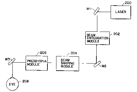
FIGURE 1 is a schematic view of a portion of one excimer laser system where
the setting of the axis of the laser ray is performed;
-10a-
CA 02353689 2006-02-22
FIGURE 2 is a schematic view of the path of the laser beam and the optics used
in the laser system embodiment of FIGURE 1;
FIGURE 3A shows the laser beam system performing an ablation on the cornea,
and the mask protecting the center area of the cornea;
FIGURE 3B shows the ring for the ablation zone;
FIGURE 3C shows the way the ablation of the cornea appears finally;
-10b-
CA 02353689 2006-02-22
FIGURE 4 shows a geometric circular zone illustration which is useful in the
description of forming desired presbyopia correction profiles through ablation
of certain volumes
within specific illustrated zones;
FIGURE 4A illustrates a side view of one preferred ablation profile formed in
accordance with desired parameters of the invention which is representative of
a single profile equation,
which profile equation is useful in forming presbyopic corrective directive
means for determining the
final corrective contour based on input patient data and, preferably, a choice
or values within a
plurality of range parameters;
FIGURES 5A-5D show representative examples of a variety of profiles which
represent the predecessors leading up to the profile depicted in FIGURE 4A;
FIGURE 5E represents another view of the ablation profile representation of
FIGURE 4A and which presents an illustrative view of what can appear on a
visual screen or the like
associated with a laser control system;
FIGURE 6 shows a schematic view of another embodiment of a presbyopia
correction system with presbyopia corrective contour control means forming
part of the overall
presbyopia correction system; and
FIGURE 7 shows a preferred central point for the non-ablated central region of
the
profile that is positioned nasal and superior to the center of the pupil.
Detailed Description of the Invention
A system in accordance with the invention includes a corneal stroma material
removal
system (e.g., an excimer laser system) with presbyopic corrective contour
control means, an automatic
corneal shaper, a pneumatic fixation ring, a mask and an air source. A
preferred automatic corneal
shaper for use in the system in accordance with this invention is the
Automatic Corneal Shaper
described in the inventor's US Patent No. 5,133,726, issued on July 28, 1992.
Using only a local anesthesia, the eye is fixed by the fixation ring which
also functions
as a guide for the automatic corneal shaper. The fixation or retaining ring,
as illustrated in the above
mentioned US Patent No. 5,133,726, permits total control of the eye movement.
The pupil of the eye
will be a reference point for making a very central stromal ablation on the
cornea or some other
suitable reference point can be used such as the vision axis or nasal-superior
center point described in
greater detail below with respect to the laser system zones.
Once the eye is fixed, a keratectomy is performed using the automatic corneal
shaper.
The keratectomy may be partial, which means that a cornea flap technique is
used. This means that
an end portion of the corneal disc remains attached to the cornea base, which
thereby permits its
repositioning in an easier and surer way, once the ablation is performed. When
the flap is retracted,
the corneal stroma becomes exposed, which is ideal tissue on which to practice
the ablation. The
-11-
CA 02353689 2001-05-04
WO 00/27324 PCT/US99/26242
superficial layers of the cornea remain untouched. In this way, undesirable
healing is avoided, and
inaccuracy in the post operative correction and regression is also avoided.
In one embodiment, an excimer laser system is used with presbyopic corrective
control means, which, preferably, features a controllable excimer laser that
accurately allows an
ablation of 0.24 tm/pulse such that an annular ablation can be made on the
stroma having a presbyopic
corrected external diameter of about 3.5 mm or less (with or without
additional exterior and interior
smoothing), with a central zone as small as 1 mm diameter and varying between
1 to 2 or 1 to 3 mm.
The annular ablation produces a central protrusion of the stroma such that
when the corneal flap is
repositioned at its initial position, this stromal curvature change is
transmitted to the forward corneal
surface, thereby indirectly transforming the corneal surface into a multifocal
surface, which is, in fact,
myopic in its central part. This is what helps make it possible for the
patient to read without optic
correction after the procedure, regardless of the age of the patient or the
loss of accommodation.
The annular ablation can be made in isolated form, for presbyopia correction,
or it
can be made together with hyperopia, myopia and astigmatism surgery, either
isolated or combined.
After the ablation is made, the procedure continues with exhaustive cleaning
of the interface using a
balanced saline solution, a brush and aspiration, in order to assure that the
interface is free from
impurities, epithelial cells or foreign particles. Thereafter, the flap is
replaced in the bed, adequately
oriented in order to avoid altering its natural position. The edges of the
flap are dried using air for
several seconds to obtain adherence of the flap, such that the patient may be
permitted to leave the
operating room with no bandages and to obtain less than 24 hours recovery
time.
The surgical procedure in accordance with the invention should be carried out
in a
sterile area (i.e., a surgery room), because the cornea will be touched not in
a superficial manner as
would be required for a PRK photo-ablation for the correction of myopia.
Rather, in the presbyopia
corrective surgical technique in accordance with the invention, a corneal flap
is lifted in a laminar way
in order to work directly on the stroma. Therefore, surgical fields are
located in order to isolate the
working area and also a blepharostat is provided in order to maintain the eye
sufficiently exposed so
as to be able to practice the surgery.
A marker is advantageously used to aid in the practice of the invention. The
marker
used in this new technique has the shape of a bullock eye having two
concentric circles (thereby
forming an inner ring and an outer ring) in which its external portion has a
diameter of about 10.5 mm
and its inner part, in one embodiment of the invention, has a diameter of
about 3 to 5 mm. This
marker is impregnated with a coloring product, such as gentian violet,
methylene blue, or the like. The
marker is placed on the patient's eye. The intern al ring has the function of
centering the marker,
having as a reference point, the pupil or a previously marked or determined
reference location. In this
manner, the external ring is automatically marked and in turn this will be
used as a reference when
positioning of the pneumatic fixation ring. In addition to these two rings,
the marker also has a para-
radial line joining both rings. The para-radial lines are useful for
adequately orienting the corneal flap.
-12-
SUBSTITUTE SHEET (RULE 26)
CA 02353689 2001-05-04
WO 00/27324 PCT/US99/26242
Alternatively, in the case where a completely separated corneal disk is
removed for the surgical
procedure instead of using a corneal flap, the para-radial lines are used in
order to assist in positioning
the disk in the right place, that is, epithelial toward the exterior and
stroma toward the inner part, and
once located in this manner, it may now be oriented in adequate form.
The pneumatic fixation ring comprises two main components. The ring itself
which
will be adapted to the eye by means of a bottom vacuum chamber, allowing it in
this manner to hold
the eye in place and to increase the intra ocular pressure. This makes it
easier to make the necessary
cut in the cornea in a uniform manner. The fixation ring also has a central
orifice through which the
cornea protrudes. In its top portion, there is provided on the fixation ring a
line of toothed protrusions
which contact with the pinions of the automatic corneal shaper (see US Patent
No. 5,133,726). This
allows the corneal shaper to be displaced in a horizontal way for performing
the laminar cut in the
cornea. The second component of this ring is a handle which places the bottom
vacuum chamber of
the fixation ring in communication with a vacuum pump. The vacuum pump is
responsible for suction
fitting the ring on the patient's eye. This handle also is used to manipulate
the eye once the ring is fixed
to the eye.
The next step of the surgical procedure is performed by the automatic corneal
shaper,
as noted above. The shaper is positioned over the fixation ring, and once the
pinions of the shaper are
in contact with the toothed protrusions of the ring, the shaper motor is
started, and the shaper is moved
horizontally and uniformly over the cornea. The cutter of the shaper will make
the laminar cut very
accurately in its thickness, in the manner described in US Patent No.
5,133,726.
Preferably, the motor of the shaper is stopped at a predetermined position of
the cut
so as to have a thin portion of cornea still fixed to one side. When this thin
portion is lifted, the corneal
stroma will appear. The corneal stroma is the place where the object of the
surgery will be practiced,
because it has the great advantage that once the corneal flap is repositioned
after the stromal ablation,
all the natural structures of the eye will be preserved in their original
place, but with a change in
topography, thereby avoiding unwanted healings and other alterations that
would be present if this
system is not used. As an alternative, microkeratome to that described in US
Patent No. 5,133,726,
reference is made to Chiron Vision's Hansatome Microkeratome for forming the
corneal flap.
Once the exposed stromal surface is examined, it must be dried prior to the
ablation
action of an excimer laser, because any remaining fluid on the stroma will be
considered by the laser
ray as corneal tissue. This would result in an irregular ablation; that is,
different depths of ablation
would be produced on the stroma.
One main element of a preferred embodiment of the system for the correction of
presbyopia, is an excimer laser system, in view of its ability to accurately
ablate a desired profile. One
embodiment of an excimer laser system is illustrated in Figures 1 and 2, and
the illustrated embodiment
is one that will perform the correction of this visual defect by providing a
stromal ablation in the
required manner with respect to location and depth in order to create a
multifocal surface in the cornea
-13-
SUBSTITUTE SHEET (RULE 26)
CA 02353689 2001-05-04
WO 00/27324 PCT/US99/26242
that allows good far sight, as well as good near sight. This good near sight
of a person is usually lost
during a person's later years due to a presbyopic physical lack of
accommodation due to, for example,
loss of elasticity of the lens.
The system of the present invention includes the novel combination of the
above
elements in order to obtain an annular shaped ablation within a corneal area
which is not used for far
sight. These are the theoretical and real bases of the system in accordance
with the invention for
presbyopia correction. There can be different ways to obtain the results, as
will be described below.
In one embodiment, the laser is directed toward a zone where the ablation must
be
done. The laser is directed with a circular movement of the laser beam (e.g.,
a flying spot system) so
that the ablation is made with the required width and depth, to thereby obtain
the desired change in
curvature. The variation in depth can be achieved, for example, by adding or
subtracting to the number
of repeat circular motions and/or varying the energy levels from one
circumferential track to the next.
For this, the apparatus that sends the laser ray beam includes an eye follower
system in order to follow
any movement of the eye, so that an irregular ablation ring does not result.
In another embodiment, as shown in Figure 3A, the laser beam ray is sent
toward the
center of the chosen area, having as a reference point the center of a pupil,
although other fixed
reference points can be relied upon such as the nasal and superior unit shift
described above and below.
A mask is positioned over the central area so that it prevents the laser rays
from touching the corneal
stroma in the central area. In this manner, the ablation will be delimited at
the outside by the selected
diameter of the laser beam and at the inside by the border of the mask,
thereby leaving a ring shaped
area, as shown in Figures 3B and 3C. Using the mask, the cornea over the pupil
area will be totally
preserved.
With this in mind, one embodiment of the present invention's method for
presbyopia
correction proceeds in the following manner. Once the stroma is totally dried,
the area that is not to
be touched by the laser ray is marked. That area will be called the optic zone
or "OZ" taking into
account that one fundamental factor for the success of the operation lays on
the centering of such optic
zone. In one embodiment of the invention, the diameter of this optic zone can
be as small as 1 mm,
and preferably is between about 1 to 3 mm.
Over the marked area a mask can be provided made out of a material that stops
the
laser rays. For the mask, generally a material called polymethyl methacrylate
(PMMA) is used, and it
should have the same dimension of the mark already located.
The laser apparatus is then positioned so as to provide laser rays on-the
cornea. The
laser apparatus is set in order to obtain a laser ray having the desired
diameter. It also may be set up
so as to provide a predetermined number of pulses which will be required for
performing an ablation
having an adequate depth so that the necessary corneal curvature change is
produced, in order to obtain
the multifocal effect. During the time of action of the laser ray over the
cornea, and mainly when the
laser equipment is not provided with an eye follower system, it is convenient
to hold the eye with a
-14-
SUBSTITUTE SHEET (RULE 26)
CA 02353689 2001-05-04
WO 00/27324 PCT/US99/26242
pneumatic fixing ring in view of the fact that this permits a greater
uniformity of the ablation ring
produced.
Once the ablation step is completed, the mask is withdrawn, and the treated
zone
inspected and cleaned up completely, making sure that no epithelial cells or
foreign particles remain
on the surface. The cleaning step is normally accomplished with a very
delicate brush, with continuous
irrigating using a balanced saline solution having an osmolarity similar to
that of the cornea. This helps
to avoid the induction of an important edema therein, which would cause a
longer patient recovery
time.
Now the treated surface is ready to receive the flap which has to be
repositioned in
its place, perfectly oriented and without folds that would cause induction of
corneal astigmatism and
reduction of the sight. Once the flap is repositioned, the tissue is dried by
means of filtered air directed
mainly to the borders thereof, to thereby obtain a good bonding of the flap to
the treated surface. This
bonding may be verified or tested with tweezers.
Once the tissues are bonded, the blepharostat and the surgical fields are
withdrawn,
and the patient is asked to blink their eyes several times and to close their
eyes tightly, to further test
the bonding of the tissues. If no complications are observed, the operation is
now successfully ended.
Figures 4, 4A and 5A-5E are directed at a further refinement and improvement
in the
present invention which involves an improved presbyopic corrective profile
that is preferably
represented by a single equation (or direct or indirect derivatives or
precursors of that equation) which
profile governs or forms the basis for a preferred presbyopic corrective
directive means. The
presbyopic corrective directive means can take on a variety of forms or
component parts such as
software or hardware used in a laser system to control, for example, laser
beam power, location and
shape with respect to an exposed corneal stroma in either a direct corneal
stroma application or in
conjunction with a masking or blocking member, the adjustment and/or
manufacture of a masking or
blocking system to control what laser beam energy reaches the corneal stroma,
the means for formation
of an erodible mask and/or the erodible mask itself for controlled blocking of
what laser energy reaches
the exposed stroma, a supplemental feedback monitoring system that uses the
equation or precursor
or derivative thereof as the basis for a fixed or desired reference profile
that the feedback monitoring
system may rely upon in checking the progress of ablation, or any other
control facet that is directly
or indirectly related to the formation of a desired presbyopic profile contour
in accordance with the
present invention.
Figure 4 illustrates a geometric circular zone configuration which is useful
in
describing both the formation of the present invention's profile equation and
the application of that
equation in forming the basis for the presbyopic corrective directive means of
the present invention.
Figure 4 is derived from the notion that the process for the correction of
presbyopia is based upon
changes induced on the corneal surface in relation to a visual axis of an eye,
preferably by a laser system
under specific ablation profile control. Such profiles can be defined by
reference to the illustrated
-15-
SUBSTITUTE SHEET (RULE 26)
CA 02353689 2001-05-04
WO 00/27324 PCT/US99/26242
geometric circular, zones and ablation volumes in these zones with
predetermined specific
characteristics. Figure 4 illustrates four distinct zones with circular zone A
being centered on the
desired central point for the unablated area and having diameter I (mm). Inner
annular zone B has outer
diameter H (mm) and shares a common boundary with zone A and thus has an
internal diameter I
(mm). Intermediate annular zone C has an outer diameter of G (mm) and an
internal boundary in
common with the exterior boundary of B which is of length H (mm). Outer
annular zone D has an
internal diameter in common with the outer boundary of zone C of diameter G
(mm) and an outer
periphery having the illustrated diameter F(mm). The outer diameter is
preferably taken from limbus
to limbus which is typically about 10.5 mm.
Internal circular zone A, which is centered about a desired central point of
the patient,
as described below, and has diameter I (mm), represents the zone which is to
be kept free of any laser
activity by, for example, mask positioning or controlled avoidance of ablating
laser contact within that
zone. Zone B, with outer diameter H (mm), represents the maximum ablation (or
removal) depth
zone. Maximum ablation depth represents the corresponding correlation between
diopters (i.e.,1/focal
length,m) and the maximum depth of ablation of tissue in microns. Zone C
represents the ablation
perimeter limit that covers all of the ablation treatment zone. The outermost
periphery of zone D of
diameter F is represented by the limbus to limbus diameter. Thus, to summarize
the relevant
definitions:
Internal Diameter: the specific circular area preferably at the visual axis,
of (1) mm,
in diameter, that is to be kept free of any laser activity.
Maximum Ablation Depth: the corresponding correlation between diopters and the
maximum depth of the ablation of tissue in microns.
Maximum Ablation Depth Zone: the distance (H) mm, for the area of the maximum
ablation depth.
Ablation Perimeter Limit: the distance (G) mm, that covers all the treatment
zone.
Exposed Corneal Stroma Zone: the diameter (F) which represents the limbus-to-
limbus diameter of the eye.
In arriving at a presbyopia correcting profile equation which can be used as a
basis for
determining an advantageous, final presbyopia correction profile, in
accordance with the present
invention, and which is useful for a wide latitude of different patients
(i.e., a universal equation
approach), the following precursor mathematical formulas are relied upon in
the construction of
surgical profiles in accordance with the present invention.
The base variable used as a starting point is "X", and it is a floating point
of movement
on a plane constraint that is limited by the following parameters.
Dist: The distance of a point of interest to the center of reference which is
defined by:
-16-
SUBSTITUTE SHEET (RULE 26)
CA 02353689 2001-05-04
WO 00/27324 PCT/US99/26242
disc = x2+y2
The equation for "X" for use in determining the curvature profile is as
follows:
X=zd- (kl *d) -dist
Wherein zd represents the main ablation zone B alone and without consideration
for
the transition zone C; k, represents a coefficient that defines the internal
distance I (mm) of zone A; d
represents the modifying factor for I (mm) such that the final product defines
I (mm) as the interior
zone to be kept free of any laser activity. The factor d can change in
correspondence with a change,
for example, in laser spot size when using a laser spot ablation technique.
Taking the above into consideration the very basic equation in the
determination of
curvature profile is:
F(X) =X5* (ri/2-X) *k2
Where k2 is a coefficient defined as:
k2=1*1013*diop4* (rig-(zd/2)2)
and ri is the initial ratio of curvature of the cornea.
The subsequent step in establishing the desired final curvature is achieved by
introducing an additional element to reshape the initial curvature equation
F(X) as follows:
G(X) =F(X) +F(X) * (k3/10+factor/k3) *arctan (factor-1)
Where the variable factor is a. curvature index that determines the external
slope and
k3 is the last coefficient that gives the final balance to the equation.
The foregoing equation is a source for profiles such as the one represented in
side view
by the graph illustration in Figure 4A which represents a preferred general
profile configuration of the
present invention for a typical presbyopia affected eye. As can be seen upon a
360 rotation of the
planar profile in Figure 4A, the corneal stroma will leave a centralized
unablated zone, followed by a
direct drop off with the transition between the central zone and sharp drop
off preferably having a
small radiused edge to a point of maximum deflection followed by a
continuously smoothly curving
extension in zone C which extends back to an internal boundary of an unablated
outer zone D (with
a radiused transition edge as well).
-17-
SUBSTITUTE SHEET (RULE 26)
CA 02353689 2001-05-04
WO 00/27324 - PCT/US99/26242
As the profile shown in Figure 4A illustrates the ablation level for the laser
system,
zone A is shown as a flat, horizontal line due to a zero ablation effect on
that region. Figure 4A shows
at the peripheral edge of zone A having a radiused (convex) edge which leads
into a relatively steep,
slightly concave, drop off profile section which extends to the maximum
ablation point MD of the
profile. Out from the maximum ablation point, there extends a smoothly curving
ablation profile
portion that is less steep than the drop off profile section (i.e., an
aspherical relationship wherein the
inner MD and outer MD slopes do not correspond) and extends from the maximum
ablation depth out
to the outer perimeter of zone C. As shown by Figure 4A, a straight line
approximation of the slope
differential between the profile section extending out from point MD and in
toward point MD is
represented by R1/r1 and R2/r2. Since depths R1.-R2, the ratio of slope
difference can generally be said
to be represented by r1/r2 or (G-I)/(H-I). Also, the profile section that is
defined by the lower quarter
depth sections of the inner curvature portion leading to the maximum ablation
point and the outer
curvature portion extending off from the maximum ablation point represent a
concave, cup-shaped
section within the lower quarter of depth region, with about a 1/3 of the area
of that cup-shaped section
being inward of a vertical line extending through the maximum ablation point
and the remaining 2/3
of that area outward thereof. The remainder of the less steep curvature
extending over the remaining
3/4 of depth has a smooth convex configuration which blends into the unablated
area extending
outward from zone C.
In general association with the illustrated profile in Figure 4A, the
following shows
the preferred values and ranges for the diameters F, G, H and 1.
F - limbus to limbus determination (approximate 10.5 mm)
G - 7.4 mm (preferred range of about 7.0 to 7.8 mm)
H - 2.8 mm (preferred range of about 2.4 - 3.2 mm)
I - 1.6 mm (preferred range of about 1.4 - 1.8 mm)
The maximum ablation depth for the preferred profile contour is about 38
microns
and a preferred range of depth is about 34 to 42 microns.
Figure 7 provides a schematic illustration for determining a desired nasal-
superior
center NS point for the later-to-be-defined circular non-ablation zone A shown
in Figure 4. In Figure
7 the left eye pupil P is shown schematically as well as nose N of the patent.
The up and down arrows
illustrate the superior and inferior half sections with horizontal line L1 and
vertical line L2 passing
through center point CP of pupil P. Lines L1 and L2 break up the pupil into
four quadrants with
quadrant Q representing the nasal-superior quadrant of the pupil. The radial
lines R1' and R2' defining
quadrant Q are divided into thirds by points P1, P2 and PA, PB. Nasal-superior
point NS, which
represents the center point for zone A, is defined by the intersection point
for the lines extending from
the points P1 and PA and into quadrant Q. Thus, for a typical pupil diameter
DI of about 2 mm, the
unit length out to each of P1 and P2 is .33 mm. It has been found that this
center point NS for the non-
ablated zone is preferred in the presbyopia correction process. Suitable
marking or tagging means of
- 18-
SUBSTITUTE SHEET (RULE 26)
CA 02353689 2001-05-04
WO 00/27324 PCT/US99/26242
the desired NS point can be relied upon or reliance can be placed on a
reference location system of a
laser system alone.
To help illustrate how the profile shown in Figure 4A is considered to
represent one
preferred embodiment of the present invention, a discussion of the inventive
background is provided
below.
In the obtainment of the preferred profile embodiment represented in Figure 4A
and
the corresponding directive means for correcting presbyopia in accordance with
the present invention,
a series of corrective surgeries were performed. The corrective surgeries can
be grouped as follows:
Group 1: Treated with a mask (28 eyes)
Group 2: Spheric circular ablation (163 eyes)
Group 3: Aspheric circular ablation subdivided as:
Subgroup a: Aspheric Small Zone (OZ)
< 1.3 mm, exterior limit > 7.8 mm (49 eyes)
Subgroup b: Aspheric Medium Zone (OZ)
> 1.3 mm, exterior limit < 7.8 mm (85 eyes)
Subgroup c: Aspheric Large Zone (OZ)
> 1.4 mm, exterior limit < 7.8 mm (28 eyes)
with modified position for the
maximum ablation point.
TOTAL (353 eyes)
Reference is made to Figures 3A and 5A-5E which correspond with the various
groupings
as follows:
FIGURE 3A - Mask Treatment of Group 1 (discussed above);
FIGURE 5A - Spheric Profile of Group 2;
FIGURE 5B - Aspheric Small Zone of Group 3, Subgroup a;
FIGURE 5C - Aspheric Medium Zone of Group 3, Subgroup b;
FIGURE 5D - Aspheric Large Zone of Group 3, Subgroup c; and
FIGURE 5E - Resultant Determination Profile based on work
in Figures 5A TO 5D.
Figures 5A-5B illustrate partial presbyopic correction ablation profiles in
somewhat
schematic fashion with the two solid vertical lines providing a common
reference frame for showing shifts
in, for example, the maximum deflection point, made from profile to profile.
-19-
SUBSTITUTE SHEET (RULE 26)
CA 02353689 2001-05-04
WO 00/27324 PCTIUS99/26242
The spheric profile of Group 2 is represented by Figure 5A. As can be seen by
Figure 5A,
a relatively large OZ zone is formed (as compared with the zones shown in
Figures 5B and 5C) with a
relatively steep, downward sloped profile section. The steep downward and
outward sloped profile section
leads to the maximum ablation depth followed by a similarity steep, sloped
profile section that slopes up and
out away from the maximum ablation depth. The slopes positioned inner and
outer of the maximum ablation
depth are generally the same and hence the spherical reference. The slope
angle is schematically depicted as
Oo-25 .
The aspheric, small OZ of Group 3, Subgroup a) profile shown in Figure 5B
features a
reduced OZ, as compared to Figure 5A and a less steep sloping downward and
upward profile leading to and
extending from the maximum ablation depth. As also can be seen by Figure 5B,
the maximum ablation depth
is shifted outward with respect to that which is shown in Figure 5B. The
system is aspherical as, unlike Figure
5A, the downward and upping slopes of the profile sections before and after
the MD point are different. The
slope angles are schematically depicted as 01-40 and 02-60 in Figure 5B.
Figure 5C represents Group 3, Subgroup b, which features an OZ with a diameter
intermediate of that of the larger OZ in Figure 5A and the smaller OZ of
Figure 5B. The inward and outward
slopes are relatively close to that of Figure 5B. The increase in OZ with
respect to the arrangement in Figure
5B, results in an additional outward shifting of the maximum ablation depth.
The slope angles are
schematically represented by 03 - 45 and 04 - 50 0.
Figure 5D shows an OZ diameter similar to that of Figure 5A and a similar
relatively steep
sloping section extending from the periphery of the OZ. Unlike the Figure 5A
arrangement, however, the
outward extension of the profile extending away from the maximum ablation
depth is of a less steep slope
then the arrangement in Figure 5A. The slopes are schematically represented by
05-25 and 06-50 .
Figure 5E illustrates a graphical representation of the above equation which
presents a profile
that represents a further evolution of the profile sequence shown in Figures
5A-5D, and thus is most similar
to the schematic illustration of Figure 5D. Figure 5E represents the same
profile as depicted in Figure 4A.
The differences in appearance between Figures 4A and 5E are based on the fact
that the horizontal scales are
not in direct correspondence with each other (e.g., the Figure 5E scale is
more compressed than the Figure 4A
scale, resulting in a somewhat more compressed profile appearance in Figure 5E
as compared with Figure 4A).
The Figure 5E depiction represents an example of what would appear on a
computer monitor following input
of the desired parameters and determination of the profile using the profile
determination means of the control
system, while Figure 4A is more representative of a pre-input or calculated
ablation profile configuration such
as profile sketch or digital tablet drawing that is scanned for input to a
control system.
In a preferred embodiment of the invention, the control system includes means
for
determining a desirable ablation profile which presents a plurality of fields
on a computer monitor screen.
These fields contain descriptions of patient measurable base values to be
input (e.g., limbus to limbus length)
and locations for inputting the correct value through use of a keyboard or the
like. As most measurable values
do not deviate extensively the field can present a plurality of measurement
choices in addition to the
-20-
SUBSTITUTE SHEET (RULE 26)
CA 02353689 2001-05-04
WO 00/27324 PCT/US99/26242
possibility of a keyboard input. A plurality of additional fields are also
preferably presented which are
directed at one or more of the diameters F, G, H and I, and preferably, MD as
well. Also preferably provided
are the aforementioned preferred ranges in mm (and microns for depth) on an
appropriate scale (e.g., .25 mm
scale) for allowing an operator to click on the desired value which once
chosen can be fed to an ablation
profile formation means for use by the directive means in providing the
correct laser output and position on
the corneal stroma.
In Figure 5E, the vertical axis represents the depth of ablation to be carried
out and ranges
from 0 to 40um or 0 to 4x10'5 meters and the horizontal axis represents a
scale which correlates with the
actual ablation locations of the laser system on the eye. On the input side,
any scale which can be
converted to the appropriate laser contact regions on the eye including values
that are in one-to-one
correspondence with the measured eye or a scale involving an appropriate
conversion factor in going from
the illustrated profile to the sculpture ablation in the exposed corneal
stroma can be relied upon. The same
can also be true on the display side in going from the determined profile to
the displayed profile.
Preferably, a flying spot laser system (e.g., the flying spot LASERSIGHT "LSX"
with a 200hz speed laser)
is used which has the appropriate input or control parameters based on the
desired presbyopia correcting
profile. This system, which includes a presbyopia correction directive means
in accordance with the
present invention, is one that helps in greatly reducing the time for ablation
and also the post-operative
removing, which is perhaps the major inconvenience for the treatment process
at the present time.
As noted above, one facet of the present invention has been the comparison of
the results
for those different groups and the various activities carried out that led to
those results, and using that
information in the process of providing a desired presbyopic correction
profile and associated presbyopia
correction directive means. This comparison process has involved the use of
the basic eye exams of VASC,
VACC, Sphere and Cylinder. Also, the contrast sensitivity analysis is
considered one of high importance
in the analysis of the results obtained, due to the area of treatment (the
central cornea) being an area that
is prone to creating controversy of this type on the symptoms for the patient.
It is also considered that
the variance amount from these tests is a relevant indicator on the recovery
time of the patient.
Other important subjective data for the evaluation of the patients are the
ghost images,
halos, and aberrations, which are very difficult to quantify. The contrast
sensitivity analysis noted above
is tested for far and near and with day light and night light, glare and haze
in different spatial frequencies.
It is also worthy of mention that, in the normal course of events, a
presbyopic patient shows a diminished
contrast sensitivity in comparison to a person of a less age due to particular
changes in cornea, lens, and
retina. This needs to be taken into account in considering the results of any
contrast sensitivity analysis.
In relying on a mask system such as that shown in Figure 3A, it has been
observed that
even after four years of the surgery, this group maintains a good and stable
far Visual Acuity (VA). This
is due, at least in part, to the fact that the induced refractive change is
not very significant (+/- 0.50).
-21-
SUBSTITUTE SHEET (RULE 26)
CA 02353689 2001-05-04
WO 00/27324 PCTIUS99/26242
Although the near vision has shown improvement in mask treated individuals,
the resultant improvements
are less than the desired correction of the inventor (in average 20/50). The
stability factor for determining-
follow up times is of equal significance in any presbyopia correction
procedure. At the time the Group
A patients were treated, there was not considered to exist the proper tools to
analyze the contrast
sensitivity. Contrast sensitivity analyzing tools are available presently and
were used with post mask
groups of patients. Accordingly, the characteristics of the contrast
sensitivity for the mask patients was
based on discussions with the patients, some of which described symptoms like
glare, halos, distortion and
night driving problems that only went away after a relatively long period of
time (in some patients more
than two years since treatment and a few have symptoms that still persist). In
view of this, refinement of
the presbyopic treatment and system was carried out.
An initial stage in the refinement process was a presbyopic treatment and
system
involving the spherical ablation profile of Figure 5A. The resultant good VA
of this group, for both far
and near, provided additional data for use in continuing on further refining
the presbyopic multifocal
correction process and system of the present invention. However, the main
disadvantage found using this
ablation profile of Figure 5A is the continued relatively long recovery time
needed for symptoms like
halos, glare, distortion and night driving. Also, in the same way as it is
found in hyperopic LASIK, the
patients were noted as having a myopic shift during the immediate post-
operative period, most probably
due to stromal edema.
In an attempt to reduce the recovery time for the inherent symptoms of the
earlier
treatments, the profile and corresponding system for forming the desired
profile was changed from the
spheric ablation configuration of Figure 5A to an aspheric ablation. As noted
above, this general aspheric
grouping can be broken down into three subgroups which are described below.
With respect to the aspheric ablation with small OZ < 1.3 mm, exterior limit >
7.8 mm
group, this group was found to be one that produced more undesirable results,
as compared with the other
illustrated profiles and associated profile forming systems. The major
undesirable result as compared with
the other profiles was found in a reduction in VA, with and without
correction, and marked symptoms
like those described above. Although the Figure 5B results presented very good
near vision in quantity,
the quality of this was poor due to the symptoms. Even though in the end the
patient was found to be
myopic, it was common in the early post-operative period to find hyperopia
that, if not corrected, would
only increase the VA problem. The general problems with this group are felt to
be associated mainly with
the small OZ that does not leave much room for any decentered ablation without
the penalty of significant
and undesirable alterations on the surgical outcome.
In the aspheric ablation with medium OZ profile of Figure 5C, with OZ > 1.3 mm
and
exterior limit < 7.8 mm, there was seen an improvement in the VA with and
without correction in spite
of the highest myopic shift in relation to the other groups, most probably due
to the shifting of the
maximum ablation point out further from the center of the optical zone. There
was also seen an
-22-
SUBSTITUTE SHEET (RULE 26)
CA 02353689 2001-05-04
WO 00/27324 PCT/US99/26242
improvement on the symptoms both in quantity and recovery time. As with the
other aspheric groups,
it also provided good near vision. In an effort to further improve on the
symptoms, testing was done in"
accordance with the subgroup c profile and corresponding system.
In subgroup c, the aspheric ablation and corresponding control systems for
forming that
ablation profile features a large OZ > 1.4 mm with an exterior limit < 7.8 mm
(most falling on or close
to 1.4 mm and 7.8 mm for the respective distances). While there has been less
follow up time for this
group than with the other groups, there can be seen in comparison with the
other groups, that this
subgroup c presents less symptoms with less discomfort for the patient and a
faster recovery, while still
retaining good near and far VA qualities. By moving the maximum ablation point
back in toward the
center of the OZ, while keeping a relatively steeper internal downward slope
as compared to its less
sloping outward ablation profile section, there was found to be a reduction in
the myopia induced by the
earlier treatment without altering the good near vision. This moderate induced
myopia reduced only
slightly the uncorrected VA for distance.
Under subgroup c), in the actual analysis of the patients treated, with an
acceptable post-
operative, a significant number (40%) of patients were found not to require
far or near optical correction
for the treated eye. With respect to all groups and subgroups, there has also
been seen a large number of
treated bilateral patients that do all of their normal activities without the
use of glasses for near or far after
surgery. In this group of bilateral patients, treated with a different profile
for each eye, there can be seen
the progress in the incurred changes represented by the profiles 5A-5E and 4A.
The complications that have been encountered during the various treatments are
believed
to be due to several different actors such as:
(1) Decentered ablations, which could obviously be the cause for distortions,
reduction
of the VASC with or without correction;
(2) Oversteepening of the central cornea with frequent occurrences of
keratitis,
distortion, glare, photophobia and reduction of the VA with and without
correction; and
(3) Induced astigmatism. Although not a very frequent occurrence due to the
surgery,
it is more likely related to a decentration of the ablation.
These complications have been shown to be manageable mainly by re-lifting the
flap, and
with the aid of a laser guided by a topographer on the first post-operative
treatment, a reablation to bring
the cornea more toward or to the desired shape. In doing this, there has been
achieved the reshaping, but
in most instances, more than one surgical retouch is necessary when
complications are involved. The
effectiveness of the enhancements has been demonstrated by the clinicals and
the topographies. Various
topography systems are available for use such as the EyeSysTM video Topography
System, an illustration
of which is provided in the aforementioned "Surgery For Presbyopia and
Hyperopia" reference. It also
has been demonstrated that the re-lifting of the flap has no adverse
contraindications, but instead provide
a noticeable improvement of the symptoms and on the VA of the patient.
-23-
SUBSTITUTE SHEET (RULE 26)
CA 02353689 2001-05-04
WO 00/27324 PCT/US99/26242
Relatively speaking, the potential disadvantages of the present invention's
treatments and
systems represented by Figures 3A and 5A-5E, for example, are few, especially
when considered in view- -
of the possibility of doing away with a requirement for bifocals in many of
those treated. The few
disadvantages found include:
(1) Slow post-operative recovery sometimes up to a year especially with the
early
treatments on the symptoms related with light distortion, halos, night glare
and in some instances these
symptoms will not go away unless a surgical re-intervention is performed. This
disadvantage has been
lessened significantly in the desired profiles of, for example, Figures 4A and
5E;
(2) Monocularity; as in monovision it is a disadvantage that can be easily
fixed once the
surgery is performed on both eyes; and
(3) High percentage of enhancements; this could become a disadvantage, but
with the
progress made in the changing and the modifying of the surgical profiles, the
technique of the present
invention is showing a trend to follow up a more normal and stable behavior as
with the other refractive
surgeries currently in practice.
As to the preferred profile of Figure 4A used in making the multifocal LASIK
ablation
for the presbyopia correction, the associated contour control means preferably
works in conjunction with
a laser system that involves an automatic ablation using a flying spot
scanning laser at 200hz. This type
of system will bring some advantages such as:
(1) Better profile; with this kind of ablation there is provided a much better
"Physiologic" ablation with much fewer symptoms for the patient and a faster
post-operative recover is
considered available; and
(2) Ablation time; there can be reduced the ablation time from an average of 2
minutes
(for a mask operation) to 20 seconds for an automatic ablation system, such as
the flying spot laser system
with the contour control system of the present invention. This reduction in
time is of significance from
the standpoint that a direct correlation has been observed between the
ablation time and the recovery time.
This time efficiency is even more pronounced when there is combined the
presbyopic correction treatment
of the present invention with myopia, astigmatism or hyperopia treatments.
Under the present invention, once the patient has achieved full patient
recovery, one great
advantage that has been found with the present invention is the multifocality
for the patient that enables
them with adequate vision for near and far. Also, while some degree of induced
myopia has been seen with
the different profiles, there has been found that the VA, with respect to far
correction, is better than the
average VA for the same degree of myopia on a person that has not undergone
the surgery. Also, it can
be said that for some patients, in spite of their age, and by ending up
slightly hyperopic, they still show
a more than acceptable near vision which is a clear indication of an effective
multifocality. For the patients
with a small residual myopia, they require only little addition for near
vision and have all in all better near
VA as compared to similar situations found for pseudophakic patients. Thus to
summarize, in the follow
-24-
SUBSTITUTE SHEET (RULE 26)
CA 02353689 2001-05-04
WO 00/27324 PCT/US99/26242
up of the patients on the different groups, in spite of having from the very
beginning a good or bad near
VA, they eventually show a clear stability. In the refraction it can be seen
that the residual myopic
predominant defect is greater during the first weeks and is perhaps due to
stromal edema, but this problem
has shown improvement from profile to profile for the most part. Another
significant advantage of the
present invention's presbyopic correction method and system is the possibility
to provide good presbyopic
correction from the start, regardless of the variations of the presbyopic
power that changes with the
growing age of the patient.
Figure 6 provides a schematic illustration of another laser system embodying
the present
invention. The Figure 6 illustration represents a modification of the
aforementioned VISX Star laser
system. This system features an excimer laser 200 as described above which
directs a laser beam into
contact with Mirror Ml for redirecting the beam into beam integration module
202 for beam refinement
before reaching mirror M3. Mirror M3 redirects the beam into beam shaping
module 204 with modules
202 and 204 providing two rotating hex prisms between mirrors M1 and the final
mirror M3 in an effort
to temporarily integrate the beam components for the purpose of smoothing the
shape of the beam. In
a preexisting VISX Star laser system a hyperopia module is placed between
module 204 and mirror M3 to
provide to mirror M3 a hyperopia correcting beam function upon reaching the
eye 208. In the present
invention, the hyperopia module is replaced with presbyopia module 206 that
provides, in conjunction
with the other beam control features of the illustrated lasersystem, a
presbyopic corrective directive means
for forming a desired presbyopia correcting contour with preestablished
profile in the exposed corneal
stroma of the eye in accordance with the above set forth present invention
parameters. The combination
of the presbyopic corrective directive means and the preexisting control
system of the laser system thus
represents the presbyopic correction control means of the present invention.
Also systems such as that described in 5,395,356 include a monitoring system
wherein the
degree of ablation is sensed and the control system is altered in an effort to
not deviate from the desired
ablation contour. The present invention's presbyopia correction control means
for forming
predetermined removed tissue contours in corneal stromas with its stored
profile parameters governed by
equation G(X) above, can thus also include means for comparing the sensed
parameters against the
predetermined parameters involving equation G(X) or some other reference
parameter arrangement in
accordance with the features of the present invention to provide additional
ablation formation control on
a real time basis.
While the invention has been described in terms of various preferred
embodiments and
methods for performing the procedure, those skilled in the art will recognize
that various changes and
modifications may be made without departing from the spirit and scope of the
invention, as defined in the
appended claims.
-25-
SUBSTITUTE SHEET (RULE 26)