Note: Descriptions are shown in the official language in which they were submitted.
CA 02353788 2001-05-29
WO 01/27149 PCT/KROO/00284
I
HUMAN CERVICAL CANCER 1 PROTOONCOGENE AND
PROTEIN ENCODED THEREIN
Field of the Invention
The present invention relates to a novel protooncogene and protein
encoded therein, and more particularly, to a human cervical cancer I
protooncogene and a protein derived therefrom, which can be used in diagnosis
of various cancers.
Background of the Invention
Higher animals including man each carry appraximately 100,000 genes,
but only about 15% thereof is expressed, and characteristics of individual's
biological process, e.g., genesis, differentiation, homeostasis, responses to
stimuli, control of cell segmentation, aging and apoptosis(programmed cell
death), are determined depending on which genes are expressed(= Liang, P.
and A. B. Pardee, Science, 257: 967-971(1992)).
Pathogenic phenomena such as tumorigenesis are caused by gene
mutation which brings about changes in the mode of gene expression.
Therefore, comparative studies of gene expressions in various cells have been
conducted to provide bases for establishing viable approaches to the
understanding of diverse biological phenomena.
For example, the mRNA differential display(DD) method suggested by
Liang and Pardee is effective in elucidating the nature of tumor suppressor
genes, cell cycle-related genes and transcriptional regulatory genes that
control
apoptosis(= Liang, P. and A. B. Pardee supra). Further, the DD method has
been widely used in examining the interrelationship of various genes in a
cell.
It has been reported that tumorigenesis is caused by various genetic
changes such as the loss of chromosomal heterozygosity, activation of
CA 02353788 2001-05-29
WO 01/27149 PCT/KR00/00284
2
oncogenes and inactivation of tumor suppressor genes, e.g., p53 gene(=
Bishop, J. M., Cell, 64: 235-248(1991); and Hunter, T., Cell, 64: 249-
270(1991)). Further, it has been reported that 10 to 30% of human cancer
arises from the activation of oncogene through amplification of
protooncogenes.
Therefore, the activation of protooncogenes plays an important role in
the etiology of many tumors and there has existed a need to identify
protooncogenes.
The present inventor has endeavored to unravel examine the mechanism
involved in the tumorigenesis of cervical cancer; and, has unexpectedly found
that a novel protooncogene, human cervical cancer l(HCCR-1). is specifically
overexpressed in cancer cells. This protooncogene can be effectively used in
diagnosis, prevention and treatment of various cancers, e.g., leukemia,
lymphoma, kidney, liver, lung, ovary and uterine cervix cancers.
t~ Summary of the lnvention
Accordingly, an object of the present invention is to provide a novel
protooncogene and a fragment thereof.
Other objects of the present invention are to provide:
a recombinant vector containing said protooncogene or a fragment
thereof and a microorganism transformed therewith;
a protein encoded in said protooncogene and a fragment thereof;
a kit for diagnosis of cancer containing said protooncogene or a
fragment thereof;
a kit for diagnosis of cancer containing said protein or a fragment
thereof;
an anti-sense gene having a base sequence complementary to that of
said protooncogene or a fragment thereof, and
a process for treating or preventing cancer by using said anti-sense gene.
In accordance with one aspect of the present invention, there is provided
CA 02353788 2004-11-29
3
a novel protooncogene having the nucleotide sequence of SEQ ID No:l or a
fragment thereof.
In accordance with another aspect of the present invention, there is
provided a recombinant vector containing said protooncogene or a fragment
thereof and a microorganism transformed with said vector.
In accordance with still another aspect of the present invention, there is
provided a protein having the amino acid sequence of SEQ ID No:2 or a
fragment thereof derived from said protooncogene or a fragment thereof.
IBrief Description_of the Drawing,~
The above and other objects and features of the present invention will
become apparent from the following description of the invention, when taken in
conjunction with the accompanying drawings which respectively show;
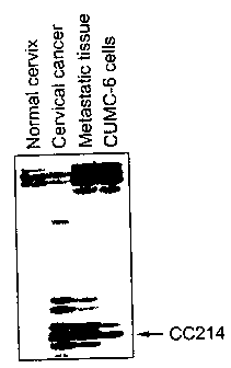
i~ Fig. 1: the DD identification of altered gene expression in normal
cervix tissue, primary cervical cancer tissue, metastatic lymph node tissue
and
CUMC-6 cervical cancer cells.
Fig. 2 : the prediction of hydrophobicity of transmembrane regions in
the protooncogene of the present invention using TMPRED program.
20 Fig. 3: the results of northern blot analyses for HCCR-1 gene expressed
in normal cervical tissues, cervical cancer tissues and cervical cancer cell
lines(CaSki and CUMC-6);
Fig. 4 : the results of northern blot analyses for HCCR-1 gene expressed
in normal lung tissue and lung cancer cell lines (NCI-H358, NCI-H460. NCI-
25 H441. NCI-H 1299, NCI-H520, NCI-H2009, and NCI-H 157);
Fig. 5A : the results of northern blot analyses for HCCR-1 gene
expressed in normal human 12-lane multiple tissues;
Fig. 5B : the results obtained with the same sample of Fig. 5A
hybridized with 0-actin.
.30 Fig. 6A : the results of northern blot analyses for HCCR-1 gene
" Trademark
CA 02353788 2001-05-29
WO 01/27149 PCT/KR00/00284
4
expressed in human cancer cell lines;
Fig. 6B : the results obtained with the same sample of Fig. 6A
hybridized with 0-actin.
Fig. 7A : the results of northern blot analyses for HCCR- I gene
~ expressed in human tumor tissues and their normal counterparts;
Fig. 7B : the results obtained with the same sample of Fig. 7A
hybridized with (3-actin.
Fig. 8 : a micrograph illustrating representative characteristics of in sicu
hybridized human cervical cancer tissues;
io Fig. 9 a phase-contrast feature of monolayer-cultured wild type
NIH/3T3 cells;
Fig. 10 : a phase-contrast feature of monolayer-cultured HCCR-1 cells;
Fig. 11 : hematoxylin-eosin staining of monolayer-cultured HCCR-l
cells;
15 Fig. 12 : a transmission electron micrograph illustrating representative
characteristics of cultured HCCR-1 cells;
Fig. 13 : tumorigenicity of HCCR-1 cells in nude mouse;
Fig. 14 : hematoxylin-eosin staining of subcutaneous tumour nodules
taken from nude mice;
20 Fig. 15 : transmission electron micrographs illustrating representative
characteristics of nude mice-derived subcutaneous tumour tissue;
Fig. 16 : phase-contrast features of monolayer-cultured nude mice-
derived HCCR-1N cells;
Fig. 17 : sodium dodecyl sulfate (SDS)-PAGE results showing protein
2i expression patterns before and after the IPTG induction;
Fig. 18 : the result of western blotting analysis of NIH/3T3 cells without
transfection(wild type), NIH/3T3 transfected with pcDNA3 vector
alone(pcDNA3) and HCCR-1 cells;
Fig. 19 : the result of western blotting analysis of human tumour tissues
30 of kidney, lung, ovary and cervix and their normal counterparts;
CA 02353788 2001-05-29
WO 01/27149 PCT/KR00/00284
Fig. 20 : the immunohistochemical study of HCCR-1-transfected
NIH/3T3 cells against reticulin fibers (x250);
Fig. 21 : the expression of epithelial marker, keratin in HCCR-I-
transfected NIH/3T3 cells (x250);
5 Fig. 22 : the expression of epithelial membrane antigen in HCCR- I-
transfected NIH/3T3 cells (x250);
Fig. 23 : the expression of mesenchymal marker, vimentin in HCCR-1-
transfected NIH/3T3 cells (x250);
Fig. 24 : the PKC activities in NIH/3T3 cells without transfection(wild-
type), NIH/3T3 transfected with pcDNA3 vector alone(pcDNA3) and NIH/3T3
transfected with HCCR-1 protooncogene (HCCR-1 cells);
Fig. 25 : the telomerase activities in 293 cells, +RNase, NIH/3T3 cells
without transfection(wild-type), NIH/3T3 transfected with pcDNA3 vector
alone(pcDNA3) and NIH/3T3 transfected with HCCR-1 protooncogene(HCCR-
i ~ I cells);
Fig. 26A : the result of RT-PCR amplification of HCCR-1 cDN A in H-
358 lung carcinoma cell lines treated with anti-sense oligodeoxynucleotides;
Fig. 26B : the results obtained with the same sample of Fig. 26A
hybridized with 0-actin.
Fig. 27 : growth curves of H-358 lung carcinoma cells treated with
sense, missense or anti-sense HCCR- I ODN, and untreated parental cells;
Fig. 28 : HCCR-1 protein expressions in fetal 16-(F 16), 18-(F I 8), 20-
(F20), postnatal 1-(P 1), 7-(P7), 14-day(P 14) and adult rat kidney tissue
extracts;
Fig. 29 : immunohistochemical staining of 20 day-old fetal rat kidney
(x42); and
Fig. 30 : differential-interference contrast microscopy of 18 day-old
fetal rat kidney illustrating HCCR-1 immunostaining in the basolateral plasma
membrane of medullary collecting duct (x220).
CA 02353788 2001-05-29
WO 01/27149 PCT/KR00/00284
6
Detailed Description of the Invention
The novel protooncogene of the present invention, i.e., human cervical
cancer 1(hereinafter "HCCR-1 protooncogene"), consists of 2118 base pairs and
s has the DNA sequence of SEQ ID NO: 1.
In SEQ ID NO: 1, the full open reading frame corresponding to base
Nos. 9 to 1088 is a protein encoding region and the predicted amino acid
sequence derived therefrom is shown in SEQ ID NO: 2 which consists of 360
amino acids("HCCR-1 protein"). Further, the region corresponding to base
iu Nos. 9 to 83 of SEQ ID NO: I encodes a signal peptide with the predicted
amino acid sequence of amino acid Nos. I to 25 in SEQ ID NO: 2: and the
region represented by nucleotide No. 435 to 494 of SEQ ID NO: I encodes a
single transmembrane domain having the predicted amino acid sequence of
amino acid Nos. 143 to 162 of SEQ ID NO: 2. This suggests that the
>> protooncogene of the present invention is a membrane-bound gene.
A single potential N-glycosylation site(corresponding to base Nos. 945
to 953 of SEQ ID NO: I and amino acid Nos. 313 to 315 of SEQ ID NO: 2) is
present at the C-terminal side of the HCCR-1 protein, which suggests that
HCCR-1 protein is a type II membrane protein. The polyadenylation signal
20 corresponds to the nucleotide Nos. 2008-2012 of SEQ ID NO:1.
The predicted extracellular domain of HCCR-1 corresponds to base Nos.
495-1088 with the predicted amino acid sequence of amino acid Nos. 163-360
consisting of 198 amino acids with 5 cysteine residues. The predicted
intracellular domain contains 117 amino acids(corresponding to nucleotide Nos.
2> 84-434 of SEQ ID NO:1 and amino acid Nos. 26-142 of SEQ ID NO:2) with
two potential protein kinase C(PKC) phosphorylation sites at Ser-42 and Ser-
48,
and two potential N-myristylation sites at Gly-34 and Gly-38. Further
computer-assisted analyses indicate that HCCR-1 is markedly hydrophobic and
possesses a characteristic single membrane-spanning domain and pre-secretory
30 signal peptide as shown in Fig. 2.
CA 02353788 2001-05-29
WO 01/27149 PCT/KR00/00284
7
In consideration of the degeneracies of codons and the preferred codons
in a specific animal wherein the protooncogene of the present invention is to
be
expressed, various changes and modifications of the DNA sequences of SEQ ID
NO:I may be made, e.g., in the coding area thereof without adversely altering
~ the amino acid sequence of the expressed protein, or in the non-coding area
without adversely affecting the expression of the protooncogene. Therefore.
the present invention also includes, in its scope, a polynucleotide having
substantially the sa.me base sequence as the inventive protooncogene, and a
fi-agment thereof. As used herein, "substantially the same polynucleotide"
to refers to a polynuleotide whose base sequence shows 80% or more, preferably
90% or more, most preferably 95% or rnore homology to the protooncogene of
the present invention.
The protein expressed from the protooncogene of the present invention
consists of 360 amino acids and has the amino acid sequence of SEQ ID NO: 2.
t~ The molecular weight of this protein is about 40 kDa. However. various
substitution. addition and/or deletion of the amino acid residues of protein
may
be performed without adversely affecting the proteiri's function. Further, a
portion of the protein may be used when a specific purpose is to be fulfilled.
These modified amino acids and fragments thereof are also included in the
20 scope of the present invention. Therefore, the present invention includes,
in its
scope, a polypeptide having substantially the same amino acid sequence as the
protein derived from the oncogene of the present invention and a fragment
thereof. As used herein, "substantially the same polypeptide" refei-s to a
polypeptide whose amino acid sequence shows 80% or more, preferably 90% or
25 more, most preferably 95% or more homology to the amino acid sequence of
SEQ ID NO:2.
The protooncogene, or the protein, of the present invention can be
obtained from human cancer tissues or synthesized using a conventional DNA
or peptide synthesis method. Further, the gene thus prepared may be inserted
30 to a conventional vector to obtain an expression vector, which may, in
turn, be
CA 02353788 2001-05-29
WO 01/27149 PCT/KR00/00284
8
introduced into a suitable host, e.g., an E. coli or yeast cell, The cells
transformed with a vector containing the HCCR-1 protooncogene or a fragment
thereof is hereinafter referred to a"HCCR-1 cell".
The transformed host may then be used in producing the inventive DNA
i or protein on a large scale. For example, E. coli JM 109 is transfected with
HCCR-1 protooncogene by using pGEM-T easy vector and the JM 109/HCCR-1
was deposited on October 11, 1999 with the Korean Collection for Type
Cultures(KCTC)(Address: Korea Research Institute of Bioscience and
Biotechnology(KRIBB), #52, Oun-dong, Yusong-ku, Taejon, 305-333, Republic
i of Korea) under the accession number, KCTC 0667BP, in accordance with the
tenms of Budapest Treaty on the International Recognition of the Deposit of
Microorganism for the Purpose of Patent Procedure.
In preparing a vector, expression-control sequences, e.g., promoter.
terminator, self replication sequence and secretion signal, are suitably
selected
15 depending on the host cell used.
The overexpression of the protooncogene of the present invention
occurs not in normal cervical and lung tissues but in cervical cancer tissues,
cervical cancer cell lines and lung cancer cell lines. This suggests that the
protooncogene of the present invention induces cervical and lung cancers.
20 Further, when a normal fibroblast cell, e.g., NIH/3T3 cell line, is
transfected
with the protooncogene of the present invention, an abnormal cells is
produced.
Morphological characterizations with optical and electronic microscopes show
that the abnormal cell has the form of a tumor cell.
When the normal fibroblast cell transfected with the protooncogene of
21- the present invention is injected into the posterial lateral aspect of a
nude mouse,
tumorigenesis is observed after about 21 days from the injection, the tumor
size
becoming 1.5 cm x 1.5 cm in 40 days. By using hematoxylin-eosin dye
method, it can be confirmed that the tumor cells are cancerous. The formation
of the epithelial carcinoma can also be confirmed by using transmission
30 electron microscopy and immunhistochemical staining methods.
CA 02353788 2001-05-29
WO 01/27149 PCT/KR00/00284
9
In addition to epithelial tissues such as cervical and lung cancer tissues,
the overexpression of the protooncogene of the present invention is also
observed in various other cancer tumors such as leukemia, lymphoma. kidney.
liver and ovarian cancers. Therefore, the protooncogene of the present
invention is believed to be a factor common to all forms of various cancer and
it
can be advantageously used in the diagnosis of various cancers and the
production of a transformed animal as well as in an anti-sense gene therapy.
A diagnostic method that can be performed using the protooncogene of
the present invention may comprise, for example, the steps of hybridizing
tc~ nucleic acids separated from the body fluid of a subject with a probe
containing
the protooncogene of the present invention or a fragment thereof. and
determining whether the subject has the protooncogene by using a conventional
detection method in the art. The presence of the protooncogene may be easily
detected by labeling the probe with a radioisotope or an enzyme. Therefore, a
cancer diagnostic kit containing the protooncogene of the present invention or
a
fragment thereof is also included in the scope of the present invention.
A transformed animal produced by introducing the protooncogene of the
present invention into a mammal, e.g., a rat, is also included in the scope of
the
present invention. In producing such a transformed animal, it is preferred to
introduce the inventive protooncogene to a fertilized egg of an animal before
the 8th cell cycle stage. The transformed animal can be advantageously used
in screening for carcinogens or anticancer agents such as antioxidants.
The present invention also provides an anti-sense gene which is useful
in a gene therapy. As used herein, the term "an anti-sense gene" means a
polynucleotide comprising a base sequence which is fully or partially
complementary to the sequence of the mRNA which is transcribed from the
protooncogene having the base sequence of SEQ ID NO: 1 or a fragment thereof
said nucleotide being capable of preventing the expression of the open reading
frame(ORF) of the protooncogene by way of attaching itself to the protein-
3o binding site of mRNA.
CA 02353788 2001-05-29
WO 01/27149 PCT/KR00/00284
An example of the anti-sense gene of the present invention is a l 8-mei-
HCCR-l anti-sense oligodeoxinucleotide(ODN) having the base sequence of
SEQ ID NO:3. Therefore, the present invention also includes, in its scope. a
polynucleotide comprising substantially the same base sequence as SEQ ID
5 NO:3 and a fragment thereof.
The present invention also includes within its scope a process for
treating or preventing cancer in a subject by way of administering a
therapeutically effective amount of the inventive anti-sense gene thereto.
In the inventive anti-sense gene therapy, the anti-sense gene of the
10 present invention is administered to a subject in a conventional manner to
prevent the expression of the protooncogene. For example, the anti-sense
ODN is mixed with a hydrophobized poly-L-lysine derivative by electrostatic
interaction in accordance with the method disclosed by Kim, J.S. et al.(J.
Conirolled Release, 53, 175-182(1998)) and the resulting mixed anti-sense
ODN is administered intravenously to a subject.
The present invention also includes within its scope an anti-cancei-
composition comprising the anti-sense gene of the present invention as an
active ingredient, in association with pharmaceutically acceptable carriers.
excipients or other additives, if necessary. The pharmaceutical composition of
the present invention is preferably formulated for administration by
injection.
The amount of the anti-sense gene actually administered should be
determined in light of various relevant factors including the condition to be
treated, the chosen route of administration, the age and weight of the
individual
patient, and the severity of the patient's symptoms.
The protein expressed from the inventive protooncogene may be used in
producing an antibody useful as a diagnostic tool. The antibody of the present
invention may be prepared in the form of a monoclonal or polyclonal antibody
in accordance with any of the methods well known in the art by using a protein
having the amino acid sequence of SEQ ID NO:2 or a fragment thereof.
Cancer diagnosis may be carried out using any of the methods known in the art,
CA 02353788 2001-05-29
WO 01/27149 PCT/KR00/00284
11
e.g., enzyme linked immunosorbentassay(ELISA), radioimmunoassay(R1A),
sandwich assay, western blot or immunoassay blot on polyacrylic gel, to asses
whether the protein is expressed in the body fluid of the subject. Therefore,
a
cancer diagnostic kit containing the protein having the amino acid sequence of
~ SEQ ID NO:2 or a fragment thereof is also included in the scope of the
present
invention.
A continuously viable cancer cell line may be established by using the
protooncogene of the present invention, and such a cell line may be obtained.
for example, from tumor tissues formed on the back of a nude mouse by
injecting fibroblast cells transformed with the protooncogene of the present
invention. The cell lines thus prepared may be advantageously used in
searching for anti-cancer agents.
The following Examples and Test Examples are given for the purpose of
illustration only, and are not intended to limit the scope of the invention.
Example 1: Cultivation of tumor cells and separation of total RNA
Step 1-1 : Cultivation of tumor cells
For differential display of mRNA, normal cervical tissues, untreated
primary cervical cancer tissues and metastatic common iliac lymph node tissues
were obtained from cervical cancer patients who underwent radical
hysterectomy. The human cervical cancer cell line used in the differential
display method was CUMC-6 cell line described by Kim et al.,(Gynecol. Oncol.,
62: 230-240(1996)).
Cells from the above-described tissues and CUMC-6 were maintained
on Waymouth's MB 752/ 1 medium (Gibco) supplemented with 2 mmol/L of
glutamine, 100 IU/ml of penicillin, 100 g/ml of streptomycin, and 10% of
fetal
bovine serum (Gibco). Only the cell suspensions with greater than 95%
viability, as assessed by trypan blue dye exclusion described by
Freshney("Culture of Animal Cells: A Manual of Basic Technique" 2nd Ed., A.
CA 02353788 2004-11-29
12
R. Liss, New York(1987)) were used in the present experiments.
Step 1-2 : Isolation of total RNA and differential display of mRNA
Total RNAs were extracted from normal cervical tissues, primary
cervical cancer tissues, metastatic common iliac lymph node tissues and
~. ._
CUMC-6 cells obtained in Step I-1 using a commercial system (RNeasy total
RNA kit) provided by Qiagen (Qiagen Inc., Germany) and the removal of DNA
contaminants from the RNAs was accomplished using Message clean* kit
(GenHunter Corp., Brookline, MA).
Example 2- Differential display reverse transcription(DDRT)-PCR
Differential display reverse transcription was performed in accordance
with the reverse transcription-polymerase chain reaction (RT-PCR) method
i~ described by Liang and Pardee(1992), supra, with minor modifications.
First, reverse transcription was carried out using 0.2 g each of the total
RNAs obtained in Step 1-2 of Example I and one of the three primers, i.e., 1-1-
TI1G. H=TI1C, or H-T11A, as anchored oligo-dT primers (RNAimage kit.
GenHunter Cor., MA, USA).
20 Then PCR was conducted using the same anchored primers and one of
the arbitrary 5' 13 mer (RNAimage primer sets 1-4, H-AP 1-32) in the presence
of 0.5 mM [a-35S]-labeled dATP (1200 Ci/mmol). The PCR thermal cycle
was repeated 40 times, the cycle being composed of: 95 C for 40 sec., 40 C
for 2 min. and 72 C for 40 sec., and finally the reaction was canied out at
2i 72 C for 5 min.
PCR-amplified fragments were resolved in 6% polyacrylamide
sequencing gels. Differentially expressed fragments were identified by
inspection of autoradiograms.
Band of more than 200 base pairs, CC214, were excised from the dried
* Trademarks
CA 02353788 2004-11-29
13
sequencing gel. The CC214 eDNA was eluted by boiling for 15 min and
reamplified with the same primer pairs and PCR conditions as used in the above
amplification step except that no [a 35S]-labeled dATP and 20 M dNTPs were
used.
;
Example 3 : Cloning
The reamplified CC214 PCR product obtained as above was inserted
into the pGEM-T Easy*vector using an TA cloning system (Promega, USA) in
M accordance with the manufacturer's instructions.
Step 3-1 : Ligation
2 l of the reamplified CC214 PCR product obtained in Example 2, 1 1
of pGEM-T easy vector (50 ng), I l of T4 DNA ligase l OX buffer solution and
15 I l of T4 DNA ligase(3 weiss units/41; T4 ligase, Promega, USA) were
charged into a 0.5m1 tube and distilled water was added thereto to a final
volume of 10 1. The ligation reaction mixture: was incubated overnight at
14 C.
20 Step 3-2 : TA cloning transformation
TA cloning transformation was performed using the following protocol.
L'. coli JM109(Promega, WI, USA) was incubated in 10 ml of LB
broth(Bacto-tryptori lOg, Bacto-yeast* extract 5g, NaCl 5g) until the optical
density at 600nm reached about 0.3 to 0.6. The culttired mixture was kept at
25 0 C for 10 minutes and centrifuged at 4000 rpm at 4 C for ] 0 minutes. The
supernatant was removed and cells were harvested. The harvested cell pellet
was exposed to 10m1 of O.1M CaCIZ at 0 C for 30 minutes to 1 hour to obtain
competent cells. The resultant was centrifuged at 4000 rpm at 4 C for another
minutes and the collected cells were suspended in 2m) of 0.1 M CaCl2 at 0 C .
* Trademarks
CA 02353788 2001-05-29
WO 01/27149 PCT/KROO/00284
14
200 l of the competent cell suspension was transferred to a microfuge
tube and 2 l of the ligation product obtained in Step 3-1 was added thereto.
The mixture was incubated in a water bath at 42 C for 90 seconds and rapidly
cooled to 0 C . Added thereto was 800 l of SOC medium (Bacto-trypton 2.0g,
Bacto-yeast extract 0.5 g, IM NaCI I ml, 1M KCI 0.25 ml, TDW 97 ml, 2M
Mg2" 1 ml, 2M glucose lml) and the mixture was incubated at 37 C for 45
minutes at 220 rpm in a rotary shaking incubator.
LB agar plates containing ampicillin(50u1/ml) were prepared by
spreading 25g1 of X-gal (40mg/mi stock in dimethylformamide) on top of agar
with a glass spreader. 251i1 of the transformed cells thus obtained was spread
thereon and the plates were incubated at a 37 C incubator overnight. White
colonies were loaded on an LB agar plate containing ampicillin and transformed
E. coli, i.e., JM 109/ CC214 were selected and incubated in a terrific
broth(TDW
900 ml, Bacto-trypton 12 g, Bacto-yeast extract 24 g, glycerol 4 ml. 0.17M
i4; KH2PO4, 0.72 N KZHPO4 100 ml).
Example 4: Separation of recombinant plasmicl DNA
The CC214 plasmid DNA of the transformed L: coli was separated by
employing Wizard' Plus Minipreps DNA Purification Kit(Promega, USA) in
accordance with the manufacturer's instructions.
A portion of the plasmid DNA thus separated was treated with ECoRI
enzyme and subjected to gel electrophoresis to confirm the insertion of CC214
partial sequence in the plasmid.
Example 5: Sequence Analysis of DNA
The CC214 PCR product obtained in Example 2 was subjected to PCR
in accordance with the conventional method and the cloned, reamplified CC214
CA 02353788 2004-11-29
PCR fragments were subjected to sequence analysis according to the dideoxy
chain termination method using a Sequenase*version 2.0 DNA sequencing kit
(United states Biochemical, Cleveland, OH) in accordance with the
manufacturer's instructions.
The base sequence of the DNA corresponds to nucleotide Nos. 1883-
2088 in SEQ ID NO:1 and is designated "CC214".
The differential display reverse transcription polymerase chain
reaction(DDRT-PCR) of the 206 bp cDNA fragment, i.e., CC214 obtained
above was carried out using a 5' arbitrary primer H-AP21 and a 3' H-T11 C
Iu anchored primer and resolved by electrophoresis. Identification of altered
gene expression by DD in the primary cervical cancer, metastatic lymph node
tissue and CUMC-6 cells is shown in Fig 1. As can be seen in Fig. 1. the 206
bp cDNA fragment, i.e., CC214 was expressed in the cervical cancer, metastatic
tissue and CUMC-6 cervical cancer cells but not in the normal tissue.
'S
Example 67 Full length cDNA sequence analysis of the HCCR-1
12rotooncogene
A bacteriophage Xgt11 human lung embryonic fibroblast eDNA library
Miki, T. et al., Gene, 83:137-146(1989)) was screened by plaque
hybridization with 'ZP-labeled CC2I4 as a probe. The full-length HCCR-1
cDNA clone, containing a 2118 bp insert in pCEV-LAC vector was obtained
from the human lung embryonic fibroblast cDNA library and registered at
GenBank on November 5, 1999 under the accession number AF195651.
HCCR-1 clone inserted into XpCEV vector (&& Miki, T. et al.. supra)
was excised out of the phage in the form of the ampicillin-resistant pCEV-LAC
phagemid vector (&& Miki, T. et al., supra) by Not I cleavage.
To make a HCCR-1 plasmid DNA, pCEV-LAC vector containing
HCCR-1 gene was ligated with T4 DNA ligase and ligated clone was
o transformed into E. coli JM109.
* Trademarks
CA 02353788 2001-05-29
WO 01/27149 PCT/KR00/00284
16
The transformed E. coli JM109/HCCRI thus obtained was deposited on
October 11, 1999 with the Korean Collection for Type
Cultures(KCTC)(Address: Korea Research Institute of Bioscience and
Biotechnology(KRIBB), #52, Oun-dong, Yusong-ku, Taejon, 305-333, Republic
4; of Korea) under the accession number, KCTC 0667BP, in accordance with the
terms of Budapest Treaty on the International Recognition of the Deposit of
Microorganism for the Purpose of Patent Procedure.
The full sequence of HCCR-1 consists of 2118 bp which is identified in
SEQ ID NO:1.
iu In SEQ ID NO:1, the full open reading frame of the HCCR-1
protooncogene of the present invention corresponds to nucleotides No. 9 to
1088 and is predicted to encode amino acid sequence shown in SEQ ID NO:2
which consists of 360 amino acids. Further, the region corresponding to
nucleotide Nos. 9 to 83 of SEQ ID NO:I encodes a signal peptide having 25
ti amino acids corresponding to amino acid Nos. I to 25 of SEQ ID NO:2; the
region of nucleotide Nos. 435 to 494 of SEQ ID NO: I encodes a single
transmembrane domain whose amino acid sequence corresponds to amino acid
Nos. 143 to 162 in SEQ ID NO:2. This indicates that the protooncogene of the
present invention is a membrane-bound gene.
20 A single potential N-glycosylation site(corresponding to base Nos. 945
to 953 of SEQ ID NO: 1 and amino acid Nos. 313 to 315 of SEQ ID NO: 2) is
present at the C-terminal side of the HCCR-1 protein, which suggests that
HCCR-1 is a type II membrane protein. The polyadenylation signal
corresponds to the nucleotide Nos. 2008-2012 of SEQ ID NO:1.
25 The predicted extracellular domain contains 198 amino acids with 5
cysteine residues. The predicted intracellular domain contains 117 amino
acids(corresponding to nucleotide Nos. 84-434 of SEQ ID NO:1 and amino acid
Nos. 26-142 of SEQ ID NO:2) with two potential PKC phosphorylation sites at
Ser-42 and Ser-48, and two potential N-myristylation sites at Gly-34 and Gly-
3o 38. Further computer-assisted analyses indicated that HCCR-1 is markedly
CA 02353788 2004-11-29
17
hydrophobic and possesses a characteristic single membrane-spanning domain
and pre-secretory signal peptide as shown in Fig. 2. In Fig. 2, the X-axis
represents the amino acid sequence number of the peptide of the present
invention and the Y-axis, the hydrophobicity of the peptide.
i
Exam l~e 7: Northern blot analysis of the HCCR-l gene in various cells
Total RNAs were extracted from various tissues and cell lines as in
Example 1.
To determine the level of HCCR-1 gene expression, 20 g denatured
total RNAs from each tissue or cell lines were electrophoresed through 1 io
formaldehyde agarose gel and transferred to nylon membranes (Boehringer-
Mannheim, Germany). The blots were hybridized with a"P-labeled random-
primed HCCR-1 full cDNA probe which was prepared using a rediprime l I
random prime labeling system(Amersham, England). The northern blot
analysis was repeated twice and the results were quantified by densitometry
and
normalized with j3 -actin.
Fig. 3 shows the results of northern blot analyses for HCCR-l gene
expressed in normal cervical tissues, cervical cancer tissues and cervical
cancer
cell lines(CaSki and CUMC-6). As can be seen in Fig. 3, the transcription
level of HCCR-1 is high in the cervical cancer tissues and cancer cell lines
(CaSki(ATCC CRL 1550) and CUMC-6), but very low or undetactable in the
normal cervical tissues.
Fig. 4 shows the results of northern blot analyses for HCCR-1 gene
2~ expressed in normal lung tissues and seven lung cancer cell lines, i.e..
H358(ATCC NCI-H358), H460(ATCC NCI-H460), H441(ATCC NCI-H441).
H299(ATCC NCI-H299), H520(ATCC NCI-H520), H2009(ATCC NCI-H2009),
and H157(ATCC NCI -H157). As shown in Fig. 4, HCCR-1 transcription
level in high level in lung cancer cell lines H358, H460, H1299, H520, and
3o H157, but not detectable in the normal lung tissues.
* Trademark
CA 02353788 2001-05-29
WO 01/27149 PCT/KR00/00284
18
Fig. 5A shows the results of northern blot analyses for HCCR-1 gene
expressed in normal human 12-lane multiple tissues; brain, heart, skeletal
muscle, colon, thymus, spleen, kidney, liver, small intestine, placenta, lung
and
leukocyte tissues(Clontech). Fig. 5B shows the results obtained with the same
samples hybridized with a 0 -actin probe to confirm mRNA integrity. As can
be seen in Fig. 5A, HCCR-1 mRNA (-2.1 kb) is weakly present or absent in
many normal tissues, but the level of expression was high in normal kidney
tissue.
Fig. 6A shows the results of northern blot analyses for HCCR- l gene
io expressed in human cancer cell lines; HL-60, HeLa, K-562, MOLT-4. Raji.
SW480, A549 and G361(Clontech). Fig. 6B shows the results obtained with
the same samples hybridized with aP-actin probe to confirm mRNA integrity.
As can be seen in Fig. 6A, HCCR-1 is transcribed at a high level in the human
leukaemia and lymphoma cell lines such as chronic myelogenous leukaemia K-
>> 562, Burkitt's lymphoma Raji, lymphoblastic leukaemia MOLT-4 and
promyelocytic leukaemia HL-60 as well as HeLa cells.
K-562, MOLT-4 and HL-60, in particular, show higher transcription
levels as compared with normal leukocyte by factors of approximately 190, 90
and 70, respectively. HCCR-1 expression levels in colorectal cancer SW480,
20 lung cancer A549 and melanoma G361 cell lines are lower than those of
leukemia and lymphoma.
Further, northern blotting analyses of the human kidney, liver, lung,
ovary and cervix tumor tissues and their normal counterparts were carried out.
As shown in Fig. 7A, HCCR-1 was transcribed at a high level in the human
25 cancer cells, while the expression of HCCR-1 gene is barely observable in
the
normal cells. Fig. 7B shows the results obtained with the same samples
hybridized with 0-actin probe to confirm mRNA integrity.
CA 02353788 2004-11-29
19
Example 8 : Micrograph of in situ hybridized Human Cervical _ancer Tissues
For in situ hybridization, human cervical cancer tissue was fixed in
periodate-lysine-paraformaldehyde, embedded in a wax according to the
~ procedure described by Ahn et al.( Am. J Physiol. 265, F792 - F801 (1993))
and sectioned(5 m). A full-length HCCR-1 cDNA fragment was used to
synthesize a digoxigenin-labelled RNA probe. RNA in situ h_ybridization was
carried out with the anti-sense RNA probe which was prepared using a DIG
RNA Labelling Kit(Boehringer Mannheim). The sense RNA probe was used
as a negative control.
Fig. 8 shows a micrograph illustrating representative characteristics of
in sitti hybridized human cervical cancer tissues. As can be seen in Fig. 8,
the
in situ hybridized ceivical cancer tissues are confirmed to contain a high-
level
of HCCR-1 gene. No staining was detected in the surrounding normal fibrous
i> tissues.
Example 9: Construction of =ression vectQrs and transformation of cells
Step 9-1 : Preparation of a vector containing HCCR-1
An expression vector containing the coding region of HCCR-1 was
constructed as follows.
First, the entire HCCR-1 cDNA obtained in Example 6 was inserted into
the Sall restriction site of a prokaryotic expression vector, pCEV-LAC(= Miki,
T. el al., Gene, 83: 137-146 (1989)). Then, the SaII fragment was isolated
from the pCEV-LAC/HCCR-1 vector.
Then, pcDNA3 (Invitrogen) was digested with Xhol to make a
compatible end with SaII. The Sall fragment containing the full length
HCCR-1 coding sequence was inserted into the Xhol-digested pcDNA3.
Lipofectamine* (Gibco BRL) was used to introduce the resulting
pcDNA3/HCCR-l expression vector into NIH/3T3 cells(ATCC CRL 1658.
* Trademarks
CA 02353788 2001-05-29
WO 01/27149 PCTIKROO/00284
USA), followed by selection in a medium supplemented with G418 (Gibco).
The resulting NIH/3T3 cells transfected with HCCR-l was designated "HCCR-
I cells". Another population of NIH/3T3 cells containing pcDNA3 alone was
prepared as a control and designated "pcDNA3 cells".
i
Step 9-2 : NIH/3T3 fibroblast cells transfected with the HCCR-1 protooncogene
The wild type normal NIH/3T3 cell, a differentiated fibroblast cell line.
is a spindle shaped cell having a long slender nucleus and a scanty amount of
cytoplasm as shown in Fig. 9. When HCCR-1 was expressed in the NIH/3T3
io expressing HCCR-1(HCCR-1 cells) obtained in Step 9-1, the cell shape
changes
into a polygonal form with an ovoid nucleus and plump cytoplasm, as shown in
Fig. 10.
Monolayer cultured HCCR-1-transfected NIH/3T3 cells which is
stained with hematoxylin-eosin, exhibit nuclear pleiomorphism, distinct
ii nucleoli, granular chromatin patterns, tumor giant cells and atypical
mitotic
figures as shown in Fig. 11.
For transmission electron microscopy(TEM), the cells and tissues were
fixed with 2.5% glutaraldehyde in a phosphate buffer (pH 7.4). They were then
postfixed with a 2% osmium tetroxide. Specimens were dehydrated in a graded
20 series of ethanols and embedded in Epon 812. Ultrathin sections thereof
were
stained with uranyl acetate and lead citrate, and photographed by TEM(JEOL
1,200 EX, Tokyo, Japan).
The TEM picture shown in Fig. 12 reveals that cultured tumour cells
have microvilli and well-developed organelles (inset). As can be seen in Fig.
12, the HCCR-1 cell has microvilli on the cell surface, lobulated nucleus with
prominent nucleoli and well-developed rough endoplasmic reticula(rER) and
Golgi complexes (circle). In Fig 12, the scale bar corresponds to 3 m. In
higher magnification of the area indicated by circle (inset), the scale bar
corresponds to 1 m.
CA 02353788 2001-05-29
WO 01/27149 PCT/KR00/00284
21
Example 10: Tu_morigenici and metastasis of HCCR-1 protooncogene in
animal
To analyze tumourigenicity, 5 X 10⁶ HCCR-1 cells were injected
subcutaneously into the posterior lateral aspect of the trunk of 9 mice (5-
week-
old athymic nu/nu on BALB/c background). Nude mice were sacrificed when
the subcutaneous tumors reached 1.5-2.5 cm in diameter.
All 9 mice injected with HCCR-1 cells showed palpable tumors after 21
days as shown in Fig 13.
io Nude mice bearing HCCR-l allografts display characteristics of an
epithelial carcinoma. Fig. 14 shows hematoxylin-eosin staining of
subcutaneous tumor nodules taken from the nude mice. The sections of the
tumour nodules revealed typical epithelial cell nests separated by fibrous
stroma.
Example 11 : Electron microscopy of HCCR-1 protooncogene - induced tumor
tissue and establishment of new cancer cell line
Tumor tissues taken from the tumor nodules formed on the nude mouse
of Example 10 were examined with an electron microscope, which revealed that
20 tumor nodules showed well-developed organelles and tumour cells ai-e
connected by desmosomes(Fig. 15). As shown in Fig. 15, the tumor tissue
consists of tightly adhered cells with intercellular junction (circle). In
Fig. 15,
the scale bar corresponds to 3 W. In higher magnification of the area
indicated by circle illustrating desmosome (inset), the scale bar corresponds
to
25 0.5 m.
The cells obtained from the above tumor tissue was cultured in a
conventional manner using 20% fetal bovine serum and the cultured cells were
designated HCCR-1N cells which have cytological features similar to HCCR-1
cells in vitro as shown in Fig. 16.
CA 02353788 2004-11-29
22
Example 12: Determination of size of protein expressed after the transfection
of
F. coli with HCCR-1 protooncogene
A portion of HCCR-1 protooncogene corresponding to nucleotide Nos.
i 123-473 and predicted amino acid Nos. 39-155 was inserted into the multiple
cloning site of pET-32b(+) vector(Novagen) and the resulting pET-
32b(+)/HCCR-1 vector was transfected into E. coli BL21(ATCC 47092). The
transfected E. coli was incubated using an LB broth medium in a rotary shaking
incubator, diluted by 1/100, and incubated for 3 hours. ImM isopropyl Q-D-
ia thiogalacto-pyranoside(IPTG, Sigma) was added thereto to induce the protein
synthesis.
The E. coli cells in the culture were disrupted by sonication and
subjected to gel electrophoresis using 12% sodium dodecyl sulfate(SDS) before
and after the IPTG induction. Fig. 17 shows the SDS-PAGE results which
t> exhibit a protein expression pattern of the E. coli BL21 strain transfected
with
pET-32b(+)/HCCR-1 vector. After the IPTG induction, a significant protein
band was observed at about 35kDa. This 35kDa fused protein contained an
about 20kDa Trix=Tag thioredoxin protein expressed from a gene in pET-32b(+)
vector.
Example 13 = Production of antibody
The 35 kDa fused protein isolated from the E. coli BL2I strain
transfected with pET-32b(+)/HCCR-1 vector in Example 12 was purified by
*
using a His-Bind Kit (Novagen). Immunoblotting of the purified peptide
confirmed the presence of a major amount of a 35kDa protein.
Then, two 6-week old Sprague-Dawley rats each weighing about 150 g
were each subcutaneously immunized with 1 mg of the peptide thus obtained,
weekly for 3 times. Blood samples were obtained from the immunized rats
and centrifuged to obtain a polyclonal serum. The anti-HCCR-1 activity of the
* Trademark
CA 02353788 2001-05-29
WO 01/27149 PCT/KR00/00284
23
polyclonal serum was detennined and confirmed by enzyme-linked
immunosorbent assay(ELISA)(1:10,000)
Example 14 : Immunoblot confirming n ibo yspecificitv
For western blot analysis, those cells identified in Figs. 18 and 19 were
harvested and lysed in a Laemmli sample buffer in accordance with the method
described by Laemmli(Nature 227: 680-685 (1970)). The cellular proteins
were separated by 10% SDS-PAGE and then electroblotted onto nitrocellulose
membranes. The membranes were incubated with the rat polyclonal anti-
HCCR-1 serum prepared in Example 13 for 16 h. After washing, the
membranes were incubated with a blocking solution containing 1:1,000 dilution
of peroxidase-conjugated goat anti-rat immunoglobulin (Jackson
lmmunoResearch) as a secondary antibody. Proteins were revealed by an
i 5 ECL-Western blot detection kit (Amersham).
As shown in Fig. 18, HCCR-1 protein is overexpressed in HCCR-1 cells,
while only faint bands are observed for the wild type and cells transfected
with
the vector alone(pcDNA3). This result illustrates the specificity of the anti-
HCCR-1 antibodies in the polyclonal serum.
Further, the HCCR-1 antibody in the polyclonal serum recognized
approximately 40 kDa protein in human protein extracts from different tissues.
As shown in Fig. 19, human tumor tissues including carcinomas of the kidney,
lung, ovary and cervix showed increased HCCR-1 protein expression when
compared with their normal counterparts.
Example 157 I munohistochemistrv
The tumor nodules formed on the nude mouse of Example 9 were
incubated with anti-vimentin, anti-keratin, anti-EMA(epithelial membrane
3o antigen) antibodies (DAKO) and polyclonal antibody raised against HCCR-1.
CA 02353788 2004-11-29
24
respectively. Then, immunohistochemistry was carried out on 5 gm-
crvosections of the incubated tumor nodules.
Binding of primary antibody was visualized by biotinylated secondary
antibody, avidin, biotinylated horseradish peroxidase and AEC(Aminoethyl
Carbaxzole Substrate Kit) as the chromogen(HISTOSTAIN-BULK KITS*
Zymed). The immunohistochemical study revealed that HCCR-1 transfection
into NIH/3T3 cells caused the conversion of the cell nature from mesenchymal
to epithelial. The cell nests were enveloped by reticulin fibers as shown in
Fig.
20.
lu The cells showed coexpression of epithelial markers, such as keratin(Fig.
21) and epithelial membrane antigen (Fig. 22) and of the mesenchymal marker,
vimentin(Fig. 23).
Example 160 Protein kinase C and telomerase activity assavs
To ensure that HCCR-1 modulates the protein kinase C(PKC) activity in
cells, PKC assay was performed using wild-type NIH/3T3 cells, pcDNA3-
containing NIH/3T3 cells and HCCR-1-transfected NIH/3T3 cells prepared in
Step 9-1 of Example 9.
PKC activity was measured using the SignaTECT"'' Protein Kinase C
Assay System (Promega) according to the manufacturer's instructions. PKC
activity was defined as the difference of the amounts of PKC incorporated into
substrate per minute in the absence and presence of phospholipids. Each value
is the means s.d. of three independent experiments.
2i The result in Fig. 24 shows that the PKC activity of HCCR-1-
ti-ansfected NIH/3T3 cells is about 10-fold higher than the wild-type.
To explain the tumorigenesis of HCCR-1, telomerase activities in wild-
type NIH/3T3 cells, pcDNA3-containing. NIH13T3 cells and HCCR-1-
transfected NIH/3T3 cells prepared in Step 9-1 of Example 9 were measured
using the telomerase PCR-ELISA kit (Boehringer Mannheim) according to the
~
* Trademarks
CA 02353788 2004-11-29
manufacturer's instructions. Human telomerase-positive immortalized human
kidney cells (293 cells) provided in the kit were used as a positive control.
Used as a negative control was the 293 cells pretreated with RNase
(+RNase). Assays were performed with an extract amount equivalent to 1 x 10
5 cells.
Results in Fig. 25 show the average mean optical density(OD) values
from four separate experiments (means s.d.). Consistent with the previous
study (Holt, S. E., Wright, W. E. and Shay J. W. Mol. Cell Biol. 16, 2932-2939
(1996), wild-type NIH/3T3 cells showed detectable telomerase activity.
10 HCCR-l gene transfection raised the telomerase activity by a factor of
about 7
as compared with the wild-type cells. The high telomerase activity of the 293
cells was nullified by pretreatment with RNase.
Example 17: Cell c, c,y le experiments
Wild-type and HCCR-1-transfected NIH/3T3 cells cultured in a DMEM
medium at mid-log phase were growth arrested. by incubation in a DMEM
medium containing 0.5% bovine calf serum for 36 h. Cells to be analyzed for
the DNA content were harvested following trypsinization, and fixed in 70 %
ethanol. Fixed cells were then stained with propidium iodide as described by
Hedley(Floiv Cytometry, DNA Analysis frona Paraffin-enibedded Rlock.s:
Darzynkiewicz, Z. & Crissman, H. A. eds., Academic Press, San Diego, 1990).
First, 50 gg/ml of a propidium iodide staining solution (Sigma) and 100
units per ml of RNase A (Boerhinger Mannheim) were added to 2 x 10⁶ cells.
After incubation for 1 h, the cellular DNA content was determined by
fluorescence analysis at 488 nm using a FACS Caliber (Becton Dickinson). A
. ,~
minimum of I x 104 cells per sample was analyzed with Modfit 5.2 software.
In order to study whether there was an alteration in the growth
properties of HCCR-1-transfected NIH/3T3 cells, cell cycle profiles were
* Trademarks
CA 02353788 2001-05-29
WO 01/27149 PCT/KR00/00284
26
examined. The cell contents of the wild type NIH/3T3 cells and HCCR-1
transfected NIH/3T3 cells(mid-log phase) in Go/G,, S, G2/M phases were
measured and the results are shown in Table I.
~ Table I
Wild Type HCCR-1 Cell
Go/C, S G2/M Go/C, S G,/M Cell Content(%) 55.7 20.6 24 46.6 31.5 22.4
As can be seen from Table I, the percentage of wild-type and HCCR-1-
transfected NIH/3T3 cells in the S-phase was 20.6% and 31.5%. respectively
(mid-log phase). These results suggest that there was a significant shift of
the
io cell population out of the Go/G,-phase into the S-phase in HCCR-l
transfected
NIH/3T3 cells.
To assess the serum-dependent cell cycle progression, cells were
cultured in 0.5% bovine calf serum for 36 h. After incubation, cells were
released with 20% bovine calf serum and harvested at indicated times. The
15 cell contents of wild type NIH/3T3 cells and HCCR-1 transfected NIH/3T3
cells in Go/G,, S, G
2/M phases at indicated times were measured and the results
are shown in Table 11.
Table II
Time(h) Cell Content(%)
Wild Type HCCR-1 Cell
Go/C, S G2/M Go/C, S G,/M
0 77 8.0 14.9 70 21.8 8.7
12 72.2 14 14.2 66.9 24.0 9.6
24 49.6 13.4 37.2 56.7 24.7 19.2
48 58.3 18.3 23.7 52.7 30.4 17.5
CA 02353788 2001-05-29
WO 01/27149 PCT/KR00/00284
27
As can be seen from Table II, few cells remained in the S-phase in wild-
type cells measured at 0 h (8%). In contrast, a considerable number of HCCR-
I cells measured at 0 h were still in the S-phase (21.8%), suggesting that
constitutive overexpression of HCCR-1 allowed for a relative amount of
~ resistance to serum deprivation-induced Go/G, arrest. Following the release
of
cells from the growth arrest caused by serum-deprivation, there were
consistent
increases of over 10% in the S-phase populations of HCCR-1 cells as compared
to wild-type cells at measured time intervals (12 h, 24 h and 48 h).
Therefore,
overexpression of HCCR-1 could deregulate cell growth by shortening the
Iu G,,/G,-phase and increasing the S-phase population of cells.
Example 18: Construction of anti-sense oligodeoxinucleotide
Anti-sense and sense phosphorothioate oligodeoxynucleotide(ODNs)
1i targeting the translation starting site of HCCR-1 mRNA were synthesized
based
on the human HCCR-1 cDNA sequence (GenBank accession number
AF195651) by cyanoethylphosphoramidite chemistry on an automated DNA
synthesizer (Expedite Nucleic Acid System, Framingham, MA).
The sequence of 18-mer HCCR-1 anti-sense ODN was 5'-
20 CCTGGACATTTTGTCACC-3' (SEQ ID NO: 3; corresponding to nucleotide
Nos. 66 to 83 of SEQ ID NO:1). The corresponding sense sequence, 5'-
GGTGACAAAATGTCCAGG-3'(SEQ ID NO: 4), and missense sequence, 5'-
CGCGGATATTTCCTCACC-3'(SEQ ID NO: 5) were used as controls.
25 Example 19: Cancer gene therapXusing HCCR- 1 antisense ODN
Step 19-1 : Inhibition of gene expression
Exponentially growing 2 X 10⁵ H-358 lung carcinoma cells (ATCC
CRL-5807) were detached by trypsin-EDTA and seeded in a 24-well plate.
3o Lipofectamine (Gibco BRL) was used for oligodeoxynucleotide(ODN)
CA 02353788 2001-05-29
WO 01/27149 PCT/KROO/00284
28
treatment. Lipofectamine (5 l/m1 medium) was incubated with an
appropriate amount of ODN to achieve a final concentration of 100 nM ODN.
CA 02353788 2001-05-29
WO 01/27149 PCT/KROO/00284
29
Step 19-2 : Inhibition of cell growth
The growth phenotype of H-358 lung cancer cells treated with 100 nM
of sense, anti-sense or missense HCCR-1 oligodeoxynucleotide was assessed by
growth curve.
In three independent experiments, H-358 lung cancer cells were
trypisinized and plated in the presence of 100 nM of sense, anti-sense or
missense HCCR-1 ODN obtained in Example 18, and a growth medium(RPMI-
1640) containing 100 nM of HCCR-1 ODN was replaced every other day. Cells
in triplicate dishes were detached and viable cells were counted every other
day
using trypan blue dye exclusion.
As shown in Fig. 27, until I day of treatment, there were no discernable
differences in cell growth among sense('-7), missense(A) or anti-sense(o) HCCR-
>; 1 ODN-treated carcinoma cells. However, after 3 days of HCCR-1 ODN
treatment, anti-sense HCCR-1 ODN inhibited lung carcinoma cell growth in a
time-dependent manner.
After 7 days exposure to antisense HCCR-1 ODN, the extent of growth
inhibition was about 100% for H-358 lung carcinoma cells, while cells exposed
to sense or missense HCCR-1 ODN showed growth patterns similar to that of
untreated wild-type H-358 cells(control cells, 0).
Exmple 20: HCCR-1 gene as a regulator of embj3~onic kidney development
Because the acquisition of epithelial properties by the fibroblast-derived
HCCR-1 cells mimics the mesenchymal to epithelial conversion of cells during
the organogenesis of the kidney (Giordano, S. et al., Proc. Natl. Acad. Sci.
USA
90, 649-653 (1993): Tsarfaty, I., el al., Science 263, 98-101 (1994)), an
experiment was conducted to examine whether HCCR-1 is expressed in a
3o developing kidney.
CA 02353788 2001-05-29
WO 01/27149 PCT/KR00/00284
Total proteins in tissue extracts of fetal 16-, 18- and 20-day rat kidneys,
postnatal 1-, 7- and 14-day rat kidneys and adult rat kidney were subjected to
SDS-PAGE. HCCR-1 protein in HCCR-1 positive bands were detected by ECL-
Western blot detection kit employing rat polyclonal anti-HCCR-1 serum as in
~ Example 14.
The result in Fig. 28 demonstrates that HCCR-1 protein having a
relative molecular mass of approximately 40,000 (M, -40K) begins to be
overexpressed at fetal 18-day remains at a high expression level up to
postnatal
14-day, and decreases to a very low level in adult rat kidney. In Fig. 28, h'
and
10 P denote fetal and postnatal, respectively.
A 20-day-old fetal rat kidney was subjected to an immunohistochemical
staining as in Example 15. As revealed in Fig. 29 which shows a stained
section of the rat kidney(Magnification, x 42), HCCR-1 antibody stained the
collecting ducts only (medulla on the left side), which are derived from the
15 ureteric bud (Saxen, L. Organogenesis of the kidney. 88-128 (Cambridge
University Press, Cambridge, United Kingdom, 1987); Coles, H. S.. et al..
1)evelopnzent 118, 777-784 (1993)). The developing nephrons in the cortex were
not stained (nephrogenic zone on the right side).
Further, a 18-day-old fetal rat kidney was subjected to an
20 immunohistochemical staining as in Example 15 and, then, observed under a
differential-interference contrast microscope. As shown in Fig. 30, the
basolateral plasma membranes of medullary collecting duct were especially
reactive to HCCR-1 antibody(Magnification, x 220).
Because nephrogenesis is stimulated by a distinct ureteric signal.
25 diffusion-limited basolateral molecules (Barasch, J., et al., Am. J.
Physrol. 271.
F50-F61 (1996)), which trigger mesenchymal to epithelial conversion, it is
presumed that the HCCR-1 product may be a mesenchyme-derived regulatory
factor (Barasch, J. et al., Cell 99, 377-386 (1999) : Barasch, J. et al., J.
Clin.
Invest. 103, 1299-1307 (1999)) that stimulates morphogenesis of epithelia in
the
3o kidney developmental process and mediates interactions between mesenchyme
CA 02353788 2004-11-29
31
and epithelia during neoplastic transformation.
Tlie present specification includes the appended Sequence Listing of 49
nucleic acid or amino acid sequences.
While the embodiments of the subject invention have been desci-ibed
and illustrated, it is obvious that various changes and modifications can be
made therein without departing from the spirit of the present invention which
should be limited only by the scope of the appended claims.
CA 02353788 2001-05-29
WO 01/27149 PCT/KR00/00284
32
rk*rilrasT TREATY 0ic Te7i M'TMA710YAL AFxfx:.vrTM pr i7rR ixwoorrr
oc p1KKCotu;AK1lAlS lroti TUG Pl1N1i06E oF PA76Nr r+iMRDuAe
INTfiRNATIONAL FORM
RECEIPT IN THE CASE OF AN ORIGINAL DEPOSIT
lsfued purxuanc to Rule 71
TO: K)r=}, Jin Woa
I.I.%,undae Apt I18-FIA, AAgtiiurg-dong, Kangnant-ku, Secxil 135-11Q
Rcpublic of Kortd
1. õNTIFIG 1'IO-N OF TNE -WCRbORGAIyZ
~ Idcmificatian refenncr given by the Accession number Aiven by the
~Z~;A~pNAL L)h'PUSITARY
uEI'O~ITOR: AU"CHORiTY.
~ fiuchertchlp coli JM109/FiCCRi KCTC 0667BP
I_~ . fiCIF:.~71~j,C UES~J~T~j~ ,!1.''D,OR _PROPOSEU T~YONO~tlC UES1GNIATQL
The microoraanism identified under I above was rreeompanied by:
i x I ct s+:ientific descripuon
( ) a Nmposcd taxonomir desiQnaition
(klr:,ic kyth a crosa Nhar. applicable)
,. ._.....,
'fhis lnternationyl Ikpositary Authority accepts the rnicroorQaniAm identiCed
under I above,
tvhich wasc tviceived by it on October 11 1999.
N. RE~LII'~T OF 1tEOUEST FOA CO.YVER,~ION
....
1'he microorganism identified unda I above was received by this Internaannal
IkGositary
Authority on and a request to conveit the oriain8l deMsit to a deposit
under the Budapest Treaty was received by It ;on
---- - = - ~- . ..~_ ~,
1__. ~~ 1-;~'C=.R':VAT1nNA~,,,]~'O,Sir"
.,_~
Name; Korean CoHoctlon for Type Culturee SiQnaturr(s) of tx:rson(s) having the
power
to represtnt the Intunxticrnal Demsitaiy
Authotity of authorized 4ff'rcisl(s):
Address' Korea IZesearch Instltute of
Biotcience and 8iotochn,oiaIIy
(KRIBB)
*52, Oun-dong, Yusong-ku,
Taejon 303-333. BAE, Kyung Sook, Director
Repuhliv of Korea Date' October 1.4 1999
CA 02353788 2001-12-10
SEQUENCE LISTING
(1) GENERAL INFORMATION
(i) APPLICANT: KIM, Jin Woo
Hyundai Apt., 118-804,
Apkujung-dong, Kangnam-ku,
Seoul 135-110 (KR)
(ii) TITLE OF INVENTION: HUMAN CERVICAL CANCER 1 PROTOONCOGENE AND
PROTEIN ENCODED THEREIN
(iii) NUMBER OF SEQUENCES: 7
(iv) CORRESPONDENCE ADDRESS:
(A) ADDRESSEE: CASSAN MACLEAN
(B) STREET: Suite 401 - 80 Aberdeen Street
(C) CITY: Ottawa
(D) PROVINCE: Ontario
(E) COUNTRY: Canada
(F) POSTAL CODE: K1S 5R5
(v) COMPUTER READABLE FORM:
(A) MEDIUM TYPE: Floppy disc
(B) COMPUTER: IBM PC compatible
(C) OPERATING SYSTEM: PC-DOS/MS-DOS
(D) SOFTWARE: KOPATIN 1.5
(vi) CURRENT APPLICATION DATA:
(A) APPLICATION NUMBER: CA 2,353,788
(B) FILING DATE: March 30, 2000
(C) CLASSIFICATION:
(vii) PRIOR APPLICATION DATA:
(A) APPLICATION NUMBER: KR 1999/44811
(B) FILING DATE: October 15, 1999
(C) CLASSIFICATION
(viii) ATTORNEY/AGENT INFORMATION:
(A) NAME: CASSAN, Lynn S.
(B) REGISTRATION NUMBER:
(C) REFERENCE/DOCKET NUMBER: 37543-0076
(ix) TELECOMMUNICATION INFORMATION:
(A) TELEPHONE: (613) 238-6404
(B) TELEFAX: (613) 230-8755
(2) INFORMATION FOR SEQ ID NO. 1:
(i) SEQUENCE CHARACTERISTICS:
(A) LENGTH: 2118
(B) TYPE: DNA
(vi) ORIGINAL SOURCE: Homo sapiens
(xi) SEQUENCE DESCRIPTION: SEQ ID NO. 1:
ctgtgaag atg gcg ctc tcc agg gtg tgc tgg gct cgg tcg gct gtg tgg 50
Met Ala Leu Ser Arg Val Cys Trp Ala Arg Ser Ala Val Trp
1 5 10
Page 1 of 6
CA 02353788 2001-12-10
ggc tcg gca gtc acc cct gga cat ttt gtc acc cgg agg ctg caa ctt 98
Gly Ser Ala Val Thr Pro Gly His Phe Val Thr Arg Arg Leu Gln Leu
15 20 25 30
ggt cgc tct ggc ctg gct tgg ggg gcc cct cgg tct tca aag ctt cac 146
Gly Arg Ser Gly Leu Ala Trp Gly Ala Pro Arg Ser Ser Lys Leu His
35 40 45
ctt tct cca aag gca gat gtg aag aac ttg atg tct tat gtg gta acc 194
Leu Ser Pro Lys Ala Asp Val Lys Asn Leu Met Ser Tyr Val Val Thr
50 55 60
aag aca aaa gcg att aat ggg aaa tac cat cgt ttc ttg ggt cgt cat 242
Lys Thr Lys Ala Ile Asn Gly Lys Tyr His Arg Phe Leu Gly Arg His
65 70 75
ttc ccc cgc ttc tat atc ctg tac aca atc ttc atg aaa gga ttg cag 290
Phe Pro Arg Phe Tyr Ile Leu Tyr Thr Ile Phe Met Lys Gly Leu Gln
80 85 90
atg tta tgg gct gat gcc aaa aag gct aga aga ata aag aca aat atg 338
Met Leu Trp Ala Asp Ala Lys Lys Ala Arg Arg Ile Lys Thr Asn Met
95 100 105 110
tgg aag cac aat ata aag ttt cat caa ctt cca tac cgg gag atg gag 386
Trp Lys His Asn Ile Lys Phe His Gln Leu Pro Tyr Arg Glu Met Glu
115 120 125
cat ttg aga cag ttc cgc caa gac gtc acc aag tgt ctt ttc cta ggt 434
His Leu Arg Gln Phe Arg Gln Asp Val Thr Lys Cys Leu Phe Leu Gly
130 135 140
att att tcc att cca cct ttt gcc aac tac ctg gtc ttc ttg cta atg 482
Ile Ile Ser Ile Pro Pro Phe Ala Asn Tyr Leu Val Phe Leu Leu Met
145 150 155
tac ctg ttt ccc agg caa cta ctg atc agg cat ttc tgg acc cca aaa 530
Tyr Leu Phe Pro Arg Gln Leu Leu Ile Arg His Phe Trp Thr Pro Lys
160 165 170
caa caa act gat ttc tta gat atc tat cat gct ttc cgg aag cag tcc 578
Gln Gln Thr Asp Phe Leu Asp Ile Tyr His Ala Phe Arg Lys Gln Ser
175 180 185 190
cac cca gaa att att agt tat tta gaa aag gtc atc cct ctc att tct 626
His Pro Glu Ile Ile Ser Tyr Leu Glu Lys Val Ile Pro Leu Ile Ser
195 200 205
gat gca gga ctc cgg tgg cgt ctg aca gat ctg tgc acc aag ata cag 674
Asp Ala Gly Leu Arg Trp Arg Leu Thr Asp Leu Cys Thr Lys Ile Gln
210 215 220
cgt ggt acc cac cca gca ata cat gat atc ttg gct ctg aga gag tgt 722
Arg Gly Thr His Pro Ala Ile His Asp Ile Leu Ala Leu Arg Glu Cys
225 230 235
ttc tct aac cat cct ctg ggc atg aac caa ctc cag gct ttg cac gtg 770
Phe Ser Asn His Pro Leu Gly Met Asn Gln Leu Gln Ala Leu His Val
240 245 250
Page 2 of 6
CA 02353788 2001-12-10
aaa gcc ttg agc cgg gcc atg ctt ctc aca tct tac ctg cct cct ccc 818
Lys Ala Leu Ser Arg Ala Met Leu Leu Thr Ser Tyr Leu Pro Pro Pro
255 260 265 270
ttg ttg aga cat cgt ttg aag act cat aca act gtg att cac caa ctg 866
Leu Leu Arg His Arg Leu Lys Thr His Thr Thr Val Ile His Gin Leu
275 280 285
gac aag gct ttg gca aag ctg ggg att ggc cag ctg act gct cag gaa 914
Asp Lys Ala Leu Ala Lys Leu Gly Ile Gly Gln Leu Thr Ala Gln Glu
290 295 300
gta aaa tcg gct tgt tat ctc cgt ggc ctg aat tct acg cat att ggt 962
Val Lys Ser Ala Cys Tyr Leu Arg Gly Leu Asn Ser Thr His Ile Gly
305 310 315
gaa gat agg tgt cga act tgg ctg gga gaa tgg ctg cag att tcc tgc 1010
Glu Asp Arg Cys Arg Thr Trp Leu Gly Glu Trp Leu Gln Ile Ser Cys
320 325 330
agc ctg aaa gaa gct gag ctg tct ctc ttg ctg cac aac gtg gtc ctg 1058
Ser Leu Lys Glu Ala Glu Leu Ser Leu Leu Leu His Asn Val Val Leu
335 340 345 350
ctc tcc acc aac tac ctt ggg aca agg cgc tg aatgaaccat ggagcggatg 1110
Leu Ser Thr Asn Tyr Leu Gly Thr Arg Arg
355 360
gcattgtcct gcagtcgtat agtatagcag tgcaggaaca aacagcactt gccagcaaag 1170
tctgtgtgta ctgttaagtg tgtgggaggc agagagagga gcaggggcca tgggcttcac 1230
agcatggcac acctgtggga actgcagaca ttcctctcac agctagaact gaaacaaacc 1290
ctcttgctag gggtggtccg tgtgaggtgt catcctgtcc ccctcataat tactaatagc 1350
tggaactggc agcagcctct actgggcttt tactgtgatg tgttcagttc atgtcctagg 1410
aagtcagctt ttgccccagg tgggaatcct tatttggctt aggactgatc cacttccatg 1470
ttacttacat ctgtgggttt ttgttgttgc tgttagaaaa tttttggctg gtgaaaacag 1530
cactcctttg gctggagcac ttgtgtccat gcatgtactt gggtgtttcc ctccatcctt 1590
tctgatatga ccaaaaatca agttgttttg ttttttgtca ccttcactgg catgggctaa 1650
ccacttcttt ttcaaaccct ctgaacacct ttttctgatg ggtaacttgc aggaatattc 1710
tattggaaaa gataacagga agtacaagtg cttcttgacc ccttcctcaa tgtttctagc 1770
cttcactctc cattgtcttt tctgggctgt attacagccc tctgtggatc ttcaactctg 1830
ctgcctccac tgtgatgcag cagtccaact gtaactgaca gtggctgcct tctctgggcc 1890
atggatcaca cctgtaaggt actaattact gcccagcctg gggagatcag gagaggtctg 1950
catagttagt aagttgggtt tagcttttgt gtgtgcatca gtgacttaga gttctgtaat 2010
aacttattgt aaatgcatga agcactgttt ttaaacccaa gtaaagactg cttgaaacct 2070
Page 3 of 6
CA 02353788 2001-12-10
gttgatggaa aaaaaaaaaa aaaaaaaaaa aaaaaaaaaa aaaaaaaa 2118
(3) INFORMATION FOR SEQ ID NO. 2:
(i) SEQUENCE CHARACTERISTICS:
(A) LENGTH: 360
(B) TYPE: PRT
(vi) ORIGINAL SOURCE: Homo sapiens
(xi) SEQUENCE DESCRIPTION: SEQ ID NO. 2:
Met Ala Leu Ser Arg Val Cys Trp Ala Arg Ser Ala Val Trp Gly Ser
1 5 10 15
Ala Val Thr Pro Gly His Phe Val Thr Arg Arg Leu Gln Leu Gly Arg
20 25 30
Ser Gly Leu Ala Trp Gly Ala Pro Arg Ser Ser Lys Leu His Leu Ser
35 40 45
Pro Lys Ala Asp Val Lys Asn Leu Met Ser Tyr Val Val Thr Lys Thr
50 55 60
Lys Ala Ile Asn Gly Lys Tyr His Arg Phe Leu Gly Arg His Phe Pro
65 70 75 80
Arg Phe Tyr Ile Leu Tyr Thr Ile Phe Met Lys Gly Leu Gln Met Leu
85 90 95
Trp Ala Asp Ala Lys Lys Ala Arg Arg Ile Lys Thr Asn Met Trp Lys
100 105 110
His Asn Ile Lys Phe His Gln Leu Pro Tyr Arg Glu Met Glu His Leu
115 120 125
Arg Gln Phe Arg Gln Asp Val Thr Lys Cys Leu Phe Leu Gly Ile Ile
130 135 140
Ser Ile Pro Pro Phe Ala Asn Tyr Leu Val Phe Leu Leu Met Tyr Leu
145 150 155 160
Phe Pro Arg Gln Leu Leu Ile Arg His Phe Trp Thr Pro Lys Gln Gln
165 170 175
Thr Asp Phe Leu Asp Ile Tyr His Ala Phe Arg Lys Gln Ser His Pro
180 185 190
Glu Ile Ile Ser Tyr Leu Glu Lys Val Ile Pro Leu Ile Ser Asp Ala
195 200 205
Gly Leu Arg Trp Arg Leu Thr Asp Leu Cys Thr Lys Ile Gln Arg Gly
210 215 220
Thr His Pro Ala Ile His Asp Ile Leu Ala Leu Arg Glu Cys Phe Ser
225 230 235 240
Asn His Pro Leu Gly Met Asn Gln Leu Gln Ala Leu His Val Lys Ala
Page 4 of 6
CA 02353788 2001-12-10
245 250 255
Leu Ser Arg Ala Met Leu Leu Thr Ser Tyr Leu Pro Pro Pro Leu Leu
260 265 270
Arg His Arg Leu Lys Thr His Thr Thr Val Ile His Gln Leu Asp Lys
275 280 285
Ala Leu Ala Lys Leu Gly Ile Gly Gln Leu Thr Ala Gln Glu Val Lys
290 295 300
Ser Ala Cys Tyr Leu Arg Gly Leu Asn Ser Thr His Ile Gly Glu Asp
305 310 315 320
Arg Cys Arg Thr Trp Leu Gly Glu Trp Leu Gln Ile Ser Cys Ser Leu
325 330 335
Lys Glu Ala Glu Leu Ser Leu Leu Leu His Asn Val Val Leu Leu Ser
340 345 350
Thr Asn Tyr Leu Gly Thr Arg Arg
355 360
(4) INFORMATION FOR SEQ ID NO. 3:
(i) SEQUENCE CHARACTERISTICS:
(A) LENGTH: 18
(B) TYPE: DNA
(vi) ORIGINAL SOURCE: Artificial Sequence
(xi) SEQUENCE DESCRIPTION: SEQ ID NO. 3:
cctggacatt ttgtcacc 18
(5) INFORMATION FOR SEQ ID NO. 4:
(i) SEQUENCE CHARACTERISTICS:
(A) LENGTH: 18
(B) TYPE: DNA
(vi) ORIGINAL SOURCE: Artificial Sequnce
(xi) SEQUENCE DESCRIPTION: SEQ ID NO. 4:
ggtgacaaaa tgtccagg 18
(6) INFORMATION FOR SEQ ID NO. 5:
(i) SEQUENCE CHARACTERISTICS:
(A) LENGTH: 18
(B) TYPE: DNA
(vi) ORIGINAL SOURCE: Artificial Sequence
(xi) SEQUENCE DESCRIPTION: SEQ ID NO. 5:
Page 5 of 6
CA 02353788 2001-12-10
cgcggatatt tcctcacc 18
(7) INFORMATION FOR SEQ ID NO. 6:
(i) SEQUENCE CHARACTERISTICS:
(A) LENGTH: 20
(B) TYPE: DNA
(vi) ORIGINAL SOURCE: Artificial Sequence
(xi) SEQUENCE DESCRIPTION: SEQ ID NO. 6:
gggagatgga gcatttgaga 20
(8) INFORMATION FOR SEQ ID NO. 7:
(i) SEQUENCE CHARACTERISTICS:
(A) LENGTH: 20
(B) TYPE: DNA
(vi) ORIGINAL SOURCE: Artificial Sequence
(xi) SEQUENCE DESCRIPTION: SEQ ID NO. 7:
gcttccggaa agcatgatag 20
Page 6 of 6