Note: Descriptions are shown in the official language in which they were submitted.
CA 02358446 2001-07-13
WO 00/42908 PCT/US00/01465
1
NON-INVASIVE CARDIAC OUTPUT AND PULMONARY FUNCTION
MONITORING USING RESPIRED GAS ANALYSIS TECHNIQUES
AND PHYSIOLOGICAL MODELING
CROSS REFERENCE TO RELATED APPLICATIONS
This application claims priority from U.S. Provisional Patent Application
Serial No.
60/116,648 entitled "Non-Invasive Cardiac Output Monitor Using Respired Gas
Analysis
Techniques And Physiological Modeling", filed January 21, 1999, and from U.S.
Provisional
Patent Application Serial No. 60/140,763 entitled "Non-Invasive Cardiac Output
and Pulmonary
Function Monitoring Using Respired Gas Analysis Techniques And Physiological
Modeling",
filed June 24, 1999. The disclosures of these provisional applications are
incorporated herein by
reference in their entireties.
BACKGROUND OF THE INVENTION
Field of the Invention
The present invention relates to methods and apparatus for determining cardiac
output,
the amount of blood the heart is pumping, as well as identifying pulmonary
functions, without
resorting to invasive techniques which introduce foreign objects and the like,
into a body.
Description of the Related Art
Monitoring the cardiovascular system to determine myocardial performance is of
paramount importance in patient care, regardless of whether the patient is
located in the
physician's office, emergency or operating room, intensive care unit, at an
accident scene or in
transit (e.g., in an ambulance). Although routine cardiac monitoring usually
begins with a
determination of the patient's heartlpulse rate and blood pressure, in the
case of patients who are
experiencing cardiac difficulties or distress, additional diagnostic details
regarding the operation
of the heart are needed. Such additional monitoring may quickly progress to
include an
electrocardiogram (EKG) and the measurement of hemodynamic variables such as
cardiac output.
The term cardiac output is defined as the mean or average total blood flow in
the
circulatory system per unit time. Cardiac output is associated with the
strength of the heart and
is consequently an important parameter in assessing the condition of a
patient's health.
Knowledge of cardiac output level and trends have important diagnostic value
in that they
CA 02358446 2001-07-13
WO 00/42908 PCT/US00/01465
2
provide the clinician with information to help him/her assess how well the
myocardium is
functioning so as to provide the basis for the timely delivery and
prescription of appropriate
therapeutic modalities. Pulmonary function relates to the ability of the body
to make use of
oxygen and to eliminate wastes such as carbon dioxide. This parameter is
strongly affected by
physiological conditions such as deadspace and shunts, and the ability to
quantify these
conditions forms a major part of cardiopulmonary therapy.
Owing to the uncertainties of the geometry of blood vessels (e.g., diameter,
compliance,
etc.) and the dynamic nature of the heart itself, conventional flowmetering
techniques such as
flow resistance measurement or velocity (e.g., Doppler and ultrasonic)
measurements have
proven unreliable in estimating cardiac output. As a result, cardiac output is
routinely measured
invasively; that is, by surgically placing an instrument into the arteries
near the heart.
The current state-of the-art, and arguably the "gold standard" for cardiac
output
measurement, is considered to be either the Direct Fick or thermodilution
technique using a flow-
directed catheter (Swan-Ganz catheter). The catheter is physically threaded
through a large vein
(femoral, internal jugular, etc.) into and through the right atrium and right
ventricle of the heart
into the pulmonary artery located between the heart and the lungs. At that
point, thermal dilution
techniques may be used to quantify the blood flow. Unfortunately, because of
the invasive nature
of the technique, the potential risk to the patient of hemorrhage, dysrhythmia
or cardiac arrest is
relatively high. Consequently, the routine use of invasive techniques such as
thermal dilution to
measure cardiac output is presently limited to specific clinical situations
where the benefits far
outweigh the risks.
A significant number of patients (as many as two percent) do not survive the
surgery
associated with the catheter insertion procedure itself. Hence, this technique
is limited to those
situations where patients are extremely ill and the increased risk for
increased morbidity and
mortality is acceptable. Efficacy studies in recent medical literature report
data that raises
questions as to the risk-benefit ratio of the information provided by invasive
cardiac output
measurement and whether invasive cardiac output measurement is in the best
interest of the
patient. In addition to the intrinsic danger of the invasive procedure, the
monetary cost of the
procedure is relatively high, as it is in itself a surgical procedure and, as
with almost all surgical
procedures, its hands-on labor intensity by expensive medical personnel
results in high costs. It
is estimated that nearly $200 million is spent in the U.S. alone for invasive
cardiac procedures,
CA 02358446 2001-07-13
WO 00/42908 PCT/US00/01465
3
equipment and materials, and recent medical literature also questions the cost
effectiveness of
these invasive techniques.
Adding to the increasing concern that the cost-benefit ratios may not be in
the patient's
best interest is the fact that many patients may not be adequately monitored
and consequently are
being put at risk from lack of diagnostic information. Presently, no reliable,
accepted, costs
effective, non-invasive techniques are available for continuous monitoring;
thus, only the sickest
and highest risk patients are candidates for continuous cardiac monitoring.
This leaves a huge
population that goes unmonitored, of which it is well known that significant
numbers encounter
cardiac distress of one kind or another during non-cardiac-related procedures.
There are many
clinical situations such as most routine surgery/anesthesia, outpatient care,
emergency medicine,
and home care where monitoring cardiac output is not routine, but if it were,
would be of
significant benefit to patient care. There are significant complications that
require treatment,
many of which may have been prevented had myocardial function monitoring been
available and
appropriate responses initiated. Current estimates of the costs of aftercare
treatment for such
cardiac complications exceed $22 billion in the U.S. alone.
Non-invasive and less invasive techniques are therefore highly desirable.
Unfortunately,
because of the variability and complexity of the physiology of the circulatory
system and the
pathology of disease, no currently used non-invasive or less-invasive
methodologies are known
to be capable of obtaining reliable cardiac output values. Although less-
invasive methodologies
such as impedance cardiography, Doppler-shift techniques, and non-invasive
rebreathing and
single-breath Fick techniques are or have been available commercially to
measure cardiac output,
in their current implementations, they all suffer from significant problems
and/or disadvantages.
In general, all of these techniques are extremely expensive, require a highly
trained technical
staff, and are limited to a few well-defined clinical situations. In addition,
each technique has
unique specific limitations.
More particularly, impedance cardiography requires the correct placement of
electrodes
on the neck and abdomen that are excited by a high frequency (e.g., 100 kHz)
current and the
subsequent monitoring of the resulting impedance changes between the
electrodes. The
impedance changes of the chest are used to determine the cardiac stroke volume
resulting from
the expansion and contraction of the cardiac volume. Cardiac output can be
calculated by
combining this volume with heart rate in an appropriate algorithm. The
limitations of this
CA 02358446 2001-07-13
WO 00/42908 PCT/US00/01465
4
technique include: the need/ability to correctly place the electrodes,
accurate accounting for the
volume changes resulting from the inhalation and exhalation of the lungs, and
patient movement.
Furthermore, the high impedance electrodes act as antennas that pick up
considerable amounts
of electromagnetic interference (EMI), thereby interfering with the
measurements.
S The Doppler-shift technique is based on the effect of the shift in frequency
of sound from
a stationary source that is reflected by a moving object. With this method,
the average velocity
of the blood flowing in an artery can be readily measured. However, to
determine the volumetric
flowrate, the cross-sectional area of the artery must be known. Obviously,
soft tissue
visualization techniques such as MRI are not practical at this time for
general use, and ultrasound
imaging generally tends not to be accurate enough, although it is used to
provide a relative
measure in some applications such as transesophageal-echocardiography. Costs
are prohibitively
high, and in this age of managed care cannot be considered practical for
routine use. Esophageal
Doppler techniques are also plagued with inevitable patient motion artifacts.
A variety of indirect Fick techniques, including breath holding, single breath
and
rebreathing, have been proposed over the years to estimate cardiac output from
various
measurements of respired and tracer gasses. Breath holding and single-breath
techniques using
tracer gasses have had limited success but are not suited to continuous
monitoring. A single-
breath technique proposed by Kim et al. in "Estimation of true venous and
arterial PC02 By Gas
Analysis of a Single Breath," J. Appl. Physiol., Vol. 21, No. 4, pp. 1338-
1344, (1966),
incorporated herein by reference in its entirety, probably had the greatest
potential because of the
promise of breath-by-breath monitoring. However, general acceptance has been
lacking due to
technology limitations for precise, real time, simultaneous respiratory gas
measurements and
limited experimental validation of their underlying assumptions.
Perhaps most popular and most widely accepted of the indirect Fick techniques
have been
C02 rebreathing techniques. Presently, the only commercially available non-
invasive device is
the Novametrix Non-Invasive Cardiac Output (1~TIC0) monitor, which monitors
respired carbon
dioxide production combined with partial rebreathing (inhaling air with
elevated carbon dioxide
levels) in a variation on the well-known Fick Principle. (The Fick Principle
relates essentially
to a statement of flow continuity and mass balance over the cardiovascular
system.) More
specifically, a non-dispersive infrared C02 sensor and a venturi-type
flowmeter measure the C02
concentration and respired volumetric flowrate and hence C02 production. The
Fick equation
CA 02358446 2001-07-13
WO 00/42908 PCT/US00/01465
is used to calculate cardiac output as the ratio of the carbon dioxide
produced to the arteriovenous
difference of carbon dioxide content in blood. NICO is reported to have
reasonable correlation
with direct Fick and indicator dilution measurements in patients with normal,
healthy lungs with
minimal deadspace and/or no pulmonary shunts.
5 However, the NICO system's reliance on the products of metabolism (i.e., the
Novametrix sensors can measure only carbon dioxide) results in questionable
accuracy in the
presence of shunts and deadspace in the lungs; the accuracy of the NICO system
is also
compromised because it must rely on compensatory algorithms that are highly
dependent on
physiological conditions and unknown metabolic and respiratory parameters.
Consequently,
results are poor for patients with pulmonary andlor obstructive airway disease
due to the effects
of V/Q mismatching caused by increased pulinonary shunts and deadspace. These
effects
invalidate the assumption that PeC02 can be used to approximate the values for
P"C02 and
PaC02. The dilutional effects of a significant shunt on the pulmonary
capillary blood flow
invalidate the assumption that systemic cardiac output is equal to pulmonary
capillary blood
flow. Reasonable success has been achieved in compensating for shunts by
measuring the degree
of 02 saturation in a peripheral artery with a pulse oximeter. The major
disadvantages of this
technique are: bulk of the rebreathing apparatus and the time required to
collect the data to
calculate cardiac output. This latter disadvantage precludes the use of this
device for continuous,
or even breath-by breath monitoring; consequently, dynamic changes may not be
detected quickly
enough for preventive measures to be taken. Furthermore, since the rebreathing
may take longer
than a recirculation time, readings may be affected by accumulated CO2.
While monitors for the continuous, breath-by-breath, measurement of C02, 02,
and
anesthetic agents are commercially available, all are lacking in one or all of
the following
attributes: reliability, ease of operation, accuracy, the need for
calibration, small size, and low
acquisition cost and life-cycle cost. For example, C02 monitors using non-
dispersive IR
spectroscopy can cost over $1000 for hand-held versions and as much as $20,000
(with
additional high life-cycle costs associated with the periodic calibration and
maintenance of the
equipment) for a full-spectrum operating room gas monitoring. Mass
spectroscopy and Raman
scattering systems are even more costly and bulky. In addition to cost,
physical size, and
inconvenience of operation (calibration) conventional systems have found
limited use of gas
monitoring in the field for such things as validation of endotracheal (ET)
tube placement during
CA 02358446 2001-07-13
WO 00/42908 PCT/US00/01465
6
emergency intubation and patient transport. Extubation, leading to severe,
irreversible
consequences, frequently occurs during patient transport, yet no monitors
meeting the above
characteristics have been available.
Consequently, there remains a need for a reliable, cost-effective, non-
invasive cardiac
output monitoring system capable of continuously measuring cardiac output and
pulmonary
function on a breath-by-breath basis using measurements of inspired and
respired gasses. The
availability of cardiac output measurement to routinely monitor the Iarge
population currently
without benefit of such monitoring could significantly reduce the huge
aftercare costs and
morbidity and mortality resulting from undiagnosed cardiac complications in
non-cardiac-related
procedures. A lightweight, rugged device would be ideally suited for use in
field environments
such as the ambulance and MEDEVAC transport, as well as the doctor's office,
clinic, emergency
and operating rooms and in intensive care units (ICU).
Since there is no currently acceptable noninvasive cardiac output monitor
available for
routine use, there remains a need for a technique to accurately measure
cardiac output and
eliminate risk of infection or invasive trauma to the patient. Further, any
technique that is
economical, reliable, accurate, and simple to operate and maintain becomes a
candidate for
routine utilization. Moreover, a device that is lightweight and small, opens
the market to
ambulatory monitoring, sports and physical fitness, and home care of cardiac
patients. Finally,
such a device would complement rural and military telemedicine where remotely
located
specialists can diagnose and treat patients given sufficient patient data.
SUMMARY OF THE INVENTION
Therefore, in light of the above, and for other reasons that become apparent
when the
invention is fully described, an object of the present invention is to provide
a non-invasive
cardiac output monitoring system that uses measurements of inspired and
respired gasses in the
determination of cardiac output.
It is another object of the present invention to utilize a mathematical model
of the human
physiology that will compensate for variations in physical and disease states
in determining
cardiac output from respired gasses.
CA 02358446 2001-07-13
WO 00/42908 PCT/US00/01465
7
It is a further object of the present invention to measure uptake and release
of inert and/or
insoluble indicator gasses that are not metabolized and absorbed in order to
eliminate the vagaries
of the metabolic and absorption processes in determining cardiac output.
It is yet a further object of the present invention to use a gas analyzer that
measures or
assays all the gasses that are inhaled and respired, not just an indicator gas
alone, thereby
allowing for a complete description of the uptake, distribution and release of
the gasses, that then
allows for accurate inputs to the physiological model.
It is still a further object of the present invention to provide for a very
low cost
implementation of the technology in order to promote widespread use and to
improve the general
standards of care for patients.
It is another object of the present invention to measure, in a real time,
breath-by-breath
situation, oxygen and carbon dioxide concentration from which both mixed
venous and arterial
concentrations of carbon dioxide can be determined.
Another object of the present invention is to measure on a real time, breath-
by-breath
basis the anatomical and physiological deadspace of the lungs by combining
breathing mass flow
measurement with concentration waveform analysis.
Still another object of the present invention is to provide a low cost means
for
determining the cardiac output and pulmonary function of a human being on a
breath-by breath
basis while accurately accounting for disease states as well as physical
conditions.
Yet another object of the present invention is to provide a cardiac output
monitoring
device that measures attributes of respired gasses on a breath-by-breath
basis, which
measurements can be used with any of the known Fick techniques for determining
cardiac output
non-invasively.
A fundamental aspect of the present invention is the use of a respired gas
analyzer that
is capable of simultaneously quantifying the concentrations of several gasses
in real time and a
true, real time mass flowmeter to calculate uptake, production and expiration
of gasses to provide
measurements with known relationships to cardiac output and pulmonary
function. A gas
analyzer suitable for use in the present invention is disclosed in pending
U.S. Patent Application
Serial No. 09/104,997 entitled "Method and Apparatus For Real Time Gas
Analysis" filed June
26, 1998 by Tadeusz M. Drzewiecki, and a pending provisional U.S. Patent
Application. Serial
No. 60/121,370 entitled "Methods and Apparatus for Real Time Fluid Analysis"
filed February
CA 02358446 2001-07-13
WO 00/42908 PCT/US00/01465
8
25, 1999 by the same inventor. The subject matter disclosed in those
applications is incorporated
herein by reference in its entirety.
The combination of a real time mass flowmeter and an inexpensive gas analyzer
capable
of simultaneously determining concentrations of multiple gasses in real time
permits for the first
time accurate determination of cardiac output on a breath-by-breath basis from
analysis of
respired gasses. More particularly, the cardiac output monitoring system of
the present invention
can be used with any of the Fick-principle-based non-invasive techniques that
have been
proposed in the art for measuring cardiac output from respired gasses, but
that have heretofore
been impractical, prohibitively expensive, inaccurate and/or unreliable.
Moreover, the
parameters measured and the extensive information provided in real time by the
cardiac output
monitoring system of the present invention allow known techniques to be
refined and extended
to more accurately account for pulmonary factors such as shunts and deadspace
in the
determination of cardiac output.
The gas analyzer disclosed in the aforementioned Drzewiecki patent
applications
simultaneously and in real time assays gasses, allowing accurate
quantification of all the
constituents of respiratory gas mixtures. Because the gas analyzer measures
physical properties
of a gas mixture, including density and viscosity, a conventional flowmeter
can be compensated
for changes in gas properties, not only as a function of temperature but also
for changes in
composition. This allows the use of any one of a variety of low cost pressure-
drop-type (fixed
or variable orifice) flowmeters to accurately measure respired flows over a
wide range of gas
compositions, with equivalent accuracy of expensive mass flowmeters. Thus,
artifacts caused
by breathing in products of combustion or other gasses (e.g., anesthetics)
that would affect the
computation of gas uptake/production by giving erroneous volumetric or mass
flows are
eliminated. In this manner, the volumetric gain or loss from the lungs can be
quantified
throughout the respiratory cycle to provide the necessary data to accurately
calculate cardiac
output using the Fick Principle. By overcoming the technical difficulty of
measuring the
concentrations of oxygen and carbon dioxide simultaneously (with standard
errors that cancel
rather than add as they do with independent sensors) an accurate measure of
cardiac output can
be obtained using single-breath techniques, such as that disclosed by Kim.
Furthermore, by
providing an improved methodology for estimating alveolar C02 and OZ
concentration values
that includes the effects of physiological (including alveolar) deadspace, by
using the Bohr
CA 02358446 2001-07-13
WO 00/42908 PCT/US00/01465
9
equation combined with the considerable work of Fletcher on analyzing C02-
volume waveforms,
in combination with an iterative anatomical/physiological model of gas
exchange that converges
on measured expiratory gas concentrations, the Kim technique provides accurate
results under
significantly broader conditions to include exercise and disease states.
Finally, by including a
pulse oximeter to measure 02 saturation, pulmonary shunts can be compensated
for directly in
the expression derived for the 02 tension rather than by trying to estimate a
value for shunts
directly and thereby adjust the C02 values.
The cardiac output monitor of the present invention poses essentially no risk
to the
patient, is easy to use, is inexpensive to manufacture and has virtually no
low life-cycle costs
(e.g., no recalibration is ever required), thereby making it economical to
operate, and can be sized
and packaged to be handheld while maintaining an instrument (e.g., waveforms,
etc.) level output
capability.
By measuring physical properties such as density, viscosity and specific heat
with very
simple but highly precise pressure, flow, temperature and frequency
transducers, the assay of the
constituent concentrations is precisely calculated. The unique combination of
concentrations that
make up a gas mixture with given measured properties (viscosity, density,
specific heat) is
determined by deconvolving the fundamental relationships that define mixture
property values
in terms of their constituent concentrations. State-of the-art, low cost,
ultra-high dynamic range,
microelectromechanical system (MEMS) pressure transducers, and a highly
precise platinum
RTD temperature sensor integrated with a specially designed fluidic oscillator
flowrneter,
measure the pressure drop, temperature and flow in a microfluidic capillary
viscometer, orifice
densitometer, and sonic microcalorimeter (specific heat sensor) integrated on
a precision micro-
injection molded Laboratory-on-a-Chip (LOAC). A high-speed microprocessor
provides
solutions to the governing equations and drives an LCD. Because the
concentrations are
determined from physical first principles, the gas analyzer never requires
calibration or
maintenance, which is a major advantage for field-use devices.
According to one embodiment of the present invention, a known amount of an
essentially
inert, insoluble, indicator gas is tracked during respiration. The input
parameters (cardiac output,
deadspace, shunts) to a validated physiological model (software) are iterated
to obtain a matching
time-history of the released (exhaled) indicator gas. The use of a model that
allows for individual
variations and disease states, as well as variations in body mass and uptake
and distribution
CA 02358446 2001-07-13
WO 00/42908 PCT/US00/01465
parameters, results in a credible as well as accurate output, that, because it
matches the measured
values, represents a reasonable estimate of the parameters. The values of
cardiac output, shunts
and deadspace that match the measured values are thus among the outputs of the
monitoring
system of the present invention.
5 In another, more general embodiment of the present invention, the use of a
tracer gas is
dispensed with and the analysis of the consumed oxygen and produced carbon
dioxide and their
concentration waveforms is used to provide a measure of the mixed venous and
arterial
concentrations of carbon dioxide and a measure of the anatomical and
physiological deadspace,
leaving the physiological model to be used only to correct for and measure
pulmonary shunts
10 corresponding to a particular disease state.
The low cost, affordable, accurate respired gas analysis technology that
constitutes the
basis of the present invention provides a mechanism for determining the
concentrations of the
constituents of a gas mixture by measurement of certain independent physical
and/or
thermodynamic properties such as density, viscosity, specific heat, dielectric
constant, refractive
index, electromagnetic radiation absorptivity, etc., of the mixture and
determining the assay of
the mixture that produces the measured values of the mixture properties. This
technology, when
applied to the measurement of cardiac output, offers significant cost and
diagnostic advantages
over other technologies that utilize the well-known and accepted Fick
principle. For example,
currently available non-invasive methods (e.g., the aforementioned NICO
system) are limited to
the use of only one indicator gas (carbon dioxide or oxygen) at a time. Since
these gasses are
products of metabolism or are themselves metabolized, the algorithms used are
necessarily
complex and not well validated because they must be able to accurately
consider the metabolism
process. By being able to use essentially inert (non-metabolizing) gasses,
nitrogen and/or
anesthetic gasses may be used as indicators and, moreover, the presence of
more than one can
be monitored simultaneously. This enables the clinician to select the
indicator(s), or
combinations thereof, appropriate to a particular clinical situation. For
example, a denitrogenated
patient on pure oxygen (and anesthetic gasses) in the operating room (OR) is a
good candidate
for a nitrogen indicator, whereas, a patient in the intensive care unit (ICU),
who is breathing air
or air and oxygen, may be a candidate for an anesthetic agent or another inert
indicator such as
helium. By choosing indicators that are not present in the body, the problem
of accounting for
CA 02358446 2001-07-13
WO 00/42908 PCT/US00/01465
11
residual indicator is eliminated. That is, one may account for all of the
indicator injected as it
is released and exhaled.
The above and still further objects, features and advantages of the present
invention will
become apparent upon consideration of the following definitions, descriptions
and descriptive
figures of specific embodiments thereof wherein like reference numerals in the
various figures
are utilized to designate like components. While these descriptions go into
specific details of the
invention, it should be understood that variations may and do exist and would
be apparent to
those skilled in the art based on the descriptions herein.
BRIEF DESCRIPTION OF THE DRAWINGS
Fig. 1 is a graph showing the linearity and accuracy of the gas analyzer of
the cardiac
output monitor of the present invention over a range of 02, N2, and COZ and
concentrations.
Fig. 2 is a graph depicting typical measurements of rebreathed 02, N2, and C02
measured
by the gas analyzer of the cardiac output monitor of the present invention.
Fig. 3 is a graph showing the Hamilton respiratory flowmeter pressure-flow
relationship
as a function of density for 02, N2, and C02.
Fig. 4 is perspective view of an experimental (breadboard) metabolic sensor
used
measure cardiac output in accordance with the present invention.
Fig. 5 is a graph showing typical measured respiration parameters; including
instantaneous respired flow rate and OZ and C02 concentrations.
Fig. 6 is a graph illustrating the rates of OZ consumption and C02 production,
their ratio,
and total volumes consumed and produced for a single breath.
Fig. 7 is a graph showing the single breath C02 versus 02 concentrations for
the alveolar
plateau region.
Fig. 8 is a graph illustrating a typical alveolar C02 concentration versus
instantaneous
respiratory exchange ratio.
Fig. 9 is a graph showing cardiac output versus 02 consumption resulting from
a variety
of research studies.
Fig. 10 is a graph showing expired C02 fraction versus expired volume that is
useful for
determining deadspace.
CA 02358446 2001-07-13
WO 00/42908 PCT/US00/01465
12
Fig. 11 is a flowchart illustrating an iterative technique for determining
cardiac output,
shunts and deadspace in accordance with an exemplary embodiment of the present
invention.
DESCRIPTION OF THE PREFERRED EMBODIMENTS
The following detailed explanations of Figures 1-11 and of the preferred
embodiments
reveal the method and apparatus of the present invention.
The main components of the cardiac output monitor of the present invention
are: a low-
cost respiratory gas analyzer, a respiratory gas flowmeter, and the
appropriate numerical
algorithms necessary to make the calculations of cardiac output and the
physiological corrections.
The multiple medical gas respiratory gas analyzer, examples of which are
disclosed in the
aforementioned Drzewiecki patent applications, has the capability to quantify
gas concentrations,
including inhaled and end-tidal concentrations (approximating arterial and
mixed venous blood
partial pressures), of any constituent of respiratory gas mixtures of a known
number of possible
constituents, in real time on a breath-by-breath basis. A respiratory
flowmeter, described
hereinbelow, accurately determines the volumetric and mass flow rates of any
gas/gasses as
calculated from the product of measured total respiratory flow and the
measured volumetric
concentration. With this flowrneter, the resulting inhaled/exhaled volumes of
the respiratory gas
mixture are quantified in real time on a breath-by-breath basis.
The gas analyzer of the cardiac output monitoring system of the present
invention can
determine in real time the individual concentrations of fluid constituents in
a mixture of N fluids
by measuring independent properties of the mixture. In particular, N equations
that, from first
principles, relate the individual fluid concentrations to measured properties
of the mixture, are
solved for the N unknown individual concentrations of the fluids in the
mixture. N-1 properties
of the mixture are measured by N-1 sensors, which from cost considerations are
preferably fluidic
sensors, but may be any other technology devices, and N-1 of the N equations
are formed from
the determined properties. The Nth equation is the constitutive equation which
requires that the
sum of the unknown concentrations of the N known constituents be equal to
unity.
For example, as described in greater detail in the aforementioned Drzewiecki
patent
applications, the individual concentrations of four gasses in a mixture of
four known gasses can
be determined by measuring the ambient pressure, temperature and flow rate of
the sample flow
of the mixture, the subsequent pressure drop of the mixture sample flow across
a capillary and
CA 02358446 2001-07-13
WO 00/42908 PCT/US00/01465
13
across an orifice which may be the supply nozzle of the flowmeter oscillator,
and finally the
acoustic velocity in the mixture. The sample flow rate can be measured by
passing the flow
through a fluidic feedback oscillator and measuring the output frequency
period which is
proportional to transit time. The acoustic velocity can be measured using a
sonic oscillator. From
these measurements, the density, viscosity and specific heat of the mixture
are computed, and the
four unknown concentrations of the four known gasses are determined by solving
in real time
four independent equations (i.e., an equation relating mixture density to the
concentrations, an
equation relating mixture viscosity to the concentrations, an equation
relating mixture specific
heat to concentrations, and the constitutive equation).
Preferably, the oscillator flowmeter, sonic oscillator and the capillary are
formed as a
disposable sensor module comprising a single small, thin, plastic lamination.
By attaching (in
a separable manner) pressure and temperature sensors at appropriate points,
all necessary
measurements can be performed. Any one of the oscillator nozzles can serve as
the orifice,
thereby eliminating the need for a separate orifice. The disposable sensor
module is connected
via a separable interface to a replaceable transducer module containing the
transducers and
amplifiers used to measure the characteristics of the mixture, as well as
containing the vacuum
line for drawing a sample.
Advantageously, low cost, fluidic sensors measure the flow, density, viscosity
and speed
of sound in gas mixtures. Low-cost micro-electro-mechanical systems (MEMS)-
based electronic
pressure transducers, low-cost integrated circuit temperature transducers, and
ultra-low cost
piezo-electric film microphones provide electronic inputs to a microprocessor.
The gas analyzer
of the present invention requires no user calibration or maintenance and may
be integrated into
existing monitoring systems. For example, the gas analyzer can be added along
the same flow
path as other sensors or can be added in a separate flow path.
Although fluidic sensors are preferable for the aforementioned reasons, the
gas analyzer
can be implemented with other types of sensors. For example, piezo-
electrically-driven surface
acoustic wave (SAVE devices have been used to determine density and speed of
sound, ultrasonic
devices can density, and electro-chemical devices can measure viscosity.
Depending on their
relative cost and accuracy advantages, these devices may be advantageously
used in place of
fluidic sensors.
One of the important advantages of the gas analyzer of present invention is
the ability to
CA 02358446 2001-07-13
WO 00/42908 PCT/US00/01465
14
simultaneously determine the individual concentrations of N gasses in a
mixture of N known
gasses by using inexpensive sensors to measure properties of the mixture as a
whole and by
solving N independent equations relating to the properties of the mixture. The
number of gasses
whose individual concentrations can be determined can be increased by
incorporating into the
gas analyzer additional sensors that measure additional independent properties
of the mixture as
a whole. If additional properties of the mixture can be independently measured
by any means
and related to unknown concentrations, concentrations of additional gasses can
be determined.
In general, if N-1 independent properties of the mixture of gasses can be
measured, then N
equations can be developed and solved for N gas concentrations (the Nth
equation being the
constitutive equation).
Operation of the gas analyzer of the cardiac output monitor of the present
invention has
been experimentally verified. On a dry gas basis, a patient exhales a mixture
of 02, C02 and
enriched N2 (a fixed mix of N2, CH4, Ar, and trace gasses). A very low
sidestream flow (~40
ml/min) sensor comprising a capillary viscometer and an orifice densitometer
has demonstrated
1 S analysis accuracy of better than t0.5 vol%, with a resolution of less than
10.25 vol%, for C02
and 02. Fig. 1 shows the accuracy and linearity that is inherent to this
device from tests using
twelve randomly selected known-assay mixtures of 02, CO2 and N2. Analysis
accuracy and
resolution for gasses with significantly different physical properties, such
as the volatile
inhalation anesthetic agents Halothane and Isoflurane, in mixtures of 02, C02
is ~O.OSvol% with
a resolution of less than 200 ppm. Response is real time (T9o<200ms). Typical
responsivity is
evidenced by the capability to measure breathing in real time. Typical of this
is the record of a
rebreathing procedure shown in Fig. 2. Starting with pure 02 the 02 decreases
as C02
concentration increases, and N2 increases as the tissues excrete N2 during
denitrogenization.
(Note that C02 does not equilibrate probably because recirculation adds to the
venous level.)
Experimental results indicate that the gas analyzer provides assays of C02 and
anesthetic
agents with resolution and accuracy comparable to those of IR devices, assays
of 02 that are
significantly more accurate than that of fuel cell, paramagnetic and Clark
electrode OZ sensors,
and assays of N2 that are superior to Raman systems. Also, with the exception
of mass
spectroscopy and Raman scattering, this gas analyzer is the first to offer
real time quantification
of nitrogen, which can be a valuable safety feature in the operating room by
detecting breathing
circuit leaks and disconnections, as well as air emboli.
CA 02358446 2001-07-13
WO 00/42908 PCT/US00/01465
In addition to gas concentrations, which can be calculated using the
aforementioned gas
analyzer, a determination of cardiac output from respired gasses using a Fick-
based technique
typically involves a determination of the respired flow rate. For example,
certain Fick techniques
require the accurate quantification of both oxygen consumption and carbon
dioxide production.
5 The rate of consumption, or production, is the product of the instantaneous
concentration and
the flow rate. Measurement of respired flow rate (pneumotachometry) is
conventionally made
with one of a variety of devices: turbine meters, rotometers, fixed or
variable orifices, capillaries,
hot wire/film anemometers, ultrasonic/acoustic transit time sensors, etc. With
the exception of
ultrasonic/acoustic devices, the accuracy of the flow through a fixed (nozzle,
venturi) of variable
10 (rotometer, flap) orifice depends on an intimate knowledge of, primarily,
density as noted in the
Bernoulli orifice equation,
~' - pQ2/(Cd2 A2) ( 1 )
where OP is the pressure drop, p is the density, Q is the volumetric flow, cd
is the discharge
coefficient (which typically is viscosity dependent) and A is the cross-
sectional area. (A similar
15 density-dependent equation can be derived for turbine meters where the
density dependence
comes from the conversion of the fluid kinetic energy to motion of the moving
part/vane.)
Since the gas analyzer of the present invention measures density and viscosity
of the
respired gas, a very low cost flowmeter, such as a bi-directional Hamilton
variable area orifice
device can be used to accurately compute Q because density is inherently
known. The pressure
flow relationship for this device for the three constituent gasses in air
(nitrogen, oxygen, and
carbon dioxide), is shown in Fig. 3 which clearly demonstrates the device's
density dependence
(e.g., high density C02 has lowest flow at the same pressure drop) and the
error incurred if
density is unknown. The pressure-flow relationship for this device, where the
area is a function
of the pressure drop via the displacement of a wedge-shaped flap can be shown
to be:
Q2 = [L2I2AF2k2p] [OP3 - OPS/4AFZk2L2] (2)
where L is the characteristic dimension of the flap, AF is the area of the
moving flap and k is the
effective spring constant of the cantilevered flap, and p , again, is the
density.
Given the accurate determination of volumetric flow, the product of the flow,
Q, with the
CA 02358446 2001-07-13
WO 00/42908 PCT/US00/01465
16
individual gas concentrations during the exhaled breath gives C02 production
and OZ
consumption, the ratio of which is respiratory quotient, R, a term that is
critical in the
determination of cardiac output, as detailed hereinbelow.
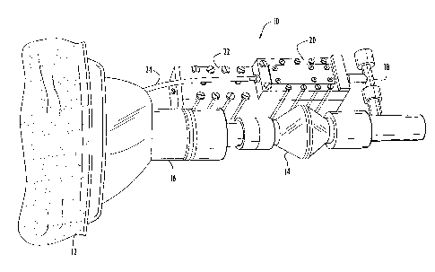
Fig. 4 illustrates an experimental cardiac output monitor 10 constructed to
demonstrate
the feasibility of readily acquiring the data required to implement the
methodologies of the
present invention. Cardiac output monitor 10 includes a face mask 12 for
receiving respired
gasses from a subject. A respiratory flowmeter 14, coupled to the face mask
via a humivent 16,
measures the volumetric respiratory flow. A sample port 18 at the output of
flowmeter 16
provides a sample flow to the gas analyzer. The gas analyzer includes a gas
sensor 20 containing,
for example, the aforementioned fluidic sensors for measuring properties of
the sample gas, and
a transducer module 22. A vacuum line 24 draws the sample gas through the gas
sensor 20 of
the gas analyzer. The transducer module 22 and flowmeter 14 provide
measurements to a
processor (not shown) which determines cardiac output and pulmonary functions
in accordance
with Fick techniques implemented in software.
By applying a mathematical algorithm that combines and integrates
concentration and
volume over time, both the rate and amount of uptake (gain) or release (loss)
of any constituent
(indicator) can be determined in quasi-real time, that is, once every several
breath cycles as
opposed to real time which implies instantaneous, continuous reading. Cardiac
output (the mean
blood flow pumped by the heart) can thus be calculated using the well-
established and accepted
Fick Principle in real time using only breath-by-breath concentration
information. It should be
understood that a number of different non-invasive techniques for measuring
cardiac output from
respiratory gasses based on the Fick Principle have been proposed over the
years. The system
of the present invention can employ any one or combination of these Fick-based
techniques,
including modifications and improvements thereto, to determine cardiac output.
Because of the ability to measure the rate and amount of uptake as well as the
end-tidal
partial pressures of unique indicators such as nitrogen, nitrous oxide,
anesthetic agents, and other
inert gasses in real time, and/or concentrations of respired gasses such as
oxygen and carbon
dioxide, it has been possible to develop this unique noninvasive cardiac
output monitor using the
Fick Principle. While conventional gas analysis technology, such as IR
spectroscopy, could be
used to measure nitrous oxide and anesthetic agent concentrations, it is the
unique ability to
inexpensively measure nitrogen and other inert gasses such as helium that
offers both a non-
CA 02358446 2001-07-13
WO 00/42908 PCT/US00/01465
17
invasive but also a non-affecting, non-toxic approach. Furthermore, it is the
ability to measure
the density and viscosity properties of the inspired and respired gas mixture
(e.g., with other
technology one would have to measure the concentrations of all, not just some,
constituents
simultaneously) that allows for the measurement of the inspired and respired
volumetric flowrate
independent of the properties. By combining these gas analysis measurement
techniques with
accurate models and simulations of uptake and distribution of the gasses in
the body, the current
concept can be further refined to consider pulinonary shunts and deadspace as
well as other
individual-specific physiological and disease states.
In the invasive application of the Fick Principle technique classically
referred to as the
indicator dilution technique, a known amount of an easily detectable substance
(indicator) is
injected into the bloodstream and its presence is monitored a short, known,
distance downstream.
When the indicator and blood are completely mixed, the concentration-time
relationship of the
indicator provides information to determine the blood flow rate, which, by
definition, is the
cardiac output.
In accordance with one embodiment of the present invention, a non-invasive
variation of
the Fick technique is used and is suited to analysis of respiratory gasses
with the gas analyzer of
the present invention. Specifically, rather than directly (invasively)
injecting an indicator into
the bloodstream, a known volume/mass bolus of an indicator gas (e.g.,
nitrogen, nitrous oxide,
sevoflurane, desflurane, or helium) is introduced into the inhaled respiratory
gas stream of a
patient breathing circuit. The blood will take up this gas via respiratory
transfer in the alveoli of
the lungs. In order to minimize the time it takes for the indicator to fill
the alveolar space and
to reduce the time required for the diffusion of the indicator into the
bloodstream, the patient is
ventilated at a predetermined, sufficient, breathing rate. The bolus of
indicator gas is introduced
during one or more quick inspired breaths. The blood will take up only a
portion of the bolus of
respiratory indicator gas, but that portion will behave in a manner similar to
indicators injected
directly into the bloodstream. The transfer of gas to the blood occurs because
there is a
difference in the partial pressure of the gas in the alveoli and in the blood
stream. The alveolar
partial pressure is higher than that in the blood, there being no (or a
different amount of) gas in
the blood. In the first inspired breath/breaths, the indicator enters the
lungs. Subsequent
inspired breaths will be free of indicator but will ensure uniform mixing in
the lungs and will also
start to dilute and evacuate the lungs. Within the first four or five exhaled
breaths, the indicator
CA 02358446 2001-07-13
WO 00/42908 PCT/US00/01465
18
gas that has not been taken up in the blood from the lungs will be completely
(to over ninety-nine
percent) removed.
The amount of indicator exhaled in these initial breaths is measured using the
property
detecting, multiple gas analyzer in conjunction with a respiratory flowmeter
inline with the
breathing circuit. The amount of indicator gas taken up by the blood is
determined by subtracting
the measured exhaled amount from the known input bolus. It is valid to perform
this
computation and measurement in the four or five breaths it takes to purge the
lungs of the
indicator gas, because there is no release of the indicator from the blood
since the blood has not
yet returned to the lungs after circulating through the body. The indicator
gas is chosen to be one
that has a low solubility in the tissues and so remains primarily in the
blood. Nitrogen is such
a gas. When the blood carrying the indicator gas returns to the lungs, there
now exists a reverse
difference in partial pressure between the blood and the alveoli. This time,
the partial pressure
in the blood is higher because there is no indicator in the alveoli. The end-
tidal values (which
approximate the arterial blood gas partial pressure) of indicator gas in each
exhaled breath are
measured and the amount and time history of the gas released from the
bloodstream after it has
circulated through the cardiovascular system is similarly measured. The total
volume of exhaled
indicator and the time it took to be released from the bloodstream, the area
under the exhaled
indicator gas-time curve, compared with the amount of indicator taken up, can
be used to
calculate the cardiac output using the Stewart-Hamilton relationship if no
shunt or deadspace
effects are present. The Stewart-Hamilton relationship is discussed and
derived hereinbelow.
Indicator dilution techniques can be subdivided according to the method of
application.
Two commonly used techniques are continuous infusion and bolus injection. The
continuous
infusion method was first described by Stewart. A major disadvantage of this
technique is that,
in the closed circulatory system, the indicator eventually saturates the blood
stream and any time
history information is lost. The injection of a bolus of indicator, on the
other hand, does not
result in such saturation, and, although lower concentrations must be
resolved, is used more often
in clinical practice and is the technique that is a basis for a preferred
embodiment of the present
invention.
The principle of both methods can be illustrated by means of a simplified flow
system in
which a constant flow of fluid with a flow rate Qo and uniform velocity
profile is assumed for a
system with a single inlet, injection site, and outlet, detection site. Ideal,
(i.e., complete) cross
CA 02358446 2001-07-13
WO 00/42908 PCT/US00/01465
19
sectional mixing is assumed throughout the volume of the system between
injection and detection
site.
For continuous infusion of indicator gas with injection rate q; into a steady
blood flowrate
Qo, the volumetric concentration measured, co, after complete mixing has
occurred, is constant
and determined by the ratio of volumetric flowrate of indicator to flowrate of
blood,
(3)
co = q;/Qo
or, after rearranging and solving for the flowrate (e.g., cardiac output),
Qo = q;/co . (4)
When a bolus of indicator is injected into the blood stream, the relation
between flowrate
and concentration is obtained by using the condition of conservation of
indicator. In an
infinitesimally small time interval dt, the volume of indicator dV; passing
out of the outlet equals
the concentration of indicator at that time, c;(t), multiplied by the volume
dVb of the blood
passing by:
dV; = c;(t) dVb (5)
The blood volume dVb equals the blood flow rate, Qo(t), multiplied by the time
interval dt,
leading to
dV; = Qo(t) c(t) dt (6)
With use of the condition of conservation of mass, the total volume of
indicator injected will pass
the detection site, yielding:
Zo V~ = l Qo(t) ~~(t) dt
0
CA 02358446 2001-07-13
WO 00/42908 PCT/US00/01465
For constant flow, the flow rate can be separated from the integral and solved
for so that,
Qo - Vv (8)
l ~~(t) dt
5 0
This equation is referred to as the Stewart-Hamilton equation. From this
equation, the
flow rate can be determined by the quotient of the total amount of indicator
gas injected and the
area under the measured indicator dilution curve. The c;(t) curve has an
asymmetrical shape,
which is caused by the continuation of the mixing process between indicator
and transport
10 medium, even when the first portion of indicator has already passed the
detection site.
In the non-invasive dilution-indicator technique of the present invention, the
indicator is
a known gas constituent of a respiratory gas mixture. Preferred indicators
are: nitrogen, nitrous
oxide, helium and a relatively insoluble volatile anesthetic (sevoflurane,
desflurane). At some
initial time zero, a bolus of indicator gas is injected into the respiratory
breathing gas and
15 ventilated into the patient over a short time less than the inspiration
period. Following injection,
the ventilatory cycle is continued and the exhaled gas is analyzed and amount
of the indicator gas
exhaled during each breath is quantified. From the amount of gas measured in
the exhaled
volume, the content of indicator in the blood are calculated using Henry's
Law. The measured
concentration of indicator as a function of time elapsed is integrated over
time to determine the
20 total amount of indicator exhaled during the time interval. By
incorporating this data (amount
of indicator taken up by the blood and the area under the exhaled indicator
time curve) into
equation (8), the cardiac output is calculated. This technique is analogous to
both dye and
thermal dilution methods, but has the advantage of being low risk and
noninvasive.
The assumptions used in the derivation of the Stewart-Hamilton equation for
indicators
include: (1) an open flow system (i.e., the indicator passes through only
once); (2) no loss of
indicator (i.e., no uptake in tissues); (3) complete cross-sectional mixing
resulting in a uniform
concentration distribution; and (4) constant flow (no transients). In clinical
situations these
conditions are rarely fulfilled when using direct injection of an indicator
substance into the blood
stream. Since the circulatory system is necessarily closed, recirculation of
indicator will occur.
Depending on the solubility of the indicator, loss of indicator cannot always
be avoided.
Depending on the injection and detection sites, adequate mixing may not always
be achieved.
CA 02358446 2001-07-13
WO 00/42908 PCT/US00/01465
21
And, most importantly, constant flow does not exist due to the pulsating
cardiac action and
effects of spontaneous or mechanical ventilation.
These factors are overcome in the present invention. By introducing into the
blood stream
a relatively small volume of indicator gas that is clear of indicator over a
short period (i.e., less
S than the recirculation time), recirculation does not come into play. By
using an insoluble gas;
either nitrogen or very small amounts of relatively insoluble agents such as
helium, nitrous oxide
or sevoflurane or desflurane, the loss of indicator is extremely small or
negligible and again any
effects of recirculation are not significant. Since nitrogen is relatively
insoluble compared to the
other indicators, and the tissue compartments and venous blood are saturated,
at equilibrium and
constant, changes in arterial blood nitrogen content reflect directly the
changes in the arterio-
venous difference. By introducing the indicator into the respiratory gas
during inhalation, mixing
is not an issue, because the transfer through the microscopic capillaries in
the alveoli ensures
uniformity in the blood stream. Finally, by averaging at least three estimates
over the ventilatory
cycle, the effects of variable flow are minimized.
Although it is possible to accurately measure the variables described above,
additional
consideration is required to account for the presence of pulmonary shunts and
deadspace in the
lungs. Shunts are regions where blood vessels (capillaries) bypass the alveoli
and as a result do
not permit blood to come into contact with the indicator gas. Thus, in the
presence of shunts, any
cardiac output that is computed by the above-described procedure, while
accurately computing
the blood flow to the alveoli, could underestimate the actual blood flowrate
because the shunt
flow is not determined. Deadspace, on the other hand, is a region of the lungs
where no transfer
of indicator (or oxygen, etc.) takes place. This relates to what is known as
lung capacity. Alveoli
that do not contribute to blood gas transfer serve only to dilute the end-
tidal value of the
indicator. That is, when indicator is given up by the blood in working
alveoli, gas with the
indicator mixes with gas without the indicator from the deadspace. Therefore,
in the presence
of deadspace, any computation of cardiac output by the above-described
procedure would
overestimate blood flow because of lower end-tidal readings which suggest
shorter residence
time in the lungs of the blood and, hence, higher blood flow or cardiac
output. The inability to
compensate adequately for these two factors (i.e., shunts and deadspace) is a
major shortcoming
of indirect Fick techniques and probably the reason why such techniques have
not been
successful in the past. _
CA 02358446 2001-07-13
WO 00/42908 PCT/US00/01465
22
In accordance with one embodiment of the present invention, in order to
overcome this
serious deficiency and at the same time provide a method for quantifying
shunts and deadspace,
the measured time-history of exhaled indicator is compared to results for the
same time-history
from a detailed physiological model of the human cardio-vascular system that
contains all the
necessary parameters to describe the uptake, distribution and release of
respired gasses. The Gas
Uptake Distribution (GUS) model, developed by one of the present inventors, is
a computer
model that meets this requirement. The GUS model simulates respiratory and
anesthetic gas
uptake and distribution using a simplified, but functionally accurate,
anatomical and
physiological model of the human body. The simulation utilizes the patient
parameters of weight
and percent body fat, in conjunction with a parametric description of patient
physiology (e.g., 02
consumption, pulmonary shunt, systemic shunt, cardiac output, deadspace),
ventilation mode
(controlled, spontaneous, minute ventilation) and gas delivery (air, 02, N20,
anesthetics). Real
time predictions of the concentrations, and uptakes or losses of any one of
nine respiratory and
anesthetic gasses in twelve compartments can be observed. The twelve
compartments consist
of eleven tissue compartments and one patient breathing circuit of either an
anesthesia machine
or mechanical ventilator. While the exemplary GUS model described herein
involves twelve
compartments, it will be understood that the GUS model can involve any number
of suitable
compartments sufficient to accurately model respiratory and anesthetic gas
uptake and
distribution in order to quantify the effects of deadspace and shunts.
The GUS model is integrated with the gas analyzer system microprocessor and is
used
to predict, in real time, the rate of exchange of an indicator between the
alveolar gas and blood,
the end tidal partial pressure of the indicator during normal and non-uniform
ventilation. The
simulation uses inputs based upon known patient data to make an initial
prediction of alveolar
partial pressure of the indicator. This initial approximation is then modified
to predict the
measured rates of uptake (gain) and release (loss) and end-tidal partial
pressures of the indicator.
Because the respiratory gas analyzer, in conjunction with a respiratory
flowmeter (spirometer),
can actually measure metabolic rates and 02 consumption, and the ventilation
parameters are
known inputs, the only unknowns are the physiological parameters of shunts,
deadspace and
cardiac output. These are manipulated in real time using fuzzy logic as the
simulation progresses
to develop an output matching the measured parameters. The resulting values of
shunts,
deadspace and cardiac output represent an accurate estimate of the actual
values. Once these
CA 02358446 2001-07-13
WO 00/42908 PCT/US00/01465
23
values have converged at some value of cardiac output that incorporates shunts
and deadspace,
that value of cardiac output is reported as the output of the monitor.
The present invention offers significant advantages over other technologies
that utilize
the Fick Principle for determining cardiac output. As previously explained,
the system of the
present invention has the capability to determine cardiac output using any of
the many recognized
Fick techniques. By implementing the present invention, the clinician is not
limited to a single
application of the Fick Principle but is offered more flexibility by selection
of appropriate
techniques, methodologies, indicators, and algorithms for the calculation of
cardiac output that
fit the clinical situation. For example, currently available methods are
limited to only one
indicator (carbon dioxide or oxygen) at a time (e.g., the NICO system). With
the present
invention, any respiratory or anesthetic gas may be used as an indicator and
more than one gas
can be used simultaneously. This enables the clinician to select the
appropriate indicator for the
clinical situation.
The determination of the rate of uptake (gain) or release (loss) of a
substance (indicator)
in the alveolar gas is determined by multiplying the concentration of the
substance (indicator) at
the subject's airway by the airway flow and integrating the product over the
respiratory cycle.
Using this procedure the inspired and expired volume (mass) of the substance
(indicator) is
determined on a breath-by-breath basis. The gain/loss of the substance
(indicator) can then be
calculated as the difference between the inspired and expired substance
volumes divided by the
duration of each breath. This can be expressed as:
V;(t) _ (1/TI) / V;(t)c;(t)dt - { 1/(TE- TI)}/ V;(t)c;(t)dt {integrals} (9)
where V;(t) = airway flow volume per unit time
c;(t) = airway substance concentration
TI = end of inspiration (time)
TE = end of expiration (time)
The fraction of the substance (indicator) at the patient's airway, c;(t), is
multiplied by the airway
flow, Vi(t), and the product is integrated over the respiratory cycle. Using
this procedure, the
inspired and expired volume of the constituent (indicator) is determined on a
breath-by-breath
basis. The uptake (gain) or production (loss) can then be calculated as the
difference between
CA 02358446 2001-07-13
WO 00/42908 PCT/LJS00/01465
24
the inspired and expired volumes of the substance (indicator) divided by the
duration of each
breath. These values are then inserted directly into the Fick Equation for
calculating cardiac
output.
The Fick Principle described hereinabove was first articulated in 1870 by
Adolf Fick and
is easily derived from the basic laws of transport phenomena with the
application of the
Conservation of Mass to blood flowing through an organ system. The result of
this derivation
simply states that the flow of blood through an organ is equal to the total
uptake or release of any
substance (indicator) by the organ divided by the arteriovenous concentration
difference of the
substance (indicator). Another way of expressing the Fick principle is that
the size of a stream
may be readily calculated if we know the amount of substance (indicator) that
enters or leaves
the stream and the concentration difference resulting from such entry or
removal. Both of these
statements are true whether the substance (indicator) is a respiratory gas
such as oxygen, carbon
dioxide, nitrous oxide, or an intravenously injected substance such as cold
normal saline.
The application of the Fick Principle to the measurement of pulmonary
capillary blood
flow enables the clinician to approximate cardiac output during steady-state
conditions at normal
levels of pulmonary shunting of blood. A mathematical application of the
Conservation of Mass
to a cardio-pulmonary gas exchange model yields an expression for direct and
indirect Fick
techniques that has become known as the Fick Equation:
Q' = N'/(Ca-Cv) ( 10)
where: Q' = pulmonary blood flow (cardiac output) (1 blood/min)
N' = rate of gain of substance (indicator) from alveolar gas (ml/min)
Ca = concentration of substance (indicator) in arterial blood (ml/1 blood)
Cv = concentration of substance (indicator) in mixed venous blood (ml/1 blood)
In order to apply the Fick Equation to determine pulmonary blood flow,
techniques that
can either directly measure or indirectly estimate the rate of exchange of a
substance (indicator)
between the blood and alveolus must be readily available. The same is true for
determining the
concentrations of the substance (indicator) in both the mixed venous and
arterial blood. By
incorporating this data into the Fick Equation, it is possible to calculate
the pulmonary blood
flow and thus cardiac output.
CA 02358446 2001-07-13
WO 00/42908 PCT/US00/01465
Numerous methods have been developed to determine each of the variables
required for
the calculation. These methods vary in computational complexity, equipment
utilized, and the
supervision required. Some methods involve invasive techniques (direct Fick)
while others
utilize various empirical correlations between noninvasive measurements and
the variables of
5 interest (indirect Fick). Various indicators including oxygen, carbon
dioxide, nitrous oxide, and
anesthetic gases have been used in these respiratory Fick techniques.
The use of direct (invasive) Fick techniques provides the most accurate prior
known
measurement of cardiac output. Unfortunately, direct techniques require
sampling of both mixed
venous and arterial blood in order to determine the amount of indicator in
each. This requires
10 the insertion of needles and catheters into the systemic circulation and
heart in order to obtain
the specimens required for analysis.
Indirect (noninvasive) Fick techniques rely on measurements of alveolar gas
concentrations to estimate the amount of each indicator in arterial and mixed
venous blood.
Indirect (noninvasive) determinations of arterial substance (indicator)
content have been made
15 based on the partial pressure of the gas sampled at the patient's mouth at
the end of a tidal
expiration.
For the indirect Fick techniques, two assumptions are made. The first
assumption is that
the partial pressure of the gas sampled at the end of a tidal expiration
(PiET) provides a good
representation of the alveolar gas. Secondly, the alveolar gas is in
equilibrium with the substance
20 (indicator) in the arterial blood leaving the lungs. If these assumptions
are valid, PiET can be
used to estimate the arterial partial pressure of the substance (indicator).
However, it is important
to emphasize that by incorporating these assumptions, the accuracy of the
calculation of cardiac
output is limited during conditions a large pulmonary shunt and/or a large
alveolar deadspace
fraction. When these conditions exist, appropriate corrections to the
estimation of cardiac output
25 must be made.
To correct for the potential effects of a large pulmonary shunt andlor
alveolar deadspace,
a somewhat more detailed model of the cardiopulmonary system than the one used
for the
derivation of the Fick Equation is necessary. This model must allow for
pulmonary shunting of
blood flow and include an alveolar deadspace compartment. A pulmonary shunt
represents
pulinonary blood flow that perfuses regions of the lungs that are not being
ventilated. An
alveolar deadspace compartment is defined as a region of the lung that is
being ventilated but is
CA 02358446 2001-07-13
WO 00/42908 PCT/US00/01465
26
not being perfused. Application of the Fick principle for the indicator at the
blood-lung interface
results in:
Q'T-Q's = Q'T(1- f Q's/Q'T}) = N/(Cv - Cal) (11)
where Q'T = Total pulmonary blood flow (ml blood/min)
Q's = Blood flow that does not undergo gas exchange - shunt (ml blood/min)
N = Loss of indicator to alveolar gas (ml/min)
Cv = Concentration of indicator in mixed venous blood (ml/ml blood)
Cal = Concentration of indicator that has undergone gas exchange of indicator
with alveolar gas (ml/ml blood)
Similarly, a mass balance of the indicator at the subject's airway, assuming
that the
alveolar deadspace empties in parallel, results in:
Cm*VT = CA(VT VD) + CD*VD (12)
where CA = concentration of indicator in alveolar gas (ml/ml gas)
Cm = Concentration of indicator measured at the subject's airway (ml/ml gas)
CD = Concentration of indicator measured in deadspace compartment (ml/ml gas)
VT = Tidal volume (ml)
VD = Alveolar deadspace (ml)
Solving for CA yields:
CA = (Cm*VT- CD*VD)/(VT-VD) (13)
With the utilization of the various measurement techniques to determine the
appropriate
variables for these equations, cardiac output can be calculated with
correction for any effects of
shunt and deadspace incorporated into the final value.
The measurement of the variables used to calculate cardiac output is
accomplished by
utilizing three common Fick techniques: direct Fick, indirect Fick, and
indicator-dilution. The
variables to be measured are the uptake/release (gain/loss) and the
arteriovenous concentration
CA 02358446 2001-07-13
WO 00/42908 PCT/US00/01465
27
difference of the indicator. The present invention enables the clinician to
measure these variables
in numerous clinical situations. This approach offers significant advantages
over other methods
utilizing the Fick Principle for determining cardiac output.
As discussed above, in order to apply the Fick Equation to determine pulmonary
blood
flow (cardiac output), the rate of exchange of a substance (indicator) between
the alveolar gas
and blood and the arteriovenous difference of the substance (indicator) must
be measured. Each
of these variables is determined from the measurement of two other variables.
The rate of
exchange of the indicator can be determined by measuring the amount of
indicator (constituent)
that is present in both the inhaled and exhaled breath. The difference in
amount between inspired
and expired constituent is the net gain or loss of the indicator that is
exchange between the
alveolus and the blood. The arteriovenous difference of the indicator is
determined by
measuring/estimating the amount of indicator (constituent) that is present in
both the arterial and
mixed venous blood. The difference in these two values is the arteriovenous
difference in the
blood.
In accordance with the present invention, the determination of the rate of
uptake (gain)
or release (loss) of a substance (indicator) in the alveolar gas is determined
by multiplying the
concentration of the substance (indicator) at the subject's airway by the
airway flow and
integrating the product over the respiratory cycle. Using this procedure the
inspired and expired
volume (mass) of the substance (indicator) is determined on a breath-by-breath
basis. The
gain/loss of the substance (indicator) can then be calculated as the
difference between the
inspired and expired substance volumes divided by the duration of each breath.
This can be
expressed as:
V'(t) = 1/TI // V'(t)F(t)dt- {1/(TE -TI)}// V'(t)F(t)dt {integrals} (14)
where V' (t) = airway flow
F(t) = airway substance concentration
TI = end of inspiration (time)
TE = end of expiration (time)
A microcomputer is used to multiply the fraction of the substance (indicator)
at the patient's
airway, F(t), by the airway flow, V(t), and to integrate the product over the
respiratory cycle.
Using this procedure, the inspired and expired volume of the constituent
(indicator) is determined
CA 02358446 2001-07-13
WO 00/42908 PCT/US00/01465
28
on a breath-by-breath basis. The uptake (gain) or production (loss) can then
be calculated as the
difference between the inspired and expired volumes of the substance
(indicator) divided by the
duration of each breath. This value is then inserted directly into the Fick
Equation for calculating
cardiac output.
Because of the increased capability and versatility of the present invention,
at least three
unique techniques for measuring cardiac output noninvasively using the Fick
Principle are
possible. The first is an adaptation of the Soluble Inert Gas Technique. The
second is an
adaptation of the indicator-dilution method. The third is an integrated
predictive computer model
technique incorporating the computer simulation (e.g., the aforementioned Gas
Uptake
Simulation (GUS) model). As previously described, the GUS model can also be
used to make
correction for the presence of increased shunt and deadspace.
Several noninvasive respiratory applications of the Fick Principle are based
upon the
uptake and distribution of physiologically inert and soluble gases. When a
soluble gas is inhaled,
the partial pressure of the gas in the pulmonary capillary blood equilibrates
with the partial
pressure of the gas in the alveolar space. The volume of gas absorbed by the
blood can be
calculated directly from the partial pressure of the gas in the alveoli, the
solubility of the gas in
blood, and the quantity of blood that has equilibrated with gas. This can be
expressed
mathematically using Henry's Law as:
Vgas = Sgas*PAgas*Q (15)
where:
Vgas = volume of gas absorbed by the pulmonary capillary blood (ml)
Sgas = solubility coefficient of the gas in blood (ml gas/ml blood/mm Hg)
PAgas = alveolar partial pressure of gas (mm Hg)
Q = volume of blood equilibrated with alveolar gas (ml blood)
Differentiating the expression with respect to time and rearranging terms
result in
Q' = V'gas /(Sgas*PAgas) (16)
CA 02358446 2001-07-13
WO 00/42908 PCT/US00/01465
29
Equation (16) represents a special case of the Fick Equation where the mixed
venous gas
concentration is zero. The absence of the mixed venous term is due to the fact
that under normal
conditions the indicators used are not present in blood.
The use of a soluble inert gas as the indicator in the Fick Equation therefore
eliminates
the need to determine the mixed venous gas concentration provided that the
mixed venous gas
concentration is zero at the start of the measurement procedure. However, this
zero concentration
assumption requires that the measurement procedure be completed before the
blood recirculation
time.
Primarily two gases have been used as indicators in inert gas techniques,
nitrous oxide
and acetylene. For the present invention, nitrous oxide and the newer volatile
anesthetic agents
(sevoflurane, desflurane) are preferred over acetylene. These gases have a
relatively low
solubility in lung tissue and higher solubility in blood. Since high
concentrations of these gasses
in the blood are undesirable, low levels of inspired gas concentration must be
used. This low
level of inspired indicator concentration decreases the magnitude of both
V'gas and PAgas in
Equation (16), and therefore increases the sensitivity of the blood flow
estimate to error in the
determination of these parameters.
A number of techniques have been developed to increase the accuracy in
measuring both
V'gas and PAgas. These techniques have been discussed and include breath-
holding techniques,
rebreathing techniques and single-breath techniques.
One of the limitations of these techniques is the assumption that the venous
concentration
of the indicator gas is zero. This assumption not only limits the duration of
the measurement of
V'gas and PAgas due to venous recirculation, but also requires a waiting
period between
determinations to allow mixed venous blood content to decrease to zero.
However, it is possible
to overcome this limitation by using nitrogen as the indicator. Since nitrogen
is relatively
insoluble compared to the other indicators, and the tissue compartments and
venous blood are
saturated, at equilibrium and constant, changes in arterial blood nitrogen
content would reflect
directly the changes in the arteriovenous difference.
The above-described embodiment is a major improvement over existing non-
invasive
techniques in that it provides means for compensating for the effects of
shunts and deadspace.
However, the fuzzy logic used in estimating the effects of shunts and
deadspace may not resolve
certain ambiguities that may arise. Another, more general embodiment of the
present invention
CA 02358446 2001-07-13
WO 00/42908 PCT/US00/01465
involves separately estimating the deadspace (both physiological and
anatomical) so that only
shunts need to be iterated in the physiological model software. In addition,
in some cases, it may
be inconvenient to provide indicator gasses that need to be tracked;
accordingly, the second
embodiment utilizes the unique instantaneous relationships between oxygen and
carbon dioxide
5 to provide much the same information.
In "Deadspace and the Single Breath Test for Carbon Dioxide During Anesthesia
and
Artificial Ventilation," Br. J. Anaesth (1984), 56, 109, incorporated herein
by reference in its
entirety, Fletcher et al. teach that the anatomical deadspace (i.e., the
deadspace of the passages
in the lungs that bring the ventilatory gasses to the alveoli) can be
accurately measured by
10 measuring the total volume of expired gasses before any change occurs from
that inspired in
measured concentration of either oxygen and carbon dioxide. Further, Fletcher
teaches that the
physiological deadspace (i.e., the measure of the alveoli that do not
participate in transfer of
gasses to the blood) can be estimated quite accurately by measuring the volume
of expired gasses
during alveolar release. Using relatively simple algorithms, this deadspace is
related to the slope
15 of the concentration-volume trace as end-tidal values are approached.
Kim et al. teach in the aforementioned paper that the value of the mixed
venous carbon
dioxide partial pressure is related to the value of the exhaled carbon dioxide
concentration at the
point when the ratio of the change of carbon dioxide concentration to the
change in oxygen
concentration (i.e., the instantaneous respiratory quotient) is approximately
0.3. This ratio value
20 is a function of physiological conditions as well as disease state;
however, by applying the
method of Fletcher et al. to account for deadspace, the variation may be
limited to shunts alone.
Since cardiac output (not including the effects of shunts) is thus defined by
the Fick equation to
be the ratio of the carbon dioxide produced to the difference in mixed venous
and arterial carbon
dioxide partial pressures, this portion of the calculated cardiac output may
be introduced into the
25 physiological model software (e.g., GUS) and a shunt value iterated that
results in the respired
breath waveform as measured. In cases where it is reasonable to assume that
shunts do not play
a major part (e.g., when they are not present or remain constant during the
period of monitoring),
the data from the gas analyzer and respiratory flow meter provide cardiac
output directly, with
accuracy that is superior to existing modified-Fick techniques. A detailed
example of a modified
30 Kim single-breath technique is now provided.
Given that the system of the present invention can inexpensively and reliably
measure
CA 02358446 2001-07-13
WO 00/42908 PCT/US00/01465
31
real time, simultaneous OZ and C02 concentrations (and N2 if needed) as well
as consumption
and production right at the patient breathing circuit, single-breath
techniques can reliably be use
to non-invasively measure cardiac output and provide continuous determination
of cardiac output
on a breath-by-breath basis.
As described in the aforementioned paper, Kim et al. developed a method of
estimating
"true" mixed-venous and arterial C02 partial pressures by the analysis of
simultaneous exhaled
02 and C02 partial pressures (Feo2 and Feco2) on the alveolar plateau of a
single extended
exhaled breath. Given either 02 consumption or C02 production cardiac output
can then be
estimated using the Fick equation. Several studies have been undertaken since
then that have
attempted to validate and improve the technique, mostly from a data processing
perspective.
However, this methodology heretofore has not received wide acceptance
primarily for three
reasons. The first reason is the difficulty associated with obtaining
simultaneous realtime 02 and
C02 data, putting the technique into a very high cost category. This
limitation has been
overcome in the present invention with the use of the aforementioned real time
mufti-gas
analyzer. The second reason is the extensive calculations that must be
performed. This
limitation has been overcome in the present invention by using state-of the-
art microprocessor
technology. The third reason is the concern about the validity of the
assumptions that were made
in the original methodology. These concerns focus upon the potential effect of
any body gas
stores, alveolar deadspace and pulmonary shunts vis a vis disease state, the
shape of the
oxyhemoglobin and carbon dioxide dissociation curves, the necessity for a
relative steady state
(also required theoretically for any Fick and single-injection indicator-
dilution methods), and the
existence of any ventiladon/perfusion (V'/Q) abnormalities (the effect of
sequential emptying of
the alveoli). Most of the assumptions made by the original investigators have
subsequently been
validated, but concerns have been expressed over using the technique in
patients with abnormal
ventilation/perfusion ratios (V'/Q). These concerns have generally been
overcome by the
significant number of studies that have categorically confirmed that there is
sufficient
information in the respired C02 data alone that correlates with cardiac
output. The former have
been studies based on the initial efforts of Fletcher who identified a very
accurate technique to
account for deadspace, and have culminated with the very recent work of Arnold
et al. who
conducted detailed correlations of exhaled C02 waveforms at various
work/exercise levels to
cardiac output. These latter studies clearly confirm that the COZ waveform has
strong predictive
CA 02358446 2001-07-13
WO 00/42908 PCT/US00/01465
32
elements relating to cardiac output.
In view of these studies, the present inventors have determined that if the
true
instantaneous alveolar values of 02 and C02 that account for lung volume and
physiological
deadspace can be computed/measured, the true instantaneous respiratory
exchange ratio can be
determined directly for the alveoli, thereby allowing computation of the
actual blood flow being
passed through the lungs. Pulmonary and systemic shunting is accounted for
with knowledge of
the arterial oxygen saturation, which is routinely measured in a peripheral
artery with a pulse
oximeter. According to the methodology of the present invention, the single-
breath technique
is augmented to account for V'/Q abnormalities. This is accomplish by modeling
the physiology
and, therefore, having a virtual patient on the side to compare with. The
inputs of deadspace and
cardiac output to the physiological model are iterated until the model
converges on the measured
metabolic outputs.
Using the cardiac output monitor 10 shown in Fig. 4, experimental data was
collected and
the computations required to determine cardiac output were performed, as shown
in Fig. 5-8.
Fig. 5 shows the typically measured respiration parameters, inspired and
exhaled 02 and C02
concentrations and instantaneous respired flowrate, for several breaths. From
this data all other
parameters are computed. Fig. 6 illustrates the rates of 02 and COZ
consumption and production,
their ratio (respiratory quotient), and total volumes consumed and produced
for the second breath.
Fig. 7 shows the single breath Fco2 plotted against Fo2 for the alveolar
plateau region. This data
is processed to obtain a relationship between alveolar C02 concentration and
instantaneous
exchange ratio, R, (accounting for deadspace) shown in Fig. 8. Note that the
data spans the range
of exchange ratio between approximately 0.2 and 0.8 so that only a minor
extrapolation to the
steady state respiratory exchange ratio (Rss) is required. Rss is calculated
by integrating the
product of the concentration and the flowrate. For this case, Rss = 0.889;
however, it should be
noted that it also equals the ratio of the C02 production to OZ consumption
rates shown in Fig.
6. Using the mixed venous and arterial values estimated from the points when R
= 0.32 and R
= 0.889, an arterio-venous difference of 0.9% (6.84 mmHg) is obtained. The
minute oxygen
consumption from Fig. 6 is 75.9m1 in the breath of 7.925s or 575m1/min. This
results in a cardiac
output for that particular breath of approximately 10.17 liters/min, which is
consistent with the
active state of the subject, and falls within expected ranges as shown in the
data of numerous
other researchers in Fig. 9.
CA 02358446 2001-07-13
WO 00/42908 PCT/US00/01465
33
The diagnostic capability of this methodology is considerably greater than
merely
providing cardiac output. Metabolic rate has been computed and can be reported
to the
user/clinician. Deadspace and pulmonary shunts can be quantified; information
that would be
of significant interest to the respiratory therapist as well as the
pulmonologist.
In Kim's technique, the lungs act as an aerotonometer that measures the
partial pressure
of the mixed venous blood, and hence gas content. The essential feature of
Kim's method is the
application of the Haldane principle to determine the mixed venous and
arterial C02 partial
pressures (Pvco2 and PACO2) from simultaneous measurements of C02 and OZ
concentrations from
a single exhalation that is long enough to ensure that alveolar gasses have
equilibrated and that
enough data points can be collected on the alveolar plateau (where the exhaled
gasses are
representative of the alveolar gasses) for a statistically meaningful
regression analysis to be
performed. Henry's Law of Solubility and the dissociation curves relate
alveolar partial pressures
and the content in the blood for CO2 and O2. Using the cardiac output monitor
of the present
invention, with sample rates of 30 - 200 per second, data taken under normal
respiratory rates of
8-12 breaths per minute yield satisfactory results; thus, patient cooperation
is not required.
Assuming that mixed-venous C02 tension Pvco2 remains constant during this time
(blood
normally does not recirculate in the period of one breath), then arterial C02
tension (Paco2) will
rise and eventually, if the breath is long enough, will approach Pvco2.
Haldane observed that the
arterial value is equal to the mixed venous value when the instantaneous
respiratory exchange
ratio, R = V'~o2/V'o2 = 0.32. A detailed discussion of data supporting this
observation is
presented by Kim et al. and will not be repeated here.
Kim, however, made the assumption that the exhaled concentrations were equal
to the
alveolar values. Clearly, this would hold true only if there were no alveolar
deadspace. The
relationship of Pco2 with R should, of course, be correct for the air exhaled
from a fully
ventilated and perfused alveolus. This means that the exhaled 02 and C02
tensions must be
corrected to true or "ideal" alveolar values in order to account for the
dilutionary effects of the
unmetabolized gasses in the deadspace. The "ideal" alveolar partial pressure
is a term used
herein to indicate that any effects of physiologic deadspace have been
incorporated. Physiologic
deadspace is defined as a region of the lung that is being ventilated but is
not being perfused.
The Bohr equation allows correction of the exhaled tensions (of gas X, that
is, COZ and OZ) for
physiologic deadspace, if deadspace and expiratory volume, VE can be
quantified.
CA 02358446 2001-07-13
WO 00/42908 PCT/US00/01465
34
VPHYSD~ VE = LPAX' PEX~ / LPAX - PIx~ (17)
In accordance with the present invention, the physiologic dead space volume,
Vp~sD, is
quantified using Fletcher's analysis of the single breath C02 concentration
(i.e., the expired
volume waveform), which is collected by the cardiac output monitor (see Fig.
5). The exhaled
gas tidal volume, VE, is determined by integrating the expired flowrate using
the property-
compensated pneumotachometer. The gas analyzer of the present invention
measures the partial
pressures in the exhaled gas, Pte, and the inhaled gas, Pte. This then leaves
the "ideal" alveolar
partial pressure, Pte, as an unknown.
Fletcher considered physiological deadspace (VPHYSD) in terms of two
components:
convective airway and alveolar deadspace. Convective airway deadspace (VDAW)
extends from
the lips to the interface between the inspired and alveolar gas. Alveolar
deadspace (VD,~,v)
includes deadspace caused by ventilation-perfusion ratio mismatching within
terminal respiratory
units; ventilation-perfusion ratio mismatching between units; venous admixture
or right to left
shunt; and temporal deadspace. Classical methods for estimating VP~sD are
based on collection
of expired air over a period of a few minutes during steady state with
simultaneous sampling of
arterial blood. The partial pressure of C02 is measured from both Samples and
VPHYSD IS
calculated by using Bohr's equation. Fletcher determines this deadspace with a
single breath test
of C02 (SBT-C02).
In accordance with Fletcher's technique, a plot of expired C02 fraction
(FECO2) against
expired volume based on the SBT-COZ is generated, as shown in Fig. 10. Phase I
defines
conducting airway deadspace. Phase II defines the transition between
conducting airway and
alveolar gas. Phase III defines the alveolar plateau, representing C02 rich
gas from the alveoli.
Phase II and III together represent the effective tidal volume (VTeff) that
contributes to gas
exchange. Fletcher then divides the single breath into regions X, Y, and Z
(Fig. 10). Area X
represents tidal elimination of carbon dioxide (VTCOa) with effective part of
VT. Area Y
represents wasted ventilation as a result of VD~,v.
Area Z represents wasted ventilation as a result Of VDAW. The areas are used
to express
the deadspace fractions: VDAW /VT = Z/(X + Y + Z); VD~,v /VT = Y/(X + Y + Z);
VDAW / VT~,v
= Y/(X + Y), and VpgYgD /VT = Y + Z/(X + Y + Z). The dashed line at the top of
the graph
CA 02358446 2001-07-13
WO 00/42908 PCT/US00/01465
represents the fraction of the C02 of a gas in equilibrium with arterial blood
(Facoa). The partial
pressure of C02 in the exhaled breath is not equal to the partial pressure of
C02 in the alveoli,
because of admixture of gas from ventilation-perfusion ratio mismatched
alveoli and gas from
alveoli with efficient gas exchange. If Faco2 is not known, the value can be
estimated by
5 extrapolating the curve out to an exhaled volume equivalent to 15% of the
predicted Total Lung
Capacity. Extrapolation is effective for normal individuals and patients with
airway disease, but
not patients with pulmonary embolism.
During mechanical ventilation, gas compression increases VD,~,v and equipment
dead
space alters VDAW, and Phase II may be difficult to distinguish from Phase
III. To extract
10 information from readily obtained data, the present inventors propose a
method closely related
to Bohr's concept in which end-tidal carbon dioxide (F~co2) is related to the
exhaled carbon
dioxide (FECOa). VTCOa (area X) is related to the volume of C02 hypothetically
eliminated by a
breath in which the whole effective volume has C02 fraction FECO2 equal to
area ABCDA. The
ratio, X/areaABCDA = VTC02~LVTEFF' FEC02~ describes the efficiency of the
effective volume in
15 eliminating C02.
Fundamentally, one needs to know alveolar concentrations in order to calculate
deadspace, but one needs to know deadspace to calculate alveolar
concentrations. To resolve this
dilemma, the present inventors propose to develop a "virtual patient"
consisting of a
physiological model of respiratory gas uptake and distribution which will be
used to mimic the
20 real patient, matching the patient's actual measured gas exchange values.
One such model is
aforementioned GUS model, which simulates respiratory gas uptake and
distribution using a
simplified anatomical and physiological model. The simulation uses patient
parameters (e.g.,
weight, percent body fat), patient physiology (e.g., 02 consumption, pulmonary
shunt, systemic
shunt, cardiac output, deadspace), ventilation mode (e.g., controlled,
spontaneous, minute
25 ventilation), and gas delivery (e.g., air, O2, N20, anesthetics).
Predictions of the concentrations
and uptakes or losses of the respiratory or indicator gasses in twelve (or any
appropriate number)
compartments can be observed. Again, the compartments include eleven tissue
compartments
and one patient breathing circuit of either an anesthesia machine or
mechanical ventilator.
The GUS model is integrated into the computational scheme and is used to
predict real
30 time rate of exchange of OZ and C02 between the alveolar gas and blood, as
well as the exhaled
partial pressures during normal and nonuniform ventilation, and iterated until
what the model
CA 02358446 2001-07-13
WO 00/42908 PCT/US00/01465
36
output corresponds to what the patient is producing. A specific example of the
iterative process
is outlined in the flowchart shown in Fig. 11 and described below.
First, an initial guess is made for physiologic deadspace by calculating the
value by
Fletcher's technique using the measured C02-volume waveform and the measured
end-tidal C02
tension as an initial guess for the alveolar value. From this estimate and
Bohr's equation;
alveolar 02 is obtained and the procedure outlined below for computing cardiac
output is
performed. The resultant value of cardiac output is provided as an input to
the GUS model, and
a prediction of alveolar and exhaled partial pressures of 02 and C02 is made.
If the partial
pressures agree with the measured values then the cardiac output is accepted.
If the values differ,
the alveolar C02 values computed by GUS are used as the next input and the
iteration continues.
When the simulation has converged to within a predetermined difference with
the measured
values, cardiac output is accepted and represents the value that has been
corrected for shunt and
deadspace.
Given the measurements of instantaneous [P;;(t), PE; (t)] and end-tidal
partial pressures
[P~;] of 02 and C02 and the "ideal" alveolar partial pressures (from Bohr's
equation using the
first estimate of deadspace from the GUS model) for each incremental volume as
a function of
time during the exhaled breath, an equation for the instantaneous respiratory
exchange ratio (R)
is derived from a mass balance of inhaled and exhaled N2 (which is not taken
up), from which,
both the mixed-venous and arterial values of COZ concentration are computed.
From the
definition of the instantaneous respiratory exchange ratio, R, (Rss is the
steady state respiratory
quotient),
R = [CAC02(t) V~Ao(t)- CIC02 V~AI(t)]~[CI02(t) V~AI(t)- CA02(t) V~Ao(t)]~ (18)
After some considerable manipulation, a relationship between the alveolar
concentrations of C02
and 02, the general alveolar air equation, which includes the effects of all
deadspace is derived,
PACO2(t) _ (P~ - P4~)[R Floa + FICO2]~[1 - (1 - R) FIO2] - PAOa(t)[R + (1 -
R)FICO2]~[1 - (1 - R)Floa].
(19)
Measured values of PA~o2, and PAOZ are used to determine R and the PACO2 at R
= 0.32
defines the true mixed venous value. The value at R = Rss defines the true
arterial value. Note
CA 02358446 2001-07-13
WO 00/42908 PCT/US00/01465
37
that the arterial value of C02 does not necessarily coincide with the end-
tidal value: a cause for
inaccuracy when using indirect Fick techniques.
The Oz consumption and C02 production (V'o2 and V'co2) for the breath is
measured.
Pulmonary capillary blood flow, QPCBF, is then determined by the indirect Fick
technique with
the assumptions: 1) instantaneous R reflects gas exchange between pulmonary
capillary blood
and alveolar gas and 2) the COZ dissociation curve is linear, or at least
parallel. Using a value
of, for example, 4.7m1/liter per mmHg PaCOa for the slope of the whole-body
COZ dissociation
curve at rest, the pulmonary capillary blood flow, QPCBF, is:
QPCBF = V'o2(Rss - 0.32) / [4.7(Pvo2 -Pa~o2)]. (20)
The slope of the C02 dissociation curve can be refined if arterial 02
saturation and hemoglobin
are known or can be adjusted for the metabolic conditions given the
approximate level of COZ.
The pulmonary capillary blood flow approximates the systemic cardiac output
when no
significant pulmonary shunt is present. A pulmonary shunt is blood flow that
perfuses
unventilated regions of the lungs. A pulmonary shunt dilutes pulmonary
capillary blood flow.
Using conservation of mass, the systemic cardiac output, Qco, can be expressed
as:
QCO = QPCBF + QS (21)
where Qs is the pulmonary shunt blood flow. The mass balance for OZ in the
blood is,
CaQco = CvQs + CCQPCBF (22)
where Ca is the systemic arterial 02 content, Cv is the mixed venous 02
content, and Cc is the
pulmonary capillary 02 content. From these relationships an equation for
determining the flow
through the pulmonary shunt can be derived, where 02 content can be expressed
as a Po2 + 1.39
Hb4 Sato2, a is solubility of 02 in blood, Hb4 is hemoglobin level, Sat is OZ
blood saturation, and,
Poi is partial pressures of 02 in arterial, capillary or venous blood. The
arterial OZ saturation
(Satao2) is measured directly using a pulse oximeter. Since the amount of the
dissolved 02 in the
arterial, venous and capillary blood (aPo2) is relatively small compared to
the amount combined
CA 02358446 2001-07-13
WO 00/42908 PCT/US00/01465
38
with hemoglobin, it is ignored. The equation for cardiac output thus becomes,
Qco = QPCBF [Satco2 - Satvo2] / [Satao2 - Satvo2]. (23)
In order to solve the equation, capillary and mixed venous 02 saturation are
obtained from the
02 dissociation curve for hemoglobin. The Margaria equation provides a
convenient form:
Sato2= [{m-1}K4Poa4+KPoz{KI'o2+ 1}3]/[{m-1}K4Poa4+ {KPo2+ 1}4 (24)
where, Sato2 = fractional oxygen saturation, Po2 = 02 partial pressure, m =
empirical constant
= 124, K = empirical constant dependent on pH and temperature. DeFilippe's
data shows that
the adjustable constant, K, for 6.8 <- pH <_7.8 and 293°K <-T <_
318°K, is,
K = [0.796{pH - 7.0}/7.0 + 0.152{pH - 7.0}/7.0 + 0.0211 }]exp[-17.72{T -
293}/T]. (25)
The pH effect is especially important because pH varies along the length of
the capillary due to
C02 transport. For normal blood (protein content ~7%), the relationship of pH
to Pco2 is:
pH = -0.3782 In Pco2 + 8.806 (26)
so that the pH may be calculated from the values of Pcoz obtained from the
analysis of pulmonary
capillary blood flow. Once the various OZ contents have been calculated, Qco
may be calculated
since QpCBF has already been determined. Qco is the value reported to GUS if
the solutions do
not match and to the clinician when convergence has been achieved.
While the cardiac output monitoring system of the present invention has been
described
in conjunction with the use of specific Fick techniques and methodologies, it
will be understood
that the system of the present invention can be used with any technique that
is based on the Fick
principle and that determines cardiac output from alveolar gas concentrations,
including, but not
limited to any: oxygen-based Fick techniques, carbon dioxide-based Fick
technique, differential
carbon dioxide Fick techniques, Fick techniques involving computer modeling
using respiratory
gasses, and soluble insert gas Fick techniques. An overview of such techniques
is provided by
CA 02358446 2001-07-13
WO 00/42908 PCT/US00/01465
39
Capek in "Fick Techniques," Enc. of Med. Devices and Instr., Wester, J. D.
(editor), Wiley, NY,
pp: 1302-1314 (1988), the disclosure of which is incorporated herein by
reference in its entirety.
The particular Fick technique algorithms used to process the measurements
provided by the
cardiac output monitor of the present invention are implemented in software;
thus, any of a
variety of techniques can be performed with the same apparatus simply by
running different
software on the processor. Moreover, software corresponding to a number of
different Fick
techniques can be stored in a memory of the cardiac output monitor, allowing
the clinician to
select a particular Fick technique that is desired or particularly well-suited
to the clinical
situation, or to determine cardiac output from more than one Fick technique.
Having described preferred embodiments of a new and improved method and
apparatus
for non-invasively determining cardiac output and pulmonary function using
respired gas analysis
techniques and physiological modeling, it is believed that other
modifications, variations and
changes will be suggested to those skilled in the art in view of the teachings
set forth herein. It
is therefore to be understood that all such variations, modifications and
changes are believed to
fall within the scope of the present invention as defined by the appended
claims. Although
specific terms are employed herein, they are used in a generic and descriptive
sense only and not
for purposes of limitation.