Note: Descriptions are shown in the official language in which they were submitted.
CA 02360250 2001-08-08
WO 00/47127 PCT/US00/03674
1
IMMEDIATE POST-EXTRACTION IMPLANT
TECHNICAL FIELD
This invention relates generally to dental implants, and, more particularly,
to
submergible screw-type implants.
BACKGROUND ART
Screw-type implants are well known in the art. U.S. Pat. No. 3,499,222 of L.I.
Linkow
et al. (the "`222 patent") discloses screw-type implants that may be buried in
the alveolar ridge crest
bone of a patient in an edentulous region. The implant has a threaded lower
portion that may be
screwed into an opening created in the bone after the tissue has been
displaced. A coronal portion
protrudes above the bone and is used to support an artificial dental
appliance, e.g., an artificial tooth
or bridge.
More recently, submergible implants have been created in which the threaded
portions
of the implants can be completely embedded in the bone. They may then be
covered with tissue and
allowed to remain in place while new bone grows around the implant and through
vent holes in it.
Once it is firmly anchored in new bone, the tissue is reopened and an upper
post portion is screwed
into the implant portion and is used to mount the artificial dental device. An
example of this type of
implant can be found in U.S. Patent No. 4,713,004 of L.I. Linkow et al. (the
"`004 patent").
A prior surgical method for installing an implant portion involved creating an
incision
in the tissue covering the alveolar ridge crest bone. This underlying bone was
then exposed and a
cylindrical bore was drilled into the bone at a depth sufficient to hold the
implant portion of the
device. The bore was made slightly smaller in diameter than the implant device
and was at an angle
that would allow it to engage the major portion of the available bone. Then a
bore tap is used to
create threads in the bore, after which the implant device was threaded into
the remaining bone.
Alternatively, an implant may be embedded and not covered with tissue. This
eliminates the need to reopen the tissue later to mount an artificial dental
device.
It is also well known in the art, e.g., from the `004 patent, that a channel
through
threads on the implant will create a cutting edge so the implant becomes self-
tapping (e.g., end
cutting). When installing an implant portion in the patient's bone, it is
advantageous if the implant
is self-tapping because it causes the implant to be anchored more securely. If
such a self-tapping
CA 02360250 2001-08-08
WO 00/47127 PCT/USOO/03674
implant is used, a bone tap is not needed and the implant is threaded directly
into the bone utilizing
the self-tapping threads created by the channel along the length of its
threads.
Also, it is advantageous if the bone chips created during a self-tapping
operation are
deposited into the bore or opening, because these autogenous chips promote
faster bone regenerative
growth. The channel guides these bone chips, which are created during the self
threading of the
implant, toward the base of the bore in the bone. In particular, during the
insertion procedure with
a self-tapping implant, bone chips are removed from the walls of the bore
while forming the grooves
in the bone that match the threads in the implant. These bone chips drop along
the channel to the base
of the bore and help to promote the growth of new bone that firmly anchors the
implant in place.
When a tooth is extracted, it leaves behind a rather large conically-shaped
cavity,
which does not lend itself to the insertion of a cylindrical implant if an
artificial tooth is to be
substituted for the removed tooth. One technique for overcoming this problem
of the extracted tooth
is to expose the cavity in the bone, fill the extraction site with bone graft
material, such as autogenous,
allographic or xerographic material, and then cover the site with gum tissue
for a period of time
sufficient for new bone to grow into and fill the cavity, e.g., with a mixture
of the grafted bone and
newly grown bone. Then a cylindrical bore is drilled at the site and a dental
implant is installed in
the usual manner. However, this requires that the patient live with an
edentulous area without a
functional prosthesis for a long period of time.
As an alternative, the implant can have a shape that is not cylindrical, but
instead is
conical or U-shaped, in order that it more nearly fits the dimensions of an
extraction site. Such
implants may be found in U.S. Patent No. 4,521,192 of L.I. Linkow (the "`192
patent"), and U.S.
Patent No. 2,609,604 of B.F. Sprague (the "`604 patent"). As the slope of the
conical shape of the
extraction site cannot be predicted in advance, these implants cannot be made
self tapping. As a
result, no pressure can be applied to these implants for a significant period
of time, i.e., until existing
bone has grown around the implant to anchor it in place. In addition, as a
self-tapping implant is not
used, there may not be intimate contact between the implant and the new bone,
so the implant may
eventually fail, even if a significant amount of time is allowed to pass
before an artificial tooth is
mounted on the implant and it is put into use.
It would be of great benefit when replacing extracted teetll with dental
implants to use
an implant that compensates for the shape of the extraction socket, is at
least partially self-tapping for
initial implant stability and assures relatively intimate contact between the
implant and new bone so
2
CA 02360250 2001-08-08
WO 00/47127 PCT/US00/03674
that the implant can be put into service relatively soon after the procedure
and still have a low
probability of subsequent failure.
SUMMARY OF THE INVENTION
The present invention is directed to a dental implant that may be used at the
site of a
recent tooth extraction and can be put into service in a reasonably short
period of time. This implant
has a lower self-tapping portion and an upper portion covered with a sintered
material that is
osteopromotive and osteoretentive so to promote adhesion between the implant
and the surrounding
bone.
In a preferred form of an illustrative embodiment, the implant is of the
submergible
screw type with an upper portion having a conical shape and a lower
cylindrical portion having
threads. A longitudinal channel or slot extends through the threads on the
lower cylindrical portion
so as to make the threads self-tapping. The channel is wider toward its apical
end.
One side of the channel is at a right angle or acute angle to the implant
circumference
so as to create a cutting edge that forms the self-tapping capability for the
implant. The other side of
the channel can be at an oblique angle to the circumference.
At least a portion of the exterior surface of the upper conical portion of the
implant is
sintered with a plurality of spherical projections made of a material suitable
for bone integration, the
spaces in between forming a porous surface.
At the lower or apical portion of the implant there is a vent or opening to
allow for
autogenous bone chips created during self-tapping to enter therein when the
implant is screwed into
the bone socket.
BRIEF DESCRIPTION OF THE DRAWINGS
The foregoing and other features of the present invention will be more readily
apparent
from the following detailed description and drawings of an illustrative
embodiment of the invention,
in which:
FIG. I is a schematic cross section of the side of a patient's face showing
the alveolar
ridge crest with a screw type implant according to the present invention
installed therein;
FIG. 2 is an enlarged view of an illustrative embodiment of the implant
portion of the
device of FIG. 1;
FIG. 3 is a top view of the implant portion of FIG. 2;
3
CA 02360250 2001-08-08
WO 00/47127 PCT/USOO/03674
FIG. 4 is a cross-sectional view through the implant portion of FIG. 2 along
line 4--4
and longitudinally down the centerline of FIG. 3 depicting the cross-sectional
shape of the lower
implant portion according to the present invention;
FIG. 5 is a cross-sectional view through the implant portion of FIG. 2 along
line 5--5
and longitudinally down the centerline of FIG. 3 depicting the cross-sectional
shape of the upper
implant portion according to the present invention;
FIG. 6 illustrates the placement of the implant of the present invention in a
tooth
extraction site; and
FIG. 7 is an enlarged view of another illustrative embodiment of the implant
portion
of the device of FIG. 1.
DESCRIPTION OF ILLUSTRATIVE EMBODIMENTS
The design requirements for dental implants placed into immediate extraction
sites
differ significantly from the design of general implants used presently for
placement in edentulous
jawbones. Today all implants used in immediate extraction sites are either
threaded, coated with a
surface material or sintered. However, these implants do not provide the best
design for immediate
fresh extraction sites. Such immediate extraction sites require an implant
designed specifically to
address the morphology of the bony defect created during the extraction of a
tooth.
The implant system of the present invention is at least a t%vo part screw-type
dental
implant 3 (FIG. 1), having a threaded cylindrical lower portion 10 that is
buried in the bone 5 of the
patient and an upper portion 9, preferably generally conically-shaped, that is
attached thereto. The
upper portion 9 is covered by soft tissue 7. A post or abutment 2 is shown in
dotted line extending
from upper portion 9 and supporting an artificial tooth structure 4 to
complete the implant system.
As shown in FIG.1 the implant screw lower portion 10 is located in a bore in
the alveolar crest bone
5, at an angle that causes it to be in the center of the thickest portion of
good available bone. The
abutment 2 is attached both to the implant portion and the artificial tooth 4
and may have an angular
offset to the implant so that the artificial tooth is in proper alignment with
the rest of the teeth.
In FIGS. 2, 4 and 5, the screw implant 3 of FIG. 1 is illustrated in more
detail. This
screw implant portion contains threads 13 in the lower portion 10 that extend
over the top two-thirds
of this lower portion 10. These threads may have a flat bottom and be angled
up to form a Christmas
tree shape in cross section. The lower half of the implant portion 10 contains
a cavity 14. as can be
seen in Fig. 4. This cavity is open at its bottom. Also, spaced about the
lower end of the implant
4
CA 02360250 2008-01-03
WO 00/47127 PCT/US00/03674
portion 10 are holes or vents 16, which penetrate from its exterior to the
interior cavity 14. The
purpose of these vents is to allow new bone to grow through and into the
center cavity 14 in order to
firmly anchor the implant in the patient's bone 5.
A channel 18 in alignment with at least one vent 16 extends through niost of
the
threads, but not the top thread. The channel does not pass through the top
thread in order to prevent
tissue from growing down the channel. This channel has two purposes. First,
the channel 18 and the
vent 16 create cutting edges on the adjacent threads that make the implant
self tapping. Also, the
channel provides a path by which bone chips created during the threading of
the implant into the bone
may pass down to the vent 16 and enter the cavity 14 where they promote the
growth of new bone.
To facilitate this, the channel 18 widens toward the bottom of the implant.
Thcsc features are
described in U.S. Patent No. 4,713,004:.
The upper portion 9 of the screw implant 3 preferably has a generally conical
or fluted
shape. For example, the base of portion 9where it contacts portion 10 may have
a dianieter of about
3.0 mm, while the top of the portion 9 may have a diameter of about 3.5 mm. In
a preferred
embodinient shown in FIG. 7 the upper end 11 of the upper portion 9 is tapered
upwardly. This
provides a more gently contoured surface to minimize soft tissue irritation.
In FIG. 5 the upper part of the implant portion is shown partly broken away
and partly
in section to illustrate an interior cavity 28 and the shape of the threads
13. The top surface 29 (Fig.
2) of the conically-shaped upper implant portion 9, has a disk-shaped
transition cap 26 froni which
there extends a hexagonally-shaped projection 27, as sliown more clearly in
FIG. 3. This hexagonal
shape allows a tool, e.g., a wrench, to be used to rotate the implant portion
so as to thread it into the
patient's bone 5. This upper portion 9 also defines the threaded aperture 28
(shown in dashed lines
in Fig. 2 and solid line in Fig. 5) that extends from the top surface 29, at
the hexagonal projection 27,
to the junction with the lower portion 10. Aperture 28 is used to connect the
abutment 2 to the
implant portion 9.
In a preferred embodiment, the implant's upper portion 9 may have a plurality
of
spherical projections 22 sintered to at least a portion of its exterior
surface. The spaces between the
spheres form micropores into which bone will grow. Preferably, the pores are
between about 200 and
350 microns. The projections 22 are made of a niaterial suitable for bone
integration and should
preferably be either a metal (e.g., titanium), a polymer, a composite, or a
copolymer. Methods of
sintering spherical projections, or beads, onto a metal's surface is generally
known in the art. In -
particular, titanium bead sintering services have traditionally been provided
by the FPD Company of
c
CA 02360250 2008-01-03
WO 00/47127 PCT/US00/03674
McMurray, Pennsylvania. Sintering is also described in general in the
Encyclopedia of Chemical
Technolo v, Vol. 16,4th ed., John Wiley S. Sons (1995) at pp. 327-329,
Just proximal to the vent 16, there is approximately 3 mni of thread that is
designed
so as to allow for imnlediate fixation within the bone to prevent movenlent of
the implant. Thus, the
implant 3 can be used for relatively ininiediate replacement of a tooth that
has been renioved. Shown
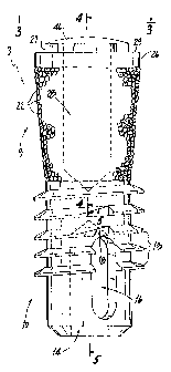
in FIG. 6 is the general shapc 60 of an extraction site 40. As this site is a
cavity that is not
cylindrical, it cannot be the site of a conventional cylindricai screw type
implant unless bone is first
regrown in the cavity and a new cylindrical bore is drilled in the new bone, a
process that could take
many weeks or nionths.
Accordina to the installation process for use of the screw implant of the
present
invention, an incision is nlade in the gum tissue 7, if any, covering the
extraction site to expose the
underlying bone S. Then, a small cylindrical bore 42 is drilled at the base of
the conical extraction
site 40. This bore 42, which is shown in dashed lines in Fig. 6, is made with
about the diameter of
the unthreaded part of the lower portion 10 of the implant 3. It is made deep
enough so that the
bottom t-,vo or three tunis of the tlireads 13 can engage the bone surrounding
the bore 42 at a level
below the original bottom of the extraction site. The bone chips created
during the formation of the
bore 42 arc preferably saved for later use.
According to the installation method of the present invention, the implant 3
is placed
in the site 40 so the unthreaded portion rests in the bore 42. Then, using a
wrench or similar tool
engaged with the hexagonal projection 27, the implant is rotated. As a result,
the cutting edge on
lowest thread of the implant at the vent 16 engages the bone surrounding bore
42 and begins to self-
tap into the bore. When the implant is rotated sufficiently, its base rests
against the bottom of bore
42. Because the implant lias been self-tapped into the bone, it is now firmly
anchored at the implant
site. Also, bone chips have fallen down or been pushed down the channel during
this process. These
chips have collected in the cavity 14 and will act to promote the growth on
new bone in the cavity and
through the vents to further anchor the implant in the future. '
The space 44 between tiie rest of the extraction site 40 and the implant 3,
especially
about the upper portioii 9, is now back filled with autogenous bone chips 6
saved from the creation
ofbore 42 or bone graft materials, such as bovine (xerographic) bone,
synthetic bone (alloplastic, e.g.,
ceramic or plastic), allographic bone, or a combination thereof. This material
may be resorbable or
non-resorbable, solid or microporous. One synthetic bone material is disclosed
in U.S. Patent No.
6
CA 02360250 2001-08-08
WO 00/47127 PCT/USOO/03674
4,728,570 of A. Ashman et al., and sold under the trade name Bioplant"~' HTR'.
In addition, one or
more bioactive substances that are medico-surgically useful may be
incorporated into the synthetic
bone. Various compositions of such are disclosed in U.S. Patent No. 5,356,629
of Sander et al.
The bone graft material 6 is packed loosely so that it fills the voids along
with the
bleeding from the surgical site and makes intimate contact with the microbeads
22, which are
preferably sintered titanium beads, on the surface of the upper portion 9. It
may be advisable to use
a surgical dressing to hold the bone chips in place. The dressing may be a
surgical adhesive or glue,
surgical foil, collagen, skin, or similar biocompatible material. In time, new
bone will grow around
and through the bone graft material 6, or replace it, thereby further
anchoring the implant in place.
This unique design of the implant 3 thus allows for immediate installation in
a fresh tooth extraction
site 40 and specifically addresses the requirements of extraction sites. r
The implant design can permit either a single or two-stage installation. For a
single-
stage installation, the abutment 2 is installed during the initial
installation of the implant. The
abutment 2 extends through the sutured gum. The artificial tooth 4 may or may
not be installed at the
same time.
A two-stage or submergible implant is shown in Fig. 6. Once the bone graft
material
6 is in place, a cap 46 is screwed into the threaded aperture 28 in the top of
the implant to make sure
the growth of new bone does not extend into the aperture. If bone does grow
into this aperture, it can
be very difficult to remove. Gingival tissue 7 is then sutured over the
implant. Time is be allowed
to pass so that new bone grows and firmly anchors the implant in place before
the rest of the implant
system is installed and the device is put into use.
At the second stage, the gingival tissue 7 is reopened. Often, bone has grown
over the
submerged implant and must be removed by a burr before the abutment 2 can be
installed. However,
ifbone grows up over the edges of the collar 26, there is no need to remove it
because it becomes part
of the permanent abutment. The cap 46 is then removed from aperture 28 and
replaced with the
threaded shaft of an abutment 2. The threaded end of the abutment 2 is engaged
with the
threaded aperture 28 and is rotated so that it is firmly secured in the
implant portion and is extending
in the proper direction. With this firm attachment completed, the artificial
tooth 4 can then be
attached over the abutment cylinder 2.
Whether a single-stage or two-stage (submergible) procedure is used, the
abutment 2,
which may be straight or have an angled shaft, is selected so as to cause the
artificial tooth 4 to be
7
CA 02360250 2001-08-08
WO 00/47127 PCT/US00/03674
correctly aligned with the other teeth of the patient. Therefore, the dentist
or oral surgeon must be
provided with a variety of such abutments that are at standard angles.
Besides being used to mount a single tooth, the implants according to the
present
invention can be used as supports for a permanent bridge or a removable
bridge.
While the invention has been particularly shown and described with reference
to a
preferred embodiment thereof, it will be understood by those skilled in the
art that various changes
in form and details may be made therein without departing from the spirit and
scope of the invention.
8