Note: Descriptions are shown in the official language in which they were submitted.
CA 02363473 2001-11-20
-1-
ANOSCOPE
BACKGROUND OF THE INVENTION
Field of Invention
The present invention relates to a speculum medical device to permit access
to treatment sites in the rectum and colon, and, in particular, to an anoscope
that is useful in the treatment of hemorrhoids.
Background of the Invention
A medical device to permit access to an orifice of the human body is referred
to generally as a speculum. Equipment for dilating and inspecting the anus,
rectum and colon of a patient is often referred to as an anoscope. An
anoscope generally comprises a hollow tubular member that is inserted into
the anus and colon. The tubular body dilates the anus and covers the skin of
the anal canal. The inner or distal end of the equipment enters the rectum and
colon. The distal end is often formed with a gap, notch or slot which is
positioned over the site of interest. The interior of the tubular member
provides a viewing and access passage for the doctor to inspect and perform
procedures at the site of interest. Anoscopes are particularly useful in the
inspection and treatment of hemorrhoids. The hemorrhoidal tissue is centred
in the notch at the distal end and the tissue tends to bulge into the tubular
interior of the anoscope for ready access by a doctor. The tissue may be
inspected, sutured or other procedures performed such as a biopsy. An
example of a typical anoscope is disclosed in United States Patent No.
4,834,067 to Block.
A common surgery performed in conjunction with an anoscope is a
hemorrhoidectomy which involves excision of the hemorrhoid tissue and
suturing of the surgically produced wounds. The patient is usually placed
under general anesthesia, which entails an inherent degree of risk. In
CA 02363473 2009-09-14
-2-
addition, the patient invariably experiences acute pain for many days during
the period of recovery, especially when defecating.
The treatment of hemorrhoids by elastic band ligation is credited to Biaisdell
who described this technique in Diseases of the Colon and Rectum in 1963.
The technique involves placing an elastic band on tissue in the rectum above
the area of the hemorrhoid where there is little sensation. The tissue trapped
in the band being cut off from its blood supply degenerates and is sloughed,
and the elastic band along with the sloughed tissue is passed with the bowel
motions. More importantly, however, the resulting healing process causes the
tissue in the vicinity to become fixed and prolapse of the hemorrhoidal tissue
is minimized. Furthermore, the elastic band ligation technique has been found
to give relief of hemorrhoidal symptoms.
Commonly owned United States Patent No. 5,741,273 entitled "Elastic band
ligation device for treatment of hemorrhoids" discloses a special tool that is
useful for elastic banding of hemorrhoids to avoid suturing. An advantage of
the patented ligation device is that the device may be used without directly
seeing the site for banding. Thus, it may be used without an anoscope or any
other type of scope or viewing technique. The ligation device is sufficiently
small that it can be inserted into the rectum of a patient without major
discomfort. For best results, however, the elastic band ligation device is
used
with an anoscope so that the doctor see the site of interest immediately prior
to banding.
In order to provide adequate room for manipulation of the elastic band
ligation
device or any other inspection or treatment instrument within the interior of
an
anoscope, it has previously been preferable to use larger diameter anoscopes
that are often uncomfortable for the patient. With anoscopes having a
diameter larger than 2 to 2 1/2 inches, depending on the patient, it is
generally
necessary to perform the procedure in an operating room under a general
anesthetic which increases the risk and cost of the procedure. The cost of the
CA 02363473 2009-09-14
-3-
anesthetic, the operating room and the recovery room can be in the order of
$1000 per procedure.
There is therefore a need for an anoscope that is designed to be of
sufficiently
small diameter that it can be used without requiring a general anesthetic
while
at the same time permitting manipulation of a medical instrument inserted into
the anoscope for improved access to the site of interest in the distal notch
of
the anoscope.
SUMMARY OF THE INVENTION
The present invention provides an anoscope that is of appropriate diameter
for insertion without discomfort into the anus of a patient which includes a
proximal notch in addition to the distal notch. The proximal notch remains
external to anus and accommodates pivoting of a medical instrument inserted
into the interior of the anoscope off the longitudinal axis of the anoscope to
improve access to the site of interest in the distal notch.
Accordingly, the present invention provides an anoscope comprising a tubular
member of sufficient length for insertion into the rectum of a patient having
a
longitudinal axis with a proximal end and a distal end at opposite ends of the
longitudinal axis, a first longitudinal slot extending from the distal end
toward
the proximal end a distance along the tubular member, and a second
longitudinal slot extending from the proximal end toward the distal end a
distance along the tubular member, said first and second longitudinal slots
being separated by about 180 on the surface of the tubular member.
The present invention also provides an anoscope comprising a tubular
member having a proximal end and a distal end, a first longitudinal slot
extending from the distal end to part way along the tubular member, and a
second longitudinal slot extending from the proximal end to part way along the
tubular member, said first and second longitudinal slots being separated by
about 180 on the surface of the tubular member.
CA 02363473 2009-09-14
-4-
The present invention also provides an anoscope for use with a medical
instrument insertable within the anoscope, comprising a tubular member
having a longitudinal axis and a proximal end and a distal end, a first
longitudinal slot extending from the distal end toward the proximal end, and a
second longitudinal slot extending from the proximal end toward the distal
end, the first and second longitudinal slots being separated by about 1800 an
the surface of the tubular member, the first longitudinal slot being adapted
to
permit access to a site to be treated and the second longitudinal slot being
adapted to accommodate pivoting of the medical instrument at an angle to the
longitudinal axis of the tubular member.
In a further aspect, the present invention provides an anoscope comprising a
tubular member of sufficient length for insertion into the rectum of a patient
having a longitudinal axis with a proximal end and a distal end at opposite
ends of the longitudinal axis, with a longitudinal proximal slot extending
from
the proximal end toward the distal end a distance along the tubular member
adapted to accommodate pivoting of a medical instrument at an angle to the
longitudinal axis of the tubular member.
The anoscope of the present invention is preferably disposable and formed
from a transparent, medical grade plastic to improve visibility for the
doctor.
The distal and proximal notches do not intersect and are spaced apart so that
rigidity of the tubular member is maintained to prevent collapse of the
tubular
member against the pressure exerted by rectal muscles of the patient.
Other aspects and features of the present invention will become apparent to
those ordinarily skilled in the art upon review of the following description
of
specific embodiments of the invention in conjunction with the accompanying
figures.
BRIEF DESCRIPTION OF THE DRAWINGS
CA 02363473 2001-11-20
-5-
Aspects of the present invention In drawings which illustrate embodiments of
the
invention,
Figure 1 is a front perspective view of a first embodiment of an anoscope
according to the present invention;
Figure 2 is a rear perspective view of the anoscope of Figure 1 a;
Figure 3 is an additional rear perspective view from a different angle to
clearly show the distal and proximal notches of the anoscope;
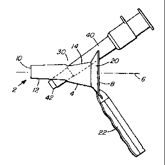
Figure 4 is a side elevation view of the anoscope with an inserted ligation
tool;
Figure 5 is a cross-section taken through Figure 4;
Figure 6 is a rear elevation view of the combined anoscope and ligation
tool of Figure 4;
Figure 7 is an elevation view of an obturator device for use with the
anoscope of the present invention to permit insertion into the anus
of a patient.
Figure 8 is a perspective view of an alternative embodiment of the
anoscope of the present invention having only a proximal slot.
DETAILED DESCRIPTION OF THE PREFERRED EMBODIMENTS
Referring to Figures 1-3, there are shown various views of a preferred
embodiment of an anoscope 2 according to the present invention. Anoscope
2 comprises a tubular member 4 of sufficient length for insertion into the
rectum of a patient. Tubular member 4 defines a hollow body having a
longitudinal axis 6 with a proximal end 8 and a distal end 10 at opposite ends
CA 02363473 2001-11-20
-6-
of the longitudinal axis. A first longitudinal slot 12 extends from distal end
10
toward proximal end 8 a distance along the tubular member. Similarly, a
second longitudinal slot 14 extends from proximal end 8 toward the distal end
a distance along the tubular member. First and second longitudinal slots 12
and 14 are separated by about 1800 on the surface of the tubular member.
Tubular member 4 is formed with an outwardly flaring generally frusto-conical
flange 20 adjacent the proximal end 8 which is intersected by slot 14. Tubular
member 4 tapers inwardly towards distal end 10 to facilitate insertion of the
tubular member into the anus and rectum of a patient. In the illustrated
embodiment, the taper occurs over two stages to divide the tubular member
into a first generally conical portion 21 near the proximal end and a second
conical portion 23 adjacent the distal end. The body of tubular member 4 is
preferably formed from a transparent medical grade plastic that can be readily
disposed of after use.
An obturator 24 (Figure 7) is used with the anoscope to provide a single
composite structure during insertion. The obturator slides lengthwise within
anoscope 2 to extend along axis 6. The obturator has a domed or bullet-nose
front portion 24a that protrudes from proximal end 10. The obturator has a
body that is generally conical in shape to conform to the shape of the
interior
of the tubular member in order to fill slots 12 and 14. The obturator is
somewhat longer than the tubular member so that flange 24b protrudes from
flange 20 to serve as a gripping surface 24b to remove the obturator when the
anoscope has been inserted.
Preferably, a handle 22 extends from flange 20 at proximal end 8 for grasping
by the doctor on insertion of the anoscope. Handle 22 is preferably located
on the same side of the tubular member as the first longitudinal slot 12 to
ensure that the tubular body of the anoscope is as strong and rigid as
possible.
CA 02363473 2001-11-20
-7-
As best shown in Figure 5, handle 22 is preferably hollow to accommodate
the insertion of an illumination device, such as a penlight, into the interior
of
the handle. The penlight serves to illuminate by internal reflection of light,
the
transparent plastic walls of the anoscope to allow the doctor an improved
lighted view of the tissues dilated by the anoscope. Such illumination means
is conventionally a part of examining and operating anoscopes.
Referring to Figure 4, first longitudinal slot 12 and second longitudinal slot
14
are preferably dimensioned to overlap in a region 30 intermediate the ends of
tubular member 4. This arrangement is preferred as it maintains the structural
rigidity of the tubular body against the involuntary muscular contractions of
the
patient and readily permits pivoting of a medical instrument inserted into the
interior of the anoscope at an angle to the longitudinal axis 6 of the tubular
member.
Figures 4, 5 and 6 illustrate a medical instrument 40 inserted into the
interior
of anoscope 2 and pivoted at an angle to the longitudinal axis 6 of the
anoscope. Figure 5 is a section view taken through Figure 4 and Figure 6 is a
rear end view of the arrangement shown in Figure 4. The illustrated medical
instrument 40 is the elastic band ligation device described and claimed in
commonly owned United States Patent No. 5,741,273. This instrument is
particularly suited for use with the anoscope of the present invention as it
is
important that the tip 42 of the instrument be correctly positioned over a
hemorrhoid site in distal slot 12 to allow the hemorrhoid to be drawn by
suction into the interior of 42 for the subsequent banding operation. It will
be
appreciated by those skilled in the art that the anoscope of the present
invention will find application with other medical devices that are introduced
through the interior of the anoscope and that require being pivoted off the
axis
6 of the anoscope through proximal slot 14 for optimal access to the site of
interest at distal slot 12
The anoscope of the present invention is of a sufficiently small diameter that
it
can be inserted without resorting to a general anesthetic. The reduced
CA 02363473 2001-11-20
-8-
interior space within the anoscope for manipulating a medical instrument is
substantially offset by longitudinally extending slot 14 which co-operates
with
slot 12 to permit improved maneuverability of the instrument by pivoting of
the instrument to an angle with respect to the longitudinal axis 6 of the
anoscope. For example, in Figures 4 and 5, instrument 40 is shown pivoted
at an angle of approximately 350 to axis 6 of the anoscope which significantly
improves the ability of a doctor to position working tip 42 of the instrument
correctly at the site of interest in slot 12 despite the smaller diameter of
the
tubular body of the anoscope.
In use, the anoscope 2 is inserted into the anus and the rectum of a patient.
During insertion, obturator 24 fills the proximal end 10 of the anoscope and
slots 12 and 14. Frusto-conical flange 20 is positioned adjacent and shields
the patient's anus. The tubular body of the anoscope dilates the anus and
covers the skin of the anal canal. The proximal end 10 enters the rectum.
After insertion of the anoscope, the obturator is withdrawn from proximal end
8 to clear slots 12 and 14. The tubular member 4 maintains the anus firmly
distended despite a tendency of the sphincter to contract. Particularly when
no anaesthetic is used, this tendency of the anus to contract is strong.
Transparent tubular member 4 assures a clear view of the hemorrhoid. When
slot 12 of proximal end 10 is centered at a hemorrhoid, the tissue bulges into
the space provided by the slot so that the tissue is positioned for inspection
and/or treatment whether by elastic band ligation, suturing or some other
technique. Despite any involuntary muscular constriction, the tubular member
maintains the anal canal dilated to maintain an adequate view of the rectum.
Moreover, flange 20 and the tubular body act as a shield for the anus and the
anal canal against the instrument used in the procedure. This is particularly
important for a fully conscious patient when no anaesthetic is used and when
some local anaesthetic is used. The tissue of the rectum is virtually
insensitive
to pain.
In a preferred arrangement that is suitable for use with the majority of
patients, the anoscope of the present invention is preferably 31/2 inches long
CA 02363473 2001-11-20
-9-
with an internal diameter that tapers from 1 3/8 inches at proximal end 8 to
3/4
inches at distal end 10. Slot 12 extends for approximately 1 7/8 inches along
the body and slot 14 extends for approximately 2 inches along the body of the
tubular member. Flange 20 is about about 2 1/4 inches in diameter at its
widest point.
Figure 8 is a perspective view of an alternative embodiment of the anoscope
of the present invention having only a longitudinally extending proximal slot
140. The presence of a proximal slot alone is sufficient to enjoy the
advantage of improved maneuverability of a medical instrument inserted into
the interior of the anoscope. Slot 140 permits angling of the instrument off
the
longitudinal axis 6. To increase the angle to which the inserted medical
instrument can be pivoted, distal conical portion 23 is preferably shortened
as
compared to longer portion 23 in the embodiment of Figure 1.
While specific embodiments of the invention have been described and
illustrated, such embodiments should be considered illustrative of the
invention only and not as limiting the invention as construed in accordance
with the accompanying claims.
