Note: Descriptions are shown in the official language in which they were submitted.
CA 02364042 2001-08-31
WO 00/51683 PCT/US00/05468
ATRIAL ABLATOR HAVING BALLOON AND SENSOR
BACKGROUND OF THE INVENTION
Field of the Invention
The field of the invention relates generally to a medical device system and
associated methods of
manufacture and use. More particularly, the invention relates to a medical
device assembly including a sensory system
integrated with an expandable member of the assembly, as well as relates to
associated methods of manufacturing
and using the assembly.
Description of Related Art
Many local energy delivery devices and methods have been developed for
treating the various abnormal tissue
conditions in the body, and particularly for treating abnormal tissue along
body space walls which define various body
spaces in the body. For example, various devices have been disclosed with the
primary purpose of treating or
recanalizing atherosclerotic vessels with localized energy delivery. Several
prior devices and methods combine energy
delivery assemblies in combination with cardiovascular stent devices in order
to locally deliver energy to tissue in order
to maintain patency in diseased lumens such as blood vessels. Endometriosis,
another abnormal wall tissue condition
which is associated with the endometrial cavity and is characterized by
dangerously proliferative uterine wall tissue
along the surface of the endometrial cavity, has also been treated by local
energy delivery devices and methods.
Several other devices and methods have also been disclosed which use catheter-
based heat sources for the intended
purpose of inducing thrombosis and controlling hemorrhaging within certain
body lumens such as vessels. Detailed
examples of local energy delivery devices and related procedures such as those
of the types described above are
disclosed in the following references: U.S. Patent Nos. 4,672,962 to
Hershenson; U.S. Patent Nos. 4,676,258 to
InoKuchi et al.; U.S. Patent No. 4,790,311 to Ruiz; 4,807,620 to Strul et al.;
U.S. Patent No. 4,998,933 to Eggers et
al.; U.S. Patent No. 5,035,694 to Kasprzyk et al.; U.S. Patent No. 5,190,540
to Lee; U.S. Patent No. 5,226,430 to
Spears at al.; and U.S. Patent No. 5,292,321 to Lee; U.S. Patent No. 5,449,380
to Chin; U.S. Patent No. 5,505,730
to Edwards; U.S. Patent No. 5,558,672 to Edwards et al.; and U.S. Patent No.
5,562,720 to Stern et al.; U.S. Patent
No. 4,449,528 to Auth at al.; U.S. Patent No. 4,522,205 to Taylor et al.; and
U.S. Patent No. 4,662,368 to Hussein et
al.; U.S. Patent No. 5,078,736 to Behl; and U.S. Patent No. 5,178,618 to
Kandarpa.
Other prior devices and methods electrically couple fluid to an ablation
element during local energy delivery
for treatment of abnormal tissues. Some such devices couple the fluid to the
ablation element for the primary purpose
of controlling the temperature of the element during the energy delivery.
Other such devices couple the fluid more
directly to the tissue-device interface either as another temperature control
mechanism or in certain other known
applications as a carrier or medium for the localized energy delivery.
Detailed examples of ablation devices which use
-1-
CA 02364042 2001-08-31
WO 00/51683 PCT/US00/05468
fluid to assist in electrically coupling electrodes to tissue are disclosed in
the following references: U.S. Patent No.
5,348,554 to lmran et al.; U.S. Patent No. 5,423,811 to lmran et al.; U.S.
Patent No. 5,505,730 to Edwards; U.S.
Patent No. 5,545,161 to lmran et al.; U.S. Patent No. 5,558,672 to Edwards et
al.; U.S. Patent No. 5,569,241 to
Edwards; U.S. Patent No. 5,575,788 to Baker et al.; U.S. Patent No. 5,658,278
to lmran et al.; U.S. Patent No.
5,688,267 to Panescu et al.; U.S. Patent No. 5,697,927 to lmran et al.; U.S.
Patent No. 5,722,403 to McGee et al.;
U.S. Patent No. 5,769,846; and PCT Patent Application Publication No. WO
97132525 to Pomeranz et al.; and PCT
Patent Application Publication No. WO 98/02201 to Pomeranz et al.
Atrial Fibrillation
Cardiac arrhythmias, and atrial fibrillation in particular, persist as common
and dangerous medical ailments
associated with abnormal cardiac chamber wall tissue, and are often observed
in elderly patients. In patients with
cardiac arrhythmia, abnormal regions of cardiac tissue do not follow the
synchronous beating cycle associated with
normally conductive tissue in patients with sinus rhythm. Instead, the
abnormal regions of cardiac tissue aberrantly
conduct to adjacent tissue, thereby disrupting the cardiac cycle into an
asynchronous cardiac rhythm. Such abnormal
conduction is known to occur at various regions of the heart, such as, for
example, in the region of the sino-atrial (SA)
node, along the conduction pathways of the atrioventricular (AV) node and the
Bundle of His, or in the cardiac muscle
tissue forming the walls of the ventricular and atrial cardiac chambers.
Cardiac arrhythmias, including atrial arrhythmia, may be of a multiwavelet
reentrant type, characterized by
multiple asynchronous loops of electrical impulses that are scattered about
the atrial chamber and are often self
propagating. In the alternative or in addition to the multiwavelet reentrant
type, cardiac arrhythmias may also have a
focal origin, such as when an isolated region of tissue in an atrium fires
autonomously in a rapid, repetitive fashion.
Cardiac arrhythmias, including atrial fibrillation, may be generally detected
using the global technique of an
electrocardiogram (EKG). More sensitive procedures of mapping the specific
conduction along the cardiac chambers
have also been disclosed, such as, for example, in U.S. Patent No. 4,641,649
to Walinsky et al. and in PCT Patent
Application Publication No. WO 96/32897 to Desai.
A host of clinical conditions can result from the irregular cardiac function
and resulting hemodynamic
abnormalities associated with atrial fibrillation, including stroke, heart
failure, and other thromboembolic events. In
fact, atrial fibrillation is believed to be a significant cause of cerebral
stroke, wherein the abnormal hemodynamics in
the left atrium caused by the fibrillatory wall motion precipitate the
formation of thrombus within the atrial chamber.
A thromboembolism is ultimately dislodged into the left ventricle which
thereafter pumps the embolism into the
cerebral circulation where a stroke results. Accordingly, numerous procedures
for treating atrial arrhythmias have
been developed, including pharmacological, surgical, and catheter ablation
procedures.
Several pharmacological approaches intended to remedy or otherwise treat
atrial arrhythmias have been
disclosed, such as, for example, those approaches disclosed in the following
references: U.S. Patent No. 4,673,563 to
Berne et al.; U.S. Patent No. 4,569,801 to Molloy et al.; and "Current
Management of Arrhythmias" (1991) by
-2-
CA 02364042 2001-08-31
WO 00/51683 PCT/US00/05468
Hindricks, et al. Such pharmacological solutions, however, are not generally
believed to be entirely effective in many
cases, and are even believed in some cases to result in proarrhythmia and long
term inefficacy.
Several surgical approaches have also been developed with the intention of
treating atrial fibrillation. One
particular example is known as the "maze procedure," as is disclosed by Cox,
J. L. et al. in "The surgical treatment of
atrial fibrillation. I. Summary" Thoracic and Cardiovascular Surgery 101(3),
pp. 402-405 (1991); and also by Cox, JL
in "The surgical treatment of atrial fibrillation. IV. Surgical Technique",
Thoracic and Cardiovascular Surgery 101(4),
pp. 584-592 (1991). In general, the "maze" procedure is designed to relieve
atrial arrhythmia by restoring effective
atrial systole and sinus node control through a prescribed pattern of
incisions about the tissue wall. In the early
clinical experiences reported, the "maze" procedure included surgical
incisions in both the right and the left atrial
chambers. However, more recent reports predict that the surgical "maze"
procedure may be substantially efficacious
when performed only in the left atrium. See Sueda et al., "Simple Left Atrial
Procedure for Chronic Atrial Fibrillation
Associated With Mitral Valve Disease" (1996).
The "maze procedure" as performed in the left atrium generally includes
forming vertical incisions from the
two superior pulmonary veins and terminating in the region of the mitral valve
annulus, traversing the region of the
inferior pulmonary veins en route. An additional horizontal line also connects
the superior ends of the two vertical
incisions. Thus, the atrial wall region bordered by the pulmonary vein ostia
is isolated from the other atrial tissue. In
this process, the mechanical sectioning of atrial tissue eliminates the
arrhythmogenic conduction from the boxed region
of the pulmonary veins to the rest of the atrium by creating conduction blocks
within the aberrant electrical conduction
pathways. Other variations or modifications of this specific pattern just
described have also been disclosed, all sharing
the primary purpose of isolating known or suspected regions of arrhythmogenic
origin or propagation along the atrial
wall.
While the "maze" procedure and its variations as reported by Dr. Cox and
others have met some success in
treating patients with atrial arrhythmia, its highly invasive methodology is
believed to be prohibitive in most cases.
However, these procedures have provided a guiding principle that electrically
isolating faulty cardiac tissue may
successfully prevent atrial arrhythmia, and particularly atrial fibrillation
caused by arrhythmogenic conduction arising
from the region of the pulmonary veins.
Less invasive catheter-based approaches to treat atrial fibrillation have been
disclosed which implement
cardiac tissue ablation for terminating arrhythmogenic conduction in the
atria. Examples of such catheter-based
devices and treatment methods have generally targeted atrial segmentation with
ablation catheter devices and
methods adapted to form linear or curvilinear lesions in the wall tissue which
defines the atrial chambers. Some
specifically disclosed approaches provide specific ablation elements which are
linear over a defined length intended to
engage the tissue for creating the linear lesion. Other disclosed approaches
provide shaped or steerable guiding
sheaths, or sheaths within sheaths, for the intended purpose of directing tip
ablation catheters toward the posterior
left atrial wall such that sequential ablations along the predetermined path
of tissue may create the desired lesion. In
-3-
CA 02364042 2001-08-31
WO 00/51683
PCT/US00/05468
addition, various energy delivery modalities have been disclosed for forming
atrial wall lesions, and include use of
microwave, laser, ultrasound, thermal conduction, and more commonly,
radiofrequency energies to create conduction
blocks along the cardiac tissue wall. Detailed examples of ablation device
assemblies and methods for creating lesions
along an atrial wall are disclosed in the following U.S. Patent references:
U.S. Patent No. 4,898,591 to Jang et al.;
U.S. Patent No. 5,104,393 to lsner et al.; U.S. Patent No. 5,427,119; U.S.
Patent No. 5,487,385 to Avitall; U.S.
Patent No. 5,497,119 to Swartz et al.; U.S. Patent No. 5,545,193 to Fleischman
et al.; U.S. Patent No. 5,549,661 to
Kordis et al.; U.S. Patent No. 5,575,810 to Swanson et al.; U.S. Patent No.
5,564,440 to Swartz et al.; U.S. Patent
No. 5,592,609 to Swanson et al.; U.S. Patent No. 5,575,766 to Swartz et al.;
U.S. Patent No. 5,582,609 to
Swanson; U.S. Patent No. 5,617,854 to Munsif; U.S. Patent No 5,687,723 to
Avitall; U.S. Patent No. 5,702,438 to
Avitall. Other examples of such ablation devices and methods are disclosed in
the following PCT Patent Application
Publication Nos.: WO 93/20767 to Stern et al.; WO 94/21165 to Kordis et al.;
WO 96/10961 to Fleischman et al.; WO
96/26675 to Klein et al.; and WO 97137607 to Schaer. Additional examples of
such ablation devices and methods are
disclosed in the following published articles: "Physics and Engineering of
Transcatheter Tissue Ablation", Avitall et al.,
Journal of American College of Cardiology, Volume 22, No. 3:921-932 (1993);
and "Right and Left Atrial
Radiofrequency Catheter Therapy of Paroxysmal Atrial Fibrillation,"
Haissaguerre, et al., Journal of Cardiovascular
Electrophysiology 7(12), pp. 1132-1144 (1996).
In addition to those known assemblies summarized above, additional tissue
ablation device assemblies have
been recently developed for the specific purpose of ensuring firm contact and
consistent positioning of a linear ablation
element along a length of tissue by anchoring the element at least at one
predetermined location along that length,
such as in order to form a "maze"-type lesion pattern in the left atrium. One
example of such assemblies is that
disclosed in U.S. Patent No. 5,971,983, issued October 26, 1999. The assembly
includes an anchor at each of two
ends of a linear ablation element in order to secure those ends to each of two
predetermined locations along a left
atrial wall, such as at two adjacent pulmonary veins, so that tissue may be
ablated along the length of tissue
extending therebetween.In addition to attempting atrial wall segmentation with
long linear lesions for treating atrial arrhythmia, other
ablation device and method have also been disclosed which are intended to use
expandable members such as balloons
to ablate cardiac tissue. Some such devices have been disclosed primarily for
use in ablating tissue wall regions along
the cardiac chambers. Other devices and methods have been disclosed for
treating abnormal conduction of the left-
sided accessory pathways, and in particular associated with "Wolff-Parkinson-
White" syndrome ¨ various such
disclosures use a balloon for ablating from within a region of an associated
coronary sinus adjacent to the desired
cardiac tissue to ablate. Further more detailed examples of devices and
methods such as of the types just described
are variously disclosed in the following published references: Fram et al., in
"Feasibility of RF Powered Thermal Balloon
Ablation of Atrioventricular Bypass Tracts via the Coronary Sinus: In vivo
Canine Studies," PACE, Vol. 18, p 1518-
1530 (1995) ; "Long-term effects of percutaneous laser balloon ablation from
the canine coronary sinus", Schuger CD
-4-
CA 02364042 2007-03-27
_
et al., Circulation (19921 86:947-954; and "Percutaneous laser balloon
coagulation of accesSory
pathways", McMath LP at al., Diagh Ther Cardlovesc Interven 1991; 1425:165-
171.
ferhythmlas OrigLiaL,c; from Foci Jag 1,Y_Wl.a.
Various modes of atrial fibrillation have also been observed to be focal In
nature, caused by
the rapid and repetitive firing of an isolated center within cardiac muscle
tissue associated, with the
atriUr.ri. Such foci may act as either a trigger of atrial fibrillatory
paroxysmal or may even sustain the
fibrillation. Various disclosures have suggested that focal atrial arrhythmia
often originates from at
least one tissue region along one or more of the pulmonary veins of the left
atrium, and even more
particularly in the superior pulmonary veins.
Less-invasive percutaneous catheter ablation techniques have been disclosed
which use
end-electrode catheter designs with the intention of ablating and thereby
treating focal arrhythmias in
the pulmonary veins. These ablation procedures are typically characterized by
the incremental
application of electrical energy to the tissue to form focal lesions designed
to terminate the
Inappropriate arrhythmogenic conduction.
One example of a focal ablation method intended to treat focal arrhythmia
originating from
a pulmonary vein is disclosed by Haissaguerre, et al. in "Right and Left
Atrial Radiofrequency
Catheter Therapy of Paroxysmal Atrial Fibrillation" in Journal of
Cardiovascular Electrophysiology
7(12), pp. 1132-1144 (1996). Haissaguerre, et al. discloses radiofrequency
catheter ablation of drug-
refractory paroxysmal atrial fibrillation using linear atrial lesions
complemented by focal ablation
targeted at arrhythmogenic foci in a screened patient population. The site of
the arrhythmogenic foci
were generally located just inside the superior pulmonary vein, and the focal
ablations were
generally performed using a standard 4mm tip single ablation electrode.
Another focal ablation method of treating atrial arrhythmias is disclosed in
Jais at al., "A
focal source of atrial fibrillation treated by discrete radiofrequency
ablation," Circulation 95:572-578
(1997). Jais et el. discloses treating patients with paroxysmal arrhythmias
originating from a focal
source by ablating that source. At the site of arrhythmogenic tissue, in both
right and left atria,
several pulses of a discrete source of radiofrequency energy were applied in
order to eliminate the
fibriliatory process.
Other assemblies and methods have been disclosed addressing focal sources of
arrhythmia in pulmonary veins by ablating circumferential regions of tissue
either along the
pulmonary vein, at the ostlum of the vein along the atrial wall, or encircling
the ostium and along the
atrial wall. More detailed examples of device assemblies and methods for
treating focal arrhythmia
as just described are disclosed in PCT Patent Application Publication No. WO
99102096 to
Diederich at at., and also in the following pending U.S. Patent Applications;
USSN# 081889,798 for
"Circumferential Ablation Device Assembly" to Michael D. Lesh et al, filed
July 8, 1997, now U.S.
Patent No. 6,024,740, issued February 15, 2000; USSN# 08/889,835 for "Device
and Method for
Forming a Circumferential Conduction Stock in a Pulmonary Vein" to Michael D.
Lash, filed July 8,
1997, now U.S. Patent No. 6,012,457 issued January 11, 2000; and USSN#
091199,736 for
- 5 -
CA 02364042 2007-03-27
"Circumferential Ablation Device Assembly" to Chris J. Diederich at al., filed
February 3, 1998, now
U.S. Patent No. 6,177,101 issued September 12, 2000.
Another specific device assembly and method which is intended to treat focal
atrial
fibrillation by ablating a circumferential region of tissue between two seals
in order to form 'a
conduction block to isolate an arrhythmogenic focus within a pulmonary vein Is
disclosed in US.
Patent No. 5,938,660 and a related PCT Patent Application Publication No. WO
99/00064.
Thermocouples have been used with prior ablation catheter to monitor and
regulate the
ablation process. A difficulties arises, however, with monitoring and
regulating the ablation process
with one or more thermocouples where ablation occurs though an inflatable
balloon, such as with the
device assembly disclosed in PCT Patent Application-Publication No. WO
99/02096 to Diederich at
al: Thermocouples are usually mounted to the catheter shaft, and if ablation
occurs at an interface
between the balloon and the tissue which it engages, the thermocouples do not
accurately measure
the temperature because of their remote distance relative to the ablation
site. Accordingly, a peed
exists for an improved approach for mounting a thermocouple onto a catheter In
proximity to the
ablation site.
SUMMARY OF TLIE INVENTION
In one mode, the present invention provides a medical device system for
ablating a
circumferential region of tissue In order to form a circumferential conduction
block at a location where
a pulmonary vein extends from an atrium in a patient's heart. Such conduction
block may be formed
in order to, for example: electrically isolate a focal source of arrhythmia In
the pulmonary vein from
the rest of the atrium; or connect linear lesions such that a pattern of
conduction blocks may be
formed to isolate a posterior region of the atrial wall from the rest of the
atrium.
In one aspect, the tissud ablation apparatus of the present invention ablates
a substantial
portion of a circumferential region of tissue at a location in a patient's
body where a pulmonary vein
extends from an atrium in a patient'. The ablation apparatus includes an
elongate body with a
proximal end portion and a distal end portion. An ablation member is coupled
to the elongate body.
The ablation member comprises an expandable member coupled to the distal end
portion of the
elongate body, wherein the expandable member Is adjustable from a collapsed
position to an
expanded position_ The expandable member is adapted to engage the substantial
portion of the
. circumferential region of tissue when in the expanded position. The
ablation member also has an
ablation element that is adapted to ablate at least a portion of the
substantial portion of the
circumferential region of tissue. The ablation member further includes a
sensor that is coupled to the
, expandable member at a location at least when the expandable member is in
the expanded position.
A conductor is coupled to the sensor in a manner that does not substantially
affect the adjustment of
the expandable member from the collapsed positioned to an expanded position.
In a preferred form,
the conductor also Is coupled to a coupler at the proximal end portion of the
elongated body.
In one variation of the ablation apparatus, the expandable member includes an
inflatable
balloon. The expandable member may be coupled to the expandable member by an
adhesive.
Further, the adhesive may encapsulate
- 6 -
CA 02364042 2001-08-31
WO 00/51683 PCT/US00/05468
the sensor. A variety of ways are disclosed for reducing tension on the
location where the sensor is coupled to the
expandable member. For example, the wire may form a loop or a helix to reduce
tension on the attachment point.
In one embodiment, the expandable member has an outer surface that is adapted
to contact the substantial
portion of the circumferential region of tissue along an ablative path when
the expandable member is adjusted to the
expanded position. The location where the sensor is coupled to the expandable
member may be provided along the
outer surface. Alternatively, the expandable member may have an inner surf ace
and an outer surface, wherein the
outer surface is adapted to contact the substantial portion of the
circumferential region of tissue, and wherein the
location, at which the sensor is coupled to the expandable member, is provided
along the inner surface of the
expandable member.
In one preferred embodiment, the sensor comprises a temperature sensor. The
sensor may be coupled to the
expandable member at a location which is fixed along the ablative path. In the
alternative or in addition, the sensor
may comprise an electrode.
In another variation of the present invention, the expandable member may
include an outer layer and an inner
layer, wherein the outer layer comprises an outer surface that is adapted to
contact the substantial portion of the
circumferential region of tissue. The sensor may be disposed between the outer
and inner layers in such an
embodiment. In a further variation, a passageway may be provided between the
outer layer and the inner layer,
wherein the sensor is slideably engaged within the passageway. The passageway
may be filled with a fluid.
Alternatively, the passageway may be incorporated into the elastomeric wall of
the expandable member, wherein the
sensor and conductor are slideably engaged within the channel. In one
variation, the expandable member has an
interior space which is in fluid communication with the passageway in order to
facilitate adjustment of the expandable
member from a collapsed to an expanded state. In another variation, the
passageway may include a stenting member
to prevent the passageway from collapsing under pressure from the interior of
the expandable member.
In another preferred embodiment, the coupling location on the expandable
member further comprises a
reinforcement disposed on either the inner or outer surf ace of the expandable
member.
In one additional variation, the expandable member of the present invention
may comprise a spline to which
the sensor is coupled, the spline being expandable or self-expanding. In some
cases, the apparatus may include both an
inflatable balloon and a spline, wherein the coupling location for the sensor
is located along the spline.
The apparatus of the present invention preferably includes an ultrasound
transducer adapted to emit a
circumferential path of ultrasound ablative energy. The sensor may be
positionable within the circumferential path
when the expandable member is in the expanded position.
Also disclosed is a method of monitoring the ablation of a substantial portion
of a circumferential region of
tissue at a location where a pulmonary vein extends from an atrium. The method
involves positioning an ablation
member, which has an ablation element, along the location where the pulmonary
vein extends from the atrium. The
ablation element is activated to ablate the substantial portion of the
circumferential region of tissue. This can be done
-7-
CA 02364042 2001-08-31
WO 00/51683 PCT/US00/05468
simultaneously or through a sequential series of ablation steps (temporal
and/or spatial). Temperature is monitored
along the substantial portion of the circumferential region of tissue. The
ablation element is deactivated when the
temperature along the substantial portion of the circumferential region of
tissue has reached either a first
predetermined value or a second predetermined valve for a predetermined period
of time. The initial act of positioning
may further comprise adjusting an expandable member from a collapsed position
to an expanded position, such that at
least a portion of the expandable member contacts the substantial portion of
the circumferential region of tissue. In a
further variation, when the expandable member is adjusted from the collapsed
position to the expanded position, a
sensor, which is coupled to the expandable member, is preferably placed
sufficiently close to the substantial portion of
the circumferential region of tissue to allow the sensor to monitor the
temperature of the substantial portion of the
circumferential region of tissue.
While various aspects and features of the present invention have particular
utility in the context of tissue
ablation apparatuses and ablation processes, such aspects and features also
can be practiced apart from such devices
and methods. Thus, in accordance with another mode of the present invention, a
medical article is provided comprising
an elongate body with a proximal end portion and a distal end portion. An
expandable member is coupled to the distal
end portion of the elongate body, and the expandable member is adapted to be
adjusted between a collapsed position
and an expanded position. A sensor is coupled to the expandable member at a
location at least when the expandable
member is in the expanded position, and a conductor is coupled to the sensor
in a manner that does not substantially
affect the adjustment of the expandable member from the collapsed position to
the expanded position. In a preferred
form, the conductor is also coupled to a coupler at the proximal end portion
of the elongated body.
The present invention also involves methods of manufacturing the medical
apparatuses described herein. In
one mode, the method involves inverting an expandable member, coupling the
sensor to the expandable member, and
then returning the expandable member to its normal position. One or more ends
of the expandable member can be
attached to an elongated body of the medical apparatus after the sensor is
coupled to the expandable member. In one
variation, one end of the expandable member is attached to the elongated body
before the expandable member is
inverted.
In another mode, an end of the sensor is formed to have a larger sized shape
than a conductor to which it is
attached. In one form, for example, the sensor is shaped to have a loop
configuration and in another form the sensor is
shaped to have a serpentine configuration. The sensor is embedded in a bonding
agent affixed to the expandable
member or within the material of the expandable member itself.
In an additional mode, the expandable member is formed with at least one
reinforcement location. The
location(s) can be formed on inner or outer sides of the expandable member.
The sensor is coupled to the expandable
member at the reinforcement location.
In a further manufacturing mode, the expandable member is formed with a
passageway. The sensor and/or a
conductor, which is connected to the sensor, is slideably inserted within or
through the passageway.
-8-
WO 00/51683 CA
02364042 2001-08-31
PCT/US00/05468
These manufacturing methods, as well as others described herein, can be used
to coupled the sensor to the
expandable member so as to position the sensor in a desired location when the
expandable member is adjusted to an
expanded position, but not to substantially affect such adjustment (e.g.,
affect the shape of the expandable member
when in the expanded position and/or impede the adjustment of the expandable
member to the expanded position).
Further aspects, features and advantages of the present invention will also
become apparent from the
following description of preferred embodiments of the invention.
BRIEF DESCRIPTION OF THE DRAWINGS
The advantages and features of the disclosed invention will readily be
appreciated by persons skilled in the
o art from the following detailed description when read in conjunction
with the drawings listed below.
Fig. 1A shows an example of a circular ablation path.
Fig. 1B shows an example of an elliptical ablation path.
Fig. 1C shows an example of an irregular ablation path.
Fig. 1D shows an example of a stepped ablation path.
Fig. 2A shows an ablation catheter with position sensing capability operably
connected to an ablation control
system and a position sensing system. An expandable member of the catheter is
illustrated in an expanded state.
Fig. 2B shows details of an ablation member in the expanded state at a distal
end of the ablation catheter of
Fig. 2A, including a sensor.
sensor. Fig. 3 shows an ultrasonic position sensing system that uses
an ablation element as an ultrasonic position
Fig. 4A shows the cylindrical ultrasonic wavefronts produced by a uniformly
circumferential (cylindrical)
ultrasonic transducer.
Fig. 4B shows a downrange time-domain response produced by an ultrasonic
sensing system having an
ultrasonic transducer configured as a transceiver using a short-pulse
transmitter.
Fig. 4C shows a downrange time-domain response produced by an ultrasonic
sensing system having an
ultrasonic transducer configured as a transceiver using a modified-pulse
transmitter.
Fig. 5A shows an ultrasonic position sensing system that uses an ultrasonic
sensing element proximal to the
ablation element.
ablation element.Fig. 5B shows an ultrasonic position sensing system that uses
an ultrasonic sensing element distal to the
Fig. 5C shows an ultrasonic position sensing system that uses two ultrasonic
sensing elements, one ultrasonic
element is proximal to the ablation element and one ultrasonic element is
distal to the ablation element.
Fig. 5D shows an ultrasonic position sensing system between two ablation
elements enclosed by a single
balloon.
-9-
CA 02364042 2001-08-31
WO 00/51683 PCT/US00/05468
Fig. 5E shows an ultrasonic position sensing system between two ablation
elements where each ablation
element is enclosed by a separate balloon.
Fig. 5F shows an ultrasonic positioning sensing system located next to an
ablation element and between a pair
of inflatable balloons.
Fig. 513 shows a pair of ultrasonic elements of a positioning sensor system
located next to an ablation element
that is disposed between a pair of inflatable balloons.
Fig. 6A shows a downrange time-domain response of a single-transducer
ultrasonic sensing system when the
transducer is positioned in the atrium.
Fig. 6B shows a downrange time-domain response of a single-transducer
ultrasonic sonar system when the
transducer is partially inserted into the ostium.
Fig. 6C shows a downrange time-domain response of a single-transducer
ultrasonic sonar system when the
transducer is fully inserted into the ostium.
Fig. 6D shows a downrange time-domain response of a multi-transducer
ultrasonic sensing system when a
proximal ultrasonic transducer is not positioned in the ostium and a distal
transducer is positioned in the ostium.
Fig. 7A shows an ultrasonic sensing system centered in a cavity or ostium and
a corresponding downrange
time-domain response.
Fig. 7B shows an ultrasonic sensing system positioned off-centered in a cavity
or ostium and a corresponding
a downrange time-domain response.
Fig. 8A is a side view of an array of ultrasonic transducers disposed around a
catheter, and Fig. 8B is a cross-
sectional view of the catheter shown in Fig. 8A and illustrates ultrasonic
wavefronts produced by the array of ultrasonic
transducers.
Fig. 9A shows downrange time-domain responses of the ultrasonic sensors shown
in Fig. 8A when the catheter
is centered in an ostium, as schematically illustrated.
Fig. 9B shows downrange time-domain responses of the ultrasonic sensors shown
in Fig. 8A when the catheter
is positioned off-center in an ostium, as schematically illustrated.
Fig. 10A is a transverse cross-section drawing showing construction of a
cylindrical ultrasonic transducer having
inner and outer electrodes.
Fig. 10B shows a partial side elevational view of a circumferential ablation
catheter for use with a position
monitoring system, and shows the ablation element to include a single
cylindrical ultrasound transducer, such as that
illustrated in Fig. 10A, which is positioned along an inner member within an
expandable balloon that is shown in a
radially expanded condition.
Fig. 10C shows a transverse cross-sectional view of the circumferential
ablation catheter shown in Fig. 10B
taken along line 10C-10C shown in Fig. 10B.
-1 0-
CA 02364042 2001-08-31
WO 00/51683 PCT/US00/05468
Fig. 10D shows a transverse cross-sectional view of the circumferential
ablation catheter shown in Fig. 10B
taken along line 100-10D shown in Fig. 10B.
Fig. 10E shows a perspective view of the ultrasonic transducer of Fig. 10B in
isolation, similar to that shown
in Fig. 10A, and further shows electrical leads coupled to the transducer.
Fig. 1OF shows a modified version of the ultrasonic transducer of Fig. 10E
with individually driven sectors.
Fig. 106 shows a perspective view of an ultrasonic transducer in an overall
assembly wherein the electrical
leads are coupled from a coaxial cable assembly to the ultrasound transducer
in a strain-relief design.
Fig. 10H shows a side view of a similar circumferential ablation catheter to
the catheter shown in Fig. 10B,
and shows the distal end portion of the circumferential ablation catheter
during one mode of use in forming a
circumferential conduction block in a pulmonary vein in the region of its
ostium along a left atrial wall (shown in cross-
section in phantom).
Fig. 101 shows a similar side view of a circumferential ablation catheter and
pulmonary vein ostium (shown in
cross-section in phantom) as that shown in Fig. 10H, but with the
circumferential ablation catheter having a balloon
with a tapered outer diameter.
Fig. 10J shows a similar view to that shown in Figs. 10H-I, although showing
another circumferential
ablation catheter wherein the balloon has a "pear"-shaped outer diameter with
a contoured surface along a taper
which is adapted to seat in the ostium of a pulmonary vein.
Fig. 10K shows a cross-sectional view of one circumferential conduction block
which may be formed by use
of a circumferential ablation catheter such as that shown in Fig. 10J, and
shows in phantom another circumferential
conduction block including a region of tissue within the pulmonary vein.
Fig. 10L shows a side view of the distal end portion of another
circumferential ablation catheter for use with
a position monitoring assembly, wherein an outer shield or filter is provided
along the balloon's outer surface in order to
isolate sonic transmissions from the inner ultrasound transducer to only a
narrow circumferential area which
circumscribes a narrow circumferential band along an intermediate region of
the working length of the balloon.
Fig. 10M shows a similar view as that shown in Fig. 101, although showing the
distal end portion of another
circumferential ablation catheter which includes a heat sink as an equatorial
band within the circumferential path of
energy emission from an inner ultrasound transducer.
Fig. 10N shows a transverse cross-sectional view of an additional
circumferential ablation catheter, and
shows the ablation element to include a single transducer sector segment which
is positioned along an inner member
within an expandable balloon which is further shown in a radially expanded
condition.
Fig. 100 shows a transverse cross-sectional view of an a further
circumferential ablation catheter for use
with a position monitoring assembly, and shows the ablation element to include
a single curvilinear section that is
mounted so as to position its concave surface facing in a radially outward
direction.
-11-
WO 00/51683 CA 02364042 2001-08-31 PCT/US00/05468
Fig. 11A is a perspective view showing the construction of a circular array of
ultrasonic transducers having a
common inner electrode.
Fig. 11B is a cross-sectional view of the circular array of the ultrasonic
transducers of Fig. 11A.
Fig. 11C is a cross-section drawing showing the construction of a circular
array of ultrasonic transducers having
a common inner electrode and a grooved piezoelectric element.
Fig. 110 is a cross-section drawing showing the construction of a circular
array of ultrasonic transducers having
independent inner and outer electrodes.
Fig. 12 shows a skew-sensing catheter positioned in a body lumen (e.g., an
ostium) in a skewed orientation.
Fig. 13 shows a display produced by data from a skew-sensing catheter.
Fig. 14 shows a position-sensing catheter having ultrasonic transducers
positioned to provide Doppler
measurements of blood velocity in the body lumen (e.g., ostium).
Fig. 15 shows a thermocouple attached to an ablation member to provide
temperature feedback for ablation
control and position control.
Fig. 16A shows a segmented view of a left atrium and pulmonary veins extending
from the atrium, and shows a
perspective view of one type of ablation catheter with a circumferential
ablation member having a balloon in an
unexpanded condition positioned within the left atrium.
Fig. 16B shows a sequential mode of use for the ablation catheter shown in
Fig. 16A, although shows the
circumferential ablation member after being advanced over a guidewire and
positioned at one desired location at a location
where the pulmonary vein extends from the left atrium with the balloon
expanded and engaged to the surrounding wall
during ablation to form a circumferential conduction block.
Fig. 16C shows a segmented view of a left atrium and pulmonary veins with one
type of circumferential lesion
formed after ablation with a circumferential ablation member according to the
modes of Figs. 16A-B.
Figs. 16D-E respectively show sequential modes of using another ablation
catheter in a partially segmented view
of a left atrium and pulmonary veins similar to that shown in Figs. 16A-B,
wherein Fig. 16D shows a circumferential
ablation member having a balloon inflated and positioned within the left
atrium, and wherein Fig. 16E shows the ablation
member after being advanced with the balloon inflated until being positioned
at a desired location wherein the expanded
balloon engages the pulmonary vein, the vein ostium, and a region of tissue
along the posterior left atrial wall surrounding
the ostium (Fig. 16E).
Fig. 16F shows a segmented view of a left atrium and pulmonary veins with one
type of circumferential lesion
formed during ablation with a circumferential ablation member according to the
modes shown in Figs. 16D-E.
Fig. 160 shows a segmented view of a left atrium and pulmonary veins with a
similar circumferential lesion as
that shown in Fig. 16F, although further showing the inclusion of such lesion
in a pattern with other lesions formed along
the posterior left atrial wall in a patient.
-12-
CA 02364042 2001-08-31
WO 00/51683 PCT/US00/05468
Fig. 16H shows another circumferential ablation member for use with the
position monitoring assembly, and
includes a pear-shaped expandable balloon with a contoured outer surface and
an ablation element forming a
circumferential band along a distal facing taper of the contoured outer
surface.
Fig. 161 shows a circumferential ablation catheter with a circumferential
ablation member similar to that shown
in Fig. 16H after using a position monitoring assembly to position the
ablation member at a desired location with the
balloon engaged to tissue in a similar manner as shown f or the
circumferential ablation member in Fig. 16E, except that
Fig. 161 shows the circumferential band formed by the ablation element to be
coupled to the circumferential region of
tissue along the posterior left atrial wall and surrounding the pulmonary
vein's ostium.
Fig. 16J shows one type of circumferential lesion formed according to the mode
shown in Fig. 161.
Fig. 161< shows a segmented view of a left atrium and pulmonary veins with a
similar circumferential lesion as
that shown in Fig. 16F, although further showing the circumferential lesion in
combination with other lesions formed along
the posterior left atrial wall in a patient in order to form one type of
lesion pattern for preventing atrial arrhythmia.
Fig. 16L shows a schematic view of another lesion pattern which may be formed
by use of a circumferential
ablation member coupled to a position monitoring assembly.
Fig. 17A shows a circumferential ablation member that includes an expandable
cage with a pattern of electrodes
and that is adapted for use with a position monitoring assembly in order to
ablate a circumferential region of tissue such as
according to the modes of use and in order to produce the circumferential
conduction blocks variously shown throughout
Figs. 16A-L.
Fig. 17B shows a circumferential ablation member for use with a position
monitoring assembly and which
includes proximal and distal insulators over the working length of a balloon
such that a narrow, circumferential band
circumscribing the working length is left uninsulated to thereby isolate the
ablatively coupling between an ablation source
within the balloon and a circumferential region of tissue engaged to the
narrow uninsulated band.
Fig. 18A shows an ablation catheter having an expandable member (such as a
balloon) in a collapsed position
and a thermocouple attached to an inside wall of the expandable member.
Fig. 18B shows the ablation catheter of Fig. 18B where the expandable member
is in an expanded position.
Fig. 18C is an enlarged view of the area A-A noted in Figs. 18A-B which
illustrates one technique for
attaching the thermocouple to the inside wall of the expandable member.
Fig. 18D is a plan and cross-sectional view of another thermocouple
configuration and technique for
attachment.
Fig. 18E is a plan view of a thermocouple configured as an oval loop.
Fig. 18F is a plan view of a thermocouple configured as a "T" shape.
Fig. 18G is a plan view of a thermocouple configured in a serpentine or "S"
shape.
Fig. 18H is a plan view of a thermocouple configured as a hook shape.
Fig. 181 is a plan view of a thermocouple configured as a spherical ball.
-13-
CA 02364042 2001-08-31
WO 00/51683
PCT/US00/05468
Fig. 18J shows a partially sectioned perspective view of the distal end
portion of a circumferential ablation
catheter during one mode of assembling a thermocouple to the inner surface of
a balloon in an assembly similar to that
shown in Figs. 18A-B.
Fig. 18K shows a perspective view of a further sequential mode of assembling
the assembly shown in Fig.
18J.
Figs. 19A-D show various sectional perspective and enlarged cross-sectioned
views of the area within circle
B-B of Figs. 19A-B of an ablation catheter having an expandable member and a
thermocouple attached to the
expandable member by a looped coupling between the thermocouple member and a
relatively flexible adhesive
attachment.Fig. 20 shows a reinforcement area wherein the inner wall of a
balloon is thickened near a stress point, such
as a point where a thermocouple, electrode, or other element is attached to
the balloon, the reinforcement is
configured to strengthen the balloon at the stress point while still
maintaining a relatively smooth outer surface of the
balloon.
Fig. 21 shows a reinforcement area wherein the reinforcement includes
thickening the outer wall of the
balloon.
Figs. 22A, 22B and 22C shows side view, a longitudinal cross-sectional view,
and an enlarged cross-sectional
view, respectively, of a thermocouple (or an electrode) protruded through an
aperture in the wall of a balloon such that
the thermocouple is disposed on the outside of the balloon.
Fig. 23 shows an enlarged cross-sectional view of a thermocouple/balloon
interface wherein the
thermocouple is shaped to lay along the outside wall of a balloon and secured
by a bead of adhesive or other material.
Fig. 24 shows an enlarged view of a bump integrally formed along the outer
surface of a balloon wall, and
shows a thermocouple seated outwardly through a channel extending through the
bump, and further shows the
thermocouple potted with adhesive in a depression on the outside aspect of the
bump.
Fig. 25 shows a similar view of a balloon wall, bump, and thermocouple as that
shown in Fig. 24, although
showing the channel formed only partially through the wall of the balloon such
that the thermocouple terminates
within the bump and is potted within the channel by use of only an adhesive
along the inner surface of the balloon.
Fig. 26A shows an enlarged cross-sectional view of a thermocouple wire and a
thermocouple disposed
between two layers of a multi-layer balloon
Figs. 26B-0 variously show longitudinal perspective (Figs. 26B-C) and partial
axial cross-sectional (Fig. 20)
views of various aspects for one particular mode of a thermocouple/multi-layer
balloon assembly such as the assembly
shown in Fig. 26A.
Figs. 26E-F show two different modes for a thermocouple/multi-layer balloon
assembly, wherein Fig. 26E
shows the thermocouple wire bound within a channel formed along the balloon in
fluid isolation from the interior of the
balloon, and Fig. 26F shows the thermocouple wire unbound and moveable within
the channel when that channel is
-14-
CA 02364042 2001-08-31
WO 00/51683 PCT/US00/05468
adapted by means of a port to communicate with the balloon inflation medium in
order to equilibrate pressures
between the channel and the balloon chamber.
Fig. 260 shows a partial longitudinal cross-sectional view of a
thermocouple/balloon assembly according to
Fig. 26F, showing the port positioned along the proximal taper region of the
balloon.
Figs. 27A-B show partial axial cross-sectional (Fig. 27A) and longitudinal
cross-sectional (Fig. 27B) views of
another thermocouple/multi-layer balloon assembly wherein a stenting member is
positioned within the thermocouple
channel in order to prevent binding of the thermocouple during balloon
inflation.
Figs. 28A-B show partially cross-sectioned longitudinal (Fig. 28A) and axial
(Fig. 28B) views of another mode
for positioning a thermocouple at a desired location within a balloon of a
circumferential ablation assembly, and
variously show a plurality of thermocouples on spline members that have a
first shape when the balloon is deflated and
a second shape that positions the thermocouples at desired locations when the
balloon in inflated.
Figs. 28C-D show partially sectioned longitudinal views of a
thermocouple/balloon assembly similar to that
shown in Figs. 28A-B, although showing more detail regarding shaft
construction of such assembly, and wherein Fig.
28D further shows a third thermocouple extending from one of the other
thermocouples' spline members and
positioned near an ultrasound transducer of the overall circumferential
ablation member assembly.
Figs. 28E-F show partially sectioned longitudinal views of a
thermocouple/balloon assembly wherein an
internal balloon positioned within an external balloon and beneath the
thermocouples is shown in a deflated mode for
the assembly (Fig. 28E) and an inflated mode wherein the thermocouples are
shown forced outwardly by the inflated
internal balloon and against the inner surface of the inflated outer balloon's
wall (Fig. 28F).
Figs. 280-H show an exploded partially sectioned longitudinal view of another
thermocouplelballoon assembly
in a deflated condition (Fig. 280) and an inflated condition (Fig. 28H).
Figs. 29A and 29B show longitudinal side views of additional
thermocouple/balloon assemblies similar to that
shown in Fig. 28A, although Fig. 296 shows the elongated thermocouple members
to include a stretchable zone
adapted to allow for the thermocouple members to elongate such that the
thermocouples maintain their relative
position along the length of the ablation balloon member when the balloon
inflates.
Fig. 30 shows a longitudinal perspective view of another thermocouple/balloon
assembly which is similar to
those shown in Figs. 29A-B, except that Fig. 30 shows the distal ends of the
elongated thermocouple members
attached to a slideable collar disposed around the ablation catheter shaft
distal to the balloon.
Fig. 31 shows a longitudinal perspective view of a temperature-sensing
circumferential catheter system
wherein the distal end of a temperature-sensing catheter is attached to a
collar disposed around a guide member that
protrudes from the distal end of a separate circumferential ablation catheter.
Fig. 32 shows a longitudinal perspective view of an ablation catheter system
having steerable deployable
temperature sensing members extending from lumens in the catheter shaft.
-15-
CA 02364042 2001-08-31
WO 00/51683 PCT/US00/05468
Fig. 33 shows a longitudinal perspective view of an ablation catheter system
having deployable temperature
members slideably deployed and controlled from the proximal end of the
catheter shaft, where the distal end of each of
each deployable temperature member is attached to a collar slideably disposed
on the catheter shaft.
Fig. 34A shows an ablation catheter having a thermocouple bundle disposed
within an expandable member,
where the thermocouples are positioned to provide a profile of the
temperatures inside the expandable member.
Fig. 34B shows additional details of the thermocouple bundle shown in Fig.
34A.
Fig. 35 shows a circumferential ablation catheter system provided with a
thermocouple sensor between two
external electrodes positioned along the balloon's working length to be used
for mapping the conductivity of the
pulmonary vein and to ascertain the effectiveness of the ablation.
Fig. 36 shows a further assembly of multiple thermocouples at particular
positions within a balloon member
in order to monitor various parameters during operation of a circumferential
ablation catheter system.
Fig. 37 shows a device of a circumferential ablation catheter provided with a
multi-layer balloon having
thermocouples affixed between the balloon layers.
In the drawings, the first digit of any three-digit number generally indicates
the number of the figure in which
the element first appears. Where four-digit reference numbers are used, the
first two digits indicate the figure number.
DETAILED DESCRIPTION OF THE PREFERRED EMBODIMENT
Definitions of Terms
The following terms will have the following meanings throughout this
specification.
The terms "body space," including derivatives thereof, is herein intended to
mean any cavity or lumen within
the body which is defined at least in part by a tissue wall. For example, the
cardiac chambers, the uterus, the regions
of the gastrointestinal tract, and the arterial or venous vessels are all
considered illustrative examples of body spaces
within the intended meaning.
The term "body lumen," including derivatives thereof, is herein intended to
mean any body space which is
circumscribed along a length by a tubular tissue wall and which terminates at
each of two ends in at least one opening
that communicates externally of the body space. For example, the large and
small intestines, the vas deferens, the
trachea, and the fallopian tubes are all illustrative examples of lumens
within the intended meaning. Blood vessels are
also herein considered lumens, including regions of the vascular tree between
their branch points. More particularly,
the pulmonary veins are lumens within the intended meaning, including the
region of the pulmonary veins between the
branched portions of their ostia along a left ventricle wall, although the
wall tissue defining the ostia typically presents
uniquely tapered lumenal shapes.
The terms "circumference" or "circumferential", including derivatives thereof,
as used herein include a
continuous path or line which forms an outer border or perimeter that
surrounds and thereby defines an enclosed region
of space. Such a continuous path starts at one location along the outer border
or perimeter, and translates along the
-16-
CA 02364042 2001-08-31
WO 00/51683 PCT/US00/05468
outer border or perimeter until it is completed at the original starting
location to enclose the defined region of space.
The related term "circumscribe," including derivatives thereof, as used herein
includes a surf ace to enclose, surround,
or encompass a defined region of space. Therefore, a continuous line which is
traced around a region of space and
which starts and ends at substantially the same location "circumscribes" the
region of space and has a
"circumference" which includes the distance the line travels as it translates
along the path circumscribing the space.
Still further, a circumferential path or element may include one or more of
several shapes, and may be for
example circular, oblong, ovular, elliptical, or otherwise planar enclosures.
A circumferential path may also be three
dimensional, such as for example two opposite-facing semi-circular paths in
two different parallel or off-axis planes
that are connected at their ends by line segments bridging between the planes.
For purpose of further illustration and example, Figs. 1A-1D show
circumferential paths 160, 162, 164, and
166, respectively. Each path 160, 162, 164, 166 translates along a portion of
a pulmonary vein wall and
circumscribes a defined region of space, shown at 161, 163, 165, and 167,
respectively, each circumscribed region of
space being a portion of a pulmonary vein lumen.
The term "transect", including derivatives thereof, as used herein includes a
way to divide or separate a
region of space into isolated regions. Thus, each of the regions circumscribed
by the circumferential paths shown in
Figs. 1A-D transects the respective pulmonary vein, including its lumen and
its wall, to the extent that the respective
pulmonary vein is divided into a first longitudinal region located on one side
of the transecting region, shown for
example at region "X" in Fig. 1A, and a second longitudinal region on the
other side of the transecting plane, shown
for example at region "Y" also in Fig. 1A.
Therefore, a "circumferential conduction block" according to the present
invention is formed along a region of
tissue that follows a circumferential path along the pulmonary vein wall,
circumscribing the pulmonary vein lumen and
transecting the pulmonary vein relative to electrical conduction along its
longitudinal axis. The transecting
circumferential conduction block therefore isolates electrical conduction
between opposite longitudinal portions of the
pulmonary wall relative to the conduction block and along the longitudinal
axis.
The terms "ablate" or "ablation," including derivatives thereof, are hereafter
intended to include the
substantial altering of the mechanical, electrical, chemical, or other
structural nature of tissue. In the context of
ablation applications shown and described with reference to the variations of
the illustrative device below, "ablation"
is intended to include sufficient altering of tissue properties to
substantially block conduction of electrical signals from
or through the ablated cardiac tissue.
The term "element" within the context of "ablation element" is herein intended
to include a discrete element,
such as an electrode, or a plurality of discrete elements, such as a plurality
of spaced electrodes, which are positioned
so as to collectively ablate a region of tissue.
Therefore, an "ablation element" according to the defined terms can include a
variety of specific structures
adapted to ablate a defined region of tissue. For example, one suitable
ablation element for use in the present
-17-
CA 02364042 2001-08-31
WO 00/51683 PCT/US00/05468
invention may be formed, according to the teachings of the embodiments below,
from an "energy emitting" type of
structure which is adapted to emit energy sufficient to ablate tissue when
coupled to and energized by an energy
source. Suitable "energy emitting" ablation elements for use in the present
invention may therefore include, for
example: an electrode element adapted to couple to a direct current ("DC") or
alternating current ("AC") current source,
such as a Radio Frequency ("RF") current source; an antenna element which is
energized by a microwave energy
source; a heating element, such as a metallic element or other thermal
conductor which is energized to emit heat such
as by convection or conductive heat transfer, by resistive heating due to
current flow, or by optical heating with light;
a light emitting element, such as a fiber optic element which transmits light
sufficient to ablate tissue when coupled to
a light source; or an ultrasonic element such as an ultrasound crystal element
which is adapted to emit ultrasonic
sound waves sufficient to ablate tissue when coupled to a suitable excitation
source.
In addition, other elements for altering the nature of tissue may be suitable
as "ablation elements" under the
present invention when adapted according to the detailed description of the
invention below. For example, a cryogenic
ablation (cryoblation) element adapted to sufficiently cool tissue to
substantially alter the structure thereof may be
suitable if adapted according to the teachings of the current invention.
Furthermore, a fluid delivery element, such as a
discrete port or a plurality of ports which are fluidly coupled to a fluid
delivery source, may be adapted to infuse an
ablating fluid, such as a fluid containing alcohol, into the tissue adjacent
to the port or ports to substantially alter the
nature of that issue.
Suitable "energy emitting" ablation elements for use in the present invention
include, for example, but
without limitation: an electrode element adapted to couple to a Direct Current
(DC) or Alternating Current (AC) current
source, such as a Radio Frequency (RF) current source; an antenna element
which is energized by a microwave energy
source; a heating element, such as a metallic element or other thermal
conductor which is energized to emit heat such
as by convection or conductive heat transfer, by resistive heating due to
current flow, or an ultrasonic element such as
an ultrasound crystal element which is adapted to emit ultrasonic sound waves
sufficient to ablate tissue when
coupled to a suitable excitation source.
Embodiments of the Invention
The following describes several ablation devices of a medical device system.
Several of the disclosed devices
employ sensors (e.g., thermocouples, electrodes, etc.) used with an expandable
member of the medical article to sense a
variety of parameters relating to the progression and efficacy of the ablation
process, and illustrate a variety of ways in
which such sensors can be used with the expandable member.
Several of the disclosed devices also include a position monitoring system
that allows a clinician to precisely
locate a distal end of the medical device within a body space by using
feedback information provided by the system. Such
feedback information is indicative of the position of the distal end of the
medical device within the body space. The
following devices of the position monitoring system are particularly well
suited for applications involving positioning an
ablation member at an area where a pulmonary vein extends from a left atrium
and relative to a targeted circumferential
-18-
CA 02364042 2001-08-31
WO 00/51683 PCT/US00/05468
region of tissue within the area, and therefore these devices are described in
this context. Various aspects of the present
invention, however, can be readily adapted by those skilled in the art for
applications involving positioning medical articles
within other body spaces.
Before describing the various devices of the position monitoring system and
the variety of ways in which a
sensor can be coupled to and/or used with an expandable member, a description
of a preferred ablation catheter assembly
is provided.
In the context of the illustrative application, catheter-based cardiac
arrhythmia therapies generally involve
introducing an ablation catheter into a cardiac chamber, such as in a
percutaneous transluminal procedure, wherein an
ablation element on the catheter's distal end portion is positioned at or
adjacent to the aberrant conductive tissue. The
ablation element is used to ablate the targeted tissue thereby creating a
lesion.
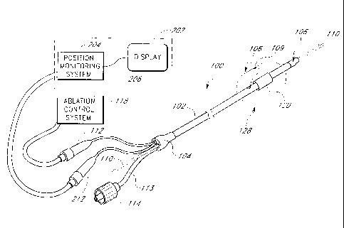
Fig. 2A shows an exemplary ablation catheter assembly 100 operably connected
through an electrical connector
112 to an ablation control system 118. The catheter assembly 100 includes an
elongated delivery member 102 with a
proximal end portion 104 and a distal end portion 106. The distal end portion
106 supports an ablation member 128
including an ablation element 120 and an expandable member 108. The expandable
member can also include a sensor 109
that is explained below.
The delivery member 102 desirably includes a plurality of lumens (some of
which are illustrated in Fig. 2B).
Various wires and electrical leads are routed to the distal end portion 106
through at least some of these lumens. In a
preferred device, these lumens generally run the length of the delivery member
102; however, for some applications, the
lumens can be shorter. In one example, a guidewire 110 runs through a lumen in
the delivery member 102 from the
proximal end portion 104 to the distal end portion 106. The proximal end
portion 104 also connects through a tube 113
to a screw connector 114. By introducing fluid into the tube 113 through the
screw connector 114, a physician can
inflate the expandable member 108, as known in the art.
In some modes of the catheter assembly, as seen in Fig. 2B, the delivery
member 102 includes a distal port
121, which is distal to an ablation member 128. In addition, there is a
proximal port 122, which is provided proximal of
the ablation member 128. The proximal port 122 connects to a proximal port
lumen 123, and the distal port 121
connects to a distal port lumen 124. The distal port 121 allows the clinician
to introduce fluids into the patient, take fluid
samples from the patient, and take fluid pressure reading on the distal side
of the ablation member 128. Similarly, the
proximal port 122 allows the clinician to introduce fluids into the patient,
take fluid samples from the patient, and take
fluid pressure reading on the proximal side of the ablation member 128. These
ports 121, 122 and lumens 123 and 124
are particularly useful when pressure or X-ray positioning techniques are
employed, as explained below; however, the
catheter assembly 100 need not include such ports and lumens when only an A-
mode or Doppler position monitoring
system is used with the catheter assembly.
-19-
CA 02364042 2001-08-31
WO 00/51683 PCT/US00/05468
In the illustrated device, the delivery member 102 also includes a guidewire
lumen 125 that is sized to track over
the guidewire 110. The lumen 125 terminates at a distal port 127 located on
the distal end 106 of the delivery member
102.
When constructed for use in transeptal left atrial ablation procedures, the
delivery member 102 desirably has
an outer diameter provide within the range of from about 5 French to about 10
French, and more preferably from about
7 French to about 9 French. The guidewire lumen 125 preferably is adapted to
slideably receive guidewires ranging
from about 0.010 inch to about 0.038 inch in diameter, and preferably is
adapted for use with guidewires ranging from
about 0.018 inch to about 0.035 inch in diameter. Where a 0.035 inch guidewire
is to be used, the guidewire lumen
125 preferably has an inner diameter of 0.040 inch to about 0.042 inch. In
addition, where the delivery member 102
includes an inflation lumen 130 for use with an inflatable balloon (a
preferred form of the expandable member 108),
the inflation lumen 130 preferably has an inner diameter of about 0.020 inch
in order to allow for rapid deflation
times, although this may vary based upon the viscosity of inflation medium
used, length of the lumen 130, and other
dynamic factors relating to fluid flow and pressure.
In addition to providing the requisite lumens and support for the ablation
member 128, the delivery member
102 for the illustrative application also is adapted to be introduced into the
left atrium such that the distal end portion
106 can be placed within the pulmonary vein ostium in a percutaneous
translumenal procedure, and even more
preferably in a transeptal procedure as otherwise herein provided. Therefore,
the distal end portion 106 is preferably
flexible and adapted to track over and along a guidewire seated within the
targeted pulmonary vein.
In a further construction, the proximal end portion 104 is adapted to be at
least 30% more stiff than the
distal end portion 106. According to this relationship, the proximal end
portion 104 may be suitably adapted to
provide push transmission to the distal end portion 106 while the distal end
portion 106 is suitably adapted to track
through bending anatomy during in vivo delivery of the distal end portion 106
of the device into the desired ablation
region.
Notwithstanding the specific device constructions just described, other
delivery mechanisms for delivering
the ablation member 128 to the desired ablation region are also contemplated.
For example, while the Fig. 2A
variation is shown as an "over-the-wire" catheter construction, other
guidewire tracking designs are suitable
substitutes, such as, for example, catheter devices which are known as "rapid
exchange" or "monorail" variations,
wherein the guidewire is only housed coaxially within a lumen of the catheter
in the distal region of the catheter. In
another example, a deflectable tip design may also be a suitable substitute to
independently select a desired pulmonary
vein and direct the transducer assembly into the desired location for
ablation. Further to this latter variation, the
guidewire lumen and guidewire of the variation depicted in Fig. 2A may be
replaced with a "pullwire" lumen and
associated fixed pullwire which is adapted to deflect the catheter tip by
applying tension along varied stiffness
transitions along the catheter's length. Still further to this pullwire
variation, acceptable pullwires may have a
-20-
CA 02364042 2001-08-31
WO 00/51683 PCT/US00/05468
diameter within the range from about 0.008 inch to about 0.020 inch, and may
further include a taper, such as, for
example, a tapered outer diameter from about 0.020 inch to about 0.008 inch.
As discussed above, the distal end portion 106 of the delivery member supports
an ablation member 128. The
ablation member 128 includes an expandable member 108 and an ablation element
120. The expandable member 108
cooperates with the ablation element 120 to position and anchor the ablation
element 120 relative to a circumferential
region of tissue at a location where a pulmonary vein extends from the left
atrium, which is targeted for ablation.
In the illustrated device, the expandable member 108 is an inflatable balloon.
The balloon has a diameter in a
collapsed state roughly the same as the outer diameter of the delivery member
distal end portion 106. The balloon 108
can be expanded to a diameter generally matching the diameter of the
circumferential region of tissue, and may be
expandable to a plurality of expanded positions in order to work with
pulmonary vein ostia and/or pulmonary veins of
various sizes. It is understood, however, that the ablation catheter assembly
can also include other types of expandable
members, such as, for example baskets, cages and like expandable structures.
The expandable balloon 108 may be constructed from a variety of known
materials, although the balloon
preferably is adapted to conform to the contour of a pulmonary vein ostium
and/or pulmonary vein lumenal wall. For this
purpose, the balloon material can be of the highly compliant variety, such
that the material elongates upon application of
pressure and takes on the shape of the body lumen or space when fully
inflated. Suitable balloon materials include
elastomers, such as, for example, but without limitation, silicone, latex, or
low durometer polyurethane (for example a
durometer of about 80A).
In addition, or in the alternative to constructing the balloon of highly
compliant material, the balloon can be
formed to have a predefined fully inflated shape (i.e., be preshaped) to
generally match the anatomic shape of the body
lumen in which the balloon is inflated. For instance, as described below in
greater detail, the balloon can have a distally
tapering shape to generally match the shape of a pulmonary vein ostium, and/or
can include a bulbous proximal end to
generally match a transition region of the atrium posterior wall adjacent to
the pulmonary vein ostium. In this manner, the
desired seating within the irregular geometry of a pulmonary vein or vein
ostium can be achieved with both compliant and
non-compliant balloon variations.
Notwithstanding the alternatives which may be acceptable as just described,
the balloon is preferably
constructed to exhibit at least 300% expansion at 3 atmospheres of pressure,
and more preferably to exhibit at least
400% expansion at that pressure. The term "expansion" is herein intended to
mean the balloon outer diameter after
pressurization divided by the balloon inner diameter before pressurization,
wherein the balloon inner diameter before
pressurization is taken after the balloon is substantially filled with fluid
in a taut configuration. In other words,
"expansion" is herein intended to relate to the change in diameter that is
attributable to the material compliance in a
stress/strain relationship. In one more detailed construction, which is
believed to be suitable for use in most conduction
block procedures in the region of the pulmonary veins, the balloon is adapted
to expand under a normal range of
-21-
CA 02364042 2001-08-31
WO 00/51683 PCT/US00/05468
pressure such that its outer diameter may be adjusted from a radially
collapsed position of about 5 millimeters to a
radially expanded position of about 2.5 centimeters (or approximately 500%
expansion).
The ablation element 120 cooperates with the expandable member 108 such that
the ablation element 120 is
held in a generally fixed position relative to the target circumferential
region of tissue. The ablation element can be located
outside or inside the expandable member, or can be located at least partially
outside the expandable member. The ablation
element, in some forms, also includes a portion of the expandable member. For
instance, the ablation catheter assembly in
Figs. 2A-B includes an ultrasonic transducer located within the expandable
member 108. In one device, the ultrasonic
transducer excites a portion of the expandable member 108 during ablation. The
specific construction of the ultrasonic
transducer and the associated construction of the delivery member shaft that
supports the transducer, is described below
in connection with Figs. 10A-0.
As noted above, the ablation element can also take many other forms. For
instance, the ablation element can
include one or more electrodes exposed on the exterior of the expandable
member and adapted to contact the targeted
tissue. Figs. 16A, D, H and 17A illustrate devices of this type of ablation
element, which are described below. The
electrode(s) can also be positioned within the expandable member with an
electrical path established between the
electrode and the tissue by an electrolytic solution (e.g., saline), as
discussed in more detail in connection with Fig.
17B below. In either of these modes, as illustrated in Fig. 2A, the ablation
element 120 is typically connected to the
electrical connector 112 and to a ground patch (not shown). A circuit thereby
is created which includes the ablation
element 120, the patient's body, and the ground patch that provides either
earth ground or floating ground to the
ablation control 118. In the circuit, an electrical current, such as a Radio
Frequency (RF) signal may be sent through
the patient between the ablation element 120 and the ground patch.
Fig. 2B shows details of the distal end portion 106 of the catheter assembly
100 and, in particular, shows
the ablation element 120 located circumferentially about an axial centerline
of the delivery member 102. A pair of
wires 129 connect the ablation element 120 to a connector 112 at the proximal
end of the catheter (shown in Fig.
2A) The connector 112 is coupled to a corresponding cable of the ablation
control system 118. If the ablation
element 120 includes more than one electrode, the conductor lead can connect
to all of the electrodes or energy
sources, or separate conductors can be used so as to allow for independent
control of each electrode or energy source
under some modes of operation.
The tissue ablation catheter 100 assembly also desirably includes feedback
control. For instance, the
expandable member 108 can include one or more thermal sensors 109 (e.g.,
thermocouples, thermistors, etc.) that are
provided to either the outer side or the inside of the expandable member 108.
Monitoring temperature at this location
provides indicia for the progression of the lesion. If the temperature sensors
are located inside the expandable member
108, the feedback control may also need to account for any temperature
gradient that occurs through wall of the
expandable member 108.
-22-
CA 02364042 2001-08-31
WO 00/51683 PCT/US00/05468
If the sensors 109 are placed on the exterior of the expandable member 108,
they may also be used to record
electrogram signals by reconnecting the signal leads to different input port
of a signal-processing unit. Such signals can be
useful in mapping the target tissue both before and after ablation.
The thermocouples and/or electrodes desirably are blended into the expandable
member 108 in order to present a
smooth profile. Transition regions, which are formed by either adhesive or
melted polymer tubing, "smooth out" the
surface of the expandable member 108 as the surface steps up from the outer
surface of the expandable member 108 to
the thermocouple surface. Various constructions to integrate the thermocouples
and/or electrodes into the expandable
member, as well as various approaches to using thermocouples and electrodes
with an expandable member, are described
in detail below.
The illustrated ablation catheter assembly 100 is designed for treatment of
the more common forms of atrial
fibrillation, resulting from perpetually wandering reentrant wavelets. Such
arrhythmias are generally not amenable to
localized ablation techniques, because the excitation waves may circumnavigate
a focal lesion. Thus, the catheter
assembly 100 uses the ablation element 120 to form a substantially
circumferential lesion, or lesions, to segment the
atrial tissue so as to block conduction of the reentrant wave fronts.
During a surgical procedure, a clinician guides the ablation catheter assembly
100 into the left atrium. The
clinician then manipulates the catheter so that the ablation member enters the
pulmonary vein from the left atrium. The
goal of the surgical procedure is to position the ablation member just inside
the pulmonary vein, at the pulmonary vein
ostium. Once the expandable member 108 is positioned at desired site within
the ostium and relative to the targeted region
of circumferential tissue, the ablation element 120 is activated to ablate the
targeted tissue and thereby form the desired
lesion.
Access to the atrium is gained using techniques known in the art. After access
to the atrium is obtained,
another guidewire or guide member is advanced into the pulmonary vein. This is
typically done through a guiding
introducer which is coaxial within a transeptal sheath seated in the fossa
ovalis, or by using a deflectable guidewire or
catheter such as those disclosed in U.S. Patent No. 5,575,766 to Swartz.
Alternatively, the guidewire should have
sufficient stiffness and maneuverability in the left atrial cavity to
unitarily select the desired pulmonary vein distally of the
transeptal sheath seated at the fossa ovalis. The guidewire is advanced into
the pulmonary vein ostium to a suitable
anchoring position.
The ablation catheter 100 is then slid over the proximal end of the guidewire
110 and advanced until the
ablation member of the ablation catheter 100, including the ablation element
120, is positioned at the area where the
pulmonary vein extends from the left atrium. A combination of pushing and
pulling alternatively on the guidewire 110
and the ablation catheter 100 may be employed to facilitate advancement and
positioning of the ablation catheter
100.
Delivery of energy (e.g., thermal, RF, ultrasonic, electrical, etc.) to the
tissue of the pulmonary vein ostium is
commenced once the ablation element 120 is positioned at the desired ablation
region. Good coupling of the energy
-23-
CA 02364042 2001-08-31
WO 00/51683 PCT/US00/05468
produced by the ablation element 120 with the tissue facilitates creation of a
continuous lesion. Energy from the ablation
control system 118 (Fig. 2A) is typically delivered to the ablation element
120 via electrical conductor leads. The
ablation control system 118 includes a current source for supplying current to
the ablation element 120, a monitoring
circuit, and a control circuit. The current source is coupled to the ablation
element 120 via a lead set (and to a ground
patch in some modes). The monitor circuit desirably communicates with one or
more sensors (e.g., temperature and/or
current sensors) which monitor the operation of the ablation element 120. The
control circuit is connected to the
monitoring circuit and to the current source in order to adjust the output
level of the current driving the ablation
element 120 based upon the sensed condition (e.g., upon the relationship
between the monitored temperature and a
predetermined temperature set point).
Position Monitoring System
Fig. 2A shows an ablation catheter with position monitoring capability
operably connected to an ablation control
system 118 and a position monitoring system 202. The position monitoring
system 202 includes a sensor control system
204 and a display 206. The sensor control system 204 communicates with one or
more sensor elements 220 located in,
or near the expandable member 108. In one variation, the ablation element 120
and sensor element 220 are combined in a
single element that provides both sensing and ablation capabilities. In other
variations, separate elements are used for the
ablation element 120 and the sensor element(s) 220. Various exemplifying
device embodiments of the position sensor
220 are described in connection with Figs. 3-14.
Amplitude Monitoring
Fig. 3 illustrates the basic operation of an ultrasonic position monitoring
system. In Fig. 3, the sensor 320 is
embodied as a single, circumferentially symmetric ultrasonic transducer 320.
The sensor 320 can be the ultrasonic
ablation element, as illustrated in Figs. 2A and 3, or a separate ultrasonic
transducer in addition to an ultrasonic ablation
element, as illustrated in Figs. 5A-E, which are described below. The
transducer 320 is shown positioned in a pulmonary
vein 322, and the transducer 320 is operably connected to a sensor control
system 304. In an exemplary device, the
sensor control system is a Panametrics Model 5073PR. The sensor control system
304 includes a transmitter 305, a
receiver 306, and a diplexer 307. An output from the transmitter 305 is
provided to a transmitter port (port 1) of the
diplexer 307. An output from a receiver port (port 3) of the diplexer 307 is
provided to an input of the receiver 306. A
transducer port (port 2) of the diplexer 307 is provided through a connector
308 to the transducer 320. An output from
the receiver 306 is provided to the display 202.
A diplexer, such as the diplexer 307, is commonly used in radar and sonar
systems to isolate the transmitter
output from the receiver input. Energy provided to the transmitter port of the
diplexer (port 1) is provided to the transducer
port (port 2) of the diplexer 307, but not to the receiver port of the
diplexer (port 3). Energy provided from the transducer
320 to the transducer port of the diplexer (port 2) is provided to the
receiver port (port 3) of the diplexer 307, but not to
the transmitter port (port 3) of the diplexer.
-24-
CA 02364042 2001-08-31
WO 00/51683 PCT/US00/05468
The diplexer 307 can be a circulator or an electronically controlled switch
controlled by a timing generator. The
timing generator sets the switch to connect the transmitter 305 to the
transducer 320 for a first time period. The timing
generator then sets the switch to connect the receiver to the transducer 320
for a second time period. By switching the
transducer 320 between the transmitter 305 and the receiver 306, the diplexer
307 effectively "timeshares" the
transducer 320 between the transmitter 305 and the receiver 306.
The transmitter 305 generates a signal that drives the transducer 320. When
the diplexer 307 connects the
transmitter 305 to the transducer 320, the drive signal from the transmitter
305 causes the transducer 320 to emit an
ultrasonic sound wave. The ultrasonic sound wave propagates through the
interior of the expandable member 108,
through the wall of the expandable member 108, and reflects off of the inner
wall of the ostium 322. The reflected
ultrasonic energy returns to the transducer 320 and causes the transducer 320
to generate an echo signal. The echo
signal is provided through the diplexer 307 to the receiver 306. The receiver
306 amplifies and processes the echo signal
to produce a display signal. The display signal is provided to the display
202.
Fig. 4A is an axial cross section showing the transducer 320 centered in the
ostium 322. The transducer
transmits a radiated wave 406. For a cylindrically symmetric transducer 320,
the radiated wave 406 will approximate a
cylindrical wave that expands away from the transducer 320. When the
cylindrical wave reaches the ostium 322, the
wave will be reflected in a substantially cylindrically symmetric fashion to
produce a reflected wave 408 that is similar to
a cylindrical wave as well. The reflected wave 408 propagates back to the
transducer 320.
Reflections will occur when the ultrasonic sound wave propagating in a medium
strikes a transition (or interface)
in the acoustic properties of the medium. Any interface between materials
having different acoustic properties will cause
a portion of the wave to be reflected.
Fig. 4B shows a downrange, time-domain, amplitude mode (A-mode) plot of the
response produced on the
display 202 by the system shown in Fig. 3. The x-axis of the plot shown in
Fig. 4B is a time axis where t = 0
corresponds to the time when the diplexer 307 connects the transducer 320 to
the receiver 306. During a time period just
before t = 0, the transducer 320 is connected to the transmitter 305, and the
transmitter produces a transmit pulse 419.
The y-axis in Fig. 4B is an amplitude plot of the energy produced by
ultrasonic vibrations of the transducer 320. The plot
in Fig. 48 shows a ring-down signal 420 during a ring-down period 422 (a time
period 0 < t < tr). The plot also shows
an echo pulse 424 at a time tr.
The transmit pulse 419 causes the transducer 320 to vibrate (in a manner very
similar to a bell) during the ring-
down period 422 thereby producing the ring-down signal 420. The echo pulse 424
is caused by ultrasonic energy that is
reflected from the ostium 322 back to the transducer 320. During the ring-down
period 422 it is difficult to see signals
caused by reflections (such as the signal 424) because the signals produced by
reflections are typically relatively small in
amplitude and are easily masked by the relatively large amplitude portions of
the ring-down signal 420. Thus, it is difficult
to detect reflections from targets that are so close to the transducer 320
that their reflections return during the ring-down
period 422. This can limit the minimum useful range of the transducer 320.
-25-
CA 02364042 2001-08-31
WO 00/51683 PCT/US00/05468
As shown in Fig. 4C, the ring-down time of the transducer 320 can be reduced
by configuring the transmitter
305 to provide a shaped transmit pulse 449. The shaped transmit pulse drives
the transducer 320 in a manner that
reduces the amplitude of the ringing and shortens the ring-down period. Thus,
Fig. 4C shows a ring-down time 448 that is
less than the ring-down time 422 shown in Fig. 4B. Fig. 4C also shows a ring-
down signal 440 having an amplitude that
is relatively smaller than the amplitude of the ring-down signal 420 in Fig.
4B. Since the ring-down period 448 is shorter,
the shaped transmit pulse 449 allows the transducer 320 to be used to detect
targets at a shorter distance.
In an device where the transducer 320 is also used as the ablation element
120, the transmitter 305 provides
two power modes, a low-power mode used for position measurements, and a high-
power mode used for ablation. When
ablation is desired, the diplexer 307 stops switching between the receiver 306
and the transmitter 305, and stays locked
on the transmitter 305 while the transmitter operates in the high-power mode.
Ultrasonic ablation requires that the transducer 320 produce an ultrasonic
wave having relatively higher power.
Higher power typically requires a transducer 320 having a relatively large
physical size. Larger transducers often have
longer ring-down times. While the use of a shaped transmit pulse will reduce
ring-down times, for some transducers even
the use of a shaped transmit pulse does not shorten the ring-down time
sufficiently to allow the ablation element 120 to
be used for position sensing. Moreover, in some devices, the ablation element
120 is not an ultrasonic transducer, and
thus may be unsuitable for use as a position sensor. Thus, in some devices, it
is desirable to add one or more ultrasonic
transducers to be used for position sensing.
Fig. 5A shows a distal end of a catheter-based ultrasonic position monitoring
system that uses an ultrasonic
sensing element 516 proximal to the ablation element 120. (In Figs. 5A-C the
expandable member 108 is omitted for
clarity). The sensing element 516 is used for position sensing, not ablation,
and thus does not need to handle the higher
powers needed for an ablation element. This allows the characteristics of the
sensing element 516 to be tailored to
attributes needed for position sensing, such attributes typically include,
small size, low power, short ring-down times, etc.
When used in connection with the sensor control system 304 shown in Fig. 3,
the sensing element 516, rather than the
ablation transducer 120, is connected to the diplexer 307. In other respects,
the sensing element operates in a manner
similar that discussed above in connection with Figs. 4A-C.
Fig. 5B shows an ultrasonic position sensing system that uses an ultrasonic
sensing element 518 distal to the
ablation element 120. In other respects, the system shown in Fig. 5B is
similar to the system shown in Fig. 5A.
Fig. 5C shows an ultrasonic position sensing system that uses both the
proximal sensor 516 and the distal
sensor 518. The two sensors 516 and 516 are each driven by the sensor control
system 304 either in parallel (using
separate channels in the system 304) or in serial (first one than the other).
Figs. 5D-E show yet other devices of an ultrasonic sensor system wherein a
single position sensing transducer
524 is provided between a proximal ablation element 522 and a distal ablation
element 520. In Fig. 5D, both ablation
elements 520 and 522 are enclosed by a single expandable member (e.g., a
balloon) 512. In Fig. 5E an expandable
member 523 surrounds the ablation element 522 and an expandable member 525
surrounds the ablation element 520. In
-26-
CA 02364042 2001-08-31
WO 00/51683 PCT/US00/05468
Fig. 5E, the ablation elements 522 and 520 are optional, and ablation energy
may be provided by, for example, an ablation
solution introduced in the space 530 between the expandable members 523 and
525. Ablation solutions include solutions
that cause ablation, such as, for example, solutions that cause cooling,
solutions that cause heating, or solutions that
cause chemical reactions on the tissue wall in the region 530.
Fig. 5F illustrates another device of an ultrasonic sensor system. A single
positioning sensing transducer 518 is
positioned next to an ablation element 120. In the illustrated device, the
transducer is located on the distal side of the
ablation element 120; however, it can be located on the proximal side as well.
Both the sensing transducer and the
ablation element are located between a pair of expandable members 526,528. In
one variation, the expandable members
are a pair of inflatable balloons that may have differing diameters such that
the distal-most balloon seals off blood flow,
while the proximal balloon permits some flow of fluid from the space 530
between the balloons. In the illustrated
variation, the ablation member comprises at least one electrode 120 and a
fluid port 540 communicates with the space
530 between the balloons 526,528. An electrolytic solution (e.g., saline) is
introduced into the space between the
balloons. This solution electrically couples the electrode to the
circumferential region of tissue located between the
inflated pair of balloons. The sensing transducer 518 aids in the proper
positioning of this assembly within the pulmonary
vein ostium, in the manner described above.
In another variation, as seen in Fig. 513, the ultrasonic sensor system
includes a pair of positioning sensing
transducers 518. Each transducer 518 is positioned in one of the expandable
members 526,528 (e.g., inflatable balloons)
with the ablation element 120 disposed between the expandable members 526,528.
The sensor systems shown in Fig. 3, and Figs. 5A-F are all used in a similar
fashion to measure the position of
the ablation element relative to the ostium. Figs. 6A-C illustrate the use of
the A-mode ultrasonic systems to determine
position.
Fig. 6A shows an A-mode response of the ultrasonic sensor systems when an
ultrasonic transducer, such as the
ultrasonic transducer 320, is in the atrium and not in the vein. The A-mode
response in Fig. 6A shows a ring-down signal
602 and little else except a few indefinite echo returns 604. The response
show no definite echo signals because the
walls of the atrium are too far away from the transducer and/or are poorly
oriented to provide a strong echo response back
to the transducer.
Fig. 6B shows an A-mode response of the ultrasonic sensor systems when an
ultrasonic transducer, such as the
ultrasonic transducer 320, is partially in the pulmonary vein (e.g., half way
into the vein). The A-mode response in Fig. 6B
shows the ring-down signal 602 and relatively weak echo return 606. Moreover,
the echo return 606 is relatively spread-
out in time. The signal is weak because only a portion of the transducer 302
is in the vein and only the portion of the
transducer 302 in the vein receives a strong echo. The portion of the
transducer 302 outside the vein (in the atrium) does
not see a strong echo response. The echo return 606 is relatively spread-out
in time because the diameter of the vein
varies significantly near the opening into the atrial chamber.
-27-
CA 02364042 2001-08-31
WO 00/51683 PCT/US00/05468
Fig. 6C shows an A-mode response of the ultrasonic sensor systems when an
ultrasonic transducer, such as the
ultrasonic transducer 320, is fully inserted in the pulmonary vein. The A-mode
response in Fig. 6C shows the ring-down
signal 602 and relatively strong, short, echo return 608. The signal is
relatively stronger than the signal 606 because all
of the transducer 302 is inside the vein and thus all of the transducer 302 is
receiving a return echo. The signal 608 also
is relatively short because all portions of the echo return at almost the same
time (assuming the transducer 320 is near the
center of the vein).
To position the transducer 320 (and thus the ablation element 120), the
clinician (e.g., an interventional electro-
physiologist) inserts the ablation catheter while watching the display 202 for
the progression of echo signals 604, 606
and 608. The clinician stops advancing the delivery member once when he or she
sees a signal profile similar to the signal
608 depicted in Fig. 6C.
Fig. 60 shows a two-trace display produced by the two-transducer system shown
in Fig. 5C having a proximal
sensor 516 and a distal sensor 518. The two-transducer system can provide more
precise positioning of the ablation
element 120 because, as shown in Fig. 60, the proximal sensor 516 produces a
first A-mode signal having a ring-down
pulse 602 and an echo pulse 612; and the distal sensor 518 produces a second A-
mode signal having a ring-down pulse
602 and an echo pulse 610.
When inserting a two-transducer system into the ostium, the clinician will
first observe the echo pulse 610 rise
up and move towards the ring-down pulse 602 (as the distal sensor moves into
the vein). The clinician will then see the
echo pulse 612 rise up and move towards the ring-down pulse 602 as the
clinician advances the proximal transducer 516
into the pulmonary vein ostium.
As with the single sensor system, the distance between the ring-down pulse and
the echo pulse is a measure of
the distance to the target (e.g., the pulmonary wall, vein wall, etc.) that
produced the echo pulse. The unquantified signal
profile can be used by the clinician to position the ablation member 120
relative to a pulmonary vein ostium of a pulmonary
vein in order to ablate a circumferential region of tissue at a location where
the pulmonary vein extends from the left
atrium; however, this signal can also be quantified to provide information
regarding the anatomical size and shape of the
particular patient's vein ostium. The clinician can use such information to
determine whether the size of the expandable
member is sufficient to engage the wall of the vein and/or vein ostium.
The shape (width and height) of the echo pulses 608, 610 and 612 can also be
used to center the transducer
320 within the vein ostium. The echo pulses 608, 610 and 612 will be affected
by the location of the ultrasonic
transducer with respect to the axial centerline of the vein. As shown in Fig.
7A, when a cylindrical transducer, such as
the transducer 320, is located near the center of the vein, the distance from
the transducer to the wall of the vein is
essentially the same in all radial directions. Thus, when the transducer 320
is centered, the A-mode display will show the
ring-down pulse 602 and an echo pulse 702 that is "sharp", having a relatively
large amplitude and relatively short
duration. By contrast, when the transducer 320 is off-center, as shown in Fig.
7B, the distance from the transducer 320
to the wall of the vein will not be uniform. Thus, when the transducer 320 is
off-center, the A-mode display will show the
-28-
CA 02364042 2001-08-31
WO 00/51683 PCT/US00/05468
ring-down pulse 602 and an echo pulse 704 that is "smeared-out", having a
relatively smaller amplitude and relatively
longer duration.
The variation of the echo return times for an off-center transducer can
advantageously be used to measure the
position of the ablation element 120 with respect to the center of the vein.
An array having a plurality of sensors,
preferably three or more, can advantageously be used to measure the position
of the ablation element 120 with respect to
center. For example, Figs. 8A-B show a portion of a catheter assembly with an
array of four ultrasonic sensors 810-813
disposed in a circumferential pattern around the catheter 310. Each of the
ultrasonic sensors is 810-813 used to produce
an A-mode plot.
Fig. 9A shows downrange time-domain responses of the ultrasonic sensors 810-
813 when the catheter 310 is
centered in a vein. The ultrasonic sensors 810-813 produce A-mode plots 902-
905 respectively. Each of the plots 902-
905 has a ring-down It = 0) pulse 912-915, respectively, and an echo pulse 922-
925, respectively. For similar
transducers, the ring-down pulses 912-915 are similar. When the catheter 310
is centered in the vein the echo pulses
922-925 will also be similar and occur at similar downrange distances (times).
Fig. 9B shows downrange time-domain responses of the ultrasonic sensors 810-
813 when the catheter 310 is
off-centered in a vein. In Fig. 9B, the catheter 310 is above center, and thus
the top transducer 810 is closest to the
wall, the left and right transducers 811, 813 are equidistant from the wall,
and the bottom transducer 812 is furthest
from the wall. Fig. 9B shows A-mode plots 942-945, each having a ring-down
pulse 952-955, respectively, and an echo
pulse 962-965, respectively. For similar transducers, the ring-down pulses 952-
955 are similar. Of the echo pulses 962-
965, the echo pulse 962 is closest to the ring-down pulses and strongest. The
echo pulses 963, 965 are approximately
the same distance from the ring-down pulse. The echo pulse 964 is the furthest
from the ring-down pulses and the
weakest. A clinician seeing the plots 942-945 would know that the catheter 310
is above center in the vein and thus be
able to manipulate the catheter and guidewire (e.g., by torquing or pulling
the guidewire taught) to move the ablation
member closer to center of the vein. lithe ablation member is disposed on a
deflectable or steerable delivery platform, the
distal end of the delivery member can be moved to reposition the ablation
member. Alternatively, a graphical
representation, as is shown in Fig. 13 below, can be used to show the location
of the array with respect to the center of
the vein. This information can then be used to supply different power levels
to a corresponding array of ablation devices
(e.g., ultrasonic transducer segments as described below) that form at least
part of the ablation element.
The array of transducers 810-813 can be constructed by attaching separate
transducers to the catheter 310.
Alternatively, the array of transducers 810-813 can be constructed by
modifying a single transducer to produce a multi-
mode transducer.
Fig. 10A is a cross-section drawing showing construction of a single
cylindrical ultrasonic transducer having a
cylindrical inner electrode 1002, a cylindrical outer electrode 1004, and a
cylindrical piezoelectric material 1003 between
the electrodes. The piezoelectric material 1003 is a suitable material, such
as, for example quartz, PZT, and the like, that
exhibits a change in physical dimension in response to an impressed voltage.
The piezoelectric material 1003 is oriented
-29-
CA 02364042 2001-08-31
WO 00/51683 PCT/US00/05468
such that when a voltage is impressed between the electrodes 1002 and 1004,
the thickness of the piezoelectric material
1003 changes slightly. Then the polarity of the impressed voltage is
alternated at a ultrasonic frequency F the
piezoelectric material 1003 will vibrate at the ultrasonic frequency F. The
vibrations of the piezoelectric material 1003
produce ultrasonic sound waves. Since the electrodes are cylindrically
symmetric, the piezoelectric material 1003 will
vibrate radially, with cylindrical symmetry. Conversely, when an ultrasonic
wave hits the piezoelectric material 1003, the
ultrasonic wave will cause vibrations in the piezoelectric material. These
vibrations will generate a voltage between the
electrodes 1002 and 1004. Thus, the transducer is a reciprocal device that can
both transmit and receive ultrasonic
waves.
Figs. 10B-0 variously show circumferential ablation device assemblies
incorporating ultrasound transducers for
ablating a circumferential region of tissue in order to form the desired
conduction block to treat left atrial arrhythmia
according to the present invention. Such ultrasound ablation assemblies are
believed to be particularly amenable to use
with the position monitoring assemblies incorporating sensing capabilities of
the ablation transducer itself, such as for
example but not limited to an "A"-mode sensing system. However, it is further
contemplated that the particular ablation
devices of Figs. 10B-0 may also be combined with the other position monitoring
assemblies and related sensors also
herein shown and described. Furthermore, such ultrasound ablation assemblies
may also be combined with the various
ablation monitoring assemblies, such as temperature monitoring assemblies and
sensors, also elsewhere described in this
disclosure.
As common to each of the following devices, a source of acoustic energy is
provided a delivery device that
also includes an anchoring mechanism. In one mode, the anchoring device
comprises an expandable member that also
positions the acoustic energy source within the body; however, other anchoring
and positioning devices may also be
used, such as, for example, a basket mechanism. In a more specific form, the
acoustic energy source is located within
the expandable member and the expandable member is adapted to engage a
circumferential path of tissue either about
or along a pulmonary vein in the region of its ostium along a left atrial
wall. The acoustic energy source in turn is
acoustically coupled to the wall of the expandable member and thus to the
circumferential region of tissue engaged by
the expandable member wall by emitting a circumferential and longitudinally
collimated ultrasound signal when
actuated by an acoustic energy driver. The use of acoustic energy, and
particularly ultrasonic energy, offers the
advantage of simultaneously applying a dose of energy sufficient to ablate a
relatively large surf ace area within or
near the heart to a desired heating depth without exposing the heart to a
large amount of current. For example, a
collimated ultrasonic transducer can form a lesion, which has about a 1.5 mm
width, about a 2.5 mm diameter lumen,
such as a pulmonary vein and of a sufficient depth to form an effective
conductive block. It is believed that an effective
conductive block can be formed by producing a lesion within the tissue that is
transmural or substantially transmural.
Depending upon the patient as well as the location within the pulmonary vein
ostium, the lesion may have a depth of 1
millimeter to 10 millimeters. It has been observed that the collimated
ultrasonic transducer can be powered to provide a
-30-
CA 02364042 2001-08-31
WO 00/51683 PCT/US00/05468
lesion having these parameters so as to form an effective conductive block
between the pulmonary vein and the posterior
wall of the left atrium.
With specific reference now to the device illustrated in Figs. 10B-E, a
circumferential ablation device
assembly 1001' includes an elongate body 1002' with proximal and distal end
portions 1010, 1012, an expandable
balloon 1020 located along the distal end portion 1012 of elongate body 1002',
and a circumferential ultrasound
transducer 1030 which forms a circumferential ablation member which is
acoustically coupled to the expandable
balloon 1020. In more detail, Figs. 10B-D variously show elongate body 1002'
to include guidewire lumen 1004',
inflation lumen 1006, and electrical lead lumen 1008. The ablation device,
however, can be of a self steering type
rather than an over-the-wire type device.
Each lumen extends between a proximal port (not shown) and a respective distal
port, which distal ports are
shown as distal guidewire port 1005 for guidewire lumen 1004', distal
inflation port 1007 for inflation lumen 1006,
and distal lead port 1009 for electrical lead lumen 1008. Although the
guidewire, inflation and electrical lead lumens
are generally arranged in a side-by-side relationship, the elongate body 1002'
can be constructed with one or more of
these lumens arranged in a coaxial relationship, or in any of a wide variety
of configurations that will be readily
apparent to one of ordinary skill in the art.
In addition, the elongate body 1002' is also shown in Figs. 10B and 10D to
include an inner member 1003'
which extends distally beyond distal inflation and lead ports 1007, 1009,
through an interior chamber formed by the
expandable balloon 1020, and distally beyond expandable balloon 1020 where the
elongate body terminates in a distal
tip. The inner member 1003' forms the distal region for the guidewire lumen
1004' beyond the inflation and lead
ports, and also provides a support member for the cylindrical ultrasound
transducer 1030 and for the distal neck of the
expansion balloon, as described in more detail below.
One more detailed construction for the components of the elongate body 1002'
which is believed to be
suitable for use in transeptal left atrial ablation procedures is as follows.
The elongate body 1002' itself may have an
outer diameter provided within the range of from about 5 French to about 10
French, and more preferable from about 7
French to about 9 French. The guidewire lumen preferably is adapted to
slideably receive guidewires ranging from
about 0.010 inch to about 0.038 inch in diameter, and preferably is adapted
for use with guidewires ranging from
about 0.018 inch to about 0.035 inch in diameter. Where a 0.035 inch guidewire
is to be used, the guidewire lumen
preferably has an inner diameter of 0.040 inch to about 0.042 inch. In
addition, the inflation lumen preferably has an
inner diameter of about 0.020 inch in order to allow for rapid deflation
times, although may vary based upon the
viscosity of inflation medium used, length of the lumen, and other dynamic
factors relating to fluid flow and pressure.
In addition to providing the requisite lumens and support members for the
ultrasound transducer assembly,
the elongate body 1002' of the present device must also be adapted to be
introduced into the left atrium such that the
distal end portion with balloon and transducer may be placed within the
pulmonary vein ostium in a percutaneous
translumenal procedure, and even more preferably in a transeptal procedure as
otherwise herein provided. Therefore,
-31-
CA 02364042 2001-08-31
WO 00/51683 PCT/US00/05468
the distal end portion 1012 is preferably flexible and adapted to track over
and along a guidewire seated within the
targeted pulmonary vein. In one further more detailed construction which is
believed to be suitable, the proximal end
portion is adapted to be at least 30% more stiff than the distal end portion.
According to this relationship, the
proximal end portion may be suitably adapted to provide push transmission to
the distal end portion while the distal
end portion is suitably adapted to track through bending anatomy during in
vivo delivery of the distal end portion of the
device into the desired ablation region.
Notwithstanding the specific device constructions just described, other
delivery mechanisms for delivering
the ultrasound ablation member to the desired ablation region are also
contemplated. For example, while the Fig. 10B
variation is shown as an "over-the-wire" catheter construction, other
guidewire tracking designs may be suitable
substitutes, such as, for example, catheter devices which are known as "rapid
exchange" or "monorail" variations
wherein the guidewire is only housed coaxially within a lumen of the catheter
in the distal regions of the catheter. In
another example, a deflectable tip design may also be a suitable substitute
and which is adapted to independently
select a desired pulmonary vein and direct the transducer assembly into the
desired location for ablation. Further to
this latter variation, the guidewire lumen and guidewire of the Fig. 10B
variation may be replaced with a "pullwire"
lumen and associated fixed pullwire which is adapted to deflect the catheter
tip by applying tension along varied
stiffness transitions along the catheter's length. Still further to this
pullwire variation, acceptable pullwires may have
a diameter within the range from about 0.008 inch to about 0.020 inch, and may
further include a taper, such as, for
example, a tapered outer diameter from about 0.020 inch to about 0.008 inch.
More specifically regarding expandable balloon 1020 as shown in varied detail
between Fig. 10B and 10D, a
central region 1022 is generally coaxially disposed over the inner member
1003' and is bordered at its end neck regions by
proximal and distal adaptions 1024, 1026. The proximal adaption 1024 is sealed
over elongate body 1002' proximally of
the distal inflation and the electrical lead ports 1007, 1009, and the distal
adaption 1026 is sealed over inner member
1003'. According to this arrangement, a fluid tight interior chamber is formed
within expandable balloon 1020. This
interior chamber is fluidly coupled to a pressurizeable fluid source (not
shown) via inflation lumen 1006. In addition to the
inflation lumen 1006, electrical lead lumen 1008 also communicates with the
interior chamber of expandable balloon
1020 so that the ultrasound transducer 1030, which is positioned within that
chamber and over the inner member 1003',
may be electrically coupled to an ultrasound drive source or actuator, as will
be provided in more detail below.
The expandable balloon 1020 may be constructed from a variety of known
materials, although the balloon 1020
preferably is adapted to conform to the contour of a pulmonary vein ostium.
For this purpose, the balloon material can be
of the highly compliant variety or of a predefined shape, as noted above.
The ablation member, which is illustrated in Figs. 10B-E, takes the form of
annular ultrasonic transducer
1030. In the illustrated device, the annular ultrasonic transducer 1030 has a
unitary cylindrical shape with a hollow
interior (i.e., is tubular shaped); however, the transducer applicator 1030
can have a generally annular shape and be
formed of a plurality of segments. For instance, the transducer applicator
1030 can be formed by a plurality of tube
-32-
CA 02364042 2001-08-31
WO 00/51683 PCT/US00/05468
sectors that together form an annular shape. The tube sectors can also be of
sufficient arc lengths so as when joined
together, the sectors assembly forms a "clover-leaf" shape. This shape is
believed to provide overlap in heated regions
between adjacent elements. The generally annular shape can also be formed by a
plurality of planar transducer
segments which are arranged in a polygon shape (e.g., hexagon). In addition,
although in the illustrated device the
ultrasonic transducer comprises a single transducer element, the transducer
applicator can be formed of a multi-
element array, as described in greater detail below.
As is shown in detail in Fig. 10E, cylindrical ultrasound transducer 1030
includes a tubular wall 1031 which
includes three concentric tubular layers. The central layer 1032 is a tubular
shaped member of a piezoceramic or
piezoelectric crystalline material. The transducer preferably is made of type
PZT-4, PZT-5 or PZT-8, quartz or Lithium-
Niobate type piezoceramic material to ensure high power output capabilities.
These types of transducer materials are
commercially available from Stavely Sensors, Inc. of East Hartford,
Connecticut, or from Valpey-Fischer Corp. of
Hopkinton, Massachusetts.
The outer and inner tubular members 1033, 1034 enclose central layer 1032
within their coaxial space and
are constructed of an electrically conductive material. In the illustrated
device, these transducer electrodes 1033,
1034 comprise a metallic coating, and more preferably a coating of nickel,
copper, silver, gold, platinum, or alloys of
these metals.
One more detailed construction for a cylindrical ultrasound transducer for use
in the present application is as
follows. The length of the transducer 1030 or transducer assembly (e.g., multi-
element array of transducer elements)
desirably is selected for a given clinical application. In connection with
forming circumferential condition blocks in
cardiac or pulmonary vein wall tissue, the transducer length can fall within
the range of approximately 2 mm up to
greater than 10 mm, and preferably equals about 5 mm to 10 mm. A transducer
accordingly sized is believed to form a
lesion of a width sufficient to ensure the integrity of the formed conductive
block without undue tissue ablation. For
other applications, however, the length can be significantly longer.
Likewise, the transducer outer diameter desirably is selected to account for
delivery through a particular
access path (e.g., percutaneously and transeptally), for proper placement and
location within a particular body space,
and for achieving a desired ablation effect. In the given application within
or proximate of the pulmonary vein ostium,
the transducer 1030 preferably has an outer diameter within the range of about
1.8 mm to greater than 2.5 mm. It
has been observed that a transducer with an outer diameter of about 2 mm
generates acoustic power levels
approaching 20 Watts per centimeter radiator or greater within myocardial or
vascular tissue, which is believed to be
sufficient for ablation of tissue engaged by the outer balloon for up to about
2 cm outer diameter of the balloon. For
applications in other body spaces, the transducer applicator 1030 may have an
outer diameter within the range of
about 1 mm to greater than 3-4 mm (e.g., as large as 1 to 2 cm for
applications in some body spaces).
The central layer 1032 of the transducer 1030 has a thickness selected to
produce a desired operating
frequency. The operating frequency will vary of course depending upon clinical
needs, such as the tolerable outer
-33-
CA 02364042 2001-08-31
WO 00/51683 PCT/US00/05468
diameter of the ablation and the depth of heating, as well as upon the size of
the transducer as limited by the delivery
path and the size of the target site. As described in greater detail below,
the transducer 1030 in the illustrated
application preferably operates within the range of about 5 MHz to about 20
MHz, and more preferably within the
range of about 7 MHz to about 10 MHz. Thus, for example, the transducer can
have a thickness of approximately 0.3
mm for an operating frequency of about 7 MHz (i.e., a thickness generally
equal to Y2 the wavelength associated with
the desired operating frequency).
The transducer 1030 is vibrated across the wall thickness and to radiate
collimated acoustic energy in the
radial direction. For this purpose, as best seen in Figs. 10B and 10E, the
distal ends of electrical leads 1036, 1037
are electrically coupled to outer and inner tubular members or electrodes
1033, 1034, respectively, of the transducer
1030, such as, for example, by soldering the leads to the metallic coatings or
by resistance welding. In the illustrated
device, the electrical leads are 4-8 mil (0.004 to 0.008 inch diameter) silver
wire or the like.
The proximal ends of these leads are adapted to couple to an ultrasonic driver
or actuator 1040, which is
schematically illustrated in Fig. 10E. Figs. 10B-E further show leads 1036,
1037 as separate wires within electrical
lead lumen 1008, in which configuration the leads must be well insulated when
in close contact. Other configurations
for leads 1036, 1037 are therefore contemplated. For example, a coaxial cable
may provide one cable for both leads
which is well insulated as to inductance interference, as further developed
below by reference to Fig. 10G. Or, the
leads may be communicated toward the distal end portion 1012 of the elongate
body through different lumens which
are separated by the catheter body.
The transducer also can be sectored by scoring or notching the outer
transducer electrode 1033 and part of
the central layer 1032 along lines parallel to the longitudinal axis L of the
transducer 1030, as illustrated in Fig. 10F.
A separate electrical lead connects to each sector in order to couple the
sector to a dedicated power control that
individually excites the corresponding transducer sector. By controlling the
driving power and operating frequency to
each individual sector, the ultrasonic driver 1040 can enhance the uniformity
of the ultrasonic beam around the
transducer 1030, as well as can vary the degree of heating (i.e., lesion
control) in the angular dimension.
The ultrasound transducer just described is combined with the overall device
assembly according to the
present device as follows. In assembly, the transducer 1030 desirably is "air-
backed" to produce more energy and to
enhance energy distribution uniformity, as known in the art. In other words,
the inner member 1003' does not contact
an appreciable amount of the inner surface of transducer inner tubular member
1034. This is because the piezoelectric
crystal which forms central layer 1032 of ultrasound transducer 1030 is
adapted to radially contract and expand (or
radially "vibrate") when an alternating current is applied from a current
source and across the outer and inner tubular
electrodes 1033, 1034 of the crystal via the electrical leads 1036, 1037. This
controlled vibration emits the
ultrasonic energy which is adapted to ablate tissue and form a circumferential
conduction block according to the
present device. Therefore, it is believed that appreciable levels of contact
along the surf ace of the crystal may provide
-34-
CA 02364042 2001-08-31
WO 00/51683
PCT/US00/05468
a dampening effect which would diminish the vibration of the crystal and thus
limit the efficiency of ultrasound
transmission.
For this purpose, the transducer 1030 seats coaxially about the inner member
1003' and is supported about
the inner member 1003' in a manner providing a gap between the inner member
1003' and the transducer inner tubular
member 1034. That is, the inner tubular member 1034 forms an interior bore
1035 which loosely receives the inner
member 1003'. Any of a variety of structures can be used to support the
transducer 1030 about the inner member
1003'. For instance, spaces or splines can be used to coaxially position the
transducer 1030 about the inner member
1003' while leaving a generally annular space between these components. In the
alternative, other conventional and
known approaches to support the transducer can also be used. For instance, 0-
rings that circumscribe the inner
member 1003 and lie between the inner member and the transducer 1030 can
support the transducer 1030 in a
manner similar to that illustrated in U.S. Patent No. 5,606,974, issued March
4, 1997, and entitled "Catheter Having
Ultrasonic Device." More detailed examples of the alternative transducer
support structures just described are respectfully
disclosed in the following references: U.S. Patent No. 5,620,479 to Diederich,
issued April 15, 1997, and entitled "Method
and Apparatus for Thermal Therapy of Tumors," and U.S. Patent No. 5,606,974 to
Castellano, issued March 4, 1997, and
entitled "Catheter Having Ultrasonic Device."
In the illustrated device, a stand-off 1038 is provided in order to ensure
that the transducer 1030 has a
radial separation from the inner member 1003' to form a gap filled with air
and/or other fluid. In one preferred mode
shown in Fig. 10D, stand-off 1038 is a tubular member with a plurality of
circumferentially spaced outer splines 1039
which hold the majority of the transducer inner surface away from the surf ace
of the stand-off between the splines,
thereby minimizing dampening affects from the coupling of the transducer to
the catheter. The tubular member which
forms a stand-off such as stand-off 1039 in the Fig. 10D device may also
provide its inner bore as the guidewire
lumen in the region of the ultrasound transducer, in the alternative to
providing a separate stand-off coaxially over
another tubular member which forms the inner member, such as according to the
Fig. 100 device.
In a further mode, the elongate body 1002' can also include additional lumens
which lie either side by side to
or coaxial with the guidewire lumen 1004' and which terminate at ports located
within the space between the inner
member 1003' and the transducer 1030. A cooling medium can circulate through
space defined by the stand-off
1038 between the inner member 1003' and the transducer 1030 via these
additional lumens. By way of example,
carbon dioxide gas, circulated at a rate of 5 liters per minute, can be used
as a suitable cooling medium to maintain the
transducer at a lower operating temperature. It is believed that such thermal
cooling would allow more acoustic
power to transmit to the targeted tissue without degradation of the transducer
material.
The transducer 1030 desirably is electrically and mechanically isolated from
the interior of the balloon 1020.
Again, any of a variety of coatings, sheaths, sealants, tubings and the like
may be suitable for this purpose, such as
those described in U.S. Patent Nos. 5,620,479 and 5,606,974. In the
illustrated device, as best illustrated in Fig.
100, a conventional, flexible, acoustically compatible, and medical grade
epoxy 1042 is applied over the transducer
-35..
CA 02364042 2012-07-30
1030. The epoxy 1042 may be, for example, Epotek 301, Epotek 310, which is
available commercially from Epoxy
Technology, or Tracon FDA-8. In addition, a conventional sealant, such as, for
example, General Electric Silicon II
gasket glue and sealant, desirably is applied at the proximal and distal ends
of the transducer 1030 around the
exposed portions of the inner member 1003', wires 1036, 1037 and stand-off
1038 to seal the space between the
transducer 1030 and the inner member 1003' at these locations.
An ultra thin-walled polyester heat shrink tubing 1044 or the like then seals
the epoxy coated transducer.
Alternatively, the epoxy covered transducer 1030, inner member 1003' and stand-
off 1038 can be instead inserted
into a tight thin wall rubber or plastic tubing made from a material such as
Teflon , polyethylene, polyurethane,
silastic or the like. The tubing desirably has a thickness of 0.0005 to 0.003
inches.
io When assembling the ablation device assembly, additional epoxy
is injected into the tubing after the tubing is
placed over the epoxy-coated transducer 1030. As the tube shrinks, excess
epoxy flows out and a thin layer of epoxy
remains between the transducer and the heat shrink tubing 1044. These layers
1042, 1044 protect the transducer
surface, help acoustically match the transducer 1030 to the load, make the
ablation device more robust, and ensure
air-tight integrity of the air backing.
Although not illustrated in Fig. 10B in order to simplify the drawing, the
tubing 1044 extends beyond the
ends of transducer 1030 and surrounds a portion of the inner member 1003' on
either side of the transducer 1030.
A filler (not shown) can also be used to support the ends of the tubing 1044.
Suitable fillers include flexible materials
such as, for example, but without limitation, epoxy, Teflon tape and the
like.
The ultrasonic actuator 1040 generates alternating current to power the
transducer 1030. The ultrasonic
actuator 1040 drives the transducer 1030 at frequencies within the range of
about 5 to about 20 MHz, and preferably
for the illustrated application within the range of about 7 MHz to about 10
MHz. In addition, the ultrasonic driver can
modulate the driving frequencies and/or vary power in order to smooth or unify
the produced collimated ultrasonic
beam. For instance, the function generator of the ultrasonic actuator 1040 can
drive the transducer at frequencies
within the range of 6.8 MHz and 7.2 MHz by continuously or discretely sweeping
between these frequencies.
The ultrasound transducer 1030 of the present device sonically couples with
the outer skin of the balloon
1020 in a manner which forms a circumferential conduction block in a pulmonary
vein as follows. Initially, the
ultrasound transducer is believed to emit its energy in a circumferential
pattern which is highly collimated along the
transducer's length relative to its longitudinal axis L (see Fig. 10F). The
circumferential band therefore maintains its
width and circumferential pattern over an appreciable range of diameters away
from the source at the transducer.
Also, the balloon is preferably inflated with fluid which is relatively
ultrasonically transparent, such as, for example,
degassed water. Therefore, by actuating the transducer 1030 while the balloon
1020 is inflated, the circumferential
band of energy is allowed to translate through the inflation fluid and
ultimately sonically couple with a circumferential
band of balloon skin which circumscribes the balloon 1020. Moreover, the
circumferential band of balloon skin
material may also be further engaged along a circumferential path of tissue
which circumscribes the balloon, such as,
-36-
CA 02364042 2012-07-30
for example, if the balloon is inflated within and engages a pulmonary vein
wall, ostium, or region of atrial wall.
Accordingly, where the balloon is constructed of a relatively ultrasonically
transparent material, the circumferential
band of ultrasound energy is allowed to pass through the balloon skin and into
the engaged ,circumferential path of
tissue such that the circumferential path of tissue is ablated.
Further to the transducer-balloon relationship just described, the energy is
coupled to the tissue largely via
the inflation fluid and balloon skin. It is believed that, for in viva uses of
the present invention, the efficiency of energy
coupling to the tissue, and therefore ablation efficiency, may significantly
diminish in circumstances where there is
poor contact and conforming interface between the balloon skin and the tissue.
Accordingly, it is contemplated that
several different balloon types may be provided for ablating different tissue
structures so that a particular shape may
be chosen for a particular region of tissue to be ablated.
In one particular balloon-transducer combination shown in Fig. 10B, the
ultrasound transducer preferably has
a length such that the ultrasonically coupled band of the balloon skin, having
a similar length d according to the
collimated electrical signal, is shorter than the working length 0 of the
balloon. According to this aspect of the
relationship, the transducer is adapted as a circumferential ablation member
which is coupled to the balloon to form an
ablation element along a circumferential band of the balloon, therefore
forming a circumferential ablation element band
which circumscribes the balloon. Preferably, the transducer has a length which
is less than two-thirds the working
length of the balloon, and more preferably is less than one-half the working
length of the balloon. By sizing the
ultrasonic transducer length d smaller than the working length D of the
balloon 1020 ¨ and hence shorter than a
longitudinal length of the engagement area between the balloon 1020 and the
wall of the body space (e.g., pulmonary
vein ostium) ¨ and by generally centering the transducer 1030 within the
balloon's working length D, the transducer
1030 operates in a field isolated from the blood pool. A generally equatorial
position of the transducer 1030 relative
to the ends of the balloon's working length also assists in the isolation of
the transducer 1030 from the blood pool. It
is believed that the transducer placement according to this arrangement may be
preventative of thrombus formation which
might otherwise occur at a lesion sight, particularly in the left atrium.
The ultrasound transducer described in various levels of detail above has been
observed to provide a suitable
degree of radiopacity for locating the energy source at a desired location for
ablating the conductive block. However, it is
further contemplated that the elongate body 1002' may include an additional
radiopaque marker or markers (not
shown) to identify the location of the ultrasonic transducer 1030 in order to
facilitate placement of the transducer at a
selected ablation region of a pulmonary vein via X-ray visualization. The
radiopaque marker is opaque under X-ray, and
can be constructed, for example, of a radiopaque metal such as gold, platinum,
or tungsten, or can comprise a
radiopaque polymer such as a metal loaded polymer. The radiopaque marker is
positioned coaxially over an inner
tubular member 1003'.
The present circumferential ablation device is introduced into a pulmonary
vein of the left atrium in a manner
similar to that described above. Once properly positioned within the pulmonary
vein or vein ostium, the pressurized
-37.
CA 02364042 2012-07-30
fluid source inflates the balloon 1020 to engage the lumenal surface of the
pulmonary vein ostium. Once properly
positioned, the ultrasonic driver 1040 is energized to drive the transducer
1030. It is believed that by driving the
ultrasonic transducer 1030 at 20 acoustical watts at an operating frequency of
7 megahertz, that a sufficiently sized
lesion can be formed circumferentially about the pulmonary vein ostium in a
relatively short period of time (e.g., 1 to 2
minutes or less). It is also contemplated that the control level of energy can
be delivered, then tested for lesion formation
with a test stimulus in the pulmonary vein, either from an electrode provided
at the tip area of the ultrasonic catheter or on
a separate device such as a guidewire through the ultrasonic catheter.
Therefore, the procedure may involve ablation at a
first energy level in time, then check for the effective conductive block
provided by the resulting lesion, and then
subsequent ablations and testing until a complete conductive block is formed.
o In addition or in the alternative, the circumferential ablation
device may also include feedback control, for
example, if thermocouples are provided at the circumferential element formed
along the balloon outer surface. Monitoring
temperature at this location provides indicia for the progression of the
lesion. This feedback feature may be used in
addition to or in the alternative to the multi-step procedure described above.
Fig. 10G shows one particular device wherein a coaxial cable 1029 provides
electrical leads 1036, 1037
that couple to ultrasound transducer 1030 in a strain relief assembly
(designated within shadowed circle) which has
been observed to provide a robust lead-transducer coupling with minimized risk
of fracturing or otherwise degrading the
joints between these members.
More particularly, the strain relief assembly shown in Fig. 10G is formed by
separating lead 1036 and lead
1037 from coaxial cable 1029. A connector lead 1037' is then soldered to inner
surface 1035 of transducer 1030,
after which connector lead 1037' is coiled around inner member 1031 and then
soldered to the distal terminus of lead
1037 to form solder joint 1037". Lead 1036 is formed from the braided outer
jacket of coaxial cable 1029, which is
pushed to one side of cable 1029 and twisted and soldered to the outer
electrode surface of transducer 1030, as
shown at joint 1036'. In one particular device of this arrangement which has
been observed to be suitable, the
distance of gap G between the distal terminus of cable 1029 and transducer
1030 may be from 1 to 5 millimeters
wherein the coiled connector lead 1037' has an outer diameter of 0.15mm, and
the transducer has an outer diameter
from 2.0 to 2.5 millimeters and a length of approximately 5 millimeters.
According to the Fig. 10G variation, joint
1035' is strain relieved and longitudinal tension on joint 1035' is minimized
when the overall assembly is placed within
a bend, such as for example while deflecting or tracking the assembly around
tortuous bends to deliver the ablation
element to the region to be ablated.
Figs. 10H-K show various alternative devices of an ablation member for the
purpose of illustrating the
relationship between the ultrasound transducer and balloon of the member just
described above. More specifically,
Fig. 10H shows the balloon 1020 having a "straight" configuration with a
working length D and a relatively constant
diameter X between proximal and distal tapers 1024, 1026. As is shown in Fig.
10H, this variation is believed to be
particularly well adapted for use in forming a circumferential conduction
block along a circumferential path of tissue
-38-
CA 02364042 2012-07-30
which circumscribes and transects a pulmonary vein wall. However, unless the
balloon is constructed of a Material
having a high degree of compliance and conformability, this shape may provide
for gaps in contact between the desired
circumferential band of tissue and the circumferential band of the balloon
skin along the working length of the balloon
1020.
The balloon 1020 in Fig. 10H is also concentrically positioned relative to the
longitudinal axis of the elongate
body. It is understood, however, that the balloon can be asymmetrically
positioned on the elongate body, and that the
ablation device can include more than one balloon.
Fig. 101 shows another assembly, although this assembly includes a balloon
1020 which has a tapered outer
diameter from a proximal outer diameter XI to a smaller distal outer diameter
X2. (Like reference numerals have been
to used in each of these devices in order to identify generally common
elements between the devices.) According to this
mode, this tapered shape is believed to conform well to other tapering regions
of space, and may also be particularly
beneficial for use in engaging and ablating circumferential paths of tissue
along a pulmonary vein ostium.
Fig. 10J further shows a similar shape for the balloon as that just
illustrated by reference to Fig. 101, except
that the Fig. 10J device further includes a balloon 1020 and includes a
bulbous proximal end 1046. In the illustrated
device, the proximate bulbous end 1046 of the central region 1022 gives the
balloon 1020 a "pear" shape. More
specifically, a contoured surface 1048 is positioned along the tapered working
length D and between proximal
shoulder 1024 and the smaller distal shoulder 1026 of balloon 1020. As is
suggested by view of Fig. 10J, this pear
shaped device is believed to be beneficial for forming the circumferential
conduction block along a circumferential path
of atrial wall tissue which surrounds and perhaps includes the pulmonary vein
ostium. For example, the device shown
in Fig. 10J is believed to be suited to form a similar lesion to that shown at
circumferential lesion 1050 in Fig. 10K.
Circumferential lesion 1050 electrically isolates the respective pulmonary
vein 1052 from a substantial portion of the
left atrial wall. The device shown in Fig. 10J is also believed to be suited
to form an elongate lesion which extends
along a substantial portion of the pulmonary vein ostium 1054, e.g., between
the proximal edge of the illustrated
lesion 1050 and the dashed line 1056 which schematically marks a distal edge
of such an exemplary elongate lesion
1050.
As mentioned above, the transducer 1030 can be formed of an array of multiple
transducer elements that are
arranged in series andfor coaxial. The transducer can also be formed to have a
plurality of longitudinal sectors. These
modes of the transducer have particular utility in connection with the
tapering balloon designs illustrated in Figs. 101
and 101 In these cases, because of the differing distances along the length of
the transducer between the transducer
and the targeted tissue, it is believed that a non-uniform heating depth could
occur if the transducer were driven at a
constant power. In order to uniformly heat the targeted tissue along the
length of the transducer assembly, more
power may therefore be required at the proximal end than at the distal end
because power falls off as 1/radius from a
source (i.e., from the transducer) in water. Moreover, if the transducer 1030
is operating in an attenuating fluid, then
the desired power level may need to account for the attenuation caused by the
fluid. The region of smaller balloon
-39-
CA 02364042 2012-07-30
diameter near the distal end thus requires less transducer power output than
the region of larger balloon diameter near
the proximal end. Further to this premise, in a more specific device,
transducer elements or sectors, which are
individually powered, can be provided and produce a tapering ultrasound power
deposition. That is, the proximal
transducer element or sector can be driven at a higher power level than the
distal transducer element or sector so as to
enhance the uniformity of heating when the transducer lies skewed relative to
the target site.
The circumferential ablation device can also include additional mechanisms to
control the depth of heating.
For instance, the elongate body can include an additional lumen which is
arranged on the body so as to circulate the
inflation fluid through a closed system. A heat exchanger can remove heat from
the inflation fluid and the flow rate
through the closed system can be controlled to regulate the temperature of the
inflation fluid. The cooled inflation
to fluid within the balloon 1020 can thus act as a heat sink to conduct away
some of the heat from the targeted tissue
and maintain the tissue below a desired temperature (e.g., 90 C), and thereby
increase the depth of heating. That is,
by maintaining the temperature of the tissue at the balloonitissue interface
below a desired temperature, more power
can be deposited in the tissue for greater penetration. Conversely, the fluid
can be allowed to warm. This use of this
feature and the temperature of the inflation fluid can be varied from
procedure to procedure, as well as during a
particular procedure, in order to tailor the degree of ablation to a given
application or patient.
The depth of heating can also be controlled by selecting the inflation
material to have certain absorption
characteristics. For example, by selecting an inflation material with higher
absorption than water, less energy will
reach the balloon wall, thereby limiting thermal penetration into the tissue.
It is believed that the following fluids may
be suitable for this application: vegetable oil, silicone oil and the like.
Uniform heating can also be enhanced by rotating the transducer within the
balloon. For this purpose, the
transducer 1030 may be mounted on a torquible member which is movably engaged
within a lumen that is formed by
the elongate body.
Another aspect of the balloon-transducer relationship of the present device is
also illustrated by reference to
Figs. 10L-M. In general as to the variations embodied by those figures, the
circumferential ultrasound energy signal is
modified at the balloon coupling level such that a third order of control is
provided for the tissue lesion pattern (the
first order of control is the transducer properties affecting signal emission,
such as length, width, shape of the
transducer crystal; the second order of control for tissue lesion pattern is
the balloon shape, per above by reference to
Figs. 10H-J).
More particularly, Fig. 101 shows balloon 1020 to include a filter 1060 which
has a predetermined pattern
along the balloon surface and which is adapted to shield tissue from the
ultrasound signal, for example, by either
absorbing or reflecting the ultrasound signal. In the particular variation
shown in Fig. 101, the filter 1060 is patterned
so that the energy band which is passed through the balloon wall is
substantially more narrow than the band which
emits from the transducer 1030 internally of the balloon 1020. The filter 1060
can be constructed, for example, by
coating the balloon 1020 with an ultrasonically reflective material, such as
with a metal, or with an ultrasonically
-40-
CA 02364042 2012-07-30
absorbent material, such as with a polyurethane elastomer. Or, the filter 1060
can be formed by varying the balloon's
wall thickness such that a circumferential band 1062, which is narrow in the
longitudinal direction as compared to the
length of the balloon, is also thinner (in a radial direction) than the
surrounding regions, thereby preferentially allowing
signals to pass through the band 1062. The thicker walls of the balloon 1020
on either side of the band 1062 inhibit
propagation of the ultrasonic energy through the balloon skin at these
locations.
For various reasons, the "narrow pass filter" device of Fig. 101 may be
particularly well suited for use in
forming circumferential conduction blocks in left atrial wall and pulmonary
vein tissues according to the present
invention. It is believed that the efficiency of ultrasound transmission from
a piezoelectric transducer is limited by the
length of the transducer, which limitations are further believed to be a
function of the wavelength of the emitted
signal. Thus, for some applications a transducer 1030 may be required to be
longer than the length which is desired
for the lesion to be formed. Many procedures intending to form conduction
blocks in the left atrium or pulmonary
veins, such as, for example, less-invasive "maze"-type procedures, require
only enough lesion width to create a
functional electrical block and to electrically isolate a tissue region. In
addition, limiting the amount of damage formed
along an atrial wall, even in a controlled ablation procedure, pervades as a
general concern. However, a transducer
that is necessary to form that block, or which may be desirable for other
reasons, may require a length which is much
longer and may create lesions which are much wider than is functionally
required for the block. A "narrow pass" filter
along the balloon provides one solution to such competing interests.
Fig. 10M shows another variation of the balloon-transducer relationship in an
ultrasound ablation assembly
according to the present invention. Unlike the variation shown in Fig. 101,
Fig. 10M shows placement of an
ultrasonically absorbent band 1064 along balloon 1020 and directly in the
central region of the emitted energy signal
from transducer 1030. According to this variation, the ultrasonically
absorbent band 1064 is adapted to heat to a
significant temperature rise when sonically coupled to the transducer via the
ultrasound signal. It is believed that
some ablation methods may benefit from combining ultrasoundfthermal conduction
modes of ablation in a targeted
circumferential band of tissue. In another aspect of this variation,
ultrasonically absorbent band 1064 may operate as
an energy sink as an aid to control the extent of ablation to a less traumatic
and invasive level than would be reached
by allowing the raw ultrasound energy to couple directly to the tissue. In
other words, by heating the absorbent band
1064 the signal is diminished to a level that might have a more controlled
depth of tissue ablation. Further to this
aspect, absorbent band 1064 may therefore also have a width which is more
commensurate with the length of the
transducer, as is shown in an alternative mode in shadow at absorbent band
1064.
It is further contemplated that, where outer shields, absorbant bands, or
sinks are placed over and around the
ultrasound transducer (as in Figs. 10L-M), use of the transducer as a position
monitoring sensor, as described herein
according to various devices, may be affected. For example, the ultrasonic
shield or sink may produce a pronounced
signal reflecting the distance of the expanded balloon from the transducer,
which signal may mask or otherwise affect
the ability to sense the signal that represents the desired anatomical
information radially disposed from the ablation
CA 02364042 2012-07-30
region along the balloon. Therefore, signal processing or other means to
recognize distinctive characteristics of the
desired anatomical signal may be required to decipher between the anatomical
ultrasound data and sensed ultrasound
data from the shield(s) or sink(s).
In each of the devices illustrated in Figs. 10B-M, the ultrasonic transducer
had an annular shape so as to
emit ultrasonic energy around the entire circumference of the balloon. The
present circumferential ablation device,
however, can emit a collimated beam of ultrasonic energy in a specific angular
exposure. For instance, as seen in
Fig.10N, the transducer can be configured to have only a single active sector
(e.g., 1800 exposure). The transducer
can also have a planar shape. By rotating the elongate body, the transducer
1030 can be swept through 360 in
order to form a circumferential ablation. For this purpose, the transducer
1030 may be mounted on a torquible
member 1003", in the manner described above.
Fig. 100 illustrates another type of ultrasonic transducer which can be
mounted to a torquible member
1003" within the balloon 1020. The transducer 1030 is formed by curvilinear
section and is mounted on the inner
member 1003" with its concave surface facing in a radially outward direction.
The inner member 1003" desirably is
formed with recess that substantially matches a portion of the concave surface
of the transducer 1030. The inner
member 1003" also includes longitudinal ridges on the edges of the recess that
support the transducer above the inner
member such that an air gap is formed between the transducer and the inner
member. In this manner, the transducer
is "air-backed." This spaced is sealed and closed in the manner described
above in connection with the device of Figs.
10B-G.
The inverted transducer section produces a highly directional beam pattern. By
sweeping the transducer
through 360 of rotation, as described above, a circumferential lesion can be
formed while using less power than
would be required with a planar or tubular transducer.
It is to be further understood that the various modes of the ultrasound-
balloon devices just illustrated by
reference to Figs. 10B-0 may be used according to several different particular
methods such as those methods
otherwise set forth throughout this disclosure. For example, any of the
ultrasound transducer devices may be used to
form a conduction block in order to prevent or treat focal arrhythmia arising
from a specific pulmonary vein, or may
alternatively or additionally be used for joining adjacent linear lesions in a
less-invasive "maze"-type procedure.
Fig. 11A is a perspective view showing construction of a circular array of
ultrasonic transducers having the
inner electrode 1134 as a common electrode and the cylindrical piezoelectric
material 1132 as a common element. The
single outer electrode 1133, however, is separated by four longitudinal
grooves 1110 into four electrodes disposed about
the outer surface of the piezoelectric material 1132. The four electrodes
correspond to the array of four sensors, each
electrode corresponding to one sensor.
Fig. 11B is a cross-sectional drawing showing construction of a circular array
of ultrasonic transducers having
the inner electrode 1134 as a common electrode and the cylindrical
piezoelectric material 1132 as a common element.
The single outer electrode 1133, however, is separated by four longitudinal
grooves into four electrodes 1101-1104
-42-
CA 02364042 2012-07-30
WO 00/51683 PCT/US00/05468,..
disposed about the outer surf ace of the piezoelectric material 1132. The four
electrodes 1101-1104 correspond to the
array of four sensors, each electrode corresponding to one sensor.
When an AC voltage is impressed between the inner electrode 1134 and a
selected one of the four electrodes
1101-1104, the piezoelectric material vibrates in the region between the inner
electrode 1134 and the selected electrode.
For example, an AC voltage impressed between the inner electrode 1134 and the
electrode 1101 will cause the region
between the electrode 1134 and the electrode 1101 to vibrate. However, the
piezoelectric material 1132 is a single piece
of material as shown in Fig. 11B, so a vibration between the inner electrode
1134 and the electrode 1101 will also cause
some vibration in the regions between the electrode 1134 and the electrodes
1104 and 1102. The vibration in the
regions between the electrode 1134 and the electrodes 1104 and 1102 will
generate a voltage between the electrode
o 1134 and the electrodes 1104 and 1102. Thus, the sensors produced by the
electrodes 11 01-1 104 are not completely
independent of one another and there will be some coupling between the
sensors.
The coupling between the sensors produced by the electrodes 1101-1104 can be
reduced by extending the
longitudinal grooves between the electrodes into the single piece of
piezoelectric material 1132 to provide a zoned
piezoelectric material 1123, as shown in Fig. 11C. The grooves in the
piezoelectric material 1123 will tend to physically
separate the piezoelectric material 1132 into four zones 1123. Each zone will
have less mass than the single piece of
piezoelectric material 1132, and thus each of the four zones will typically
provide a faster right-down time than the single
piezoelectric material 1132.
The coupling between the sensors produced by the electrodes 1101-1104 can be
further reduced by extending
the longitudinal grooves all the way through the piezoelectric material 1132
as shown in Fig. 11D, thereby producing four
separate pieces of piezoelectric material 1124-1127.
The electrodes 1101-1104 shown in Figs. 11B-D can be driven separately thereby
providing four separate
transducers. The electrodes 1101-1104 can also be driven in unison to provide
a single transducer similar to the
transducer shown in Fig. 10A.
As discussed above, a single ultrasonic sensor is sufficient to determine when
the ablation element has entered
the ostium. An array of sensors can be used to determine that that the
catheter is off-center in the ostium. By extension,
Fig. 12 shows a skew-sensing system that uses two arrays of sensors, a
proximal array 1202 and a distal array 1204,
that can be used to determine that the longitudinal centerline of the catheter
1210 angled ("skewed") with respect to the
longitudinal centerline of the vein. Skew is detected by sensing that the
array 1202 is off-center in a first direction when
the array 1204 is off-center in a second direction. In the illustrated
variation, the ultrasonic transducer 1220 that forms
the ablation element is shown in between the proximal 1202 and distal 1204
arrays of sensor transducers.
Fig. 13 shows a pair of displays produced by data from a skew-sensing
catheter. A proximal display 1302
shows the position of the proximal array 1202. A distal display 1304 that
shows the position of the distal array 1204.
The catheter 1210 is centered in the vein, with no skew, when both the display
1302 and the display 1304 show that the
arrays are centered.
-43-
CA 02364042 2001-08-31
WO 00/51683
PCT/US00/05468
Doppler Monitoring
The position of the ablation member (and thus the ablation element) can also
be determined by measuring the
velocity of the blood flow near an ablation member because blood flows faster
in the vein than in the left atrium of the
heart. Fig. 14 shows a position-sensing Doppler catheter having ultrasonic
transducers positioned near an ablation
element 1406 to provide Doppler measurements of blood velocity near the
ablation element 1406. The ablation element
1406 is surrounded by a expandable member 1404. The Doppler catheter is shown
inside a vein 1401, and the catheter
includes a multi-lumen shaft portion 1402. A guidewire 1405 runs through one
of the lumens in the shaft 1402 and
protrudes from the distal end of the shaft 1402. The expandable member 1404 is
located near the distal end of the
shaft 1402.An ultrasonic transducer 1422 is mounted inside the expandable
member 1404 and distal to the ablation
element 1406. The ultrasonic transducer 1422 is oriented so that it has a
field of view that includes a region between the
expandable member 1404 and the inner wall of the vein 1401. Another ultrasonic
transducer 1420 is mounted inside the
expandable member 1404 and proximal to the ablation element 1406. The
ultrasonic transducer 1420 is oriented so that
it has a field of view that includes the region between the expandable member
1404 and the wall of the vein 1401. An
ultrasonic transducer 1421 is mounted to the catheter shaft 1402 outside the
expandable member 1404 and distal to the
ablation element 1406. The ultrasonic transducer 1421 is oriented so that it
has a field of view that is generally down
the channel of the vein 1401 distal to the ultrasonic transducer 1421. An
ultrasonic transducer 1423 is mounted to the
catheter shaft 1402 outside inside the expandable member 1404 and proximal to
the ablation element 1406. The
ultrasonic transducer 1423 is oriented so that it has a field of view that is
generally along the channel of the vein 1401
proximal to the ultrasonic transducer 1423. An ultrasonic transducer 1424 is
mounted on the end of the guidewire 1405.
The ultrasonic transducer 1424 is oriented so that it has a field of view that
is generally down the channel of the vein
1401 distal to the end of the guidewire 1405.
Each of the ultrasonic sensors 1420-1424 is configured to transmit an
ultrasonic sound wave into the blood
flowing in the vein 1401. The ultrasonic sensors 1420-1424 are oriented such
that the transmitted sound wave will
propagate in a vector direction that has a component parallel to the direction
of blood flow. A portion of the ultrasonic
sound wave will be reflected back to the ultrasonic sensor. The reflections
are typically caused by particles and
turbulence in the blood flow. The velocity of the blood flow is obtained by
comparing a frequency difference between
the frequency of the transmitted sound wave and the frequency of the reflected
sound wave. A relatively small
frequency difference corresponds to lower velocities, and a relatively higher
frequency difference corresponds to higher
velocities. As stated, the velocity of blood flow is typically higher in the
vein 1401 than in the atrium. As the
expandable member 1404 expands, the highest velocities are expected to occur
in the blood flowing between the
expandable member 1404 and the inner wall of the vein 1401 (corresponding to
the field of view of the sensors 1422
and 1420). When the expandable member 1404 is fully expanded inside the vein
1401, very little if any blood will
flow over the expandable member 1404 and thus the measured velocity will be
very close to zero. By contrast, if the
-44-
CA 02364042 2001-08-31
WO 00/51683 PCT/US00/05468
expandable member is expanded inside the heart cavity, little or no change
will be seen in the velocity of the blood
flow.
Thus, as the catheter is inserted into the vein 1401, each of the sensors 1420-
1424 will sense an increase
in flow velocity. As the expandable member 1404 is expanded the sensors 1420
and 1422 will sense a further
increase in flow velocity, followed by a significant decrease in flow velocity
when the expandable member 1404 plugs
the vein 1401. As the expandable member expands, the sensors 1421, 1423 and
1424 will see a general decrease in
flow velocity corresponding to the reduced blood flow. Any one of the sensors
1420-1424 is sufficient to determine
the position of the expandable member 1404 and ablation element 1406 in the
vein 1401. However, the use of more
than one of the sensors 1420-1424 will typically provide greater accuracy.
The pulmonary vein is very distensible, and even with an expandable member
1404 that inflates to well over
the "normal" diameter of the vein 1401, the compliance of the vein wall can
allow blood flow to pass around the
expandable member 1404. It is desirable to minimize this flow as it provides a
convective cooling means that
competes with the desire to heat the tissue during ablation. The Doppler
sensors 1420 and 1422 can advantageously
be used indicate that the ablation element 1406 is positioned in the vein
ostium, and to check for leakage flow around
the expandable member 1404. The Doppler measurements of leakage flow can be
used to assist the clinician when
adjusting the position of the expandable member 1404 and when adjusting the
size of the expandable member 1404
so that there is no appreciable leakage flow around the expandable member
1404.
The A-mode devices discussed in connection with Figs. 3-13, and the Doppler-
mode device discussed in
connection with Fig. 14 are not mutually exclusive. A combination of A-mode
sensing and Doppler-mode sensing can
be advantageously used to provide additional information regarding the
position of an ablation element in a pulmonary
vein ostium.
Fluid Pressure Monitoring
Fluid pressure at various locations in the patient's body can also be used to
establish the position of the
ablation catheter, either alone or in combination with the position
measurement techniques discussed herein. Pressure
measurements can include distal pressure, proximal pressure, and a
differential between distal and proximal pressure.
Distal pressure can be monitored either through fluid-filled lumen such as the
guidewire lumen or the distal
lumen connected to the distal port. The fluid-filled lumen is used as a fluid
column to which a pressure transducer is
attached. Alternatively, pressure may be monitored by piezoelectric pressure
transducers mounted on the catheter
shaft distal to the expandable member. In yet another device, a separate
pressure-monitoring guide member (e.g.,
guidewire) may be inserted through a lumen in the catheter shaft to measure
the intraluminal blood pressure.
The distal intraluminal blood pressure is relatively higher when the vein is
occluded as compared to when the
vein is not occluded. A relatively gradual rise in distal pressure is observed
as the distal end of the catheter enters
pulmonary vein. A relatively sharp increase in distal pressure is observed
when the expandable member seats in the
pulmonary vein ostium and blocks flow from the pulmonary vein into the atrium.
Seating can occur either by inserting
-45-
=
CA 02364042 2001-08-31
WO 00/51683 PCT/US00/05468
the ablation member into the pulmonary vein ostium and expanding the
expandable member to engage the wall of the
ostium, or by expanding the expandable member in the atrium and then advancing
the expanded expandable member
into the pulmonary vein ostium in a retrograde fashion. By monitoring pressure
as the expandable member is
positioned to occlude the vein, it is possible to determine that the
expandable member is properly seated in the ostium.
Proximal pressure is typically measured either by using a proximal port lumen
attached to a proximal port as
a fluid column, by using a piezoelectric pressure transducer mounted on the
catheter shaft proximal to the expandable
member, or by a pressure-monitoring guide wire inserted through the proximal
port lumen. When the expandable
member is properly seated in the pulmonary vein ostium, a pressure difference
is observed between the distal pressure
and the proximal pressure, with the distal pressure being relatively higher
than the proximal pressure. This pressure
io difference will increase relatively gradually as the expandable member
enters the ostium and it will increase relatively
rapidly as the expandable member seats in the ostium.
X-ray Visualization
A distal port or the guidewire lumen can also be used to introduce radiopaque
dies into the blood stream. By
using an X-ray system, the clinician can watch the path of the radiopaque die
in the bloodstream of the patient. When
the expandable member is in the atrium, the radiopaque die will rapidly
disperse owing to the relatively large volume of
blood and turbulence in the atrium. When the distal port is in the pulmonary
vein, the radiopaque die will be seen to
flow past the expandable member owing to the relatively uniform flow of blood
in the vein. When the expandable
member is seated against the pulmonary vein ostium, the radiopaque die will be
seen to pool, or stagnate, near the
distal port, owing to the cessation of flow in the vein.
Temperature Monitoring
The position of the ablation member can also be determined by temperature
measurement. Fig. 15 shows a
temperature sensor 1504, such as, for example, a thermocouple, solid-state
semiconductor junction, thermistor,
optical junction, and the like, attached to the expandable member 1508. The
sensor 1504 can be attached to the
inside wall of the expandable member 1508, to the outside wall of the
expandable member 108, or between layers of
the wall of the expandable member 1508. A cable 1505, such as, for example, a
wire, optical cable, waveguide, etc,
is provided to the sensor 1504. The cable 1505 is routed through a lumen 1502
in the catheter 1506 and back to a
proximal sensor element system (not shown).
When the expandable member 1508 is seated against the wall of a vein 1501, the
temperature sensor
1504 can measure the temperature of a region of tissue 1510 on the wall of the
vein 1501. When the sensor 1504 is
not mounted on the outside of the expandable member 1508, temperature
corrections can be employed to convert a
measured temperature at the sensor 1504 into an actual temperature for the
tissue 1510. Such temperature
corrections can be used to compensate for the imperfect thermal conductivity
of the materials typically used for the
expandable member 1508. Although Fig. 15 shows a single sensor 1504, multiple
temperature sensors can be used in
a similar fashion to monitor more regions of the wall of the vein 1501.
-46-
CA 02364042 2001-08-31
WO 00/51683 PCT/US00/05468
In one device, the sensor 1504 is in the path of the ablation energy produced
by the ablation element 1520,
and the ablation energy causes a temperature rise (or drop) in the sensor
1504. For example, an RE or ultrasonic
ablation element will cause heating of a thermocouple. The temperature reading
produced by the sensor 1504 is
preferably compensated to remove temperature variations caused by ablation
energy coupling into the sensor 1504. In
one device, the temperature variations caused by coupling of ablation energy
into the sensor 1504 are identified by
extracting a relatively slow temperature decay (or rise) due to thermal
conduction (with the ablated tissues) from a
relatively rapid temperature change due to the ablation energy being turned on
or off.
The sensor 1504 can be used to monitor and control the position of the
ablation element 1520. The sensor
1504 can also be used to monitor and control the temperature of the tissue
1510 during the ablation process.
When the temperature sensor 1504 (or the portion of the expandable member 1508
near the sensor 1504) is
not in contact with the wall of the vein 1501, blood flowing past the sensor
1504 will cool the sensor. Thus, for
example, if the ablation element 1520 is activated while the temperature
sensor 1504 is in the atrium, blood flowing
past the sensor 1504 will carry away the heat produced by the ablation element
1520 and the temperature sensor
will detect a relatively small temperature rise. Similarly, if the expandable
member 1508 is not properly seated in the
OStiUM to cut off the flow of blood from the vein, blood flowing past the
sensor 1504 will again carry away heat
produced by the ablation element 1520, and the temperature sensor will only
detect a relatively small temperature
rise. But, if the expandable member 1508 is properly seated in the ostium,
little or no blood will flow past the sensor
1504, the region of tissue 1510 will exhibit a relatively large temperature
rise, and the temperature sensor will
measure a relatively large temperature rise. Thus, the clinician can position
the ablation element by watching for the
expected temperature rise, and can determine if the expandable member has
properly engaged the target tissue (i.e.,
determine whether the expandable member is in contact with the target tissue).
The temperature sensor can also be used to assist in the initial positioning
of the ablation member (when in
an unexpanded state) in the pulmonary vein ostium. In this mode, the ablation
element is activated while the ablation
member is located in the atrium. As the ablation member is advanced into the
pulmonary vein ostium, it has been
observed that the sensed temperature decreases. The clinician can use this
feedback information to determine when
the ablation member is located at the pulmonary vein ostium.
In one device, the ablation element is activated at a medium power, that is,
enough power to cause a
measurable temperature rise, but not enough power to cause denature the cells
of the blood or tissue adjacent to the
ablation band of the ablation member. The clinician then advances the catheter
1506 until the sensor 1504 indicates
an abrupt temperature rise, indicating that the expandable member 1508 is
properly seated in the ostium. Once the
expandable member is properly seated, the ablation element is operated at high
power to cause ablation of the tissue
region 1510. The sensor 1504 is used to measure the temperature of the region
1510 to control the ablation process
and provide sufficient energy to produce the desire ablation without over
ablating the tissue region 1510.
-47-
CA 02364042 2001-08-31
WO 00/51683 PCT/US00/05468
Figs. 16A-L show various modes of operating a circumferential ablation member
and various circumferential
conduction blocks thereby formed. The present invention contemplates the
various combinations of these ablation
assemblies and methods with the position monitoring assemblies and related
sensor devices, and/or with ablation
monitoring assemblies such as temperature monitoring assemblies otherwise
herein shown and described, as would be
apparent to one of ordinary skill. Therefore, the inclusion of a position
monitoring assembly 1650 is variously shown
in schematic form throughout Figs. 16A-L, and furthermore various ablation
monitoring sensors and circuits, such as
temperature monitoring sensors and circuits, may be coupled to the various
ablation members shown where
appropriate.
Fig. 16A shows an ablation catheter having on its distal end a circumferential
ablation member 1601 with
the ablation element that forms an ablative circumferential band 1603 that
circumscribes an expandable member
1608 shown to be a balloon. The expandable member 1608 is shown in a radially
collapsed position adapted for
percutaneous translumenal delivery into the pulmonary vein. However,
expandable member 1608 is also adjustable to
a radially expanded position when actuated by an expansion actuator, such as,
for example, a pressurizeable fluid
source, as shown in Fig. 16B, in order to couple circumferential band 1603 to
a circumferential region of tissue and
thereby form a circumferential conduction block 1654, as shown in Fig. 16C.
More specifically, the circumferential
band 1603 is schematically shown to be coupled to the ablation actuator 1602
at a proximal end portion of the
catheter. After expandable member 1608 is adjusted to the radially expanded
position and the band 1603 engages
the pulmonary vein wall in the ablation region, the ablation element is
actuated by the ablation actuator 1602 such
that circumferential band 1603 ablates the surrounding circumferential path of
tissue, thereby forming a
circumferential lesion that circumscribes the pulmonary vein, as shown in Fig.
16C.
As shown in various other illustrative examples and modes of use throughout
Figs. 160-I, the desired
positioning of the ablation member may be varied, as would be defined by a
particular patients anatomy, and the
respectively desired ablative coupling of a particular ablation element device
via a particular mode for the expandable
member of the ablation member. More specifically, the sequential modes of
operation for ablation member 1601
shown in Figs. 16D¨E the position sensor of the position monitoring assembly
may be coupled to the expandable
member. In one mode, the engagement of the expandable member to the tissue
surrounding the pulmonary vein ostium
is the sensed by the position monitoring sensor, such as when the ablation
element is actuated before engagement, as
in Fig. 16D, and the sensor senses when the element immediately begins
ablating as the assembly is advanced into the
vein, such as when the band 1608 couples to the tissue as shown in Fig. 16E.
In another mode, by introducing an
already inflated balloon into the vein ostium as shown in Figs. 16D-E,
distinctive changes of other parameters, such as
blood flow or pressure, may also be accentuated in order to aid the position
monitoring according to related sensor
devices described above.
Moreover, as shown in Figs. 160 and 161, the desired location for the ablation
member may be at least in
part outside of the pulmonary vein (and in some circumstances as shown
partially within the vein and partially outside),
-48-
CA 02364042 2001-08-31
WO 00/51683
PCT/US00/05468
such that the ablation element is coupled to a circumferential region of
tissue along the posterior left atrial wall and
surrounding the ostium. Further to this particular circumstance, the ablative
coupling may include both tissue outside
and inside of the pulmonary vein (Figs. 160-G), or only tissue outside of the
vein and along the atrial wall (Figs. 16H-
1). Still further, as shown variously in Figs. 16F-G and 16K-L, such
circumferential lesions as variously described
may be formed in conjunction with other lesions to form particular patterns of
connected lesions in order to isolate
arrhythmogenic foci from other parts of the atrium.
Various forms of ablation elements may be suitable for use in an overall
ablation assembly as contemplated
within the present invention.
In one example, the band 1603 includes one or more conductive electrodes. In
one device, the band 1603
includes a porous skin that is adapted to allow fluid, such as hypertonic
saline solution, to pass from an internal
chamber defined by the catheter and outwardly to contact the tissues of the
ostium. Such a porous skin can be
constructed according to several different methods, such as by forming holes
in an otherwise contiguous polymeric
material, including mechanically drilling or using laser energy, or the porous
skin may simply be an inherently porous
construction, such as a porous fluoropolymer, e.g. polytetrafluoroethylene
(PTFE), cellulose, polyurethane, or other
porous material, blend, or construction. In any case, by electrically coupling
the fluid within the porous balloon skin to
an RF current source (preferably monopolar), the porous band serves as an
electrode wherein RF current flows
outwardly through the pores via the conductive fluid. In addition, it is
further contemplated that a porous outer skin
may be provided externally of another, separate expandable member, such as a
separate expandable balloon, wherein
the conductive fluid is contained in a region between the porous outer skin
and the expandable member contained
therein. Various other "fluid electrode" designs than those specifically
herein described may also be suitable according
to one of ordinary skill upon review of this disclosure.
In the alternative, or in addition to the RF electrode variations just
described, the circumferential ablation
element may also include other ablative energy sources or sinks, and
particularly may include a thermal conductor that
circumscribes the outer circumference of the working length of an expandable
member. Examples of suitable thermal
conductor arrangements include a metallic element, which can, for example, be
constructed as previously described for
the more detailed RF devices above. In one device, the thermal conductor, such
a metallic element, can be generally
either resistively heated in a closed loop circuit internal to the catheter,
or conductively heated by a heat source
coupled to the thermal conductor. In the latter case of conductive heating of
the thermal conductor with a heat
source, the expandable member may be for example a polymeric balloon skin
which is inflated with a fluid that is
heated either by a resistive coil or by bipolar RF current. In any case, it is
believed that a thermal conductor on the
outer surface of the expandable member is suitable when it is adapted to heat
tissue adjacent thereto to a temperature
between 400 and 80 C.
-49-
CA 02364042 2001-08-31
WO 00/51683 PCT/US00/05468
In a further variation, another ablation catheter for use with a position
monitoring assembly includes an
expandable member comprising a cage structure 1750 as shown in Fig. 17. The
cage 1750 includes a mesh of wires
1751 and is expandable to engage a desired ablation region in a pulmonary
vein.
The radial expansion of the cage 1750 is accomplished as follows. A sheath
1752 is secured proximally
around the wires 1751. A core 1753, which may be a metallic mandrel such as
stainless steel, extends through the
sheath 1752 and distally within the cage 1750 wherein it terminates in a
distal tip 1756. The wires 1751 are
secured to the distal tip 1756, by, for example, soldering, welding, adhesive
bonding, heat shrinking a polymeric
member over the wires, or any combination of these methods. The core 1753 is
slideable within the sheath 1752, and
may for example be housed within a tubular lumen (not shown) within the sheath
1752. By moving the sheath 1752
relative to the core 1753 and the distal tip 1756 (shown by arrows in Fig.
17), the cage 1750 is collapsible along its
longitudinal axis in order to force an outward radial bias (also shown with
arrows in Fig. 17) to the wires 1751 in an
organized fashion to formed a working length of the cage 1750, which is
expanded (not shown).
A plurality of ablation electrodes 1755 are attached to the cage 1750. Each
electrode is positioned on one
of the wires 1751 and each electrode is similarly located along the
longitudinal axis of the cage 1750. The radial bias
given to the wires 1751 during expansion, together with the location of the
ablation electrodes 1755, serves to
position the plurality of ablation electrodes 1755 along a circumferential,
equatorial band along the expanded working
length of cage 1750.
The wires 1751 are preferably electrically conductive, and are typically made
from an elastic metal alloy,
such as, for example, stainless steel, nickel-titanium, or a combination of
both. In one device, separate electrical
conductors are used in order to actuate the ablation electrodes 1755 to
efficiently emit ablative current into
surrounding tissues. The wires 1751 themselves can also serve as electrical
conductors for ablation electrodes 1755.
The wires 1751 can be coated with an electrical insulator to isolate the
electrical flow into surrounding tissues to the
site of the ablation electrodes 1755. Moreover, the ablation electrodes 1755
can be formed by removing electrical
insulation to expose a selected region on the wires 1751 to allow for current
to flow into tissue only from the exposed
region.
For the purpose of further illustration, Fig. 17B further shows another
suitable circumferential ablation
member 1750' that includes an inflatable balloon 1751' for the expandable
member. Circumferential ablation member
1750' further includes shields or insulators 1752',1756' which isolate an
ablative coupling from an ablation source
within the balloon to circumferential band 1755' that is formed along an
uninsulated portion of the balloon between
insulators 1752', 1756'. Further to this assembly, ablation actuator 1702 is
coupled to and actuates ablation source
1703 that thereby ablative couples through the inflation medium and through
the balloon 1751' to tissue engaged to
circumferential band 1755'. The ablative coupling may take many forms as
herein described for the various types of
ablation elements. For example, the band 1755' may include a porous skin such
as described above, wherein fluid
within the balloon 1751' ablative couples through the skin, such as a chemical
ablation means or an electrical ablation
-50-
CA 02364042 2001-08-31
WO 00/51683 PCT/US00/05468
means (shown in schematically in shadow by electrical current source 1702
coupled to ablation source 1703 that may
be, for example, an electrode). The insulators according to this variation do
not allow such coupling along the insulated
regions of the balloon. Furthermore, the coupling may be thermal, such as by
heating or hypothermically cooling tissue
engaged to the band 1755'. Furthermore, the insulators may be ultrasonic or
light shields, wherein the ablation source
1751 may be an ultrasonic or light emitting ablation element.
Still further, other ablation members and related ablation elements may be
used with a position monitoring
assembly or with an ablation monitoring assembly as herein described without
departing from the scope of the
invention. For example, various shaped ablation members such as looped members
that are adapted to ablate a
circumferential region of tissue may be appropriate substitutes for inclusion
in the overall assemblies herein described.
Thermocouple-Electrode Attachment
As noted above, the catheter assembly can include one or more temperature
sensors (e.g., thermocouples) to
(1) determine the position of the ablation member and/or (2) monitor tissue
ablation. Thus, such temperature sensors
can be used in conjunction with all of the position monitoring systems
described above.
The catheter assembly can also include one or more electrodes arranged to make
contact with venous and/or
cardiac tissue adjacent the targeted region of tissue. Such electrodes
desirably are arranged for electrical mapping
purposes as well as to check the integrity of the conductive block after
ablation of the region of tissue. For instance,
in one mode, an electrode is mounted distal of the ablation element and is
used to invoke an arrythemogenic condition
in venous/cardiac tissue distal of the formed lesion. This electrode can be
used by itself or in combination with one or
more electrodes that are positioned proximally of this distal-most electrode.
One or more of these proximal electrodes can be used to map the responsive
electro-physicological response
to determine whether the response transcends the formed lesion (i.e., the
produced conductive block). In one variation,
the catheter assembly includes only one distal electrode and a proximal
electrode positioned on opposite sides of the
ablation element. In another variation, the catheter assembly includes an
array of electrodes positioned along a length
of the catheter. When the expandable member lies in a collapsed position, the
distal portion of the delivery member
can be manipulated to position the array of electrodes against the tissue and
across the formed lesion. In this manner,
the integrity of the formed conduction block being formed can be monitored and
checked.
Both temperature sensors and electrodes desirably are arranged along at least
a portion of the length of the
expandable member (e.g., the inflatable balloon). The following provides a
description of several ways to attach such
sensors and electrodes to or use such sensors and electrodes with an
expandable member.
The temperature sensor devices herein shown and described are believed to be
particularly well suited for use
with highly elastomeric balloons, wherein such designs are at least in part
intended to account for and accommodate
high amounts of elongation at the balloon/sensor interface. More particular
examples of such highly compliant or
elastomeric balloons are described elsewhere in this disclosure.
-51-
CA 02364042 2001-08-31
WO 00/51683 PCT/US00/05468
Notwithstanding the highly beneficial aspects of such assemblies, the
embodiments may also be combined
with other non-compliant balloon varieties, or may be further coupled to other
ablation members not incorporating
balloons, such as for example those using expandable cages, wherein the outer
perimeter of such cage may be
interchangeably substituted with the balloon skin in the devices shown. In
other more isolated instances, the
temperature monitoring sensor assemblies herein disclosed may be combined with
certain circumferential ablation
members without reliance on any particular circumferential ablation member
design, such as in the event of deployable
thermocouple splines that may be positioned in a circumferential pattern in
order to monitor ablation in a manner that
is relatively independent of the ablation member features (see, for example,
Figs. 28A-F).
In one device, one or more thermocouples or electrodes are disposed near a
distal end of a catheter. Figs.
18A-B show a catheter having a catheter shaft 1801 and an expandable member,
such as a balloon 1802 shown in
the figure, near the distal end of the shaft 1801. In Fig. 18A the balloon
1802 is collapsed, and in Fig. 188 the
balloon 1802 is expanded. The catheter optionally includes an ablation
element, such as an ultrasonic ablation
element 1803, which may be inside or near the balloon 1802. A thermocouple
wire 1804 runs through a lumen in the
shaft 1801 and exits the shaft inside the balloon. A thermocouple 1806 on the
thermocouple wire 1804 is attached
to the balloon 1802.
Suitable shapes for the thermocouple 1806 include, but are not limited to, a
loop 1807 as shown in the plan
view of Fig. 18D, an oval loop 1811 as shown in the plan view of Fig. 18E, a
"T" configuration 1812 as shown in the
plan view of Fig. 18F, an "S" configuration 1813 as shown in the plan view of
Fig. 18G, a hook configuration 1814
as shown in the plan view of Fig. 1811 or a spherical ball configuration 1815
as shown in the plan view of Fig. 181.
Such shapes are desirable both for anchoring the thermocouple to the balloon
and for sensing the temperature of tissue
outside the balloon. That is, in each of the above shapes a portion of the
thermocouple lies generally normal to, or at
least skewed relative to, the axis of the thermocouple wire to enhance the
coupling between the thermocouple and the
adhesive that bonds it to the balloon wall, as described below. These shapes
also provide more surf ace area for the
thermocouple without lengthening the thermocouple. These thermocouples, with
more exposed area than a straight
thermocouple, are believed to have better accuracy and response time.
The thermocouple 1806 is attached to an inside wall of the balloon 1802 as
shown in Fig. 18C by a
fastener 1809. In one variation, the fastener 1809 is a bead of adhesive that
is compatible with the material used for
manufacturing the balloon 1802. Suitable adhesives include, but are not
limited to, epoxies, cyanoacetate adhesives,
silicone adhesives, flexible adhesives, etc. In alternate embodiments, the
fastener 1809 is a tape that is bonded to the
balloon, a bead of material that is molded or heat-bonded to the balloon.
The thermocouple wire 1804 preferably has sufficient flexibility so that it
does not seriously impede the
expansion of the balloon 1802. Additionally, according to one highly
beneficial aspect of the embodiment shown in
Figs. 18A-B, the thermocouple wire 1804 is provided with a looped or single-
turn spring shape so that the wire
-52-
CA 02364042 2001-08-31
WO 00/51683 PCT/US00/05468
expands with the balloon 1802, and again does not seriously impede the
expansion of the balloon 1802, as well as not
pull on the embedded thermocouple when the balloon 1802 is expanded.
In Fig. 18C, even though the fastener 1809 can be constructed using a flexible
material, the fastener 1809
is shown as providing a relatively non-adjustable or fixed attachment point
between the thermocouple 1806 (or the
thermocouple wire 1804) and the balloon 1802.
Attachment of the thermocouple to the interior wall of the balloon 1802 may be
completed according to
many modes, one of which, is provided herein for the purpose of illustration
in Figs. 18J-K. More specifically shown
in Fig. 18J, the proximal end of the balloon 1802 is secured onto the catheter
shaft at proximal adaption 1810 and is
then everted over onto the shaft to expose inner balloon surface 1808. The
thermocouple 1806 is secured to inner
surf ace 1808 in the orientation shown in Fig. 18J and by use of adhesive as
elsewhere described herein. Other
additional thermocouples can be secured in this manner as also shown and
described in more detail in Fig. 36 below.
The balloon is then rolled back into its original non-everted orientation
where distal adaption 1812 is made to the
shaft, securing the thermocouple within the interior chamber of balloon 1802,
as shown in Fig. 18K.
Figs. 19A-D show another embodiment wherein an attachment 1920 attaches the
thermocouple 1806, or
the thermocouple wire 1804, to the balloon 1802. The attachment 1920 is
adjustable and provides a relatively
flexible attachment. In the embodiment illustrated in Figs. 19C-D, the
attachment 1920 is a bead of adhesive that
includes a channel or tube 1924. The thermocouple wire 1804 is slideably
disposed in the tube so that as the balloon
1802 expands, the thermocouple wire 1804 can move somewhat with respect to the
balloon 1802. In one
embodiment, the thermocouple wire 1804 is held in the tube 1924 by one or more
beads 1922a,b of material
attached to the thermocouple wire 1804, as shown in Fig. 19D. The thermocouple
wire 1804 can also be held in the
tube by a loop or bend in the thermocouple wire as shown in Fig. 19C.
For clarity, the figures show a single thermocouple. Nonetheless, a plurality
of thermocouples can also be
attached to the balloon 1802 using the techniques shown in Figs. 18A-I and 19A-
D. One beneficial construction for
such thermocouples uses "T"-type thermocouple wire which is commercially
available from "Hudson International" and
is constructed of copper and constantine, and more particularly a 44 gauge
bifilar configuration has been observed to
be suitable.
In brief, such thermocouple wires may be cut to the desired length and then
soldered where the temperature
monitoring is to be made ¨ such solder removes insulation between the
individual strands of the bifilar and electrically
couples the leads in a manner that is sensitive to changes in temperature.
Notwithstanding the benefits provided by
such thermocouples in the present embodiments, other well-known temperature
sensors may be suitable substitutes
for the thermocouples described herein without departing from the scope of the
invention.
The attachment points are typically located in high-stress areas. In one
embodiment, the wall of the balloon
1802 may be reinforced near attachment points, such as is shown in Figs. 20-
21. More specifically, Fig. 20 shows a
reinforcement 2009 wherein the wall surface of the balloon 1802 is thickened
on an inner side near the attachment
-53-
CA 02364042 2001-08-31
WO 00/51683 PCT/US00/05468
point. Thickening the inner surface wall provides increased strength while
still maintaining a smooth outer surf ace of
the balloon 1802, thus allowing the balloon to be easily manipulated inside
the body of the patient.
Fig. 21 shows a reinforcement 2109 wherein the reinforcement includes
thickening the wall of the balloon
1802 on an outer side. The thickened areas of the outer wall surface of the
balloon may be smoothed such that the
balloon still provides a relatively smooth outer contour. In either event,
such variable wall thickness may be created by
use of molding of the balloon material, either as a thermoset or thermoplastic
material process, or by varying the
operating parameters or mandrel dimensions when the balloon is formed by a dip
coating process, or otherwise by post
processing a pre-extruded generally uniform tubing (e.g. shrinking,
stretching, laminating the tubing as appropriate to
achieve the desired dimensions and geometry).
In a further thermocouple/balloon variation, shown in Figs. 22A-C, the
thermocouple wire 1804 protrudes
through an aperture or hole 2202 in the wall of the balloon 1802 such that the
thermocouple 1806 (or an electrode) is
disposed on the outside of the balloon 1802. The thermocouple 1806 is held in
place by a bead of adhesive 2204
compatible with the balloon 1802, such as, for example, a flexible adhesive,
an elastomeric adhesive, a silicone
adhesive, etc. Placing the thermocouple 1806 on the outside of the balloon
1802 typically provides enhanced thermal
contact between the thermocouple 1806 and the pulmonary vein. The mounting
shown in Figs. 22A-C can also be
used to place an electrode in contact with the pulmonary vein by replacing the
thermocouple with an electrode and
leaving the electrode electrically conductive to tissue adjacent thereto (or
merely using the thermocouple as an
electrode). As noted above, electrodes are useful for stimulating and mapping
the electrical properties of tissue, such
as along the ablation region or elsewhere for the purpose of monitoring the
electrical cardiac signals or otherwise
assessing the formation of a conduction block, e.g. where a pulmonary vein
extends from an atrial wall.
Fig. 23 shows an additional embodiment wherein the thermocouple 1806 is laid
along an outside wall 2302
of the balloon 1802 and secured by a bead of adhesive 2304 or other material.
Fig. 24 shows yet another
embodiment, wherein a bump 2400 is formed into the wall of the balloon 1802
and the thermocouple wire 1804
extends from the inside surface of the balloon, and through the bump 2400,
wherein the thermocouple 1806 is seated
on the outside surf ace of the bump or within a recess 2402 on the outside
surf ace of the bump 2400.
Fig. 25 shows another embodiment, wherein a bump 2500 is formed in the wall of
the balloon 1802 and the
thermocouple 1806 is seated inside the bump 2500 without the need for
introducing a hole completely through the
wall of the balloon 1802. The bumps 2400 and 2500 shown in Figs. 24 and 25
provide extra strength to the wall of
the balloon 1802 at the stress point caused by the thermocouple 1806. The
bumps 2400, 2500 also help to smooth
the outside surface of the balloon with an external thermocouple 1806, thus
allowing the balloon to slide smoothly
through the patient. In the embodiments shown, the bumps 2400 and 2500 are
formed integrally with the balloon
wall 1802, such as according to the constructions described previously above,
though they may be otherwise formed
in suitable substitute constructions or methods without departing from the
scope of the invention.
-54-
CA 02364042 2001-08-31
WO 00/51683 PCT/US00/05468
Fig. 26A shows an embodiment wherein the thermocouple 1806 is disposed between
two layers of a multi-
layer balloon 2602. Like the previously disclosed balloons, the balloon 2602
is mounted on a catheter shaft 2610.
The balloon 2602 includes a two-layer wall having an outer layer 2620 and an
inner layer 2621. The outer layer
2620 is attached to the catheter shaft by an outer seal 2650, and the inner
layer 2621 is attached to the catheter
shaft by an inner seal 2652. The thermocouple wire exits a lumen 2654 in the
catheter shaft via an exit port 2656
located between the inner and outer seals. The thermocouple 1806 is disposed
between the outer layer 2620 and the
inner layer 2621.
In one embodiment, shown in Figs. 26B-D, the thermocouple wire 1804 and the
thermocouple 1806 are
slideably disposed between the inner layer 2621 and the outer layer 2620, such
that the thermocouple slides inside
the balloon 2602 as the balloon 2602 expands. When the balloon 2602 is
collapsed, as shown in Fig. 26B, the
thermocouple is in a first position. When the balloon 2602 is expanded, as
shown in Fig. 26C, the thermocouple 1806
moves a distance d (see Fig. 26B) to a second position owing to the expansion
of the balloon. In the particular
variation shown, the thermocouple 1806 and the thermocouple wire 1804 slide in
a channel 2624 (Fig. 26D) formed
between the inner layer 2621 and the outer layer 2620. The channel 2624 may be
filled with a fluid, such as in order
to facilitate movement of the thermocouples as described or to prevent air
bubbles from being trapped. It may be
further desirable to fill channel 2624 with a material having an acoustic
property similar to the acoustic properties of
the fluid used to fill and expand the balloon 2602.
It is contemplated that the channel embodiment just described by reference to
Figs. 26A-D may be further
constructed out of one continuous balloon material, versus between laminate
layers. For example, in a dip coating
process for forming balloon 2602, the thermocouple and thermocouple member may
be laid along the partially formed
balloon length and dipped over during completion of the dipping process until
it is imbedded within the resultant cured
balloon. Furthermore, a separate member such as a relatively lubricious
"beading" mandrel, more particularly a beading
mandrel constructed from polytetrafluoroethylene (PTFE) may be used during the
dip coating process as just described
for the thermocouple. According to this variation however, upon completion and
cure of the fully dip coated balloon,
the beading mandrel is then removed to leave channel 2624. The thermocouple is
then inserted within the channel
that was formed in a final thermocouple/balloon subassembly. In the event a
thermocouple bifilar construction is used
having outer dimensions of 0.003 inches by 0.006 inches (applicable to the
other thermocouple embodiments herein
described), the use of a 0.007 inches PTFE beading mandrel in such a process
is one illustrative example that is
believed to be particularly suitable.
Moreover, and further to other appropriate embodiments herein shown and
described, such
thermocouple/balloon assembly has been observed to be suitable when forming
the balloon 2602 from silicone rubber,
such as is available from "Applied Silicones", PN 40000, formed according to
the methods described with a wall
thickness of approximately 0.015 inches in an uninflated condition and
approximately 0.0025 inches in the inflated
condition at 25 millimeters outer diameter. The channel 2624 may also be
formed along the whole length of the
-55-
CA 02364042 2001-08-31
WO 00/51683 PCT/US00/05468
balloon, and then sealed distally to the thermocouple 1806 and proximally over
the thermocouple lead to complete the
thermocouple/balloon sub-assembly. In one particular construction using the
silicone material just described above,
LocliteTM 4011 has been observed to be a suitable adhesive for these seals.
It is also to be understood that the methods of construction just described
may also be applicable to the
multi-layered balloon variation previously described, and in particular
wherein the multi-layers are adhered or laminated
to each other, as would be apparent to one of ordinary skill based upon this
disclosure.
Further to the "thermocouple-imbedded-in balloon" variation just described by
reference to Figs. 26A-D, it
has been observed that pressure within the balloon chamber 2610 may at times
collapse inner and outer layers 2621,
2620 and close down channel 2624 over thermocouple 1806, thereby preventing
the desired movement of
thermocouple 1806 which in turn limits the balloon's expansion characteristics
during inflation (see Fig. 26E).
Therefore, Fig. 26F shows a further variation wherein an aperture or port 2605
is provided that allows for fluid
communication between the channel 2624 and the inflation medium within the
balloon chamber 2610. It is believed
that such communication between these regions allows their respective
pressures to equilibrate, such that the channel
2624 remains open and patent during inflation of the balloon. The port 2605 is
formed by puncturing through the
inner wall of the balloon 2602 until the channel 2624 communicates with the
balloon chamber 2610.
As shown in Fig. 266, the port 2605 is preferably formed along the proximal
taper region of the balloon
2602, in part for reliability reasons so that the induced material flaw in the
balloon from the puncture forming port
2605 is not provided along the high strain region of the working length of the
balloon. Such positioning is also believed
to aid in achieving the desired pressure equilibration whereas the
thermocouple extends distally therefrom along the
balloon and is pulled proximally in the direction of the port 2605 during
inflation.
In addition or in the alternative to providing the pressure communication and
equilibrium between the channel
2624 and the balloon chamber 2610, Figs. 27A-B show an embodiment wherein a
tube of material, such as, for
example, polyimide material, is provided along the channel 2624 between the
outer layer 2620 and the inner layer
2621. In this variation, the tube provides a stenting member 2725 for
preventing collapse of the channel 2624 and
maintaining patency of the channel during balloon inflation. Other "stenting
member" variations may also be suitable
substitutes for the particular embodiment shown in Fig. 27, such as, for
example, by use of a coil or braid
reinforcement within the inter-layer channel 2624.
Figs. 28A-H show variations of a further mode for deploying a thermocouple at
or near the balloon skin in a
circumferential ablation device assembly, wherein the thermocouple members
2810a,b are "free-floating" within the
balloon chamber in the sense that the thermocouples are deployable within the
balloon chamber independently of the
balloon skin and there is no direct coupling between the thermocouple and the
balloon skin. It is believed that these
embodiments allow for a more simple balloon construction than may be provided
by other balloon/thermocouple
designs.
-56-
CA 02364042 2001-08-31
WO 00/51683 PCT/US00/05468
In one regard, the thermocouple or thermocouples may be coupled to a delivery
assembly which is in a
collapsed state when the balloon is collapsed, and is adjustable to an
expanded state when the balloon is inflated. For
example, Figs. 28A-D show various modes of such a thermocouple delivery
assembly incorporating shaped or
shapeable thermocouple splines 2810 that may be either self expanding, or that
may be actuated to expand in order to
mechanically deploy the thermocouple such as the thermocouple 1806 to the
desired position such as against the
balloon skin as shown in Figs. 28B and D. Such deployment may be accomplished,
for example, by incorporation of a
superelastic alloy member into the spline, such as a nickle-titanium alloy
that is preshaped to expand upon balloon
inflation, or by incorporation of a shape-memory alloy, such as also for
example a nickel-titanium alloy that may be
heated either electrically or by conduction to take on the shape necessary to
deploy the thermocouples (as shown).
Another mode for independently deploying the thermocouples to interface with
an expanded balloon skin is
shown in Figs. 28E-F, wherein an internal expandable member or balloon 2850 is
positioned within the outer balloon
2602 and beneath the thermocouple spline 2810. The internal balloon 2850
inflates through the inflation port 2852
to push the thermocouple spline 2810 radially outwardly until the thermocouple
1806 provided along that spline is
positioned as desired.
Figs. 28G-H show a further embodiment wherein a mechanical means for deploying
the thermocouple 1806
and the spline 2810 to the desired orientation and position within the balloon
is incorporated into the wall of the
balloon 2602. More specifically, the thermocouple spline 2810 is slideably
housed within a channel 2860 (shown in
shadow) that is positioned along the proximal taper 2809 (Fig. 28H). The
spline 2810 extends distally from the
channel 2860 at the port 2865 positioned along the proximal taper 2809 such
that the thermocouple 1806 is free-
floating within the chamber of the balloon 2602. Upon inflation of the
balloon, the channel 2860 takes a radially
deposed orientation with the proximal shoulder or taper 2809, and deploys with
it in that orientation spline 2810,
such that the thermocouple 1806 is forced against the inner wall of the
balloon 2602, preferably along an ablation
region for the purpose of temperature monitoring there, as is shown in Fig.
2811.
Figs. 29A-B show an alternative technique for attaching thermocouples or
electrodes to a catheter 2601
and the balloon 2602. In Fig. 29A, one or more elongated flexible members 2910
are disposed around the balloon
2602. While the members 2910 are herein described in one illustrative
embodiment as being relatively "flexible", the
present invention contemplates that the members 2910 need not necessarily be
flexible, and in some particular
embodiments may be preferably relatively stiff in order to provide a
controlled motion and positioning when the balloon
2602 is inflated. Each elongated flexible member 2910 is attached to the
catheter shaft 2601 proximal to the balloon
2602, and each elongated flexible member 2910 is attached to the catheter
shaft 2601 distal to the balloon 2602.
Each elongated flexible member 2910 includes one or more thermocouples 1806
and thermocouple wires. The
elongated flexible members can be constructed: as tubes with the thermocouples
and thermocouple wires inside the
tube; as flexible printed circuits; as bundles of thermocouples and
thermocouple wires, etc. When the balloon 2602
expands, it presses the elongated flexible members 2910 against the inner wall
of the pulmonary vein.
-57-
CA 02364042 2001-08-31
WO 00/51683 PCT/US00/05468
In one embodiment, the elongated flexible members 2910 are attached to the
catheter shaft 1801 with
enough slack to allow the balloon 2602 to expand properly. In one embodiment,
the elongated flexible members 2910
are provided with sufficient stretch to allow the balloon 2602 to expand
properly. In one embodiment, shown in Fig.
29B, the elongated flexible members 2910 are provided with a stretchable zone
2950 to allow the elongated flexible
members 2910 to accommodate the expansion of the balloon 2602. The proximal
ends of the elongated flexible
members 2910 are attached to the catheter body 2601.
In an additional embodiment, shown in Fig. 30, the distal ends of the
elongated flexible members 2910 are
attached to a slideable collar 3011 disposed around the catheter shaft 2601
distal to the balloon 2602. The sliding
collar 3011 slides longitudinally along the catheter shaft 2601, thereby
allowing the elongated flexible members 2910
to accommodate the expansion of the balloon 2602.
The elongated flexible members 2910, being external to the balloon 2602, can
be provided with either or
both of the thermocouples and electrodes. The thermocouples can be used for
measuring position of the balloon, as
discussed above, and for monitoring the ablation process, as discussed above.
The electrodes can be used for ablation,
as discussed above, and for mapping the electrical properties of tissue, such
as along the wall of the pulmonary vein
either along an ablation region or distal or proximally thereto.
Fig. 31 shows an ablation catheter system that uses a separate temperature
catheter (or wire) 3132 in
addition to the catheter shaft 2601 and the balloon 2602. The distal end of
the temperature catheter 3132 is
attached to a collar 3130 disposed around a guide wire 3102 that protrudes
from the distal end of the catheter shaft
2601. The temperature catheter 3132 includes one or more thermocouples (or
electrodes) 3133.
In one surgical procedure, the guide wire 3102 is introduced into the
pulmonary vein. The temperature
catheter 3132 is then deployed over the guide wire 3102. After the temperature
catheter 3132 is in position in the
pulmonary vein, the ablation catheter 2601 is deployed over the guide wire
3102. The thermocouples 3133 on the
temperature catheter 3132 may be used to position the ablation catheter 2601
at a desired location for ablation, as
previously described for the position monitoring assemblies above (this
applies to the other temperature monitoring
embodiments as well). The temperature catheter 3132 is also used to provide
temperature feedback during the
ablation process. The temperature catheter 3132 can also be provided with
electrodes to map the conductivity of the
pulmonary vein ostium both before and after ablation, as elsewhere herein
described.
Fig. 32 shows an ablation catheter system having a catheter shaft 3201, the
balloon 2602, and one or more
deployable temperature members 3210. Each deployable temperature member 3210
is slideably disposed in a lumen
in the catheter shaft 3201 and is deployed outward from a port 3218 in the
lumen such that the deployable
temperature member 3210 can be slideably deployed and controlled from the
proximal end of the catheter shaft 3201.
Each deployable temperature members 3210 is typically deployed such that the
distal end of the deployable
temperature member 3210 is distal to the balloon 2602. One or more temperature
sensors (e.g. thermocouples) in the
-58-
CA 02364042 2001-08-31
WO 00/51683 PCT/US00/05468
deployable temperature members 3210 provide temperature feedback used to
measure position of the ablation
catheter and/or monitor the ablation process.
In one embodiment, such as that shown in Fig. 32, each deployable temperature
member 3210 is provided
with a steerable tip 3230a,b that allows the deployable temperature member to
be desirably maneuvered inside the
patient. In another embodiment, such as that shown in Fig. 33, the distal end
of each of the deployable temperature
members 3210 is attached to a portion of the catheter shaft 3201 distal to the
balloon 2602. The deployable
temperature members 3210 can be fixedly attached to the catheter shaft 3201 or
slideably attached to the catheter
shaft 3201 (e.g. attached to a collar 3315 that is slideably disposed about
the catheter shaft 3201). In this
embodiment, the temperature members slide through the openings 3318a,b in the
catheter shaft 3201.
The use of thermocouples inside an expandable member (e.g., a balloon), as
shown, for example, in Figs.
18A-K, is not limited to thermocouples attached to the balloon, but includes
thermocouples disposed inside the balloon
as shown in Figs. 34A-B. Fig. 34A shows the catheter shaft 2601, the balloon
2602, the thermocouple wire 1804,
and the thermocouple 1806. Fig. 34A also shows additional thermocouple wires
3411, 3413 provided to additional
thermocouples 3410, 3412, respectively. The thermocouple wires 1804, 3411 and
3413 are members of a
thermocouple bundle 3402 as shown in Fig. 346. Each thermocouple has two
terminals. The thermocouple wire
1804 includes two conductors for the thermocouple 1806. The thermocouple wire
3411 includes two conductors for
the thermocouple 3410. The thermocouple wire 3413 includes two conductors for
the thermocouple 3412.
Alternatively, the first terminal of each of the thermocouples 1804, 3410 and
3412 can be provided to
common thermocouple bus (not shown) and the second terminal of the
thermocouples 1806, 3410 and 3412 are
provided to single-conductor embodiments of the thermocouple wires 1804, 3411
and 3413, respectively. The
thermocouples are staggered in the bundle 3402, such that the when the balloon
2602 is expanded, the thermocouple
3412 is positioned relatively near the shaft 2601 inside the balloon 2602, and
the thermocouple 3410 is positioned
between thermocouples 1806 and 3412.
The thermocouples 1806, 3410 and 3412, being positioned at different locations
inside the balloon 2602,
are useful for measuring an axial temperature gradient across the balloon
2602. The temperature of the wall of the
pulmonary vein that is in contact with the balloon 2602 near the thermocouple
1806 can typically be calculated
relatively more accurately by using the axial temperature gradient than can be
calculated by using the temperature
measured by the thermocouple 1806 alone.
In particular, where a thermocouple is positioned within the path of ablative
coupling between an ablation
element within the balloon and the balloon/tissue interface, there may be
false temperature readings for that
thermocouple due to a response of the thermocouple itself to the ablation
energy (e.g. ultrasonic heating of the
thermocouple within an ultrasonic ablation energy path may heat the
thermocouple to a greater temperature than its
surroundings). In this case, providing multiple thermocouples at different
locations and comparing their operating
-59-
CA 02364042 2001-08-31
WO 00/51683 PCT/US00/05468
parameters (e.g. response times, etc.) may provide useful information to allow
certain such variables to be filtered and
thereby calculate an accurate temperature at the thermocouple location.
As shown in Fig. 35, an ablation catheter system can be provided with
electrodes to be used for mapping the
conductivity of the pulmonary vein and to ascertain the effectiveness of the
ablation. Fig. 35 shows the catheter
shaft 2601, the balloon 2602, the thermocouple wire 1804, and the thermocouple
1806. Fig. 35 also shows a distal
electrode 3502 and a proximal electrode 3504. The distal electrode is distal
to an ablated region of the tissue 3509
and the proximal electrode is proximal to the ablated region 3509. According
to this orientation, the distal and
proximal electrodes 3502, 3504 are positioned to allow the monitoring of an
action potential across the ablation zone
where the thermocouple is located, thereby enabling a user to confirm
formation of a conduction block either during or
after performing an ablation procedure with the assembly.
Fig. 36 further shows one particular arrangement of thermocouples within a
balloon-based circumferential
ablation member that incorporates an ultrasound ablation element 3607, such as
previously described above. More
specifically, the thermocouples 3601 and 3603 are secured to the interior wall
of the balloon 2602 with a 1800
separation about the circumference of the balloon, each thermocouple being
secured to the balloon in an orientation
previously discussed above that allows for a loop in the associated leads to
provide slack and a robust coupling during
balloon inflation. The thermocouple 3602 is coupled to a common delivery
member with the thermocouple 3601 and is
spaced from the thermocouple 3601 sufficiently to allow for a comparison of
temperature at the tissue interface and
within the ultrasound ablation path in the balloon inflation medium, such as
for example with a separation of
approximately 2 millimeters. In one construction, these two thermocouples may
be provided as a twisted pair of bifilar
leads. The thermocouple 3604 is positioned along the proximal taper of the
balloon 2602 for the purpose of
monitoring general balloon temperature outside of the ablation zone
(circumferential pattern of radiating energy from
the ablation element 3607), and may be secured to the inner surf ace of the
taper by adhesive or as otherwise
described for the various embodiments above. The thermocouple 3606 is
positioned underneath the ultrasound
ablation element 3607 (or may be between the ablation element and a spline
member, or between the spline member
and the inner catheter member 3608 according to the detailed ultrasound
constructions described above), in order to
monitor the operating parameters of the transducer such as for safety and
operating efficiency purposes. The
thermocouple 3605 is secured to an outer surface of a PET member that covers
the transducer ablation element 3607
for a similar purpose as the thermocouple 3606.
It is believed that this particular arrangement provides a useful array of
data points for monitoring various
aspects of the ablation member during ablation, as just described by way of
examples for each thermocouple.
However, various modifications to this particular arrangement of the
transducer array may be made without departing
from the scope of the invention, as is evidenced in part by the other
embodiments. For example, it is believed that
providing three thermocouples along the balloon/tissue interface at 120
radial separation may provide a high degree
of confidence in monitoring complete circumferentiality of ablation during
certain ablation procedures in and around
-60-
CA 02364042 2007-03-27
pulmonary veins, although the two-thermocouple at 180 separation shown is
believed to be
adequate in many if not most applications.
Fig. 37 provides another embodiment of a particular arrangement of
thermocouples
mounted between the balloon -wall 2602 and a silicon layer 3704 of a balloon-
based
circumferential ablation member. As illustrated, the thermocouple wires 1804
run from the orifices
3708 in the catheter shaft 2601 through the holes 3710 in the balloon wall
2602.
The embodiment illustrated in Fig. 37 can be manufactured by drilling or
punching the
hole 3710 through the balloon 2602 in order to provide a passageway for the
thermocouple wires
1804. The thermocouple 1806 Is then threaded from the interior of the balloon
2602 to the
, exterior of the balloon 2002. It should be noted that the thermocouple wires
1804 traverse the
hole 3710 in order to enter the interior of the catheter shaft 2801. The
thermocouple 1808 is.then
affixed to the exterior of the balloon by an adhesive or other compound, as
discussed previously.
The balloon/thermocouple combination is then dipped into a silicone bath to
create the layer of
silicone 3704 covering the thermocouple 1808.
While the above-description of the sensory system need not be used with an
ablation
catheter, the embodiments described herein are believed to be particularly
useful In catheter
= assemblies which are specifically adapted for ablating tissue along a
region where a pulmonary
vein extends from a left atrium in the treatment of atrial fibrillation, as
noted above. Therefore, the
assemblies arid methods of the present invention are also contemplated for use
in combination
with, or where appropriate in the alternative to, the various particular
features and embodiments
shown and described in the following co-pending U.S. Patent Applications that
also address
circumferential ablation at a location where a pulmonary vein extends from an
atrium: USSN#
08/889,798 for "CIRCUMFERENTIAL ABLATION DEVICE ASSEMBLY" to Leah at at.,
filed July
' 8, 1997, now U.S. Patent No. 6,024,740, issued February 16, 2000;
USSN#08/889,835 for
"DEVICE AND METHOD FOR FORMING A CIRCUMFERENTIAL CONDUCTION BLOCK IN A
PULMONARY VEIN" to Lash, filed July 8. 1997, now U.S. Patent No. 6,012,457
issued January
11, 2000; and USSN# 09/199,738 for "CIRCUMFERENTIAL ABLATION DEVICE ASSEMBLY'
to
Diederich at al., filed February 3, 1998, now U.S. Patent No. 8,117,101 Issued
September 12,
2000.
It is further contemplated that the embodiments shown and described herein may
be
combined, assembled together, or where appropriate substituted for, the
various features and
embodiments which are disclosed in the following co-pending non-provisional
U.S. Patent
Applications: the copending non-provisional U.S. Patents Nos. 6.997,925,
8,968,908 and
6,964,860 for "TISSUE ABLATION DEVICE ASSEMBLY AND METHOD FOR ELECTRICALLY
ISOLATING A PULMONARY VEIN OSTIUM FROM A POSTERIOR LEFT ATRIAL WALL", filed
on the same day as this Application, the co- pending non-provisional U.S.
Patent Application for
-61-
CA 02364042 2007-03-27
-----_-
"APPARATUS AND METHOD INCORPORATING AN ULTRASOUND TRANSDUCER ONTO A
DELIVERY MEMBER", filed=on the same day as this Application and issued as U.S.
Patent No.
6,607.502, and which claims priority to Provisional U.S. Patent Application
Number 60/133,680,
filed.May 11, 1999 and U.S. Patent No. 6,758,830 for "CATHETER POSITIONING
SYSTEM".
In addition, such a circumferential ablation device assembly .may be used in
combination with other linear ablation assemblies and methods, and various
related components
or steps of such assemblies or methods, respectively, in order to form a
circumferential
conduction block adjunctivety to the -formation of long linear lesions, such
as in a less-invasive
"maze"-type procedure. Examples of such assemblies and methods related to
linear lesion
formation and which are contemplated in combination with the presently
disclosed embodiments
are shown and described in the following additional co-pending U.S. Patent
Applications and
Patents: U.S. Patent No. 5.971,983, issued on October 26, 1999, entitled
"TISSUE ABLATION
DEVICE AND METHOD OF USE" filed by Lash on May 9, 1997; and USSN# 09/071907
for
"TISSUE ABLATION DEVICE WITH FLUID IRRIGATED ELECTRODE", to Scheer et al.,
filed
May 6. 1998 which issued as U.S Patent No. 6,622930 on February 18, 2003.
While a number of variations of the invention have been shown and described in
detail,
other modifications and methods of use contemplated within the scope of this
invention will be
readily apparent to those of skill in the art based upon this disclosure. It
is contemplated that
various combinations or sub-combinations' of the specific embodiments may be
made and stilt fall
within the scope of the invention. For .example, the embodiments variously
shown to be
"guidewire` tracking variations for delivery into a left atrium and around or
within a pulmonary vein
may be modified to instead incorporate a defiectable/steerable tip instead of
guidewire tracking
and are also contemplated. Moreover, all assemblies described are believed
useful when
modified to treat other tissues in the body, in particular other regions of
the heart, such as the
coronary sinus and surrounding areas_ Further, the disclosed assemblies . may
be useful in
treating other conditions, wherein aberrant electrical conduction may be
implicated, such as for
example, heart flutter. Indeed, other conditions wherein catheter-based,
directed tissue ablation
may be indicated, such as for example. In the ablation of _fallopian tube
cysts. Accordingly, it
should be understood that various applications, modifications and
substitutions may be made of
equivalents without departing from the spirit of the invention or the scope of
the following claims.
- 62 -
. =