Note: Descriptions are shown in the official language in which they were submitted.
CA 02365246 2001-07-23
WO 00/47135 PCT/US99/25282
1
LASER CUTTING OF FABRIC GRAFTS
Background of the Invention
The Field of the Invention
The present invention is related to methods and apparatus for forming a
tubular prosthesis, and more specifically, to methods and apparatus for laser
cutting a tubular fabric graft.
Description of the Related Art
Stents and vascular grafts of various designs are known for the treatment
of aneurysms as well as for the treatment of occlusive diseases of the blood
vessels or other ducts. A common type of tubular prosthesis includes a graft
made of a biocompatible material having mechanical properties that can
withstand varying internal pressures. The graft may be supported by an
internal
or external stent, or by a plurality of expandable circular wires. One such
wire-
supported intraluminal graft is disclosed in USPN 5,782,904, issued July 21,
1998.
Many grafts of the prior art, such as in the '904 patent, are made of
porous textile material, usually a crimped or resiliently circular-knitted
stocking
of polymerized ethylene-glycol-terephthalate (DacronTM). Such textile grafts
must often be blood treated, or "pre-clotted," before they are implanted to
improve initial leak-resistance and biocompatibility. In recent years,
vascular
grafts have been made of expanded polytetrafluoroethylene (PTFE) possessing a
porosity and flexibility such that no pre-treatment with blood is necessary.
In general, tubular grafts and their respective support and/or attachment
means fall into two major categories, self-expanding and pressure expandable.
Self-expanding intraluminal tubular prostheses include grafts supported and/or
attached via resilient or shape-memory material such as spring steel or
CA 02365246 2001-07-23
WO 00/47135 PCT/US99/25282
2
NitinolTM. Self-expanding material is capable of being formed in a
configuration from which it may be compressed to a radially compact diameter
for placement within a damaged vessel. At the time of use, the memory feature
of these materials causes them to self-expand from the radially compact
diameter to the expanded operative diameter.
Pressure-expandable tubular prostheses include grafts supported and/or
attached via plastically deformable material such as stainless steel that is
initially formed in its radially compact diameter. This type of material does
not
have memory, and will remain in the radially compact diameter until manually
expanded. Typically, outwardly directed pressure is exerted upon the
prosthesis
through use of a balloon so as to cause radial expansion and resultant plastic
deformation of the material to its operative diameter.
If individual circular wires are used as opposed to a cylindrical stent,
consideration must be given to the attachment means between the wires and
tubular graft such that uniform and durable support is provided. Some designs
stitch the wires to the exterior of the tubular graft. This stitching may
ultimately
fail, however, and more importantly the support provided to the tube may be
inadequate, especially when high negative pressures are present within the
lumen of the tube. USPN 5,782,904 describes the use of thin, stainless-steel
undulating wires that are woven through the fabric of the tube such that
spaced
segments of each wire are outside the tube with the remainder of that wire
inside
the tube. In this manner, fairly even support is provided to withstand varying
pressures in the lumen of the tube. One drawback, however, is the time-
intensive nature of weaving a plurality of undulating wires in specific
locations
along the tube. The weave pattern must be laboriously pre-marked on the
outside of the tube. The assembly process is made even more complex if the
graft is branched, such as a bifurcated or so-called "trouser graft."
CA 02365246 2001-07-23
WO 00/47135 PCT/US99/25282
3
In the prior art processes for forming grafts, tubular lengths of fabric or
PTFE material are cut to individual graft lengths using a heated cautery wire.
Shears or other mechanical cutters are unsuitable for fabric grafts because
the
cut ends tend to fray. The use of a heated wire, however, creates difficulties
such as fumes and excessive melting of the material, and is also fairly time-
consuming and imprecise.
Lasers are often used for cutting textile material for garments and
sailcloth, for example. Examples of the use of lasers in the textile industry
can
be seen in USPNs 4,588,871, 5,200,592, and 5,614,115. However, lasers have
not been used for forming grafts, although they have been employed to
perforate
material for bioprosthetic applications. For instance, USPN 5,326,356
discloses using a laser to render biocompatible material porous for use in
skin
grafts, and USPN 4,729,766 discloses using a laser to form indentations in the
exterior surface of an otherwise impermeable tube to encourage tissue
ingrowth.
In another example, USPN 5,628,782 discloses the use of a laser to
macroscopically perforate an outer tube for covering a fiber-wrapped vascular
graft. In all these examples, the goal is to render an otherwise impermeable
material porous.
Despite many advances in the field of tubular grafts, there remains a
need for an improve method of forming such grafts, and in particular a need to
shorten and automate the process for forming which will produce more uniform,
and more efficacious, grafts.
Summary of the Invention
The present invention provides a method of forming a tubular prosthesis
including the steps of providing a tube of biocompatible material and fitting
the
tube on a rotatable mandrel. A laser is directed onto the tube, the laser
having
sufficient power to cut through the material without excessive melting or
CA 02365246 2007-09-05
burning of the material. A graft portion of the prosthesis is formed from the
tube, and
a plurality of spaced holes are formed around the circumference of the graft.
The graft
is then removed from the mandrel, and at least one support wire is weaved
through the
spaced holes and around the circumference of the graft. In accordance with the
present
invention the holes may be undersized with respect to the thickness of the
support wires
to create an interference fit therebetweeen. In one embodiment, the material
is a
fabric and the laser is a low-powered, sealed, RF-excited laser operated so as
to cut
through the fabric material and fuse the cut ends without excessively melting
or
discoloring the material. In one embodiment, the laser is focused to have a
nominal
beam width of approximately 0.152 mm (0.006 inches), and the holes formed
thereby
are between 0.178 mm (0.007 inches) and 0.229 mm (0.009 inches). Preferably,
the
laser is a C02 laser, and emits light energy in the infra-red spectrum.
The method may be used for straight grafts, or bifurcated grafts. If
bifurcated grafts
are being formed, the mandrel comprises a trunk portion and a pair of
detachable leg
portions. In a first step in the process, the trunk portion is rotated
concentrically and
the spaced holes are formed in the trunk portion of the graft. Subsequently,
the
mandrel is reconfigured so that one of the legs rotates concentrically, with
the trunk
portion rotating off-center, and one of the graft legs is cut to size. By
repositioning
the graft on the mandrel, the other of the graft legs is cut to size.
The present invention also provides a system for forming bioprosthetic trouser
grafts,
comprising a frame having a cutting instrument mounted for linear motion
thereon. An
elongated graft-supporting mandrel is provided including a trunk portion and a
pair of
removable leg portions. The system includes a rotatable chuck and associated
secondary support spaced therefrom, both adapted to be fixed with respect to
the frame.
The chuck and secondary support are configured to rotate the mandrel
therebetween
about an axis parallel to and adjacent the linearly movable cutting
instrument. The chuck
and secondary support are preferably mounted for linear motion on a rail fixed
with
4
CA 02365246 2001-07-23
WO 00/47135 PCT/US99/25282
respect to the frame. The cutting instrument preferably comprises a low-
powered, sealed, RF-excited laser positioned to direct a beam of light energy
onto the generatrix of the mandrel facing the cutting instrument.
In another aspect, the present invention provides a mandrel kit for
5 forming bifurcated grafts. The kit includes a generally cylindrical trunk
portion
having a first and a second end. A pair of leg portions removably attach to
the
second end of the mandrel to form a bifurcated mandrel for receiving an
unfinished bifurcated graft. The kit may include a first adapter disk having a
centered cylindrical cavity therein for receiving the first end of the trunk
portion, the disk further including a centered shaft stub extending in the
opposite direction from the centered cavity. A leg portion adapter having a
pair
of off-center holes for receiving the leg portions, and a centered shaft stub
extending from the side opposite the pair of holes is also preferably
provided.
Additionally, the kit may include a second adapter disk having an off-center
cylindrical cavity therein for receiving the first end of the trunk portion
and a
centered shaft stub extending in the opposite direction from the off-center
cavity. Furthermore, a second leg portion adapter may be provided having a
centered hole for receiving one of the leg portions, and a centered shaft stub
extending from the side opposite the center hole.
Brief Description of the Drawings
Figure 1 is a detailed perspective view of one end of a wire-supported
tubular prosthesis of the prior art;
Figure 2 is a front view of a straight tubular prosthesis manufactured in
accordance with the present invention;
Figure 3 is a front view of a graft portion of the prosthesis of Figure 2;
Figure 4 is a front view of a bifurcated prosthesis manufactured in
accordance with the present invention;
Figure 5 is a front view of a graft portion of the prosthesis of Figure 4;
CA 02365246 2001-07-23
WO 00/47135 PCT/US99/25282
6
Figure 6 is a schematic elevational view of a system for forming grafts
in accordance with present invention;
Figure 7 is a perspective exploded view of a mandrel assembly for
forming bifurcated grafts for use in the system of Figure 6;
Figure 7a is an end view of a trunk portion of the mandrel assembly of
Figure 7;
Figure 8 is a perspective view of a portion of the graft forming system
with the mandrel in Figure 7 assembled and showing a first step in a method of
forming a bifurcated graft;
Figure 9 is a perspective view as in Figure 8 showing a second step in
the method of forming a bifurcated graft;
Figure 10 is a perspective view as in Figure 8 showing a third step in
the method of forming a bifurcated graft.
Description of the Preferred Embodiments
Figure 1 illustrates one end of prior art tubular prosthesis 20 having a
crimped, fabric graft portion 22 and plurality of undulating support wires 24.
The support wire 24 at the terminal end of the graft portion 22 includes
crests 26
that project beyond the graft portion. In between crests 26, valleys 28 are
forcibly woven through the material of the graft portion 22 thus defining
apertures 30. In this respect, therefore, the valleys 28 are the only portions
of
wires exposed to the exterior of the graft portion 22, with the majority of
each
wire providing internal support thereto. The wires 24 are formed of the single
strand, and are joined together at opposite ends by twisting, such as seen at
32.
The twisted portion may be covered with some form of sleeve or collar, not
shown.
As mentioned, the crests 26 of the wire 24 at the terminal end of the
graft portion 22 project beyond the graft portion, and the mouth of the graft
CA 02365246 2001-07-23
WO 00/47135 PCT/US99/25282
7
portion therebetween includes notches 34 to reduce the potential for the
material
flapping or otherwise creating flow disturbances in the lumen of the
prosthesis
20. The notches 34 are typically formed after the wire 24 at each end is woven
through the fabric of the graft portion 22, which is time-consuming.
One embodiment of a vascular prosthesis formed in accordance with
the present invention is seen at 40 in Figure 2. The prosthesis 40 is a
straight
tube and may be utilized in a variety of clinical locations, one of which
being an
extension for a bifurcated prosthesis positioned in the abdominal artery to
connect with the iliac arteries. These extension grafts typically comprise
straight or tapered cylindrical tubes, with an upstream end having a common
diameter, while the diameter of the downstream ends can vary depending on the
anatomy of the patient. The upstream ends interlock with the respective
downstream portions of the bifurcated prosthesis. By fixing the diameter of
the
upstream ends of the extension graft and the downstream ends of the bifurcated
aortic graft, a consistent interface and interlock can be achieved regardless
of
the patient's anatomy. The diameter of the downstream end of the graft
extensions can be provided in varying diameters so as to suit the diameter of
the
iliac artery into which graft portions are being implanted. The change in
diameter can be provided by a short step-down portion or a step-up portion or
by a region of taper extending along a length of the graft portion. It will be
appreciated that the graft forming techniques described herein may be adapted
for straight, tapered, or other shaped grafts.
With reference to Figure 2, the straight tubular prosthesis 40 includes a
flexible tubular structure 42 that is reinforced by wireforms 44 extending
circumferentially therearound. The flexible tubular structure 42 is foldable
and
the wireforms are radially compressible and expandable. Thus, the prosthesis
40 is configured to move between an insertion diameter, in which state the
graft
may be inserted through a femoral and iliac artery and into one of the
bifurcated
CA 02365246 2001-07-23
WO 00/47135 PCT/US99/25282
8
legs of the aortic graft, and a larger, expanded diameter (illustrated in
Figure 2).
The flexible tubular structure 42 is preferably made of a tube of woven
polyester fabric, preferably polymerized ethylene-glycol-terephthalate
(DacronT"'). Although polyester is presently preferred, other materials may be
utilized for the flexible tubular structure 42. Such materials include but are
not
limited to expanded polytetrafluoroethylene (ePTFE), coated polyester, porous
polyurethane, silicone, and spun or woven polymeric fibers. One of skill in
the
art of biocompatible grafts will readily identify other materials suitable for
application in the construction of the flexible tubular structure 42. It is
preferred
that the tubular structure be made of a material which is porous, thereby
allowing tissue ingrowth into the graft material and/or formation of an
intimal
layer, although for some applications it may be desirable to make the tubular
structure of a fluid impervious material.
If a fabric is used, it is preferably woven into the tubular configuration,
thereby eliminating seams or other internal protrusions that could interfere
with
blood flow or form locations for thrombi to occur. By employing a flexible
fabric for the tubular structure, the fabric will readily fold to accommodate
radial contraction of the graft, such as is necessary for intraluminal
introduction
of the graft.
In accordance with a presently preferred embodiment of the invention, a
number of balloon-expandable wireforms 44 are provided to furnish structural
rigidity to the graft and to secure the graft within the body lumen. Each of
the
balloon-expandable wireforms is similarly configured with an undulating
geometry such as a closed sinusoidal-like wave, with alternating crests 46 and
valleys 48. Alternatively, the balloon-expandable wireforms are configured
such that they are continuously curvilinear. An alternative method for
constructing the balloon-expandable wireforms is to configure the wireforms in
a true sinusoidal pattern. One of skill in the art will be familiar with other
CA 02365246 2001-07-23
WO 00/47135 PCT/US99/25282
9
methods for manufacturing balloon-expandable wireforms without departing
from the teachings of the present invention. In one particular embodiment, the
crests 46 and valleys 48 are formed with a radius which of about 0.025 inches.
Additionally, the amplitude of each wireform is defined as the longitudinal
distance between a crest 46 and an adjacent valley 48. In a preferred
embodiment, the amplitude of the wireforms in their expanded states is
approximately 2.61 mm (0.103 inches).
The balloon-expandable wireforms 44 of the present invention are
preferably made of an alloy of carbon, silicon, phosphorus, sulphur, chromium,
nickel, beryllium, cobalt, iron, manganese and molybdenum which is sold under
the ELGILOY trade name by Elgiloy, L.P. of Elgin, Illinois, U.S.A. Other
materials that may be utilized in making the wireforms include a nickel -
titanium shape memory alloy sold under the NITINOL trade name, for example
(by, Memry MetalsTM of California) stainless steel, and other biocompatible,
implantable metals. The wires used in manufacturing the balloon-expandable
wireforms of the present invention are preferably about 0.3 mm (0.012 inches)
in diameter.
The balloon-expandable wireforms that are positioned along the graft
extension are preferably secured to the tubular structure 42 by weaving the
wireform through holes 50 (Figure 3) formed in the tubular structure. The wire
is woven through the tubular structure 42 such that the distal tip of the
valley 48
of each wireform extends through the graft and is positioned on the outside of
the fabric structure. The weaving is accomplished by initially configuring an
elongated piece of wire into the predetermined curvilinear configuration. With
the wire so configured, it may be manually woven through the holes 50 formed
in the tubular structure 42 until the wire extends around the entire
circumference
of the tubular structure. The wire is woven such that it is primarily
positioned
along the interior of the tubular structure 42, with only small segments of
wire
CA 02365246 2001-07-23
WO 00/47135 PCT/US99/25282
exposed to the outside of the tube.
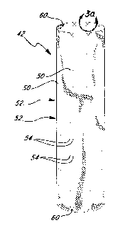
Figure 3 illustrates just the flexible tubular structure 42 without the
wireforms 44. The holes 50 can be seen in a plurality of axially spaced
5 circumferential rows 52. Each row 52 includes a plurality of pairs 54 of
holes
50, so that a portion of a wireform 44 may be threaded in and out at that
location. More specifically, as seen in Figure 2, each valley 48 is exposed to
the
exterior of the tubular structure 42 via a pair 54 of the holes 50.
The holes 50 are of a size and shape which prevents blood seepage
10 between the edge of each hole and respective wireform 44. Specifically, the
holes 50 are desirably circular and smaller in diameter than the diameter of
the
cylindrical wireforms 44 so as to form an interference fit. The tubular
structure
42 is flexible and expands slightly when a larger wireform passes through one
of the undersized holes 50. The holes 50 have a diameter of less than 95% with
respect to the wireform diameter, preferably between about 8% and 92%, and
more preferably between about 58% and 75%. Thus, if the wireform has a
diameter of 0.3 mm (0.012 inches) the holes 50 desirably have a diameter of
between about 0.025 mm (0.001 inches) and 0.279 mm (0.011 inches), and
more preferably between about 0.178 mm (0.007 inches) and 0.229 mm (0.009
inches). The process for forming the holes 50 to be precisely circular, within
proper size ranges, and located accurately will be described in greater detail
below.
In a preferred embodiment, the pairs 54 of holes 50 are spaced apart
with respect to one another with a tolerance of +0.254 mm (0.0 10 inches) and -
0.0 mm. There are preferably six to twelve rows 52 with eight pairs 54 in each
row. The total number of holes 50 may be between 100 and 200. Each hole 50
location has a tolerance of +0.254 mm (0.010 inches) and -0.254 mm (-0.010
inches).
CA 02365246 2001-07-23
WO 00/47135 PCT/US99/25282
11
As seen in Figure 2, the wireform is woven into the tubular structure 42
such that when the wire extends around the entire periphery, and the free ends
of
the wire protrude from the tube at positions adjacent to each other defining a
tail
segment 56. The loose ends are preferably held together with a crimping sleeve
58. After crimping the sleeve to secure the ends to each other and thereby
complete the circular configuration of the wireform, any portion of the wires
extending beyond the ends of the sleeve may be trimmed to cleanly finish the
tail segment.
The most proximal wireform 44a and the most distal wireform 44b are
positioned with respect to the upper and lower edge of the tubular structure
42
such that approximately one-third of the wireform extends beyond the
respective edge of the tubular structure. In particular, the proximal-most
wireform is positioned to extend above the mouth of the tubular structure 42
to
prevent any portion of the structure from oscillating, or "flapping," in
response
to the flow of blood past the edge of the graft.
As an additional measure to prevent such oscillation in the blood
stream, the proximal and distal edges of the tubular structure 42 are
configured
with rounded V-shaped notches 60 corresponding generally to the valleys 48 of
the proximal and distal wireforms, as seen in Figures 3 and 3a. Thus, the risk
of
the existence of any loose material that could potentially be affected by the
passing flow of blood is substantially reduced. The notches are formed to
precise dimensions including a depth A of about 0.686 mm (0.027 inches), a
point radius R, of about 0.279 mm (0.0 11 inches) and a fillet radius R2 of
about
1.727 mm (0.068 inches).
Desirably, the wireforms are positioned adjacent one another and are
spaced apart from each other such that the wireforms do not interfere with
each
other in either a radially expanded or contracted state. Thus, for example,
the
CA 02365246 2001-07-23
WO 00/47135 PCT/US99/25282
12
valleys of one wireform are located proximal of the crests of the next
adjacent
wireform. Preferably, the wireforms are also aligned "in phase," with peaks
along one longitudinal line and adjacent valleys aligned along a second
longitudinal line, thereby further reducing the possibility of overlap of
adjacent
wireforms. (While there may be some overlapping of the tail segments with an
adjacent wireform, because the tail segments extend on the outside of the
fabric
layer and the adjacent wirefonn is primarily on the inside of the fabric
layer, a
small degree of overlap with an adjacent wireform does not pose a problem.)
Another important feature of the straight prosthesis 40 of the present
invention
is the spacing distance between adjacent wireforms. It has been discovered in
accordance with the investigations of the present invention that optimizing
the
spacing distance between the wireforms improves the balance between kink
resistance and flexibility in the graft extensions. Too much space promotes
kinking, while too little space detracts from flexibility. These are important
features in the often tortuous path of the iliac arteries and abdominal aorta
in
which the graft extensions are to be placed. In this respect, accurate and
precise
location of the rows 52 of pairs 54 of holes 50 is essential to proper
functioning
of the prosthesis 40.
As illustrated in Figure 4, another embodiment of tubular prosthesis
fabricated by the techniques of the present invention is designated generally
at
62. This bifurcated prosthesis, sometimes referred to as a "trouser graft," is
adapted for insertion transfemorally to the situs of an aortic aneurysm in the
region where the iliac arteries branch from the abdominal aorta.
The prosthesis 62 includes a trunk portion 64 that bifurcates into two
legs 66, 68 at a septum region 70. The cylindrical tubes defined by the two
legs
66, 68 are in fluid communication with the trunk portion 64, thereby
approximating the internal configuration of the bifurcated junction of the
aortic
artery. In this preferred embodiment, one leg 66 extends longer than the other
CA 02365246 2001-07-23
WO 00/47135 PCT/US99/25282
13
leg 68 to facilitate loading of both legs into a smaller diameter catheter-
based
loader when self expanding wireforms are attached to the end of each leg.
The bifurcated prosthesis 62 comprises a flexible tubular graft portion
72 (Figure 5) reinforced by a variety of wireforms extending circumferentially
around or woven into the structure. The graft portion 72 is foldable, and the
wireforms are radially compressible and expandable. Thus, the graft is
configured to move between an insertion diameter, in which state the graft may
be inserted intraluminally into the aorta, and a larger, expanded diameter
(illustrated in Figure 4) in which state the graft may be secured within the
aorta.
The bifurcated prosthesis 62 includes two different types of wireforms:
balloon-expandable wireforms and self-expanding wireforms. This preferred
embodiment includes three balloon expandable wireforms 74a, 74b, and 74c,
which are woven into the trunk region 64 of the graft portion 72 but are
positioned primarily on the interior thereof, and a single exterior balloon-
expandable wireform 76 positioned at the distal end of the trunk region 64. A
self-expanding wireform 78 is attached to the outside of the graft portion 72
at
the septum region 70 with a self-expanding wireform 80a positioned on the
distal end of the longer leg 66 and another self-expanding wireform 80b at the
distal end of the shorter leg 68.
The balloon-expandable wireforms 74a, 74b, and 74c are preferably
made of an alloy as more specifically described above, and preferably have a
circular cross-section of about 0.3 mm (0.012 inches) in diameter. In
addition,
each of the balloon-expandable wireforms 74a, 74b, and 74c is similarly
configured with a curvilinear geometry such as the closed sinusoidal-like wave
illustrated in Figure 2, with alternating crests and valleys.
The balloon-expandable wireforms 74a, 74b, and 74c that are
positioned along the upper portion of the trunk 64 are preferably secured to
the
graft material by weaving through a plurality of holes 82. Figure 5
illustrates
CA 02365246 2001-07-23
WO 00/47135 PCTIUS99/25282
14
just the tubular graft portion 72 bereft of wireforms. The holes 82 can be
seen
in a plurality of axially spaced circumferential rows 84. Each row 84 includes
a
plurality of pairs 86 of holes 82, so that a portion of each wireform 74 may
be
threaded in and out at that location. More specifically, as seen in Figure 4,
each
valley is exposed to the exterior of the tubular graft portion 72 via a pair
86 of
the holes 82.
As in the straight tube embodiment, each wireform 74 is woven into the
graft portion 72 such that when the wire extends around the entire periphery
of
the fabric tube, the free ends of the wire protrude from the tube at positions
adjacent to each other, thereby enabling a tail segment 88 to be defined by
the
free ends. The loose ends are preferably held together with a crimping sleeve
90
positioned over them.
The distal balloon-expandable wireform 76 is attached to the graft
portion 72 in a different manner from the other balloon-expandable wireforms.
Instead of being woven into the graft portion 72, distal wireform 76 is
attached
to the fabric by tying it to the fabric with polyester thread. Other
biocompatible
threads may also be employed for securing the distal wireform 76 to the graft
portion 72. Although in this preferred embodiment, wireform 76 is tied to the
fabric structure with a thread, one of skill in the art will readily identify
other
attachment methods, including threading through the graft portion 72.
The configuration of each of the self-expanding wireforms 78 and 80 is
naturally biased towards an expanded state, such as that illustrated in Figure
4.
The self-expanding wireforms may be made of the same base material used in
the construction of the balloon-expandable wireforms, although the method of
manufacturing may differ. Thus, ELGILOY wire is preferred, with a number of
other materials acceptable for such use. Attachment of the self-expanding
wireforms 78 and 80is preferably accomplished by tying the crests and valleys
to the graft portion 72, as illustrated in Figure 4. It is presently preferred
that
CA 02365246 2001-07-23
WO 00/47135 PCT/US99/25282
each crest and valley be tied in five separate locations around the perimeter
of
the loop defining the respective crest or valley.
In the expanded state illustrated in Figure 4, the trunk portion 64 is
generally cylindrical and has mouth 91 configured with rounded V-shaped
5 notches 92 corresponding generally to the valleys of the terminal wireform
74a.
Thus, the risk of the existence of any loose material that could potentially
be
affected by the passing flow of blood is substantially reduced.
The graft portion 72 is preferably made of a tube of woven polyester
fabric, although other materials may be utilized as was described previously
10 with respect to the graft portion 42 of Figure 3.
System for Laser Forming Grafts
A system 100 for forming grafts in accordance with the present
invention is schematically shown in Figure 6. The illustrated system 100 shows
15 the basic components for forming grafts on a single mandrel, and it will be
appreciated as explained below that multiple mandrels in a full-scale
production
version may be provided. In addition, the various components for providing
motion and for cutting the grafts are exemplary only, and other means may be
used.
The system 100 comprises a frame 102 mounted on base 104, the frame
supporting a laser 106 and linear displacement mechanism 108 above a guide
rail 110. The laser 106 generates a beam 107 of light energy which is directed
at a series of mirrors and/or lenses (not shown) ultimately terminating at a
movable mirror 112 forming a portion of a cutting instrument 114. The cutting
instrument 114 comprises a vertically disposed, generally cylindrical member
having one or more focusing lenses therein and an output lens 116 on its
bottom
end. The cutting instrument 114 is fastened to a toothed belt 118 which is
driven horizontal left and right by the aforementioned linear displacement
CA 02365246 2001-07-23
WO 00/47135 PCT/US99/25282
16
mechanism 108. A flexible hose 120 attaches to a lower end of the cutting
instrument 114 and supplies a gas to an internal chamber communicating with
outlet ports (not shown) on the bottom of the cutting instrument and
surrounding the output lens 116.
The guide rail 110 supports a pair of carriages 122, 124 which slide
thereon and can be fastened in different locations along the guide rail.
Preferably, the guide rail 110 and carriages 122, 124 include respective
elements of a precision linear bearing arrangement, such as a conventional
tongue and groove linear slide. The left carriage 122 supports an upstanding
frame 126 to which is mounted a servo motor 128. The output shaft of the servo
motor 128 communicates with a drive arrangement (not shown) ultimately
turning a chuck 130 rotatably coupled to the frame 126. The drive arrangement
may include a belt drive and an encoder 131 is desirably provided to monitor
the angular position of the chuck shaft to accommodate for any belt slip and
ensure rotational accuracy. The chuck 130 extends horizontally from the frame
126 toward the right carriage 124 and an upstanding frame 132 mounted
thereon. The chuck 130 rotates about an axis 134 and includes an internal jaw
mechanism (not shown) for clamping to mandrels, as will be described below.
The right end frame 132 includes bearings which support a pair of horizontally
spaced wheels 136. The wheels 136 preferably include a pair of peripheral
elastomeric rings and are spaced apart to be in position to support a cylinder
of a
predetermined diameter rotating about the axis 134. The axis 134 is in the
same
vertical plane as the output lens 116 of the cutting instrument 114. The
cooperation between the support wheels 136 and chuck 130 will be described in
greater detail below with respect to individual mandrels and graft forming
techniques.
A computer control device 140 synchronizes the horizontal movement of
the cutting instrument 114 and the rotating movement of the chuck 130.
CA 02365246 2001-07-23
WO 00/47135 PCT/US99/25282
17
Various means are known for coordinating multiple moving elements in a
manufacturing environment, and the present invention should not be construed
as being limited to any one. One preferred embodiment of control system is a
[programmable, multi-axis digital motion control system using DC servo motors
coupled to optical rotary encoders. A 3 axis controller is required. Two axis
are
use for motion and the third is used to control the power of the laser. Custom
software is written in the native language of the controller to control the
path of
the laser beam and to modulate the laser power to produce the drilled holes
and
cuts in the fabric. The software is written in such a way to take advantage of
the
symmetry of the holes and notches in the graft. For example, grafts of
different
diameters, having the same hole and notch patterns can be processed using the
same program by changing only the number that designates the diameter of the
graft in the program. A preferred controller is the DMC- 1500 available from
Galil Motion Control Inc. of Mountain View CA. The chuck 130 is driven by a
servo motor and directly coupled to a rotary encoder to insure precision in
angular positioning of the chuck. Such precision is desirable when forming
particular grafts, such as those shown in Figures 2-5. Of course, other graft
forming applications such high precision may not be needed and a conventional
belt drive or other similar expedient may be substituted.
Apparatus for ForminQ Straight Grafts
An elongated cylindrical mandrel 150 is shown extending between the
chuck 130 and the support wheels 136. As the mandrel 150 rotates about the
axis 134, an upper generatrix of the mandrel directly faces the output lens
116.
In other words, the beam of light energy from the output lens 116 projects
directly downward and impinges on the uppermost tangential surface of the
cylindrical mandrel 150. The mandrel 158 includes a shaft stub 152 projecting
from the left end that is captured by the jaws of the chuck 130. In this
regard,
CA 02365246 2001-07-23
WO 00/47135 PCT/US99/25282
18
the support wheels 136 (inclusive of the peripheral elastomeric rings) have
outer
diameters and are spaced apart so as to contact and support the mandrel 150
for
rotation about the axis 134. Alternatively, a shaft stub may be provided on
the
right end of the mandrel 150 to rest on the wheels 136. The mandrel 150
preferably comprises stainless-steel, but may be made of other suitable
materials.
Figure 6 illustrates the graft forming system 100 in the process of
forming a plurality of straight grafts 160 (such as the graft shown in Figure
3)
out of an elongated tube of fabric material 162. The various steps in forming
the grafts 160 will be described in more detail below in the method of use
section.
Apparatus for Forming Bifurcated Grafts
With reference to Figures 7 and 7a, a mandre1200 used in forming
bifurcated grafts, such as the graft shown in Figure 5, is seen in exploded
perspective view. The mandrel 200 comprises, from left to right, a first
adapter
disk 202, a generally cylindrical trunk portion 204, and a pair of identical
legs
206. The first adapter disk 202 includes a central cylindrical cavity 208
sized to
closely receive the left end of the trunk portion 204. The right end of the
trunk
portion 204 narrows in a cone shape to a tip 210. A pair of diametrically
opposed and axially extending cylindrical reliefs 212 at the conical right end
and associated threaded pins 214 receive, respectively, the left ends of each
of
the legs 206. In this respect, the legs 206 include centered tapped holes for
mating with the threaded pins 214. The right end 218 of each of the legs 206
is
tapered and includes a slot 220 provided on the axially facing end for
receiving
a screwdriver when coupling and de-coupling the legs from the trunk portion
204. As with the elongated cylindrical mandrel 150 described above, each of
CA 02365246 2001-07-23
WO 00/47135 PCT/US99/25282
19
the components of the mandre1200 comprises stainless-steel, or other similar
expedient.
A central shaft stub 222 projects to the left from the first adapter disk
202 and is sized to be gripped by the jaws of the chuck 130. As will be more
fully described below in the method of use section, the mandrel 200 has
several
assembled states, some of them including additional adapter disks, all
rotatably
driven by the chuck 130 and underneath the cutting instrument 114. In this
manner, bifurcated grafts, such as the graft shown in Figure 5, are formed in
a
series of sequential steps.
Method of Forming Straight Grafts
With reference to Figures 3 and 6, the steps in forming straight grafts
with the system 100 of the present invention will now be described. First, a
length of biocompatible fabric tube is procured. In a preferred embodiment,
grafts of the present invention are formed from tubes of polyester
terephthalate,
commonly known in the industry by its trade name DACRON. A supplier of
biocompatible fabric tubes suitable for forming the grafts of the present
invention is Prodesco.
The fabric tube 162 is then fitted over the cylindrical mandrel 150 and
the assembly is positioned between the chuck 130 and support wheels 136. As
will be explained more clearly below with respect to preferred laser
parameters,
the fabric tube 162 desirably closely fits over the mandrel 150 without any
looseness or spaces therebetween.
To rotationally calibrate the fabric tube 162 with respect to the output
lens 116, an axial seam or selvage line is oriented to face upward. This can
be
done manually, or alternatively, the left shaft stub 152 of the mandrel 150
may
include some type of registering device limiting the insertion into the jaws
of
the chuck 130 to only one rotational orientation. In the latter instance,
assuming
CA 02365246 2001-07-23
WO 00/47135 PCT/US99/25282
the fabric tube 162 is fitted over the mandrel 150 in a predetermined
rotational
position with respect to the registering device, the control system 140 may
automatically position the selvage line at the upper generatrix of the
mandrel/tube assembly. This automated calibration technique removes any
5 guess work of the operator once the mandrel 150 is mounted in the system
100.
In other words, the calibration operation takes place off-line when the fabric
tube 162 is fitted onto the mandrel 150.
Linear calibration between the cutting instrument 114 and fabric tube
162 is accomplished by making a test cut on a paper model overlying the tube
10 162. Thus, for example, the control system 140 commands the linear
displacement mechanism to position the cutting instrument 114 at the left end
of
the fabric tube 162 adjacent the chuck 130. The laser 106 generates a beam of
light energy which is directed directly downward onto the paper model, while
simultaneously the servo motor 128 causes the mandrel 150 to rotate. In this
15 manner, a clean cut around the paper model is made and the paper model is
removed. All other linear distances are then measured from this first cut.
As seen in Figure 3, the notches 60 at the left end of the first straight
graft 160 are formed at the time of making the first cut 224 around the tube
162.
The notches 60 preferably comprise rounded indentations from the
20 circumferential edge of the graft 160. These rounded notches 60 are formed
by
synchronizing the linear movement of the cutting instrument 114 with the
rotational orientation of the mandrel 150. In a like manner, a plurality of
grafts
160 are delineated along the fabric tube 162 by the circumferential cuts
interrupted by notches 60. A typical length of fabric tube 162 produces up to
eight separate straight grafts of approximately 7.6 cm (3 inches) in length.
Of
course, as will be appreciated, the need for varying lengths of grafts 160 may
necessitate smaller or larger numbers be cut from any one tube 162.
CA 02365246 2001-07-23
WO 00/47135 PCT/US99/25282
21
After cutting the first, or calibration, line 224 on the left end of the
fabric
tube 162, a series. of axially spaced circumferential rows 52 of wire-
receiving
holes 50 are formed in the first graft 160. These holes were described
previously with respect to the straight graft 42 of Figure 3, and serve to
receive
the support wires 44 as seen in Figure 2. In this regard, the holes 50 are
preferably circular having a diameter equal to or less than the diameter of
the
support wires. A close fit is provided between the support wires 44 and the
holes 50 to prevent leakage through graft 160 upon implantation in a vessel.
In
one particular embodiment, the support wires 44 have a diameter of 0.3 mm
(0.012 inches) and the holes 166 have a diameter of between 0.025-0.279 mm
(0.001-0.011 inches), and more preferably between 0.178-0.229 mm (0.007-
0.009 inches).
Again, as seen in Figure 3, each row 52 of wire-receiving holes 50
comprises a plurality of closely spaced pairs 54 of holes, each pair of holes
being spaced farther from an adjacent pair of holes been from each other. Each
pair 54 of holes 50 thus receives either a crest or a valley of the undulating
support wires 44 on the outside of the graft, with the remainder of the
support
wires being located within the graft. This arrangement was shown in Figure 2.
Because of the computer control system 140 and synchronized precision
movement of the cutting instrument 114 and rotating mandrel 150, the location
of each of the holes 50 is very precise. Those of skill in the art in
programming
will recognize that there are variety of patterns that can be formed on the
graft
using the tools described herein. The illustrated pattern of axially spaced
circumferential rows of holes 166 is preferably formed one row at a time by
fixing the location of the cutting instrument 114 and rotating the mandrel
150.
After all of the wire-receiving holes 50 are formed, the first graft 160 is
finished by cutting the right end, including the notches 60. The process
continues with the system forming first the left end of each graft 160, then
the
CA 02365246 2001-07-23
WO 00/47135 PCT/US99/25282
22
pattern of wire-receiving holes 50, and finally the right end of each graft.
In the
embodiment shown, the right end of each graft coincides with the left end of
the
adjacent graft, with the respective notches being cut in opposite directions
and
at the same location. This of course reduces the amount cutting and associated
fabrication time. In an alternative embodiment, a space may be formed between
each of the grafts 160. Because of the close fit between the fabric tube 162
and
mandrel 150, supplemental restraints holding the tube to the mandrel may not
be
needed. Of course, various forms of straps or elastomeric rings, for example,
may be utilized to secure the fabric tube onto the mandrel.
Method of Forming Bifurcated Grafts
Figures 8-10 illustrates three snapshots or stages of forming the
bifurcated graft 72 seen in Figure 5. Initially, an unfinished bifurcated
graft is
procured from a source such as Prodesco. In forming the unfinished bifurcated
graft, the graft is preferably shrunken onto a forming mandrel having
approximately the same shape as the assembled mandre1200 (seen exploded in
Figure 7). Figure 8 illustrates an assembled mandre1200 with the adapter disk
202 being clamped by the jaws of the chuck 130, the left end of the trunk
portion 204 being inserted and retained within the cavity 208 (Figure 7), and
the
two legs 206 being screwed into the reliefs 212 on the right end of the trunk
portion. An unfinished bifurcated graft 230 shown closely fitted over the
assembled mandrel 200. Again, the size and shape of the mandrel 200 with
respect to the unfinished graft 230 is such that no looseness or spaces exist
therebetween.
The unfinished bifurcated graft 230 includes a septum region 231 which
contacts the tip 210 (Figure 7) of the conical end of the trunk portion 204 of
the
mandre1200. By sliding the bifurcated graft 230 over the legs 206 and over the
trunk portion 204, the septum region 231 eventually contacts and is stopped by
CA 02365246 2001-07-23
WO 00/47135 PCT/US99/25282
23
the tip 210. In this manner, the bifurcated graft 230 is located with respect
to
the mandre1200 as an initial step in registering the graft with respect to the
cutting instrument 114. That is, various means will be apparent one of skill
the
art for calibrating the location of the cutting instrument 114 with respect to
the
mandrel 200 mounted in the system 100, and the registration of the graft 230
with respect to the mandre1200 completes the overall calibration.
A leg adapter disk 232 is shown on the right end of the mandre1200.
The adapter disk 232 includes a pair of apertures for receiving the right ends
of
the legs 206, and a centered shaft stub 234 sized the same as each of the
legs.
The shaft stub 234 is rotatably supported by the two wheels 136. In this
manner, the mandre1200 in conjunction with the leg adapter disk 232 rotates
about the axis 134 and is supported at both axial ends.
The cutting instrument 114 is seen in Figure 8 forming the mouth 91 of
the bifurcated graft 72. After the mouth 91 is formed, the computer control
system 140 commands the servo motors and laser 106 to form the plurality of
axially spaced circumferential rows of wire-receiving holes 82, and the
notches
92. Again, these holes 82 are sized to closely receive the support wires 74
used
in the final prosthesis, as seen in Figure 4. Once the holes 82 are formed,
the
trunk portion 64 is finished and the legs 66, 68 of the graft are cut to size
in the
steps seen in Figures 9 in 10.
In Figure 9, the mandrel 200 has been reconfigured by replacing the first
adapter disk 202 with a second adapter disk 250. The second adapter disk 250
includes an off-center cavity 252 for receiving the mandrel trunk portion 204.
In addition, one of the legs 206 has been removed and the now unsupported leg
254 of the unfinished graft is shown folded back upon the trunk portion of the
graft and fastened thereto with a band 256 or other such device. The remaining
mandrel leg 206 is received by a centered hole in a second leg adapter disk
258
having a shaft stub 260 rotatably supported by the wheels 136. The remaining
CA 02365246 2001-07-23
WO 00/47135 PCT/US99/25282
24
mandrel leg 206 is oriented with respect to the second adapter disk 250 to be
aligned with the axis 134. In this manner, when the shaft stub of the second
adapter desk 250 is captured in the jaws of the chuck 130, the remaining
mandrel leg 206 rotates about the axis 134.
The cutting instrument 114 is seen above the mandrel leg 206 cutting the
short graft legs 68 to size. Any other notching or hole forming in the short
graft
leg 68 is accomplished at this time.
In Figure 10, the mandre1200 essentially remains in the same
configuration as in Figure 9, but the graft fitted thereon has been removed
and
re-fitted with the uncut leg positioned over the remaining mandrel leg 206. In
this arrangement, the short graft leg 68 extends freely to the right. There is
no
need to restrain the already cut short graft leg 68 because the cutting
instrument
114 only has to cut the longer graft leg 66 to size, and the short leg will
not be
in the way.
After the longer graft leg 66 is cut, the bifurcated graft is in the
configuration seen in Figure 5. At this point, the assorted support wires and
other hardware are added in a separate assembly step to form the prosthesis
seen
in Figure 4.
Preferred Cutting Instruments
A variety of different cutting instruments 114 may be utilized in the
formation of the grafts in accordance with present invention. A preferred
cutting instrument 114 is a laser with an appropriate power and wave length
that
will allow the removal of the graft material without damage to the mandrel
material. In addition the laser must have the ability to modulate the power
output. The laser power should be low enough to avoid excessive melting or
burning of the material in the graft, while still being strong enough to cut a
hole
therein and fuse the otherwise frayed ends of fabric material. A particularly
CA 02365246 2001-07-23
WO 00/47135 PCT/US99/25282
useful kind of laser for this invention is a low-power, sealed RF-excited COZ
laser. COz lasers are, for the most part, less expensive and more compact than
other types of lasers, have the ability to modulate the power output and have
a
wavelength that is easily absorbed by the graft material. Alternatively, a YAG
5 laser may be suitable although it is somewhat larger and more expensive than
a
COz laser and harder to control the power output.
As mentioned, the holes 50, 82 in the grafts for receiving the wire forms
are preferably circular and have a diameter of between 0.178 mm (0.007 inches)
and 0.229 mm (0.009 inches). The spot size of the laser beam therefore must
10 be sized to avoid creating larger holes than this preferred range. Laser
beams
typically have a Gaussian distribution, and thus the spot size must be
undersized to accommodate for some widening of the hole because of secondary
fringe energy present adjacent the primary beam causing the laser to cut or
drill
a larger kerf than the spot size. In one the example, therefore, a laser beam
as a
15 spot size of about 0.006 inches, and the Gaussian distribution therefore
expands that width by about 50% to result in an effective cutting width of
0.009
inches.
A specific example of laser suitable for manufacturing the grafts in
accordance with the present invention is a 25 watt CO2laser available from
20 Synrad of Mulelteo, WA. Moreover, the power must be attenuated to avoid
burning the graft material. For example, the power is preferably set at an
output
of 7% of the total. CO2lasers are particularly useful for forming grafts made
of
synthetic fabrics which efficiently absorb the light energy produced at the
infrared wavelengths characteristic of COZ lasers. In other words, the light
25 energy is absorbed primarily by the graft material, reflected or absorbed
by the
underlying mandrel.
In this regard, a word should be said about the necessity for a close fit
between the graft material and the mandrel. If looseness or spaces exist
CA 02365246 2001-07-23
WO 00/47135 PCT/US99/25282
26
therebetween, some of the light energy will continue through the hole being
formed and will either excessively heat, or be reflected by, the underlying
mandrel thus heating the underside of the edges of the hole being formed. This
excess heating of the mandrel can be detrimental to the hole forming process.
Therefore, it is particular important to size the mandrel so as to form a
tight or
close fit with the graft material. Furthermore, the mandrels are preferably
formed with curved or tapered ends to avoid snagging on the graft material and
thus to facilitate assembly thereover and reduce rejects.
Finally, though lasers are particularly useful for forming the grafts as
described herein, especially fabric grafts to prevent fraying, the advantages
of
the graft forming system may also be utilized with other cutting instruments
such as a mechanical cutter and the like. In particular, if the graft material
does
not tend to fray, such as PTFE grafts, then a blade or die stamp may be
substituted for the laser such as in the configuration of Figure 8.
Production Graft Forming
The present invention has so far been described with respect to a single
rotating chuck 130 for holding a single cylindrical or bifurcated mandrel.
This
arrangement is suitable for describing the essential elements of the
respective.
graft forming systems but may be limited in its production capacity. In the
alternative, multiple chuck devices may be used for forming a plurality of
grafts
or performing different steps simultaneously. Such devices may be obtained
from Beam Dynamics, of San Carlos, California.
In one example of a production device, four parallel chucks in 4 rotary
devices are processed in series with only one setup.. Four elongated
cylindrical
mandrels, such as the mandrel 150 shown in Figure 6, are then fitted with
tubes
of graft material and installed for rotation by each of the chucks. Each
mandrel
is processed in series by cutting eight individual grafts from the tubes,
after
CA 02365246 2001-07-23
WO 00/47135 PCT/US99/25282
27
which the mandrels and cut grafts are removed and replaced immediately by
new mandrels having uncut tubes thereon. In this manner, the time-consuming
step of cleaning and manually fitting each of the mandrels with the tubes of
graft material, and removing each of the cut grafts is accomplished off-line,
during the cutting process, optimizing the throughput of the system.
A multiple chuck machine is also well-suited for rapidly forming a
plurality of the bifurcated grafts 72 seen in Figure 5. In particular, a four
chuck
machine can be utilized to process different steps in the sequence shown in
Figures 8-10. That is, a first pair of chucks may be used to rotate mandrels
200
in the configuration of Figure 8 while cutting the mouth 91 and notches 92, as
well as the pattern of holes 82 in the trunk portion 64. While the first two
chucks are being used to perfonn the more elaborate cutting steps of Figure 8,
the second two chucks may be used to cut the legs to size, as seen in Figures
9-
10. Those of skilled in the art will recognize that a multiple-chuck machine
greatly facilitates the throughput by permitting the time-consuming manual
tasks of fitting the grafts over the mandrels and reconfiguring the mandrels
to be
accomplished off-line.
In a multiple chuck machine, multiple lasers or a single laser supplying
light beams to a series of movable reflective mirrors and cutting instruments
may be used.
Additionally, the power of the laser may be increased and then
"dumped" to result in less power fluctuations. That is, the first embodiment
suggested a 25 watt laser at 7% power. Another example is a 50 watt laser from
Coherent of Santa Clara, CA which is operated at a higher percentage of its
maximum power, perhaps 25-50%. The power is then dumped at one of the
reflective mirrors which is designed to reflect only a portion of the light
and
transmit the remainder to a light absorbent black box structure. For example,
perhaps 80% of the output beam is dumped, the remaining 20% being used to
CA 02365246 2001-07-23
WO 00/47135 PCT/US99/25282
28
form the grafts. As a result, any power fluctuations of the laser as it
continues
to operate and heat up are relatively less harmful to the process. That is,
the
fluctuations undergo the same relative reduction in power and thus the
absolute
changes in power are reduced also (to 20% in the exemplary embodiment).
The present invention may be embodied in other specific forms without
departing from its spirit or essential characteristics. The described
embodiments
are to be considered in all respects only as illustrative and not restrictive.
The
scope of the invention is, therefore, indicated by the appended claims rather
than
by the foregoing description. All changes which come within the meaning and
range of equivalency of the claims are to be embraced within their scope.