Note: Descriptions are shown in the official language in which they were submitted.
CA 02365376 2001-12-19
USE ~OF REINFORCED FOAM IMPLANTS WITH ENHANCED INTEGRITY
FOR SOFT TISSUE REPAIR AND REGENERATION
FIELD OF THE INVENTION
The present invention relates to bioabsorbable, porous, reinforced,
biocompatible
tissue repair stimulating implant devices that may comprise at least one
biological
component for use in the repair of orthopaedic type injuries, such as damage
to the
meniscus, ligaments, and tendons, and methods for making such devices.
BACKGROUND OF THE INVENTION
Individuals can sometimes sustain an injury to tissue, such as cartilage,
muscle,
bone, and sinew that requires repair by surgical intervention. Such repairs
can be
effected by suturing or otherwise repairing the damaged tissue, and/or by
augmenting
the damaged tissue with other tissue or with a tissue implant. The implant can
provide
structural support to the damaged tissue.
One example of a common tissue injury concerns damage to cartilage, for
example, the menisci of a knee joint. There are two menisci of the knee joint;
a medial
and a lateral meniscus. The meniscus is a biconcave, fibrocartilage tissue
that is
interposed between the femur and tibia of the leg. The primary functions of
the
meniscus are to bear loads, absorb shock, stabilize, and lubricate the joint.
If not treated
properly, an injury to the meniscus, such as a "bucket-handle tear," can lead
to the
development of osteoarthritis. Currently, treatment modalities for a damaged
meniscus
include removal of the meniscus and surgical repair of the damaged meniscus.
Another common tissue injury is a damaged or torn rotator cuff, which
facilitates
circular motion of the humerus bone relative to the scapula. The most common
injury
associated with the rotator cuff is a strain or tear to the supraspinatus
tendon. This tear
can be at the insertion site of the tendon with the humerus, thereby releasing
the tendon
partially, or fully (depending upon the severity of the injury), from the
bone.
Additionally, the strain or tear can occur within the tendon itself. Treatment
for a
strained tendon usually involves physical cessation from use of the tendon.
However,
depending upon the severity of the injury, a torn tendon might require
surgical
intervention as in the case of a full tear of the supraspinatus tendon from
the humerus.
-1-
CA 02365376 2001-12-19
Surgical intervention can involve the repair and/or reattachment of torn
tissue. A
prolonged recovery period often follows repair of a rotator cuff injury.
Surgical treatment of damaged tissue (e.g., the menisci, ligaments, and
tendons)
would benef t from techniques that effect a more reliable repair of tissue,
and which
facilitate more rapid healing. Thus, various implants have been used in
surgical
procedures to help achieve these benefits. Examples of such implants include
those that
are made from biologically derived tissue (e.g., allografts and autografts),
and those that
are synthetic. Biologically derived materials can have disadvantages in that
they can
contribute to disease transmission, while synthetic materials are difficult to
manufacture
in such a way that their properties are reproducible from batch to batch.
Various known devices and techniques for treating such conditions have been
described in the prior art. For example, Naughton et al. (U.S. Pat. 5,842,477)
describe
an in vivo method of making and/or repairing cartilage by implanting a
biocompatible
structure in combination with periosteal/perichondrial tissue which
facilitates the
securing of the implant.
Various tissue reinforcing materials are disclosed in U.S. Patent No.
5,891,558
(Bell et al.) and European Patent Application No. 0 274 898 A2 (Hinsch). Bell
et al.
describe biopolymer foams and foam constructs that can be used in tissue
repair and
reconstruction. Hinsch describes an open cell, foam-like implant made from
resorbable
materials, which has one or more textile reinforcing elements embedded
therein.
Although potentially useful, the implant material is believed to lack
sufficient strength
and structural integrity to be effectively used as a tissue repair implant.
Despite existing technology, there continues to be a need for devices and
methods for securing damaged tissue and facilitating rapid healing of the
damaged
tissue.
SUMMARY OF THE INVENTION
This invention relates to bioabsorbable, porous, reinforced, biocompatible
tissue
repair stimulating implants, or "scaffold," devices for use in the repair
and/or
regeneration of diseased or damaged tissue, and the methods for making and
using these
devices. The implants comprise a bioabsorable polymeric foam component having
pores with an open cell pore structure. The foam component is reinforced with
a
material such as a mesh. Preferably, the implant has sufficient structural
integrity to
-2-
CA 02365376 2001-12-19
enable it to be handled in the operating room prior to and during
implantation. These
implants should also have sufficient properties (e.g., tear strength) to
enable them to
accept and retain sutures or other fasteners without tearing. Desirable
properties are
imparted to the implant of the invention by integrating the foam component
with the
reinforcement component. That is, the pore-forming webs or walls of the foam
component penetrate the mesh of the reinforcement component so as to interlock
therewith. The implant may include one or more layers of each of the foam and
reinforcement components. Preferably, adjacent layers of foam are also
integrated by at
least a partial interlocking of the pore-forming webs or walls in the adjacant
layers. The
implants of the instant invention may optionally include at least one
biological
component that is incorporated therein.
The reinforcement material is preferably a mesh, which may be bioabsorbable.
The reinforcement should have a sufficient mesh density to permit suturing,
but the
density should not be so great as to impede proper bonding between the foam
and the
reinforcement. A preferred mesh density is in the range of about l2 to 80%.
The biological component of the present invention comprises at least one
effector
molecule and/or cell, which contributes to the healing process of an injured
tissue.
Collectively, these materials are sometimes referred to herein as "effectors."
The
effectors can be a cellular factor such as a protein or peptide (for the sake
of simplicity,
use of the term "protein" herein will include peptide), a non-protein
biomolecule (e.g.,
nucleic acids and lipids), a cell type, viruses, virus particles, a
pharmaceutical agent, or
combinations thereof. One function of the implant of the current invention is
as a carrier
for the effectors, and the effector can be incorporated within the implant
either prior to
or following surgical placement of the implant.
The invention also relates to a method of preparing such biocompatible,
bioabsorbable tissue repair stimulating implants. The implants are made by
placing a
reinforcement material within a mold in a desired position and orientation. A
solution of
a desired polymeric material in a suitable solvent is added to the mold and
the solution. is
lyophilized to obtain the implant in which a reinforcement material is
embedded in a
polymeric foam. The effector may be added to the implant, either during or
after
manufacture, by a variety of techniques.
-3-
CA 02365376 2001-12-19
The tissue repair stimulating implant can be used to treat injuries ocurring
within
the musculoskeletal system, such as rotator cuff injuries or meniscal tears.
Further, such
implants can be used in other orthopaedic surgical procedures, such as hand
and foot
surgery, to repair tissues such as ligaments, nerves, and tendons.
BRIEF DESCRIPTION OF THE DRAWINGS
The invention will be more fully understood by reference to the following
detailed description when considered in conjunction with the accompanying
drawings, in
which:
Figure 1 is a sectional view of a tissue implant constructed according to the
present invention;
Figure 2 is a sectional view of an alternative embodiment of the implant of
the
present invention;
Figure 3 is a sectional view of yet another embodiment of the implant of the
presentinvention;
Figure 4 is a perspective view of one embodiment of a mold set-up useful with
the present invention;
Figure 5 is a sectional view of a portion of the mold set-up of Figure 4;
Figure 6 is a scanning electron micrograph of a bioabsorbable knitted mesh
reinforcement material useful with the implant of the present invention; and
Figure 7 is a scanning electron micrograph of a portion of an implant
according
to the present invention.
-4-
CA 02365376 2001-12-19
DETAILED DESCRIPTION OF THE INVENTION
The present invention relates to a biocompatible'tissue repair stimulating
implant
or "scaffold" device which, preferably, is bioabsorbable, and to methods for
making and
using such a device. The implant includes one or more layers of a
bioabsorbable
S polymeric foam having pores with an open cell pore structure. A
reinforcement
component is also present within the implant to contribute enhanced mechanical
and
handling properties. The reinforcement component is preferably in the form of
a mesh
fabric that is biocompatible. The reinforcement component may be bioabsorbable
as
well. The implant optionally has incorporated therein a biological component,
or
effector that assists in and/or expedites tissue healing. Preferably, the
biological
component, if present, is housed primarily within the pores of the foam
component of
the implant.
In some surgical applications, such as for use in the repair of tissue
including a
torn ligament, tendon, rotator cuff, nerve, or meniscus, the tissue implants
of the
invention must be able to be handled in the operating room, and they must be
able to be
sutured or otherwise fastened without tearing. Additionally, the implants
should have a
burst strength adequate to reinforce the tissue, and the structure of the
implant must be
suitable to encourage tissue ingrowth: A preferred tissue ingrowth-promoting
structure
is one where the cells of the foam component are open and sufficiently sized
to permit
cell ingrowth and to house the effector. A suitable pore size to accommodate
these
features is one in which the pores have an average diameter in the range of
about 100 to
1000 microns and, more preferably, about 150 to 500 microns.
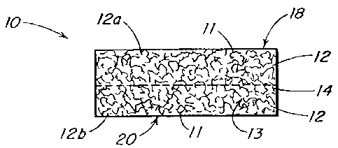
Refernng to FIGS. 1 through 3, the implant 10 includes a polymeric foam
component I2 and a reinforcement component 14. The foam component preferably
has
pores 13 with an open cell pore structure. Although illustrated as having the
reinforcement component disposed substantially in the center of a cross
section of the
implant, it is understood that the reinforcement material can be disposed at
any location
within the implant. Further, as shown in FIG. 2, more than one layer of each
of the foam
component 12a, 12b and reinforcement component 14a, 14b may be present in the
implant. It is understood that various layers of the foam component and/or the
reinforcement materials may be made from different materials and have
different pore
sizes.
-5-
CA 02365376 2001-12-19
FIG. 3 illustrates an embodiment in which a barrier layer 16 is present in the
implant. Although illustrated as being only on one surface of the implant 10,
the barrier
layer 16 may be present on either or both of the top and bottom surfaces 18,
20 of the
imp lant.
The implant 10 must have sufficient structural integrity and physical
properties
to facilitate ease of handling in an operating room environment, and to permit
it to
accept and retain sutures or other fasteners without tearing. Adequate
strength and
physical properties are developed in the implant through the selection of
materials used
to form the foam and reinforcement components, and the manufacturing process.
As
shown in FIG. 7, the foam component 12 is integrated with the reinforcement
component 14 such that the web or walls of the foam componenets that form
pores 13
penetrate the mesh of the reinforcement component 14 and interlock with the
reinforcement component. The pore-forming walls in adjacent layers of the foam
component also interlock with one another, regardless of whether the foam
layers are
separated by a layer of reinforcement materials or whether they are made of
the same or
different materials.
A variety of bioabsorbable polymers can be used to make porous, reinforced
tissue repair stimulating implant or scaffold devices according to the present
invention,
Examples of suitable biocompatible, bioabsorbable polymers include polymers
selected
from the group consisting of aliphatic polyesters, poly(amino acids),
copoly(ether-
esters), polyalkylenes oxalates, polyamides, tyrosine derived polycarbonates,
poly(iminocarbonates), polyorthoesters, polyoxaesters, polyamidoesters,
polyoxaesters
containing amine groups, poly(anhydrides), polyphosphazenes, biomolecules
(i.e.,
biopolymers such as collagen, elastin, bioabsorbable starches, etc.) and
blends thereof:
For the purpose of this invention aliphatic polyesters include, but are not
limited to,
homopolymers and copolymers of lactide (which includes lactic acid, D-,L- and
meso
lactide), glycolide (including glycolic acid), E-caprolactone, p-dioxanone
(1,4-dioxan-2-
one), trimethylene carbonate (1,3-dioxan-2-one), alkyl derivatives of
trimethyIene
carbonate, 8-valerolactone, ~3-butyrolactone, 'y-butyrolactone, E-decalactone,
hydroxybutyrate, hydroxyvalerate, I,4-dioxepan-2-one (including its dimer
1,5,8,I2-
tetraoxacyclotetradecane-7,14-dione), 1,5-dioxepan-2-one, 6,6-dimethyl-1,4-
dioxan-2-
one 2,5-diketomorpholine, pivalolactone, a, a diethylpropiolactone, ethylene
carbonate,
ethylene oxalate, 3-methyl-1,4-dioxane-2,5-dione, 3,3-diethyl-1,4-dioxan-2,5-
dione,
-6-
CA 02365376 2001-12-19
6,8-dioxabicycloctane-7-one and polymer blends thereof. Poly(iminocarbonates),
for
the purpose of this invention, are understood to include those polymers as
described by
Kemnitzer and Kohn, in the Handbook of Biodegradable Polymers, edited by Domb,
et.
al., Hardwood Academic Press, pp. 251-272 (1997). Copoly(ether-esters), for
the
purpose of this invention, are understood to include those copolyester-ethers
as
described in the Journal of Biomaterials Research, Vol. 22, pages 993-1009,
1988 by
Cohn and Younes, and in Polymer Preprints (ACS Division of Polymer Chemistry),
Vol.
30(1), page 498, 1989 by Cohn (e.g., PEO/PLA). Polyalkylene oxalates, for the
purpose
of this invention, include those described in U.S. Patent Numbers 4,208,51 l;
4,141,087;
4,130,639; 4,140,678; 4,105,034; and 4,205,399. Polyphosphazenes, co-, ter-
and higher
order mixed monomer based polymers made from L-lactide, D,L-lactide, lactic
acid,
glycolide, glycolic acid, para-dioxanone, trimethylene carbonate and E-
caprolactone
such as are described by Allcock in The Encyclopedia of Polymer Science, Vol.
13,
pages 3I-41, Wiley Intersciences, John Wiley & Sons, 1988 and by Vandorpe, et
al in
the Handbook of Biodegradable Polymers, edited by Domb, et al., Hardwood
Academic
Press, pp. 161-182 (1997). Polyanhydrides include those derived from diacids
of the
form HOOC-C6H4 -O-(CH2)m-O-C6H4-COOH, where "m" is an integer in the range
of from 2 to 8, and copolymers thereof with aliphatic alpha-omega diacids of
up to 12
carbons. Polyoxaesters, polyoxaamides and polyoxaesters containing amines
and/or
amido groups are described in one or more of the following U.S. Patent Nos.
5,464,929;
5,595,751; 5;597,579; 5,607,687; 5,618,552; 5,620,698; 5,645,850; 5,648,088;
5;698,213; 5,700,583; and 5,859,150. PoIyorthoesters such as those described
by Heller
in Handbook of Biodegradable Pol ers, edited by Domb, et al., Hardwood
Academic
Press; pp. 99-l I8 (1997).
As used herein, the term "glycolide" is understood to include polyglycolic
acid.
Further, the term "lactide" is understood to include L-lactide, D-lactide,
blends thereof,
and lactic acid polymers and copolymers.
Currently, aliphatic polyesters are among the preferred absorbable polymers
for
use in making the foam implants according to the present invention. Aliphatic
polyesters can be homopolymers, copolymers (random, block, segmented, tapered
blocks, graft, triblock, etc.) having a linear, branched or star structure.
Suitable
monomers for making aliphatic homopolymers and copolymers may be selected from
_7_
CA 02365376 2001-12-19
r
the group consisting of, but are not limited, fo lactic acid, lactide
(including L-, D-, meso
and D,L mixtures), glycolic acid, glycolide, ~-caprolactone, p-dioxanone (1,4-
dioxan-2-
one), trimethylene carbonate (1,3-dioxan-2-one), 8-valerolactone, ø-
butyrolactone, E-
decalactone, 2,5-diketomorpholine, pivalolactone, a, a -diethylpropiolactone,
ethylene
carbonate, ethylene oxalate, 3-methyl-1,4-dioxane-2,5-dione, 3,3-diethyl-1,4-
dioxan-
2,5-dione, y-butyrolactone, 1,4-dioxepan-2-one, 1,5-dioxepan-2-one, 6;6-
dimethyl-
dioxepan-2-one, 6,8-dioxabicycloctane-7-one, and combinations thereof.
Elastomeric copolymers are also particularly useful in the present invention.
Suitable elastomeric polymers include those with an inherent viscosity in the
range of
about 1.2 dL/g to 4 dL/g, more preferably about 1.2 dL/g to 2 dL/g and most
preferably
about 1.4 dL/g to 2 dL/g as determined at 25°C in a 0.1 gram per
deciliter (g/dL)
solution of polymer in hexafluoroisopropanol (HFIP). Further, suitable
elastomers
exhibit a high percent elongation and a low modulus, while possessing good
tensile
strength and good recovery characteristics. In the preferred embodiments of
this
invention, the elastomer from which the foam component is formed exhibits a
percent
elongation (e.g., greater than about 200 percent and preferably greater than
about 500
percent). In addition to these elongation and modulus properties, suitable
elastomers
should also have a tensile strength greater than about 500 psi, preferably
greater than
about 1,000 psi, and a tear strength of greater than about 50 lbs/inch,
preferably greater
than about 80 lbs/inch.
Exemplary bioabsorbable, biocompatible elastomers include, but are not limited
to, elastomeric copolymers of s-caprolactone and glycolide (including
polyglycolic acid)
with a mole ratio of E-caprolactone to glycolide of from about 35:65 to about
65:35,
more preferably from 45:55 to 35:65; elastomeric copolymers of E-caprolactone
and
lactide (including L-lactide; D-lactide, blends thereof, and lactic acid
polymers and
copolymers) where the mole ratio of s-caprolactone to lactide is from about
35:65 to
about 65:35 and more preferably from 45:55 to 30:70 or from about 95:5 to
about 85:15;
elastomeric copolymers of p-dioxanone (1,4-dioxan-2-one) and lactide
(including L-
lactide, D-lactide, blends thereof, and lactic acid polymers and copolymers)
where the
mole ratio ofp-dioxanone to lactide is from about 40:60 to about 60:40;
elastomeric
copolymers of s-caprolactone and p-dioxanone where the mole ratio of s-
caprolactone to
p-dioxanone is from about from 30:70 to about 70:30; elastomeric copolymers of
p-
_g-
CA 02365376 2001-12-19
dioxanone and trimethylene carbonate where the mole ratio of p-dioxanone to
trimethyIene carbonate is from about 30:70 to about 70:30; elastomeric
copolymers of
trimethylene carbonate and glycolide (including polyglycolic acid) where the
mole ratio
of trimethylene carbonate to glycolide is from about 30:70 to about 70;30;
elastomeric
copolymers of trimethylene carbonate and lactide (including L-lactide, D-
lact~de, blends
thereof, .and lactic acid polymers and copolymers) where the mole ratio of
trimethylene
carbonate to lactide is from about 30:70 to about 70:30; and blends thereof.
Examples
of suitable bioabsorbable elastomers are described in U.S. Patent Nos.
4,045,418;
4,057,537 and 5,468,253.
In one embodiment, the elastomer is a 35:65 copolymer ofpolyglycolic acid and
polycaprolactone, formed in a dioxane solvent and including a polydioxanone
mesh. In
another embodiment, the elastomer is a 50:50 blend of a 35:65 copolymer of
poIyglycolie acid and polycaprolactone and 40:60 E-caprolactone-co-lactide.
One of ordinary skill in the art will appreciate that the selection of a
suitable
polymer or copolymer for forming the foam depends on several factors. The more
relevant factors in the selection of the appropriate polymers) that is used to
form the
foam component include bioabsorption (or bio-degradation) kinetics; in vivo
mechanical
performance; cell response to the material in terms of cell attachment,
proliferation,
migration and differentiation; and biocompatibility. Other relevant factors,
which to
some extent dictate the in vitro and in vivo behavior of the polymer, include
the
chemical composition, spatial distribution of the constituents, the molecular
weight of
the polymer, and the degree of crystallinity.
The ability of the substrate material to resorb in a timely fashion in the
body
environment is critical. But the differences in the absorption time under in
vivo
conditions can also be the basis for combining two different copolymers. For
example, a
copolymer of 35:65 s-caprolactone and glycolide (a relatively fast absorbing
polymer) is
blended with 40:60 E-caprolactone and L-lactide copolymer (a relatively slow
absorbing
polymer} to form a foam component. Depending upon the processing technique
used,
the tW o constituents can be either randomly inter-connected bicontinuous
phases, or the
constituents could have a gradient-like architecture in the form of a laminate
type
composite with a well integrated interface between the two constituent layers.
The
-9-
CA 02365376 2001-12-19
microstructure of these foams can be optimized to regenerate or repair the
desired
anatomical features of the tissue that is being engineered.
In one embodiment, it is desirable to use polymer blends to form structures .
which transition from one composition to another composition in a gradient-
like
architecture. Foams having this gradient-like architecture are particularly
advantageous
in tissue engineering applications to repair or regenerate the structure of
naturally
occurring tissue such as cartilage (articular, meniscal, septal, tracheal,
auricular; costal,
etc.), tendon, ligament, nerve, esophagus, skin, bone, and vascular tissue.
For example;
by blending an elastomer of E-caprolactone-co-glycolide with s-caprolactone-co-
lactide
(e.g., with a mole ratio of about 5:95) a foam may be formed that transitions
from a
softer spongy material to a stiffer more rigid material in a manner similar to
the
transition from cartilage to bone. Clearly, one of ordinary skill in the art
will appreciate
that other polymer blends may be used for similar gradient effects, or to
provide:
different gradients (e.g:, different absorption profiles, stress response
profiles, or
different degrees of elasticity). For example, such design features can
establish a
concentration gradient for the biological component or effector such that a
higher
concentration of the effector is present in one region of the implant (e.g.,
an interior
portion) than in another region (e,g., outer portions). This may be effected
by
engineering an implant in which the overall pore volume is greater in a region
in which
it is desired to have a greater concentration of biological component.
The implants of the invention can also be used for organ repair replacement or
regeneration strategies that may benefit from these unique tissue implants.
For example,
these implants can be used for spinal disc, cranial tissue, dura; nerve
tissue, liver,
pancreas, kidney, bladder, spleen, cardiac muscle, skeletal muscle, skin,
fascia,
maxillofacial, stomach, tendons, cartilage, ligaments, and breast tissues.
The reinforcing component of the tissue repair stimulating implant of the
present
invention can be comprised of any absorbable or non-absorbable biocompatible
material,
including textiles with woven, knitted, warped knitted (i.e., lace-like), non-
woven; and
braided structures. In an exemplary embodiment, the reinforcing component has
a
mesh-like structure. In any of the above structures; mechanical properties of
the
material can be altered by changing the density or texture of the material, or
by
embedding particles in the material. The fibers used to make the reinforcing
component
can be monofilaments, yarns, threads, braids, or bundles of fibers. These
fibers can be
- 10-
CA 02365376 2001-12-19
made of any biocompatible material including bioabsorbable materials such as
polylactic
acid (PLA), polyglycolic acid (PGA), polycaprolactone (PCL), polydioxanone
(PD~),
trimethylene carbonate (TMC), polyvinyl alcohol (PVA), copolymers or blends
thereof.
In one embodiment, the fibers are formed of a polylactic acid and polyglycolic
acid
copolymer at a 95:5 mole ratio.
In another embodiment, the fibers that form the reinforcing material can be
made
of a bioabsorbable glass. Bioglass, a silicate containing calcium phosphate
glass, or
calcium phosphate glass with varying amounts of solid particles added to
control
resorption time are examples of materials that could be spun into glass fibers
and used
for the reinforcing material. Suitable solid particles that may be added
include iron,
magnesium, odium, potassium, and combinations thereof.
The reinforcing material may also be formed from a thin, perforation-
containing
elastomeric sheet with perforations to allow tissue ingrowth. Such a sheet
could be
made of blends or copolymers of polylactic acid (PLA), polyglycolic acid
(PGA),
polycaprolactone (PCL), and polydioxanone (PDO).
In one embodiment, filaments that form the reinforcing material may be co-
extruded to produce a filament with a sheath/core construction. Such filaments
are
comprised of a sheath of biodegradable polymer that surrounds one or more
cores
comprised of another biodegradable polymer. Filaments with a fast-absorbing
sheath
surrounding a slower-absorbing core may be desirable in instances where
extended
support is necessary for tissue ingrowth.
One of ordinary skill in the art will appreciate that one or more layers of
the
reinforcing material maybe used to reinforce the tissue implant ofthe
invention. In
addition, biodegradable reinforcing layers (e.g., meshes) of the same
structure and
chemistry or different structures and chemistries can be overlaid on top of
one another to
fabricate reinforced tissue implants with superior mechanical strength.
As noted above, a biological component may, optionally, be incorporated within
the implant. When present, the biological component can be selected from among
a
variety of effectoxs that, when present at the site of injury, promote healing
and/or
regeneration of the affected tissue. In addition to being compounds or agents
that
actually promote or expedite healing, the effectors may also include compounds
or
agents that prevent infection (e.g., antimicrobial agents and antibiotics),
compounds or
agents that reduce inflammation (e.g., anti-inflammatory agents), compounds
that
-I1-
CA 02365376 2001-12-19
prevent or minimize adhesion formation, such as oxydized regenerated cellulose
(e.g.,
INTERCEED, available from Ethicon, Inc.), hyaluronic acid, and compounds or
agents
that suppress the immune system (e.g., immunosuppressants). By way of example,
other types of effectors present within the implant of the-present invention
include
heterologous or autologous growth factors, proteins, glycoproteins, hormones,
cytokines,
glycosaminoglycans, nucleic acids, analgesics, viruses, virus particles, and
cell types. It
is understood that one or more effectors of the same or different
functionality may be
incorporated within the implant.
Examples of suitable effectors include the multitude of heterologous or
autologous growth factors known to promote healing and/or regeneration of
injured or
damaged tissue. Exemplary growth factors include, but are not limited to, TGF-
(3, bone
morphogenic protein, fibroblast growth factor, platelet-derived growth factor,
vascular
endothelial cell-derived growth factor (VEGF), epidermal growth factor,
insulin-like
growth factor, hepatocyte growth factor, and fragments thereof. Suitable
effectors
likewise include the agonists and antagonists of the agents noted above.
The proteins that may be present within the implant include proteins that are
secreted from a cell which is housed within the implant, as well as those that
are present
within the implant in an isolated form. The isolated form of a protein
typically is one
that is about SS% or_greater in purity, i.e., isolated from other cellular
proteins,
molecules, debris, etc. More preferably, the isolated protein is one that is
at least 65%
pure, and most preferably one that is at least about 75 to 95% pure.
Notwithstanding the
above, one of ordinary skill in the art will appreciate that proteins having a
purity below
about 55% are still considered to be within the scope of this invention. As
used herein,
the term "protein" embraces glycoproteins, lipoproteins, proteoglycans;
peptides, and
fragments thereof. Examples of proteins useful as effectors include, but are
not limited
to, pleiotrophin, endothelin, tenascin, fibronectin, fibrinogen, vitronectin,
V-CAM, I-
CAM, N-CAM, selectin, cadherin, integrin, laminin, actin, myosin, collagen,
microfilament, intermediate filament, antibody, elastin, fibrillin, and
fragments thereof
Glycosaminoglycans, highly charged polysaccharides which play a role in
cellular adhesion, may also serve as effectors according to the present
invention.
Exemplary glycosaminoglycans useful as effectors include, but are not limited
to,
heparan sulfate, heparin, chondroitin sulfate, dermatan sulfate, keratin
sulfate,
hyaluronan (also known as hyaluronic acid), and combinations thereof.
-12-
CA 02365376 2001-12-19
Suitable cell types that can serve as effectors according to this invention
include,
but are not limited to, osteocytes, osteoblasts, osteoclasts, fibroblasts,
stem cells,
pluripotent cells, chondrocyte progenitors, chondrocytes, endothelial cells,
macrophages,
leukocytes, adipocytes, monocytes, plasma cells, mast cells, umbilical cord
cells,
stromal cells, mesenchymal stem cells, epithelial cells, myoblasts, tenocytes,
ligament
fibroblasts, and bone marrow cells. Cells typically have at their surface
receptor
molecules which are responsive to a cognate ligand {e.g., a stimulator): A
stimulator is a
ligand which when in contact with its cognate receptor induce the cell
possessing the
receptor to produce a specific biological action. For example, in response to
a stimulator
(or ligand) a cell~may produce significant levels of secondary messengers,
like Ca+2,
which then will have subsequent effects upon cellular processes such as the
phosphorylation of proteins, such as (keeping with our example) protein kinase
C. In
some instances, once a cell is stimulated with the proper stimulator, the cell
secretes a
cellular messenger usually in the form of a protein (including glycoproteins,
proteoglycans, and lipoproteins). This cellular messenger can be an antibody
(e.g.,
secreted from plasma cells), a hormone, (e.g., a paracrine, autocrine, or
exocrine
hormone), or a cytokine.
The tissue implant of the invention can also be used in gene therapy
techniques
in which nucleic acids, viruses, or virus particles deliver a gene of interest
to specific
cells or cell types. Accordingly, the biological effector can be a nucleic
acid (e.g., DNA,
RNA, or an oligonucleotide), a virus, or a virus particle. The viruses and
virus particles
may be, or may be derived from, DNA or RNA viruses.
Once the applicable nucleic acids andlor viral agents (i.e., viruses or viral
particles) are incorporated into the tissue implant materials, the implant can
then be
implanted into a particular site to elicit a type of biological response. The
nucleic acid or
viral agent can then be taken up by the cells and any proteins that they
encode can be
produced locally by the cells. One of ordinary skill in the art will recognize
that the
protein produced can be a protein of the type noted above, or a similar
protein that
facilitates an enhanced capacity of the tissue to heal an injury or a disease,
combat an
infection, or reduce an inflammatory response. Nucleic acids can also used to
block the
expression of unwanted gene product that may impact negatively on a tissue
repair
process or other normal biological processes. DNA, RNA and viral agents are
often
-13-
CA 02365376 2001-12-19
used as effectors to accomplish such an expression blocking function, which is
also
known as gene expression knock out.
The foam component of the tissue implant may be formed as a foam by a variety
of techniques well known to those having ordinary skill in the art: For
example; the
polymeric starting materials may be foamed by lyophilization, supercritical
solvent
foaming (i.e., as described in EP 464,163 ), gas injection extrusion, gas
injection
molding or casting with an extractable material (e.g., salts, sugar or similar
suitable
materials).
In one embodiment, the foam component of the engineered tissue repair
stimulating implant devices of the present invention may be made by a polymer-
solvent
phase separation technique, such as lyophilization. Generally, however, a
polymer
solution can be separated into two phases by any one of the four techniques:
(a)
thermally induced gelation/ crystallization; (b) non-solvent induced
separation of solvent
and polymer phases; (c) chemically induced phase separation, and (d) thermally
induced
spinodal decomposition. The polymer solution is separated in a controlled
manner into
either two distinct phases or two bicontinuous phases. Subsequent removal of
the
solvent phase usually leaves a porous structure with a density less than the
bulk polymer
and pores in the micrometer ranges. See Microcellular Foams Via Phase
Separation, J.
Vac. Sci. Technolol., A. T. Young, Vol. 4(3), May/Jun 1986.
The steps involved in the preparation of these foams include choosing the
right
solvents for the polymers to be lyophilized and preparing a homogeneous
solution.
Next, the polymer solution is subjected to a freezing and vacuum drying cycle.
The
freezing step phase separates the polymer solution and vacuum drying step
removes the
solvent by sublimation and/or drying, leaving a porous polymer structure or an
interconnected open cell porous foam.
Suitable solvents that may be used in the preparation of the foam component
include, but are not limited to, formic acid, ethyl formate, acetic acid,
hexafluoroisopropanol (HFIP), cyclic ethers (e.g., tetrahydrofuran (THF),
dimethylene
fluoride (DMF), and polydioxanone (PDO)), acetone, acetates of C2 to CS
alcohols
(e.g., ethyl acetate and t-butylacetate), glyme (e.g., monoglyme, ethyl glyme,
diglyme,
ethyl diglyme, triglyme, butyl diglyme and tetraglyme), methylethyl ketone,
dipropyleneglycol methyl ether, lactones (e.g., y-valerolactone, 8-
valerolaetone; (3-
butyrolactone, ~-butyrolactone), 1,4-dioxane, 1,3-dioxolane, 1,3-dioxolane-2-
one
-14-
CA 02365376 2001-12-19
(ethylene carbonate), dimethlycarbonate, benzene, toluene, benzyl alcohol, p-
xylene,
naphthalene, tetrahydrofuran, N-methyl pyrrolidone, dimethylformamide,
chloroform,
1,2-dichloromethane, morpholine, dimethylsulfoxide, hexafluoroacetone
sesquihydrate
(HFAS), anisole and mixtures thereof. Among these solvents, a preferred
solvent is 1,4-
dioxane. A homogeneous solution of the polymer in the solvent is prepared
using
standard techniques.
The applicable polymer concentration or amount of solvent that may be utilized
will vary with each system. Generally, the amount of polymer in the solution
can vary
from about 0.5% to about 90% and, preferably, will vary from about 0.5% to
about 30%
by weight, depending on factors such as the solubility of the polymer in a
given solvent
and the final properties desired in the foam.
In one embodiment, solids may be added to the polymer-solvent system to
modify the composition of the resulting foam surfaces. As the added particles
settle out
of solution to the bottom surface; regions will be created that will have the
composition
of the added solids, not the foamed polymeric material. Alternatively, the
added solids
may be more concentrated in desired regions (i.e.; near the top, sides, or
bottom) of the
resulting tissue implant, thus causing compositional changes in all such
regions. For
example, concentration of solids in selected locations can be accomplished by
adding
metallic solids to a solution placed in a mold made of a magnetic material (or
vice
versa). .
A variety of types of solids can be added to the polymer-solvent system.
Preferably, the solids are of a type that will not react with the polymer or
the solvent.
Generally, the added solids have an average diameter of less than about 1.0 mm
and
preferably will have an average diameter of about 50 to about 500 microns.
Preferably,
the solids are present in an amount such that they will constitute from about
1 to about
50 volume percent of the total volume of the particle and polymer-solvent
mixture
(wherein the total volume percent equals 100 volume percent).
Exemplary solids include, but are not limited to, particles of demineralized
bone,
calcium phosphate particles, Bioglass particles, calcium sulfate, or calcium
carbonate
particles for bone repair, Ieachable solids for pore creation and particles of
bioabsorbable
polymers not soluble in the solvent system that are effective as reinforcing
materials or
to create pores as they are absorbed, and non-bioabsorbable materials.
-15-
CA 02365376 2001-12-19
Suitable Ieachable solids include nontoxic teachable materials such as salts
(e.g.,
sodium chloride, potassium chloride, calcium chloride, sodium tartrate; sodium
citrate,
and the like), biocompatible mono and disaccharides (e.g., glucose, fructose,
dextrose,
maltose, lactose and sucrose), polysaccharides (e.g., starch, alginate,
chitosan), water
soluble proteins (e.g., gelatin and agarose). The teachable materials can be
removed by
immersing the foam with the teachable material in a solvent in which the
particle is
soluble for a sufficient amount of time to allow leaching of substantially all
of the
particles, but which does not dissolve or detrimentally alter the foam. The
preferred
extraction solvent is water, most preferably distilled-deionized water. Such a
process is
described in U.S: Patent No. 5,514,378. Preferably the foam will be dried
after the
leaching process is complete at low temperature and/or vacuum to minimize
hydrolysis
of the foam unless accelerated absorption of the foam is desired.
Suitable non-bioabsorbable materials include biocompatible metals such as
stainless steel, cobalt chrome, titanium and titanium alloys, and bioinert
ceramic
I5 particles (e.g., alumina, zirconia; and calcium sulfate particles).
Further, the non-
bioabsorbable materials may include polymers such as polyethylene,
polyvinylacetate,
polymethylmethacrylate, silicone, polyethylene oxide, polyethylene glycol,
polyurethanes, polyvinyl alcohol, natural biopolymers ~(e.g., cellulose
particles, chitin,
keratin, silk, and collagen particles), and fluorinated polymers and
copolymers (e.g.,
polyvinylidene fluoride, polytetrafluoroethylene, and hexafluoropropylene).
It is also possible to add solids (e.g., barium sulfate) that will render the
tissue
implants radio opaque. The solids that may be added also include those that
will promote
tissue regeneration or regrowth, as well as those that act as buffers,
reinforcing materials
or porosity modifiers.
As noted above, porous, reinforced tissue repair stimulating implant devices
of
the present invention are made by injecting, pouring, or otherwise placing,
the
appropriate polymer solution into a mold set-up comprised of a mold and the
reinforcing
elements of the present invention. The mold set-up is cooled in an appropriate
bath or
on a refrigerated shelf and then lyophilized, thereby providing a reinforced
tissue
engineered scaffold. The biological component can be added either before or
after the
lyophilization step. In the course of forming the foam component, it is
believed to be
important to control the rate of freezing of the polymer-solvent system. The
type of pore
morphology that is developed during the freezing step is a function of factors
such as the
-16-
CA 02365376 2001-12-19
solution thermodynamics, freezing rate, temperature to which it is cooled,
concentration
of the solution, and whether homogeneous or heterogenous nucleation occurs.
One of
ordinary skill in the art can readily optimize the parameters without undue
experimentation.
The required general processing steps include the selection of the appropriate
materials from which the polymeric foam and the reinforcing components are
made. If a
mesh reinforcing material is used, the proper mesh density must be selected.
Further,
the reinforcing material must be properly aligned in the mold, the polymer
solution must
be added at an appropriate rate and, preferably, into a mold that is tilted at
an appropriate
angle to avoid the formation of air bubbles, and the polymer solution must be
lyophilized.
In embodiments that utilize a mesh reinforcing material, the reinforcing mesh
has
to be of a certain density. That is, the openings in the mesh material must be
sufficiently
small to render the construct sutureable or otherwise fastenable, but not so
small as to
impede proper bonding between the foam and the reinforcing mesh as the foam
material
and the open cells and cell walls thereof penetrate the mesh openings. Without
proper
bonding the integrity of the layered structure is compromised leaving the
construct
fragile and difficult to handle.
During the lyophilization of the reinforced foam, several parameters and
procedures axe important to produce implants with the desired integrity and
mechanical
properties. Preferably, the reinforcement material is substantially flat when
placed in the
mold. To ensure the proper degree of flatness, the reinforcement (e.g., mesh)
is pressed
flat using a heated press prior to its placement within the mold. Further, in
the event that
reinforcing structures are not isotropic it is desirable to indicate this
anisotropy by
marking the construct to indicate directionality. This can be accomplished by
embedding one or more indicators, such as dyed markings or dyed threads,
within the
woven reinforcements. The direction or orientation of the indicator will
indicate to a
surgeon the dimension of the implant in which physical properties are
superior.
As noted above, the manner in which the polymer solution is added to the mold
prior to lyophilization helps contribute to the creation of a tissue implant
with adequate
mechanical integrity. Assuming that a mesh reinforcing material will be used,
and that it
will be positioned between two thin (e.g:, 0.75 mm) shims it should be
positioned in a
substantially flat orientation at a desired depth in the mold. The polymer
solution is
-17-
CA 02365376 2001-12-19
poured in a way that allows air bubbles to escape from between the layers of
the foam
component. Preferably, the mold is tilted at a desired angle and pouring is
effected at a
controlled rate to best prevent bubble formation. One of ordinary skill in the
art will
appreciate that a number of variables will control the tilt angle and pour
rate. Generally,
the mold should be tilted at an angle of greater than about 1 degree to avoid
bubble
formation. In addition, the rate of pouring should be slow enough to enable
any air
bubbles to escape from the mold, rather than to be trapped in the mold.
If a mesh material is used as the reinforcing component, the density of the
mesh
openings is an important factor in the formation of a resulting tissue implant
with the
desired mechanical properties. A low density, or open knitted mesh material;
is
preferred. One particularly preferred material is a 90/10 copolymer of
PGA/PLA, sold
under the tradename VICRYL (Ethicon, Inc., Somerville, NJ). One exemplary low
density, open knitted mesh is Knitted VICRYL VKM-M, available from Ethicon,
Inc.,
Somerville, NJ.
The density or "openness" of,a mesh material can be evaluated using a digital
photocamera interfaced with a computer. In one evaluation, the density of the
mesh was
determined using a Nikon SMZ-U Zoom with a Sony digital photocamera DKC-5000
interfaced with an IBM 300PL computer. Digital images of sections of each mesh
magnified to 20x were manipulated using Image-Pro Plus 4.0 software in order
to
determine the mesh density. Once a digital image was captured by the software,
the
image was thresholded such that the area accounting for the empty spaces in
the mesh
could be subtracted from the total area of the image. The mesh density was
taken to be
the percentage of the remaining digital image. Implants with the most
desirable
mechanical properties were found to be those with a mesh density in the range
of about
12 to 80 % and more preferably about 45 to 80%.
The biological component or effector of the issue repair stimulating implant
can
be incorporated within the implant before or after manufacture of the implant,
or before
or after the surgical placement of the implant.
Prior to surgical placement, the implant comprising a foam and reinforcement
layer can be placed in a suitable container comprising the biological
component. After
an appropriate time and under suitable conditions, the implant will become
impregnated
with the biological component. Alternatively, the biological component can be
incorporated within the implant by, for example, using an appropriately gauged
syringe
_I8_
CA 02365376 2001-12-19
to inject the effectors into the implant. Other methods well known to those of
ordinary
skill in the art can be applied in order to load an implant with an
appropriate biological
component, such as mixing, pressing, spreading, and placing the biological
component
into the implant. Alternatively, the biological component can be mixed with a
gel-like
earner prior to injection into the implant. The gel-like carrier can be a
biological or
synthetic hydrogels, including alginates, cross-linked alginates, hyaluronic
acid;
collagen gel, poly(N-isopropylacrylamide), poly(oxyalkylene), copolymers of
polyethylene oxide)-polypropylene oxide), and blends thereof.
Following surgical placement, an implant devoid of any biological component
can be infused with effectors, or an implant with an existing biological
component can
be augmented with a supplemental quantity of the biological component. One
method
of incorporating a biological component within a surgically installed implant
is by
injection using an appropriately gauged syringe:
The amount of the biological component included with an implant will vary
depending on a variety of factors, including the size of the implant, the
material from
which the implant is made, the porosity of the implant, the identity of the
biologically
component, and the intended purpose of the implant. One of ordinary skill in
the art can
readily determine the appropriate quantity of biological component to include
within an
implant for a given application in order to facilitate and/or expedite the
healing of tissue.
The amount of biological component will, of course, vary depending upon the
identity of
the biological component and the given application.
FIGS. 4 and 5 illustrate a mold set up useful with the present invention in
which
mold 18 has a base 21 and side walls 22. Bottom shims 24 are disposed parallel
to each
other on an upper surface of base 21. Although parallel alignment of bottom
shims 24 is
illustrated, any number of shims; as well as any desired alignment, may be
utilized. As
further illustrated, reinforcing fabric 25 is placed over the bottom shims 24,
and held in
place by top shims 26, that are disposed parallel to each other on the
reinforcing fabric
25. Though not shown, reinforcing fabric 25 can be placed between the bottom
shims
24 and top shims 26 in a variety of ways. In one embodiment, the height of the
bottom
shims 24 can be varied so the mesh is placed nearer to the top or bottom
surface of the
sandwich construct.
-19-
CA 02365376 2001-12-19
In another embodiment, an electrostatically spun fabric burner may be added to
act as a barrier to hyperplasia and tissue adhesion, thus reducing the
possibility of
postsurgical adhesions. The fabric barrier is preferably in the form of dense
fibrous
fabric that is added to the implant. Preferably, the fibrous fabric is
comprised of small
diameter fibers that are fused to the top and/or bottom surface of the foam
component.
This enables certain surface properties of the stmcture, such as porosity,
permeability,
degradation rate and mechanical properties, to be controlled.
One of ordinary skill in the art will appreciate that the fibrous fabric can
be
produced via an electrostatic spinning process in which a fibrous layer can be
built up on
a Lyophilized foam surface. This electrostatic spinning process may be
conducted using
a variety of fiber materials. Exemplary fiber materials include aliphatic
polyesters. A
variety of solvents may be used as well, including those identified above that
are useful
to prepare the polymer solution that forms the foam component.
The composition, thickness, and porosity of the fibrous layer may be
controlled
to provide the desired mechanical and biological characteristics. For example,
the
bioabsorption rate of the fibrous layer may he selected to provide a longer or
shorter
bioabsorption profile as compared to the underlying foam Layer. Additionally,
the
fibrous layer may provide greater structural integrity to the composite so
that mechanical
force may be applied to the fibrous side of the structure. In one embodiment
the fibrous
layer could allow the use of sutures, staples or various fixation devices to
hold the
composite in place. Generally, the fibrous layer has a thickness in the range
of about 1
micron to 1000 microns. However, for some applications such as rotator cuff
and
meniscus injury repair, the fibrous layer has a thickness greater than about
1.5 mm.
In one embodiment of the present invention, the tissue repair stimulating
implant
is used in the treatment of a tissue injury, such as injury to a ligament,
tendon, nerve, or
meniscus. The implant can be of a size and shape such that it matches the
geometry and
dimensions of a desired portion or lesion of the tissue to be treated. The
implant can be
sized and shaped to achieve the necessary geometry by numerous techniques
including
cutting, folding, rolling, or otherwise manipulating the implant. As noted
above; the
biological component may be added to the implant during or after manufacture
of the
implant or before or after the implant is installed in a patent. An additional
quantity o.f
the biological component may be added after the implant is installed. Once
access is
made into the affected anatomical site (whether by minimally invasive or open
surgical
-20-
CA 02365376 2001-12-19
technique), the implant can be affixed to a desired position relative to the
tissue injury,
such as within a tear or lesion. Once the implant is placed in the desired
position or
lesion, it can be affixed by using a suitable technique. In one aspect, the
implant can be
affixed by a chemical and/or mechanical fastening technique. Suitable chemical
fasteners include glues and/or adhesive such as fibrin glue, fibrin clot, and
other known
biologically compatible adhesives. Suitable mechanical fasteners include
sutures,
staples, tissue tacks, .suture anchors, darts, screws, and arrows. It is
understood that
combinations of one or more chemical and/or mechanical fasteners can be used.
Alternatively, one need not use any chemical and/or mechanical fasteners.
Instead,
placement of the implant can be accomplished through an interference fit of
the implant
with an appropriate site in the tissue to be treated.
One of ordinary skill in the art will appreciate that the identity of the
effector(s)
that serve as the biological component may be determined by a surgeon, based
on
principles of medical science and the applicable treatment objectives.
In another embodiment, the tissue repair stimulating implant is useful in
surgical
techniques that repair ligaments, tendons, and/or nerves. In particular, the
tissue repair
stimulating implant is useful in hand and/or foot surgery.
In one exemplary use, the tissue repair stimulating implant can be used alone
to
augment tissue loss during tendon or ligament repair surgery. Tendon ends are
approximated through appropriate surgical techniques and the tissue repair
stimulating
implant is used to connect the two ends of the tissue or ligament. As a result
of the
healing process, the tendon or ligament tissue grows within the implant
device,
eventually replacing it. The implant provides the mechanical support that is
initially
necessary to ensure proper healing; and it also serves as a guide for tissue
regeneration.
The tissue repair stimulating implant can be utilized in a variety of
configurations. For example, the implant can be folded or stacked in multiple
laminates
or it can be rolled into the shape or a tube-like structure. Tendon or
ligament ends can
be joined (e.g., by suturing, stapling, clipping, adhering, or anchoring) to
ends of the
implant.
In another variation, the implant can be used to repair or replace the sheath
of a
tendon. To do so, the implant is sutured or otherwise joined to the connective
tissue,
such as the periosteum, synovium, and muscle, and wrapped around the tendon.
This
construction allows free gliding of the tendon within the sheath formed by the
implant.
-21 -
CA 02365376 2001-12-19
The implant provides the necessary structural support following surgery. Over
time,
however, the implant is resorbed and replaced by new tissue.
The following examples are illustrative of the principles and practice of this
invention. Numerous additional embodiments within the scope and spirit of the
invention will become apparent to those skilled in the art.
Example 1
This example describes the preparation of three-dimensional elastomeric tissue
implants with and without a reinforcement in the form of a biodegradable mesh.
A solution of the polymer to be lyophilized to form the foam component was
prepared in a four step process. A 95/5 weight ratio solution of 1,4-
dioxane/(40/60
PCL/PLA) was made and poured into a flask. The flask was placed in a water
bath,
stirring at 70°C for 5 hrs. The solution was filtered using an
extraction thimble, extra
coarse porosity, type ASTM 170-220 (EC) and stored in flasks.
I5 Reinforcing mesh materials formed of a 90/10 copolymer of
polyglycolic/polylactic acid (PGA/PLA) knitted (Code VKM-M) and woven (Code
VWM-M), both sold under the tradename VICRYL were rendered flat by ironing,
using
a compression molder at 80 ~CI2 min. Figure 6 is a scanning electron
micrograph
(SEM) of the knitted mesh. After preparing the meshes, 0.8-mm shims were
placed at
each end of a 15.3 x15.3 cm aluminum mold, and the mesh was sized (14.2 mm) to
fit
the mold. The mesh was then Iaid into the mold, covering both shims. A
clamping
block was then placed on the top of the mesh and the shim such that the block
was
clamped properly to ensure that the mesh had a uniform height in the mold.
Another
clamping block was then placed at the other end, slightly stretching the mesh
to keep it
even and flat.
As the polymer solution was added to the mold, the mold was tilted to about a
5
degree angle so that one of the non-clamping sides was higher than the other.
Approximately 60 ml of the polymer solution was slowly transferred into the
mold,
ensuring that the solution was well dispersed in the mold. The mold was then
placed on
a shelf in a Virtis, Freeze Mobile G freeze dryer. The following freeze drying
sequence
was used: 1) 20oG for 15 minutes; 2) -SoC for 120 minutes; 3) -5oC for 90
minutes
under vacuum 100 milliTorr; 4) 5oC for 90 minutes under vacuum 100 milliTorr;
5)
-22-
CA 02365376 2001-12-19
20°C for 90 minutes under vacuum 100 milliTorr. The mold assembly was
then
removed from the freezer and placed in a nitrogen box overnight. Following the
completion of this process the resulting implant was carefully peeled out of
the mold in
the form of a foam/mesh sheet.
Nonreinforced foams were also fabricated. To obtain non-reinforced foams,
however, the steps regarding the insertion of the mesh into the mold were not
performed.
The lyophilization steps above were followed.
Figure 7 is a scanning electron micrograph of a portion of an exemplary mesh-
reinforced foam tissue implant formed by this process. The pores in this foam
have been
optimized for cell ingrowth.
Example 2
Lyophilized 40/60 polycaprolactone/polylactic acid, (PCL/PLA) foam, as well as
the same foam reinforced with an embedded VICRYL knitted mesh, were fabricated
as
described in Example 1. These reinforced implants were tested for suture pull-
out
strength and burst strength and compared to both standard VICRYL mesh and non-
reinforced foam prepared following the procedure of Example 1.
Specimens were tested both as fabricated, and after in vitro exposure. In
vitro
exposure was achieved by placing the implants in phosphate buffered saline
(PBS)
solutions held at 37°C in a temperature controlled waterbath.
For the suture pull-out strength test, the dimension of the specimens was
approximately 5 cm x 9 cm. Specimens were tested for pull-out strength in the
wale
direction of the mesh {knitting machine axis). A size 0 polypropylene
monofilament
suture (Code 8834H), sold under the tradename PROLENE (by Ethicon, Inc.,
Somerville, NJ) was passed through the mesh 6.25 mm from the edge of the
specimens.
The ends of the suture were clamped into the upper jaw and the mesh or the
reinforced
foam was clamped into the lower jaw of an Instron model 4501. The Instron
machine,
with a 201b load cell, was activated using a cross-head speed of 2.54 cm per
minute.
The ends of the suture were pulled at a constant rate until failure occurred.
The peak
load (lbs) experienced during the pulling was recorded.
The results of this test are shown below in Table 1.
-23-
CA 02365376 2001-12-19
Table l: Suture Pull-Out Data (lbs)
Time Foam Mesh Foamed Mesh
~
0 Day 0.46 5.3 +/- 0.8 5.7 +/-0.3
7 Day - 4.0 +/-1.0 5.0 +/-0.5
For the burst strength test, the dimension of the specimens was approximately
15.25 cm x 15.25 cm. Specimens were tested on a Mullen tester (Model J,
manufactured
by B.F. Perkins, a Stendex company, a division of Roehlen Industries,
Chicopee, MA).
The test followed the standard operating procedure for a Mullen tester.
Results are
reported as pounds per square inch (psi) at failure.
The results of the burst strength test are shown in Table 2.
Table 2: Burst Strength Data (psi)
Time Point-Knitted VICRYL Meshfoamed Knitted Mesh
0 Day 1349.5 1366.8
7 Day 1109.4 1279.6
Example 3
Mesh reinforced foam implants were implanted in an animal study and compared
to currently used pelvic floor repair materials. The purpose of this animal
study was to
evaluate the subcutaneous tissue reaction and absorption of various polymer
scaffolds.
The tissue reaction and absorption was assessed grossly and histologically at
14 and 28
days post-implantation in the dorsal subcutis. In addition, the effect of
these scaffolds
on the bursting strength of incisional wounds in the abdominal musculature was
determined. Burst testing was done at 14 and 28 days on ventrally placed
implants and
the attached layer of abdominal muscle.
Lyophilized 40/60 polycaprolactone/polylactic acid (PCL/PLA) foam, as well as
the same foam reinforced with an embedded VICRYL knitted mesh were fabricated
as
described in Example I . The foam and mesh reinforced foam implant were
packaged
and sterilized with ethylene oxide gas following standard sterilization
procedures.
Controls for the study included: a VICRYL mesh control, a mechanical control
(No
-24-
CA 02365376 2001-12-19
mesh placed), and a processed porcine corium control, sold under the tradename
DermMatrix (by Advanced UroScience, St. Paul; MN) .
The animals used in this study were female Long-Evans rats supplied by Harlan
Sprague Dawley, Inc. (Indianapolis, Indiana) and Charles River Laboratories
(Portage,
Michigan). The animals weighed between 200 and 350 g. The rats were
individually
weighed and anesthetized with an intraperitoneal injection of a mixture of
ketamine
hydrochloride (sold under the tradename KETASET, manufactured for Aveco Co.,
Inc.,
Fort Dodge, Iowa, by Fort Dodge Laboratories, Inc., Fort Dodge, Iowa,) (dose
of 50
milligram~kg animal weight) and xylazine hydrochloride (sold under the
tradename
XYLAZINE, Ferments Animal Health Co., Kansas City, MO) (dose of 10
milligrams/kg
animal weight). After induction of anesthesia, the entire abdomen (from the
forelimbs to
the hindlimbs) and dorsum (from the dorsal cervical area to the dorsal
lumbosacral area)
was clipped free of hair using electric animal clippers. The abdomen was then
scrubbed
with chlorhexidine diacetate, rinsed with alcohol, dried, and painted with an
aqueous
iodophor solution of l % available iodine. The anesthetized and surgically
prepared
animal was transferred to the surgeon and placed in a supine position. Sterile
drapes
were applied to the prepared area using aseptic technique.
A ventral midline skin incision (approximately 3-4 cm) was made to expose the
abdominal muscles. A 2.5 cm incision was made in the abdominal wall,
approximately
1 cm caudal to the xyphvid. The incision was sutured with size 3-0 VICRYL
suture in a
simple continuous pattern. One of the test articles, cut to approximately 5 cm
in
diameter, was placed over the sutured incision and 4 corner tacks were sutured
(size 5-0
PROLENE) to the,abdominal wall at approximately 11:00, 1:00, 5:00 and 7:00
o'clock
positions. The skin incision was closed with skin staples or metal wound
clips.
After the surgeon completed the laparotomy closure, mesh implant, and
abdominal skin closure, the rat was returned to the prep area and the dorsum
was
scrubbed, rinsed with alcohol, and wiped with iodine as described previously
for the
abdomen. Once the dorsum was prepped, the rat was returned to a surgeon and
placed
in the desired recumbent position for dorsal implantation. A transverse skin
incision,
approximately 2 cm in length, was made approximately 1 cm caudal to the caudal
edge
of the scapula. A pocket was made in the dorsal subcutis by separating the
skin from the
underlying connective tissue via transverse blunt dissection. One of the test
materials
-25-
CA 02365376 2001-12-19
cut to approximately 2.0 x 2.0 cm square, was then inserted into the pocket
and the skin
incision closed with skin staples or metal wound clips.
Each animal was observed daily after surgery to determine its health status on
the
basis of general attitude and appearance, food consumption, fecal and urinary
excretion
and presence of abnormal discharges.
The animals utilized in this study were handled and maintained in accordance
with current requirements of the Animal Welfare Act. Compliance with the above
Public Laws was accomplished by adhering to the Animal Welfare regulations (9
CFR)
and conforming to the current standards promulgated in the Guide for the Care
and Use
of Laboratory Animals.
For the histopathology study, the rats were sacrificed after two weeks or four
weeks, and the dorsal subcutaneous implant was removed, trimmed, and fixed in
10
neutral buffered Formalin (20X the tissue volume). The samples were processed
in
paraffin, cut into 5 mm sections, and stained with Hematoxylin Eosin (H & E).
Dorsal samples for tissue reaction assessment were cut to approximate 2:0 cm
squares. Ventral samples for burst testing were cut to approximate 5.0 cm
diameter
circles.
The bursting strength of each specimen was measured together with the attached
underlying abdominal muscle layer following the method of Example 2. The
results of
the burst strength tests are shown in Table 3.
Table 3: Burst Strength (psi)
Sample 14 Days 28 Days
Mesh Reinforced Foam 81.8 +/-17.3 73 +/-4.5
DermMatrix 70 +/-4.0 70*
-~~tanaara aemation is not avauame since only one sample surmvect unti!
explant.
The histopathology study showed the mesh reinforced foam constructs had the
highest degree of fibrous ingrowth and most robust encapsulation of all the
implants
tested at both time points. This fibrous reaction was mild in extent at 28
days.
Example 4
-25-
CA 02365376 2001-12-19
This example describes another embodiment of the present invention in which
the preparation of a hybrid structure of a mesh reinforced foam is described.
A knitted VICRYL mesh reinforced foam of 60140 PLA/PCL was prepared as
described in Example 1. A sheet, 2.54 cm x 6.35 cm, was attached on a metal
plate
connected with a ground wire. The sheet was then covered with microfibrous
bioabsorbable fabric produced by an electrostatic spinning process. The
electrostatically
spun fabric provides resistance to cell infiltration from surrounding tissues
and it
enhances the sutureability of the implant.
A custom made electrostatic spinning machine located at Ethicon Inc
(Somerville, NJ) was used for this experiment. A Spellman high voltage DC
supply
(Model No.: CZE30PN1000, Spellman High Voltage Electronics Corporation,
Hauppauge, NY) was used as high voltage source. Applied voltage as driving
force and
the speed of mandrel were controlled: Distance between the spinneret and the
plate was
mechanically controlled.
A 14% solution of a 60/40 PLA/PCL copolymer produced according to Example
1 was prepared in trichloroethane chloride (TEC) solvent. The polymer solution
was
placed into a spinneret and high voltage was applied to the polymer solution.
This
experiment was performed at ambient temperature and humidity. The operating
conditions during spinning were as follows:
Spinneret voltage: 25,000 V
Plate voltage: Grounded
Spinneret to mandrel distance: 15 cm
This process resulted in a deposited porous elastomeric polymer of
approximately 10-
500 tzm in thickness on the surface of the mesh reinforced foam.
Example 5
Peel test specimens of mesh reinforced foam were made sows to separate
otherwise bonded layers at one end to allow initial gripping required for a T-
peel test
(ref. ASTM D 1876-95).
-27-
CA 02365376 2001-12-19
Copolymer foams of 40/60 polycaprolactorie/polylactic acid (PCL/PLA);
reinforced with both 90/10 copolymer of polyglycolic/polylactic acid (PGA/PLA)
knitted (Code VKM-M) and woven (Code~VWM-M) meshes, were fabricated as
described in Example 1. Test specimens strips, 2.0 cm x 11.0 cm, were cut from
the
S reinforced foam. Due to the cost of labor and materials, the size of the
specimens was
less than that cited in the above ASTM standard. The non-bonded section for
gripping
was produced by applying an aluminum foil blocker at one end to inhibit the
penetration
of polymer solution through the mesh reinforcement. The specimens were tested
in an
Instron Model 4501 Electromechanical Screw Test Machine. The initial distance
between grips was 2.0 crn. The cross-head speed for all tests was held
constant at 0.25
cm/min. The number of specimens of each construct tested was five.
The knitted VICRYL mesh .foamed specimens required less force (0.087 +/-
O.OSO in*lbfj to cause failure than did the woven VICRYL foamed specimens
(0.269 +/-
O.OS4 in*lbf). It is important to note that the mode of failure in the two
constructs was
1S different. In the woven mesh specimens, there was some evidence of peel,
whereas in
the, knitted mesh specimens, there was none. In fact, in the knitted specimens
there was
no sign of crack propagation at the interface between layers. A rate
dependency in peel
for the woven mesh specimens was noted. The test rate of 0.25 cm/min was
chosen due
to the absence of peel and swift tear of the foam at higher separation rates.
Test results
reported herein consist of tests run at this cross-head speed for both types
of mesh. A
slower speed of 0.025 cm/min was next attempted for the knitted mesh construct
to
investigate the possible onset of peel at sufficiently low separation speeds.
However, the
slower speed did not result in any change in the mode of failure.
In conclusion, the higher density of the woven mesh inhibited extensive
2S. penetration of polymeric foam and resulted in the dissipation of energy
through the
peeling of the foam from the mesh when subjected to a T-peel test at a cross-
head speed
of 0.25 cm/min. In the case of the lower density knitted mesh construct, there
appeared
to be little to no separation of foam from the mesh. In these experiments it
appeared that
the load was wholly dissipated by the cohesive tearing of the foam.
-28-
CA 02365376 2001-12-19
3
Example 6
Primary chondrocytes were isolated from bovine shoulders as described by
Buschmann, M.D. et al. (J. Orthop. Res.10, 745-752, 1992): Bovine chondrocytes
were
cultured in Dulbecco's modified eagles medium (DMEM-high glucose)
supplemented.
with 10% fetal calf serum (FCS), 10 mM HEPES, 0.1 mM nonessential amino acids,
20
g/ml L-proline, 50 glml ascorbic acid, 100 U/ml penicillin, 100 glml
streptomycin and
0.25 g/ml amphotericin B (growth media). Half of the medium was replenished
every
other day.
5 mm x 2mm discs or scaffolds were cut from reinforced foam polymer sheets
(60/40 PLA/PCL foam reinforced with 90/10 PGA/PLA} prepared as described in
Example 1. These discs were sterilized for 20 minutes in 70% ethanol followed
by five
rinses of phosphate-buffered saline (PBS).
Freshly isolated bovine chondrocytes were seeded at 5 x 10⁶ cells (in 50 ~1
medium) by a static seeding method in hydrogel-coated plates (ultra low
cluster 'dishes,
Costar). Following 6 hours of incubation in a humidified incubator, the
scaffolds were
replenished with 2 ml of growth media. The scaffolds were cultured statically
for up to
6 ,weeks in growth media.
Constructs harvested at various time points (3 and 6 weeks) were fixed in Z
0°/~
buffered formalin, embedded in paraffin and sectioned. Sections were stained
with
Safranin-O (SO; sulfated glycosaminoglycans - GAG's) or immunostained for
collagen
type I and II. Three samples per time point were sectioned and stained.
Following 3-6 weeks of culturing under static conditions, the architecture of
the
scaffolds supported uniform cell seeding and matrix formation throughout the
thickness
of the scaffolds. Furthermore, the histological sections stained positively
for Type II and
GAG and weakly for collagen Type I indicating a cartilage-like matrix.
Example 7
Lyophilized 60/40 PLA/PCL foam; as well as the same foam reinforced with an
embedded Vicryl (90/10 PGA/PLA) knitted mesh were fabricated analogous to the
method described in Example l, packaged and sterilized with ethylene oxide
gas.
Animals were housed and cared for at Ethicon, Inc. (Somerville, NJ) under an
approved institutional protocol. Three neutered male adult Neubian goats (50-
65 Kg}
were used in the study. An analgesic, Buprenorphine hydrochloride, was
administered
-29-
CA 02365376 2001-12-19
subcutaneously (0.005 mglkg) about 2-3 hrs before the start of the surgery.
Anesthesia
was induced in each goat with an intravenous bolus of Ketamine at 11.0 mg/kg
and
Diazepam at 0.5 mg/kg both given simultaneously IV. Next, animals were
intubated and
maintained in a plane of deep anesthesia with 3% Isoflurane and an oxygen flow
rate of
I 1-15 ml/kg/min. A gastric tube was placed to prevent bloating. Cefazolin
sodium (20
mg/kg) was administered intravenously preoperatively.
A medial approach to the right stifle joint by osteotomy of the origin of the
medial collateral ligament was taken to achieve full access to the medial
meniscus.
Approximately 60% of the central meniscus was excised in the red-white zone.
The
scaffold (+/- reinforced mesh) was secured in the defect (9 x 5 x Zmm) using 6
interrupted PROLENE sutures (6-0) on a C-1 taper needle (Fig. 9). The joint
capsule,
fascial; and skin layers were closed with PROLENE-0 or VICRYL 2-0 sutures.
Following the surgery, the goats were placed in a Schroeder-Thomas splint with
an
aluminum frame for 2 weeks to allow for partial weight bearing of the right
stifle.
The animals were sacrificed after two weeks, and the medial meniscus was
removed, trimmed, and fixed in 10 % neutral buffered Formalin (20X the tissue
volume). The samples were processed in paraffin, cut into 5 ~m sections, and
stained
with Hematoxylin Eosin (H & E).
At necropsy, all implants with the embedded knitted mesh structure remained
intact, whereas those without any mesh did not remain intact or were
completely lost
from the defect site. Furthermore; the histological sections show evidence of
tissue
ingrowth at the interface between the reinforced scaffolds and the native
meniscus. Due
to partial or complete loss of the non-reinforced foams from the defect site
there was
little or no tissue ingrowth into the scaffolds.
Example 8
The purpose of this study was to determine the efficacy of the synthetic
mesh/foam composite in stimulating the regeneration of the infraspinatus
tendon in an
ovine model.
Lyophilized 60/40 polylactic acid/polycaprolactone (PLA/PCL) foam reinforced
with polydioxanone (PDS) knitted mesh were fabricated as according to the
general
procedure described in Example l, except that PDS mesh was used in place of
VICRYL
mesh. The mesh reinforced foams were packaged and sterilized with ethylene
oxide gas.
-30-
CA 02365376 2001-12-19
Mesh reinforced foam implants were used to repair a defect in the rotator cuff
tendon. The middle third of the infraspinatus tendon was resected unilaterally
in 12
skeletally mature Rambouillet X Columbia ewes sheep. The tendon was resected
from
its insertion point on the greater tubercle to a length of 32 mm, which was
near the
S muscle tendon junction but still within the tendon. Three animals were used
for 2 time
points (6 and 12 weeks) such that 6 animals were used in total. The implants
were
attached to the tendon at the muscle tendon junction with mattress sutures of
USP#2
EthibondT"" (Ethicon, Inc., Somerville, New Jersey). The opposite end of the
implant
was attached through bone tunnels to a bony trough using two USP #2
EthibondT""
sutures. A bony trough 0.S cm deep was prepared in the proximal humerus using
a Hall
orthopaedic burr (Conmed Corporation, Utica, New York). The sides of the
implants
were sutured to the surrounding tendon using 2-0 PolysorbT"" (U.S. Surgical,
Norwalk,
CT) sutures (Fig. 1). The animals were euthanized 6 and 12 weeks after
implantation
and the regenerated tissue was evaluated biomechanically and histologically.
The
1 S animals were handled and maintained in accordance with current
requirements of the
Animal Welfare Act. Euthanasia was performed according to the guidelines set
forth by
the AVMA Panel on Euthanasia (J. Am. Yet. Med. Assoc., 202:229-249, 1993).
Example 9
The Numeral head and the whole infraspinatus tendon repaired according to
Example 8 was removed from the sheep and mechanically tested in a custom-made
machine. The mechanical testing on the regenerated tissue was performed within
24
hours after sacrifice and the tissue was kept moist with saline until testing.
The Numeral
head was placed in potting medium and the tendon was placed in dry ice-cooled
grips to
prevent slippage during testing. The strength of the regenerated tissue was
measured in
2S tension at a displacement rate of S00 mm/min. The maximum strength of the
repaired
tissue after 12 weeks of implantation was found to be about 1224 N.
Example 10
For the histopathology study; harvested bone-tendon-implants formed according
to Example 8 were fixed in 10 % neutral buffered formalin, trimmed following f
xation,
and decalcified in nitric acid. The samples were processed in paraffin, cut
into five
micron thick sections, and stained with hematoxylin and eosin (H&E).
-31-
CA 02365376 2004-11-03
Histopathological evaluations were performed and included the following
parameters:
1 ). Morphometric: cross-sectional area, area of biomaterial in cross-section
and area of
pre-existing infraspinatus tendon, 2). Qualitative: presence of implant in
section, tissue
response to implant (inflammatory and collagenous components), orientation of
section
and presence of native anatomical features (such as the native infraspinatus
tendon).
The morphometric measurements taken were as follows: 75.5% of the original
cross-sectional area of the implants was measured at 6 weeks, 108.5% at 12
weeks. The
total area of native infraspinatus tendon foci in the histologic sections at 6
weeks was
25.3 mm2 and at 12 weeks was 32.8 mm2. The percent difference of total native
tendon
area with control cross-sectional area was 26.3% at 6 weeks and 34.5% at 12
weeks.
The total area of new connective tissue in the tendon was 75.8 mrn2 at 6 weeks
and 90.0
mmZ at 12 weeks.
Qualitatively, there was regenerative activity in all infraspinatus tendons
and
some progression and maturation of this healing tissue between 6 and 12 weeks.
From
the standpoint of the intrinsic response of the body to these biomaterials,
there was a
moderate foreign body granulomatous reaction (with moderate numbers of
macrophages
and macrophage giant cells and lesser fibroblasts) within the interstices of
the foam
surrounding the mesh. There was no evidence of any collateral tissue damage
resulting
from this localized tissue response.
One of ordinary skill in the art will appreciate further features and
advantages of
the invention based on the above-described embodiments. Accordingly, the
invention is
not to be limited by what has been particularly shown and described, except as
indicated
by the appended claims.
-32-