Note: Descriptions are shown in the official language in which they were submitted.
CA 02366534 2001-09-18
-1-
System and method for 3D real-time sonography
The invention refers to a system and method for 3D real-time sonography
including an
ultrasonic head, a signal processor and a monitor, in which the data
collection speed of
unknown structures is only limited by the physical characteristics of sound
propagation in
the body.
In the most simple application in diagnostic ultrasound, an ultrasonic pulse
is transmitted
into tissue, followed by the returned echoes being evaluated for travel time,
in order to
define the depth and scope of a specific structure, on which reflections are
generated. For
conventional devices used in diagnostic ultrasound, ultrasonic heads are used,
of which
the most known designs comprise a linear arrangement of individual
mechanically
separated piezo-electric units. The piezo-electric units are transmitting a
series of pulses
into the tissue, followed by receiving the returned echo signals continuously
over a fixed
period of time. The identical piezo-electric units are then acting as
receivers for receiving
the echoes, with the period of time being defined by the last echo signal
received from the
deepest reflection zone. In the ultrasonic system described, generally the
same piezo-
electric units are used both as transmitters and receivers. In the images
generated, which
are superposed by a large noise proportion certain structures may become
apparent, which
in most cases may only be accurately assessed based on a consultant's profound
experience.
In the past, resolution (lateral and axial) has been the major criterion for
the capacity and
quality of ultrasonic devices. Normally, the resolution is 0.5 mm (= SOOpm).
Consequently, the development of "scanning pulse technology" has come to an
end due to
the physical limits of technologies used. Based on modern computer technology
(hardware) and up-to-date signal processing methods (software) it is now
possible to
achieve slight improvements in image quality. Another improvement in image
quality
could be achieved by specific contrast media, administered to the patient.
However, these
agents frequently impose considerable stress on patients and consequently
their
application is debatable.
CA 02366534 2001-09-18
-2-
In conventional 3D ultrasonic devices, this "classic" scanning technology is
used for
taking scans of the body in "layers", similar to the computerized tomography
(CT). Based
on the vast data volume associated with these technologies, tight limits have
been set for
"real-time data acquisition". As a rule, scans of the volumes involved require
between 0.3
s and 2 min., subject to no interfering patient movements (internally and
externally) either
not being allowed or being included as statistical interference, thus highly
affecting
accuracy.
In US patent No. 5,601,083, a unit is described, based on ellipsoidal back
projection, in
order to improve resolution. This unit comprises a receiver array, in which
each receiver
unit corresponds to one reconstruction pixel angle. The echoes scanned by the
receiver are
weighted in an amplitude function generator as a function of the
reconstruction pixel
angle. In a downstream back projection image reconstruction processor, an
image is
reconstructed and displayed from the weighted echoes.
In the latest sonographic developments, the three-dimensional representation
has been
subject to major improvements. Three-dimensional images are being computed
from
individual images by recently disclosed methods. In the past, the main problem
of these
methods was the excessive time required for computing these images. Today,
even larger
image sequences comprising more than 30 images, may be computed without any
problem
within a period of approx. 10 - 15 s due to the availability of faster
computers. However,
this is by no means a real-time display, i.e. the drawbacks described above
still remain.
Any three-dimensional ultrasonic technology is based on scanning a multitude
of two-
dimensional image layers, accurately defined in position, the total of which
results in a
volume. A specific ultrasonic head, for instance, comprises a motor,
swivelling at the push
of a button the internal array unit, depending on the type of the head, by
10° to 95°, thus
obtaining a multitude of sectional levels having the same distance from each
other. After
passing through the signal processor and quantification, the echoes scanned
are filed as
digital signals in the correct location in a high-capacity memory. Depending
on the
volume, the type of the head and the swivelling speed of the ultrasonic unit,
scanning
times are between 0,3 s and 2 min. All sectional layers may then be computed
and
displayed from the contents of the high-capacity memory within each volume,
with three-
CA 02366534 2001-09-18
-3-
dimensional images being either displayed on a monitor as individual images or
in
sequence by rotary animation.
In another method, volume data are collected externally. In this case,
movement of the
ultrasonic unit is coupled to a locator and the ultrasonic head may also be
moved
manually. Together with the image data, the image position must be recorded
and saved in
this case. Although a standard sound head may be used, the system is rather
unwieldy and
requires excessive time for collecting the image data. Due to the fact that
the distance
between individual two-dimensional images is not identical, sectional levels
may overlap,
thus causing inferior displays.
Other drawbacks of both methods can be seen in the fact that in general on the
one hand,
the ultrasonic heads may only be operated on a unit specifically provided for
this purpose,
as otherwise determination of the location will be lost. On the other, no real-
time
representation is available due to sectional levels being scanned in sequence.
For
cardiologic scans, display of a heart reaction may simply be useless after 6
to 7 seconds
under specific conditions. In many cases it is of great importance to
consultants in
particular that changes are scanned immediately. Consequently scanning efforts
are
focused at a real-time representation.
It is the task of the present invention to generate a device, realising a high
image quality
and fast data collection and 3D visualisation in real-time.
Another system and method for generating ultrasonic images is prior art from
US-
5,111,823. In this system, the transducers of an array are transmitting a send
signal to the
medium, followed by all echo signals of all reflectors being simultaneously
scanned from
the medium. The volume of the echo signals consequently increases the more
receivers are
available and the longer the send signals are. Shortening the send signals
will increase the
bandwidth and improve correlation results, with the send signals acquiring
excessively
high frequencies, although these are of a low penetration depth. Over large
distances, low
frequencies only are reflected, which may not provide any useful correlation
results. In
addition, an array of transducers will generate a complex side lobe noise, due
to side lobes
being generated at the required aperture, which may also generate echo
signals.
CA 02366534 2001-09-18
-4-
A synthetic aperture method is used for processing the complete volume data,
which
requires very fast computers having vast memory capacities. Although data are
collected
in real-time, these cannot be displayed by any means within an acceptable
period of time.
The problem is solved by means of a system for 3D real-time sonography
according to
Claim 1 and a method according to Claim 6. The system comprises an ultrasonic
head, a
signal processor and visualization device, in which the ultrasonic head
comprises a
minimum of one transmitter and separately from this a minimum of three
receivers, the
position of which in relation to the transmitters is known, processing of
signals from a
signal generator for generating a send signal of an arbitrary modulating
function, a
correlator on each receiver, each connected to the signal generator, a
computing unit for
determination of the paths of the send signal over the reflective structure to
the receivers
on each correlator and a computing unit for the calculation of space co-
ordinates of the
reflective structure, connected to each computing unit for the determination
of the paths of
the send signal over the reflective structure to the receivers.
It is fully left to the user to decide where the transmitters) and receivers
will be arranged
on the medium containing the structure to be examined. This allows finding the
best
"viewing and lighting angle" of a structure inside a medium. When a minimum of
three
receivers are arranged on one plane and defined as "sight windows", i.e. the
reference
plane for all transmitters, a shadow-free image of a structure imbedded in a
medium may
be generated. It is also left to the user to decide how many transmitters and
receivers will
be arranged. However, for a three-dimensional image, a minimum of one
transmitter and
three receivers or three transmitters and one receiver will be required.
The transmitters and receivers may, for instance, be arranged to allow the
send signal to
hit the structure from the side or in that the medium is located between the
transmitters
and the receivers. The echo signals are then mainly influenced by the
absorption capacity
of the medium and the structure to be examined. When more than one transmitter
is
available, echo signals may be received, reflecting both the absorption and
reflection
capacity of a structure.
CA 02366534 2001-09-18
-5-
In another embodiment of the system for 3D real-time sonography, an A/D
converter is
arranged between one or several transmitters and the correlator as well as
each receiver
and the correlator. This allows digitalisation of the send and receive
signals, followed by
digital processing.
The system for 3D real-time sonography may include a memory downstream from
the
AID converter for the send signal, saving the digitalised send signals, in
order to make
them available again in the same shape for any subsequent ultrasonic
transmitting
procedure. For this purpose, the memory is connected directly to the generator
or via a
control unit. The control unit may be designed for manual or automatic
triggering.
In addition, the invention comprises a method for 3D real-time sonography, in
which
ultrasonic signals are transmitted by an ultrasonic head into a medium and
echo signals are
received and displayed on a visualization device, with this method comprising
the
following steps:
a) Transmission of a send signal having an arbitrary modulating function by a
minimum of
one transmitter into a medium;
b) Receiving echo signals from a minimum of three receivers, separately
arranged in
relation to the transmitters and the position of which to the transmitters is
known;
c) correlation of echo signals to the send signal for determination of the
path lengths of the
send signal from the transmitter to each receiver over a reflective structure
in a medium,
by detecting the patterns of the send signal in the echo signals;
d) Calculation of space co-ordinates and the reflection and/or absorption
capacity of the
reflective structure from the results of step c) by means of triangulation and
e) display of space co-ordinates and the reflection and/or absorption capacity
of the
reflective structure on a visualization device.
Should more than one transmitter be used in this method, which are to transmit
a send
signal of the identical modulating function, in order to allow the receivers
to differentiate
between "viewing directions", individual transmitters must send the send
signals in
CA 02366534 2001-09-18
-6-
sequence. When send signals of different modulating functions are transmitted,
the
transmitters may simultaneously transmit their send signal into the medium.
Due to the separation of the receivers from the transmitter, the send signal
is not limited in
length. Its duration is only limited downward by the modulating function.
When the system comprises A/D converters downstream from the transmitter and
the
receivers, the send signal and the echo signals are digitalised prior to
correlation.
For correlation between the send signal and the echo signals, in which the
reflection points
of the send signal are to be found, any prior art method may be used. A simple
correlation/convolution or a pulse compression method may be applied, a
wavelet method
may be used or neural networks may be applied, in order to assist in finding
the pattern of
the send signal in the echo signals.
The intensity-modulated dots of images, i.e. 3D B-mode image, is displayed on
the
visualization device, subject to free choice of the co-ordinates. Reflection
points may be
defined in the computing unit in Cartesian co-ordinates, cylindrical co-
ordinates, polar co-
ordinates or the like.
The method for 3D real-time sonography will also be expanded when send signals
are
filed in a memory, followed by being used for the control of the signal
generator for the
regeneration of identical send signals. This step of the method is of great
benefit when a
body is scanned initially by a send signal, having a freely adjustable
modulating function,
until reflections are displayed on the monitor. The send signal of the same
modulating
function may then be accessed randomly from the memory for repeats.
Each of the echo signals is a superposition of the reflection signals from the
volume. The
echo signals are separately processed in each channel, followed by being
correlated with
the corresponding send signal. For calculation of the position of the
reflection points, the
path from the transmitter through the reflection points to individual
receivers must initially
be determined. For this purpose, the echo signals are correlated to the send
signal. At
specific points in time, the signal shows a specific signal pattern when a
reflected signal is
received. Ellipses and/or ellipsoids will be found from these points in time,
defined by the
CA 02366534 2001-09-18
_7_
path of the send signal to the reflection points and on to the receivers ,with
the ellipses or
ellipsoids, respectively, focusing on the transmitters and receivers. The
intersections of
individual ellipsoids corresponding to the receivers result in the space co-
ordinates of the
reflection points.
The decisive difference versus the conventional method is due to the fact that
individual
layers of reflections are not scanned in sequence but that all data are
collected
simultaneously. This fact is a major prerequisite for real-time sonography,
which has not
been realised in the past. It is therefore possible for the first time to even
scan moving
structures in real-time, for instance the movement of heart valves, as a 3D
image in slow
motion, thus offering very important tools to the cardiologist and
gynaecologist.
Depending on the physical situation, penetration depth is reduced with
increasing
frequency. This basic trade-off is associated in principle with the
examination of live
material. This trade-off may be reduced when ultrasonic energy is increased,
which is,
however, only acceptable to a limited degree in live material. The solution of
the invention
offers an opportunity for achieving a very high resolution concomitant with a
large
penetration depth. Ultrasonic scans may therefore be performed subject to very
low
energies and consequently minimum stress to the patient. In the conventional
method, the
maximum resolution is 1.5 mm for a penetration depth of approx. 20 cm. In the
method
according to the invention, for instance, at a penetration depth of 30 cm, the
resolution is
constantly 0.1 mm. Resolution may be increased to 0.05 mm.
An arbitrarily modulated ultrasonic signal (for instance also including a
rising or falling
frequency sequence - based on the method of echolocation used by bats and
dolphins) is
transmitted. Data of the entire image volume may be transmitted by one of
these signals,
with the time required for this being a matter of microseconds depending on
the depth of
the structure in the medium. Echo signals are scanned in "parallel" and
consequently much
more time-efficiently than in conventional methods.
Another decisive benefit lies in the fact that display of any structures
scanned includes a
much smaller noise portion. This makes display much clearer, i.e. a
consultant's profound
experience is no longer decisive for assessing a sonogram. Owing to the fact
that in the
CA 02366534 2001-09-18
_g_
first place signal processing and no image processing- as in conventional
methods - takes
place the entire data content remains intact. Falsification of the display may
therefore be
ruled out.
Another benefit, in particular for the examination of live tissue, is the
facility of using very
low energies for scanning a body. This eliminates the decisive disadvantage of
any other
past methods, due to achieving improvements in resolution simply by increasing
the
energy level.
The invention will be explained in detail in the following by means of some
figures. In
individual drawings, the same reference numbers are referring to identical or
similar
components.
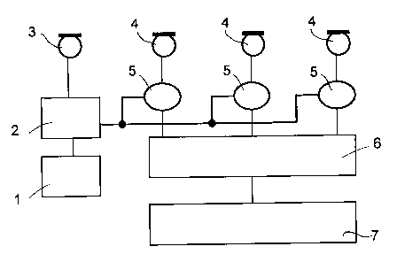
Fig. 1 shows a block diagram of a system for 3D real-time sonography according
to the
present invention, based on analogue send signals and corresponding to
analogue
processing of echo signals;
Fig. 2 shows a block diagram of a system for 3D real-time sonography according
to the
present invention based on digital processing of echo signals;
Figs. 3A and 3B show a specific send signal named "Chirp" and the
corresponding echo
signal;
Fig. 4 shows an echo signal of the "Chirp" according to Fig 3, reflected by 3
points;
Fig. 5 shows the correlation result of the "Chirp" according to Fig 3 with an
echo signal
according to Fig 4;
Fig. 6 shows an echo signal having an SNR of 0 dB;
Fig. 7 shows the correlation result of the echo signal according to Fig. 6
with a send signal
according to Fig 3; and
Fig. 8 demonstrates the method of triangulation based on three receivers for
the
calculation of space co-ordinates of a reflection point.
CA 02366534 2001-09-18
-9-
Fig. 1 is the block diagram of a system for 3D real-time sonography according
to the
invention, in which an analogue send signal is generated and echo signals are
consequently subject to analogue processing. The generator 1 generates a
carrier
frequency, modulated in a modulator 2 subject to an arbitrary function. This
send signal is
transmitted in this embodiment by a transmitter 3 into a medium or body. In
this
embodiment, the echoes reflected by any structures within the medium are
received by
three receivers 4. Therefore, for the determination of reflections initially
each of the echo
signals must be correlated to the send signal in the correlator 5. In this
process, each
reflection point in the medium is "detected" by individual receivers 4 at
another point in
time. For this purpose, the modulator 2 is connected to the correlator 5 of
each individual
receiver 4. Similar patterns in the send signal and each echo signal must be
interpreted as a
reflection. For instance, detection of these patterns may also be effected by
displacing the
send signal on the echo signals until agreement is obtained, equal to
reference to a
reflection. The result of this correlation shows a set of reflections, each
representing the
total path of the send signal from the transmitter 3 to the reflection point
and back to the
corresponding receiver 4. This means that the transmitter 3 and the
corresponding receiver
4 are in the two focal points of an ellipsoid. The space co-ordinates of these
reflection
points are calculated in the downstream computing unit 6 by a simple
triangulation
method. The starting point is that the points that are located at the same
distance from the
transmitter 3 to the reflection point and back to the receiver 4 are located
on the same
ellipsoid. The point of intersection of the three ellipsoids specifies the
space co-ordinates
on which the actual reflection occurred. Fig. 8 clearly shows this situation.
The space co-
ordinates are then displayed on the visualization display 7 at the appropriate
intensity.
Fig. 2 shows the block diagram of a system for 3D real-time sonography
according to the
invention, but subject to digital data processing. The generator 1 generates a
carrier
frequency, modulated in a modulator 2 having an arbitrary function. In this
embodiment,
the send signal is also transmitted by a transmitter 3 into any medium. As a
variation from
the first embodiment, an A/D converter 8 is arranged between the modulator 2
and each
correlator 5 as well as each receiver 4 and associated correlators 5. In
addition, an
additional memory 9 is arranged in this embodiment between the A/D converter 8
for the
CA 02366534 2001-09-18
- 10-
modulated signal and the generator 1, saving the transmitted send signal for
later
repetition. For this purpose, the memory 9 is coupled with the generator 1.
Fig. 3 shows a send signal of an increasing frequency. This is a Chirp having
a frequency
of f",;~ to fmax. The wave length of this signal decreases from left to right
in the drawing.
The entire data content of the range of interest is simultaneously scanned by
one send
signal only, followed by parallel processing in a fast computer.
Each of the receivers 4 receives the echo signals of the send signal described
in Fig. 3. Fig.
4 shows such an echo signal, received by a receiver 4, which has been
reflected in three
points. In this figure, the echo signal is not superposed by any noise
portions. Only the
first reflection point can be seen in the figure. Other reflection points can
no longer be
detected due to superpositions of the echoes in this diagram. Only after
correlation of the
echo signals with the send signal, other reflection points will appear.
Fig. 5 shows the correlation result of the "Chirp", according to Fig 3, with
the echo signal
according to Fig 4. A sample feature of this amplitude proportional to the
reflection and/or
absorption capacity is generated exactly at the reflection points.
Fig. 6 shows the run of a highly noisy echo signal, the SNR of which is 0 dB.
In this echo
signal, no reflections can be seen. After correlation to the send signal, the
signal
characteristic according to Fig 7 results. This signal is comparable with the
signal of Fig 5,
with the reflection points being clearly noticeable. This explains a major
benefit of the
method according to the invention. Even based on an SNR of -20 dB, reflection
points
were still clearly detectable in the echo signals. Only at a very unfavourable
SNR, no
evaluation was possible.
When using the system according to the invention and the method according to
the
invention for medical diagnostics, the features of the medium are of eminent
significance.
Due to its complex nature, it is very difficult to derive a simplified model,
describing the
frequency dependence of ultrasonic attenuation. In general, a linear
association is assumed
between attenuation, the signal path length and frequency. When G represents
attenuation
(in dB), f frequency (in MHz), z depth (in cm) in the medium and (3 the
attenuation
constant (in dB/[MHz cm]) of the medium, the following will be obtained:
CA 02366534 2001-09-18
-11-
G=2(3fz.
Higher frequencies are therefore more attenuated than lower frequencies. Table
1 shows
the attenuation constants for various types of tissues:
Table 1
Tissue Attenuation constant
(dB/[MHz cm])
Liver 0.6 - 0.9
Kidneys 0.8 - 1.0
Gall bladder 0.5 - 1.0
Fat 1.0 - 2.0
Blood 0.17 - 0.24
Plasma 0.01
Bones 16.0 - 23.0
Table 2 lists the attenuation (in dB) depending on the depth in tissue and the
frequency for
tissue subject to an attenuation constant of 0.7 dB/[MHz cm]).
Table 2
z(cm) 30 25 20 1 S 10 5
f(MHz) (dB)
1 42 35 28 21 14 7
2 84 70 56 42 28 14
3 126 105 84 63 42 21
3,5 147 122.5 98 73,5 49 24.5
5 210 175 140 1 OS 70 3 5
7,5 315 262.5 210 157,5 105 52.5
10 420 350 280 210 140 70
As it is possible, as shown in Figs. 4 to 7, to still detect the positions of
reflection points
even if the SNR is unfavourable, very good results may be achieved at
relatively low
CA 02366534 2001-09-18
-12-
frequencies and low sonic energies, irrespective of the attenuation in tissue,
according to
tables l and 2.
Fig. 8 shows the method of triangulation based on three receivers for the
calculation of
space co-ordinates of a reflection point. It explains how it is possible to
calculate the space
co-ordinates of reflection points by the transmission of an arbitrarily
modulated signal.
After having determined the distances of reflection points in the correlator
between each
transmitter 3 and the reflection points up to the corresponding receivers 4,
ellipsoids may
be defined, in the focal points of which the transmitter 3 and/or the receiver
4 are
arranged. Each intersection of all three ellipsoids marks the space co-
ordinates of a
reflection point. Should one transmitter and more than three receivers be
available, more
than three ellipsoids will be available for each reflection point, all
intersecting in one
point, defining the space co-ordinates of the corresponding reflection point.