Note: Descriptions are shown in the official language in which they were submitted.
CA 02366822 2001-10-01
WO 00/59411 PCT/US99/07309
SURGICALLY IMPLANTABLE KNEE PROTHESIS
TECHNICAL FIELD
The present invention pertains to prosthetic devices. More
particularly, the invention pertains to self-centering knee joint prostheses
which may
be surgically implanted between the femoral condyle and tibial plateau of the
knee.
BACKGROUND ART
Articular cartilage and meniscal cartilage provide the mobile weight
bearing surfaces of the knee joint. Damage to these surfaces is generally due
to
genetic predisposition, trauma, and/or aging. The result is usually the
development
of chondromalacia, thinning and softening of the articular cartilage, and
degenerative
tearing of the meniscal cartilage. Various methods of treatment are available
to treat
these disease processes. Each option usually has specific indications and is
accompanied by a list of benefits and deficiencies that may be compared to
other
options. Nonsteroidal anti-inflammatory drugs (NSAIDS), cortisone injections,
arthroscopic debridement, osteotomy, unicompartmental knee replacement, and
total
knee replacement have all been used to treat the disease depending on the
severity
of the process.
Currently, there is a void in options used to treat the relatively young
patient with moderate to severe chondromalacia involving mainly one
compartment
of the knee. Some patients cannot tolerate or do not want the risk of
potential side
effects of NSAIDS. Repeated cortisone injections actually weaken articular
cartilage
after a long period of time. Arthroscopic debridement alone frequently does
not
provide long lasting relief of symptoms. Unicompartmental and bicompartmental
total knee replacements resect significant amounts of bone and may require
revision
surgery when mechanical failure occurs. Revision total knee replacement
surgery
is usually extensive and results in predictably diminished mechanical life
expectancy.
-1-
CA 02366822 2001-10-01
WO 00/59411 PCT/US99/07309
Therefore, it is best to delay this type of bone resecting surgery as long as
possible.
DESCRIPTION OF THE RELATED ART
Several approaches have generally been employed in the past to
correct the aforementioned problems. The first approach involves repair of
articular
or meniscal cartilage. Repair of the articular cartilage by surgically
transplanting
autogenous or autologous osteochondral core grafts has had limited success,
but is
not always indicated. Meniscus repair using barbed "arrows" such as the Bionix
"Meniscus Arrow" has been used for "bucket-handle" tears, but is not
applicable to
other knee joint problems. Thus, these methods have limited scope and are
generally
confined to unique kinds of damage.
In the second approach, a unicompartmental or bicompartmental bone
resection is performed, replacing the bone with a load bearing prosthesis.
This
resection may be performed only on the femoral condyle, or may include the
tibial
plateau. In either case, the resection involves considerable surgical skill,
and results
in prosthetic devices physically anchored into the bone structure. Not only is
such
reconstruction expensive major surgery, but moreover, the mechanical means of
attachment may fail as the patient grows older. Examples of prostheses
utilized in
such methods are those disclosed in Ries, U.S. Patent 5,549,688; Cloutier,
U.S.
Patent 4,207,627; and Shah, U.S. Patent 5,263,987.
The third approach has been to replace the meniscal cartilage
("meniscus") with a soft, compliant material. In theory, such devices cushion
the
femoral and tibial bearings surfaces and distribute loads uniformly over a
large
portion of the knee joint due to the ability of these devices to elastically
deform.
This ability to deform can also be a detriment, however, when it is desired to
isolate
portions of the articular cartilage or bone surfaces from loads. Moreover,
such
devices are prone to tearing or disintegration under repeated stress due to
their low
tensile strength and modulus. Being flexible, they may be ejected from the
meniscular cavity if not anchored in place. Anchoring devices may create an
area
-2-
CA 02366822 2001-10-01
WO 00/59411 PCTIUS99/07309
susceptible to fatigue fracture, causing dislocation of the prosthesis and
further
damage to the knee joint.
Thus, for example, Kenny, in U.S. Patent 4,344,193, discloses a
meniscus prosthetic device of a rubbery material such as silicone rubber,
having two
prominences, which interact with a space defmed by the geometry of the femoral
condyles. This interaction involving the prominences, together with surgical
sutures
secured to surrounding soft tissue, are said to maintain the meniscus fixed in
the
proper location. A porous border, into which fibrous tissue ingrowth is
theorized
to occur, may also assist in performing the locating function. A similar
approach
is disclosed in Stone, U.S. Patents 4,880,429; 5,007,934; and 5,158,574, where
the
meniscus comprises a porous matrix of biocompatible fibers or fibers of
natural
origin to act as a "scaffold" for regrowth of native meniscal tissue. The
device is
manufactured with an outer contour substantially the same as that of a native
meniscus.
In Kenny, U.S. Patent 5,171,322, a meniscus prosthetic device is
composed of a biocompatible, deformable, flexible and resilient material
having the
shape of a natural meniscus, but having a tail which may extend through holes
bored
in the bone to anchor the device. In similar fashion, Wall, in U.S. Patent
4,502,161, discloses an extra-articular extension attached to the bone outside
the
joint; while Richmond, U.S. Patent 4,919,667 employs natural fibrous growth to
positively anchor his device, again shaped like a natural meniscus. Schwartz,
U.S.
Patent 5,344,459 utilizes a soft device of rings that are inflatable with air,
liquid, or
semisolid to provide a gel cushion between joint surfaces.
The previously described devices of the prior art second approach all
utilize soft, cushiony materials which are anchored in place by mechanical
means or
through tissue regrowth to prevent movement of the device or its extrusion
(spitting)
from the compartments. One device which differs from those previously
described,
and which has been used in knee reconstruction, is the so-called "MacIntosh
knee,"
where a hard prosthesis is located by means of protruding ridges, generally in
the
form of a cross, which nest into corresponding grooves cut into the tibial
plateau to
-3-
CA 02366822 2006-09-27
71087-654
prevent movement of the device. These devices have been
found to cause pain in the knee joint. This type of
prosthetic device and the so-called "McKeever" device
require very invasive surgical procedures, require large
arthrotomy, require bone and tissue resection, and are
irreversible processes.
SUMMARY OF THE INVENTION
The present invention pertains to a meniscal
device suitable for surgical implantation into a knee
compartment defined by the space between a femoral condyle
and the respective tibial plateau. The device is a hard,
self-centering meniscal device devoid of physical means that
fix its location. The device does not have the natural
shape of the meniscus, but rather is designed such that
articulation of the knee results in a modest amount of
lateral/medial and anterior/posterior translation, relative
to the tibial plateau, of the device.
More particularly, according to one aspect the
invention provides a unicompartmental knee prosthesis
suitable for implantation between the femoral condyle and
the associated tibial plateau of a knee compartment,
comprising a hard body having a substantially elliptical
shape in plan, a peripheral portion of said body being of
greater thickness than a central portion of said body, said
body devoid of physical means of attachment which fix its
location in said knee compartment.
According to another aspect the invention provides
a meniscal load distribution prosthesis suitable for an
arthroscopically assisted implantation into a meniscal
cavity of one compartment of a knee joint between a femoral
condyle and the respective tibial plateau, said prosthesis
-4-
CA 02366822 2006-09-27
71087-654
comprising a high modulus material having a hardness greater
than about Shore D 60, said prosthesis being substantially
elliptical in plan, and having a concave femoral meniscal
surface, a convex tibial meniscal surface, and a periphery
defined by lateral, medial, anterior, and posterior
portions, the periphery being on average of greater
thickness than a central portion of the meniscal device,
said femoral meniscal surface having a contour such that the
contour angle of said femoral meniscal surface is
approximately the same as the contour angle of said femoral
condyle, relative to the contour angle of the tibial
plateau, said tibial meniscal surface having a contour such
that the contour angle is substantially the same as the
contour angle of said tibial plateau.
In another aspect there is provided a method of
selecting a prosthesis for surgical knee reconstruction of a
patient in need thereof, said method comprising: a)
determining a proper size and shape of the novel
unicompartmental knee prosthesis; and b) selecting a
unicompartmental knee prosthesis of said proper size and
shape.
In a further aspect the invention provides a
process for the preparation of a unicompartmental prosthesis
suitable for implantation into a knee joint, between a
femoral condyle and its corresponding tibial plateau, said
process comprising: a) determining the geometry of said
femoral condyle by a non-invasive imaging technique; b)
determining the geometry of said corresponding tibial
plateau; c) molding the novel prosthesis which is
dimensioned to fit within the compartment defined on its top
and bottom surfaces by the geometry of said femoral condyle
and said tibial plateau.
-4a-
CA 02366822 2006-09-27
71087-654
BRIEF DESCRIPTION OF THE DRAWINGS
FIGURE 1 depicts the relationship between the
radius (RFC) and the femoral condyle.
FIGURE 2 illustrates the shape of the femoral
condyle in cross section.
FIGURE 3 illustrates certain spatial relationships
with respect to an embodiment of the subject invention
device.
FIGURE 4 illustrates the distorted elliptical
(kidney bean) shape of a device.
FIGURE 5 and 6 illustrate cross-sections of a
device in orthogonal planes.
-4b-
CA 02366822 2001-10-01
WO 00/59411 PCT/US99/07309
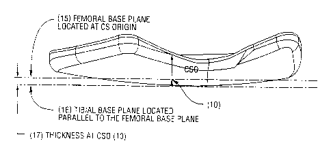
FIGURE 7 illustrates a device contour and its relationship with the
femoral and tibial base planes.
FIGURE 8 illustrates the axes and planes which may be used to
generate the shape of a meniscal device in one embodiment of the subject
invention.
FIGURE 9 illustrates the relationship of various coordinates and axes
of a device viewed perpendicular to the plane of the device.
FIGURE 10 illustrates one embodiment of a device viewed in plan.
FIGURE 11 illustrates the device of Figure 10 viewed from the side.
DESCRIPTION OF THE PREFERRED EMBODIMENTS
The prosthetic meniscal devices of the subject invention are
unicompartmental devices suitable for minimally invasive, surgical
implantation. By
the term "meniscal devices" is meant that the devices are positioned within a
compartment in which a portion of the natural meniscus is ordinarily located.
The
natural meniscus may be maintained in position or may be wholly or partially
removed, depending upon its condition. Under ordinary circumstances, pieces of
the natural meniscus which have been torn away are removed, and damaged areas
may be trimmed as necessary. In somewhat rarer instances, the entire portion
of the
meniscus residing in the meniscal cavity may be removed. Thus the term
"meniscal
device" is descriptive of the location of the device rather than implying that
it is a
replacement for, or has the shape of, the natural meniscus. Actually, as
described
hereinafter, the shape of the meniscal device is not the same as the natural
meniscus,
and in most cases, will not entirely replace the meniscus.
By the term "unicompartmental" is meant that each device is suitable
for implantation into but one compartment defined by the space between a
femoral
condyle and its associated tibial plateau. In other words, the device is not a
"bicompartmental" device which, in one rigid device, could be inserted into
both of
-5-
CA 02366822 2001-10-01
WO 00/59411 PCTIUS99/07309
the two femoral condyle/tibial plateau compartments. In many, if not most
cases,
a device will be inserted into one compartment only, generally the medial
compart-
ment, as the meniscus and associated articular surfaces in these compartments
(left
knee medial and right knee medial compartments) are most subject to wear and
damage. However, it is possible to insert two separate devices into the medial
and
lateral compartments of the same knee, or to use two such devices that are
mechani-
cally but non-rigidly linked.
The meniscal devices are translatable but self-centering. By
"translatable" is meant that during natural articulation of the knee joint,
the device
is allowed to move, or change its position. Thus, the device is devoid of
means of
physical attachment which limit its movement, for example, screws, mating
ridges
and depressions, porous areas to accommodate tissue regrowth, and the like.
By the term "self-centering" is meant that upon translation from a first
position to a second position during knee articulation, the device will return
to
substantially its original position as the articulation of the knee joint is
reversed and
the original knee position is reached. Thus, the device will not progressively
"creep" towards one side of the compartment in which it is located. Rather,
the
angle of attack of the femoral condyle and/or tibial plateau bearing surfaces
against
the meniscal device will ensure that the device reversibly translates during
articula-
tion, maintaining the meniscal device, on average, in the same location for
any given
degree of knee articulation.
Contrary to most devices which are composed of soft, compliant
material designed to assume the function of the natural meniscus which they
replace,
the present device is composed of relatively hard, relatively high modulus
material.
Suitable materials are, for example, steel, ceramics, and reinforced and non-
reinforced thermoset or thermoplastic polymers. The device need not be made of
a single material, but composite structures of steel/thermoplastic,
steel/ceramic,
ceramic/polymer, etc., may be used. Alternatively, composites of above
materials
with biologically active surfaces or components may be used. Biologically
active
components include surfaces that may contain pharmaceutical agents to
stimulate
-6-
CA 02366822 2001-10-01
WO 00/59411 PCT/US99/07309
cartilage growth or retard cartilage degeneration that may be delivered at
once or in
a timed- release manner.
Generally, portions of the devices expected to have the most wear due
to either greater movement relative to the mating surface, i.e., relative to
the femoral
condyle or tibial plateau; or high stress, may be made of stronger, more
abrasion
resistant material than the remainder when composite structures are used. This
method may be ideal for use in conjunction with cultured chondrocyte
implantation
(cartilage cells used as seeds) or osteochondral transplantation or
mosaicplasty.
Moreover, when the locus of damage to the articular cartilage or to portions
of the
bone structure are known, the relatively constant radius of the surface of the
meniscal device will bridge the defective areas at these loci, thus
redistributing load
to healthy tissue and allowing inflamed, diseased, or other damaged areas to
regenerate.
For example, a portion of the femoral condyle, tibial plateau, articular
cartilage, etc., may have been damaged or may experience tissue degeneration.
The
continued load experienced at such points and the wear experienced as the knee
flexes will substantially hinder the regeneration of healthy tissue. If
suitable
biologically active materials, chondrocytes, etc. are applied to the damages
or
degenerated surface to assist in tissue regeneration, these will, under
ordinary
circumstances, be rapidly dissipated. If a flexible, cushiony material is
inserted
within the knee compartment, the damaged area will still experience intimate
contact
with the damaged area under static loads, and will also experience continued
wear
and abrasion under non-static conditions. Under such circumstances, active
substances will be rapidly dissipated. However, more importantly, newly
regenerated articular cartilage not having the necessary density or
cohesiveness to
withstand wear, will be rapidly eroded away.
The subject invention meniscal load distributing devices may be
supplied with a contour which allows the devices to act as a surface which
distributes
the loads evenly over regions of healthy articular cartilage, in general,
abutting and
bridging surfaces where articular cartilage degeneration or damage has
occurred.
-7-
CA 02366822 2001-10-01
WO 00/59411 PCT/US99/07309
Active substances may be applied at once or in a timed-release manner to the
degenerated or damaged articular cartilage surface by means of, or in
conjunction
with, the meniscal device. Because the recess or shape of the meniscal device
protects the damaged area from loads and wear, tissue regeneration may occur
without disturbance. The regenerating tissue will have time to mature and
crosslink
into a fully developed matrix. Moreover, as regeneration proceeds, the
regenerating
tissue will assume a shape dictated by the shape of the meniscal load-
distributing
device. Growth under these circumstances has the greatest potential for dense,
ordered growth most closely replicating the original surface.
The hardness of the meniscular devices is preferably higher than
Shore 60 D. The shore hardness may range from that common for engineering
grade plastics to hardened steel and titanium, and preferably on the portion
of the
Rockwell hardness scale typical of steels, hard plastics and ceramic
materials. From
the high hardness desired of the meniscal device, it is readily apparent that
the
devices function in a manner completely different from those of the prior art
such
as Stone, Dedo, Schwartz, Richmond, and Kenny. The purpose of the devices of
the subject invention is to achieve a span-like effect to bridge the defective
areas.
However, in a composite variation, any single component (like a bioactive
material
component) may be softer than the supporting material. Rather than deforming
to
distribute a load relatively equally on the mating surfaces, the meniscal
devices of
the present invention function as rigid, substantially non-deforming, self-
centering
bearings, which do not necessarily spread the load uniformly, but rather may
concentrate the load upon desired points, spanning areas of imperfection. If a
soft
and/or low modulus elastomer or thermoplastic is used for the entire device,
not only
is the load not concentrated on healthy tissue, but moreover, damaged areas
due to
wear and/or degeneration will also be subjected to loading, decreasing the
opportunity for the body's natural regenerative capability to function.
The high modulus of the subject meniscal devices thus allows for the
provision of recessed or non-contacting areas of the device to encourage
articular
cartilage regeneration. In softer, lower modulus materials, the naturally
occurring
loads, which may exceed 1000 lbs/in2 in certain cases, will cause the softer
devices
-8-
CA 02366822 2001-10-01
WO 00/59411 PCT/US99/07309
to deform and allow ordinarily non-contacting areas to contact bone or
cartilage for
which contact is desired to be avoided. A flexural modulus of elasticity for
load
bearing portions of the meniscal devices should therefore be preferably
greater than
2x10⁵ psi, and more preferably greater than 3x10⁶ psi. Portions of the device
not
exposed to the highest loads may be made of lower modulus materials, which may
be softer as well, e.g., in a non-limiting sense, nylon, polyurethane,
polypropylene,
polyester, and the like, optionally fiber reinforced.
As indicated previously, the meniscal devices of the subject invention
may be manufactured so as to substantially contain or have deposited thereon,
a
biologically or pharmaceutically active material. This is particularly
suitable when
the device bridges a defective area of bone or articular cartilage. In such
cases, the
meniscal device may be provided with a coating containing a biologically or
pharmaceutically active material, for example one that promotes tissue
regrowth or
one that decreases inflammation. Such materials may also, and more preferably,
be
contained in a portion of the meniscal device. The portion may be filled with
medication, or may be filled with a gel, paste, or soft polymer material that
releases
medication over a period of time. Preferably, this medically active portion
does not
actually contact, or minimally contacts, the damaged tissue. This freedom from
contact is made possible by the surrounding bearing surfaces. Coatings may
also be
of a gel, paste, or polymer containing time-release medicaments. Biologically
and
pharmaceutically active materials are identified subsequently herein as
"active
materials."
The actual shape of the meniscal devices may be tailored to the
individual. Individuals with high varus (heels in, knees out - typical
degenerative
arthritis or valgus (heels out, knees in) deformation due to wear,
degeneration, or
disease, may require meniscal devices which are of considerably greater
thickness
over the portions where wear is most advanced. In youthful patients, where
trauma-
induced damage rather than severe wear or degeneration has occurred,
differences
in device thickness will be more moderate. In general, the meniscular devices
are
kidney-shaped when viewed from above, and have a negative meniscus shape when
viewed from the side, i.e.; the thickness along the periphery of the device is
greater
-9-
CA 02366822 2001-10-01
WO 00/59411 PCT/US99/07309
than the thickness along the center of the device. The kidney-shape in plan
(Figure
4) may be described generally as elliptical, the shape resembling a distorted
ellipse,
with the distortion (30) (Figure 8) generally determined by the shape and
location
of the tibial spine. The device covers not only the peripheral areas of the
meniscus
but also the central weight bearing surface of the femoral condyle and tibial
plateau.
For example, the inside (central) thickness ((17) Figure 7) may range
from about 0.010 inches (0.25mm) to 0.20 inches (5mm) over a somewhat
elliptical
area measuring, for a hypothetical average knee, about 1.0 inches (25.4mm)
along
the minor axis and 1.40 inches (35.6mm) across the major axis. The meniscal
devices are generally thicker at the posterior portion (11) of the device (the
portion
of the periphery nearest the posterior of the knee joint) as compared to the
lateral (7)
or medial (6) sides. The medial(6) side of a medial compartment device,
(lateral side
of a lateral compartment device) is generally thicker than the lateral (7)(the
side
along the tibial spine) side, and the medial (6) and anterior (4) sides are
generally
of the same thickness. The outside thickness may range up to 0.40 inches
(10mm)
in some cases.
The edges of the device are rounded rather than presenting the sharp
corners of the devices of U.S. Patent 5,158,574. This rounded periphery is
necessary due to the fact that the device will be allowed to move within the
cavity.
Movement of a device having a periphery with sharp corners would result in the
potential for severe damage to the surrounding tissue and articular surfaces,
in
addition to causing pain. The "kidney shaped" devices are termed
"substantially
elliptical" as that term is used herein. The "depression" in the elliptical
shape on the
part of the device which will be proximate to the tibial spine (30 in Fig. 4)
will vary
from patient to patient. It is possible, due to the great range of variability
of human
anatomy that this depression might be absent in devices for some patients.
However,
the overall shape in plan is substantially elliptical regardless.
As shown for the femoral and tibial surfaces of the device in Figure
1 and in Figure 2, the surfaces of the meniscal device generally are convex or
concave in a symmetrical manner, i.e., their radius of curvatures in a given
-10-
CA 02366822 2001-10-01
WO 00/59411 PCT/US99/07309
direction, are in general, relatively constant. There are generally four
directions of
radii need to describe the two surfaces, as illustrated in Figures 1-9, the
femoral
anterior to posterior (RFC)(2), the tibial anterior to posterior (RTc)(13),
the femoral
medial to lateral (RFCX)(3)and the tibial medial to lateral(RTcx)(14). Typical
values
would be RFC from 1.1-2.0 inches (28-51mm), RFCX from 0.5-1.5 inches (12.7-
38mm), RTP from 6-12 inches (15.2-30.5cm) and RTPX from 1.5-3 inches (38-
76mm).
An example of a device would have the following values: RFc=1.6 inches
(40.6mm),
RFCx =1.2 inches (30.5mm), RTP =10 inches (25.4cm) and RTpx = 2.3 inches
(58.4mm). However, it is also necessary to allow for an increasing or
decreasing
radius to accommodate a specific patient's needs. For example, the RFC of such
a
device may have a radius of 1.3 inches (33mm) at the most anterior point of
the
device but may increase in a geometric manner to a radius of 1.8 inches
(45.7mm)
at its most posterior aspect. Simultaneously, the RFCX may have a radius of
0.8
inches (20.3mm) at the most anterior point of the device but may increase in a
geometric manner to a radius of 1.3 inches (33mm) at its most posterior
aspect. Such
transitions of radii would occur in a smooth manner consistent with a bearing
surface.
The asymmetric shape of the device still allows for a good fit to the
femoral condyle as the femoral condyle has an almost constant radius of
curvature,
as shown in Figure 1, in the area that the tibial plateau moves against. This
radius
of curvature, when viewed from above, as in Figure 3, generally describes the
contour angle (or the predominent orientation of the radius of curvature along
the
anterior to posterior direction) of the femoral condyle. In addition, the
posterior rim
and the large radius of the tibial side of the device prevents the device from
"spitting" . Thus, regardless of whether the knee is in extension or flexion,
the
degree of "tightness" remains the same and the device will not restrain or
limit the
motion of the knee. Further, the surface area of the femoral side may be
smaller than
its corresponding tibial surface along the anterior, medial (medial side of a
medial
device) and posterior aspects of the device. In such a manner, the femoral
side of the
device would be closer in size to the femoral condyle, while the tibial
plateau would
remain fully covered thus, giving the device a "sloped" shape along the
aforementioned edges. Such a device shape would be suitable for use with
certain
-11-
CA 02366822 2006-09-27
71087-654
anatomical shapes as well as for use with a partially or fully intact
meniscus. The
term "substantially immune from spitting" means that the device, without any
physical attachment to the knee, will ordinarily remain in place in the knee
compartment over the normal range of activity expected of the knee.
The ability of the subject meniscal devices to translate yet be self-
centering is created by the geometry of the devices in conjunction with the
geome-
tries of the femoral condyles and tibial plateaus. The bearing surface
geometries of
the tibial plateaus and the femoral condyles define the axis of joint rotation
of the
knee. Figure 2 shows the shape of the femoral condyle in cross section. Figure
3
shows the angle (8) of the contour of the femoral condyle relative to the
tibial plateau
(5) to be such that the planes of symmetry of the respective condyles are not
orthogonal to the axis of rotation of the joint, but instead are at angles
that converge
toward the anterior portion of the particular knee compartment.
The axis of rotation of the tibia on the femur is 90 degrees to the path
of the tibial plateau against the femoral condyle. The two tibial plateaus
(medial and
lateral) are not in the same plane with each other but do act in a relatively
constant
radius to its respective femoral condyle. In other words, although the
symmetry of
the device's femoral side may be matched with the femoral condyle while the
leg is
in full extension, the rotation of the tibial plateau against the femoral
condyle is
along a constant axis of rotation (90 degrees to the axis of rotation), thus
the
angularity of the axis of symmetry of the femoral condyle relative to the axis
of
symmetry of the tibial plateau is not parallel but at some acute angle. Also,
the axis
of symmetry of the tibial plateau is not parallel to the path of rotation of
the tibia
relative to the femur but also at some mildly acute angle. Thus, the true
orientation
of the device, regardless of the relative orientations of symmetry of the
tibial side
to the femoral side is 90 degrees to the true axis of rotation as described in
Hollister
et al., "The Axes of Rotation of the Knee", CLIN. ORTHOPAEDICS AND REL. RES.,
290 pp. 259-268, J.B. Lippincott Co., m 1993,.
Any localized positions of higher loads are.self-limiting due to the ability
of the
device to translate both rotationally and laterally which rnimics the true
motion of
the natural meniscus as described by Hollister.
-12-
CA 02366822 2001-10-01
WO 00/59411 PCTIUS99/07309
The geometry provided by the meniscal device thus mimics the
geometry of the tibial plateau with the meniscus intact with respect to the
femoral
condyle and mimics the geometry of the tibial plateau with the meniscus
removed
with respect to the tibial plateau, resulting in but little translation
relative to the tibia,
except for a relatively small rotational and lateral components. With respect
to the
femoral condyle, however, the device experiences large relative movement, and
a
rotational component brought about by any difference in the contour angle (22)
of
the femoral condyle and the concave meniscal device topmost surface (femoral
surface). This rotational component further ensures that the device is self-
centering,
and cannot be "spit" from the joint. In general, the contour angle (22) of the
femoral surface of the meniscal device should be within +/- 15 , and in
general, less
than 20 , of the contour angle of the femoral condyle relative to the tibial
plateau.
Too large an angle will provide too high a centering force, and may accelerate
wear
of the femoral condyle articular cartilage or the device itself.
In the "rest position," where the knee is in full extension, the outer
contours of the meniscal device are designed to substantially mate with the
corresponding tibial and femoral surfaces. As the knee is flexed, the mating
along
the tibial surface is substantially maintained, with only a slight rotation
which is
resisted due to the fact that the contour angle or orientation of the tibial
surface of
the meniscal device and the contour angle or orientation of the tibial plateau
are the
same. However, the contoured mating surfaces of the femoral condyle and
femoral
meniscal device surfaces can become increasingly dissimilar when the joint
articulates, as the contour angles will not necessarily mate correctly
throughout the
entire articulation cycle. This can cause relative lateral or rotational
movement, in
the tibial plane, between the femoral condyle and the femoral surface of the
meniscal
device. The forces generated by the increasingly different geometry creates a
rotational moment, in the tibial plane, which is resisted along the mating
tibial
surfaces but which also results in a restoring force tending to correctly
locate the
meniscal device along the femoral condyle. Thus, the device is self-centering,
in
part, as a result of the similar contour angles of the femoral condyle and the
femoral
surface of the meniscal device.
-13-
CA 02366822 2001-10-01
WO 00/59411 PCT/US99/07309
Generally speaking, each knee presents a different geometry of the
respective femoral condyles and tibial plateaus. Even with respect to the
right and
left knees of a single individual, although bilateral symmetry dictates that
the left and
right knee components should be mirror images, this is often only an
approximation.
Thus, the shape of the affected femoral condyle and tibial plateau (while
discussed
herein in the singular, more than one pair of condyle(s)/plateau(s) may be
involved),
will have to be ascertained to determine the correct geometry of the meniscal
device
for a given patient. In some cases, it is desirable to offset the contour
angles (from
the CSO(30)) of either or both of the meniscal surfaces. This is done to bias
the
thickness of the meniscal device to the periphery of the device. This would be
done,
for instance, to accommodate the absence or presence of the meniscus or for
some
other anatomical reasons.
To implant a meniscal device that possesses the characteristics
required by the subject invention, the patient's knee joint may be examined by
a non-
invasive imaging procedure capable of generating sufficient information such
that on
appropriately sized and shaped meniscular device may be selected. While a
variety
of non-invasive imaging devices may be suitable, for example X-ray devices and
the
like, it is preferable that information as to the size and shape of the
meniscal device
be provided by magnetic resonance imaging (MRI).
Two methods of non-invasive imaging for selection of a suitable
prosthesis are preferred. In the first method, MRI or other non-invasive
imaging
scans, optionally coupled with exterior measurements of the dimensions of the
relevant tibial and femoral portions including the surfaces of the articular
cartilage
of the tibia and femur, may be used to establish a library of meniscal
prostheses
whose size and geometry differ according to the age and size of the patient,
the
patient's genetic make-up, and the like. A limited number of "standard"
meniscal
device molds are then created, from which corresponding "standard" meniscal
devices are produced.
In this first method, a non-invasive imaging scan, such as X-ray or
MRI, together with knowledge of the patient's genetic make-up, general body
type,
-14-
CA 02366822 2001-10-01
WO 00/59411 PCT/US99/07309
extent of the disease, degeneration, or trauma and the like, will enable the
surgeon
to select a meniscal device of the correct size and shape from the library for
the
patient. The device is then introduced by arthroscopically assisted
implantation,
generally limited to extensive clean-up of existing damaged tissue, e.g., torn
or
particulate natural meniscus damage. It may also be used in conjunction with
tibial
osteotomy or articular surfacing procedure such as cartilage transplantations
or
abrasion anthroplasty. Following insertion of the device, X-ray, Fluoroscopy,
or
MRI may be used to assess the correct positioning of the device both
intraoperatively
as well as postoperatively. Since the surgical procedures used are not severe,
and
also not irreversible, an unsuitable device may be readily removed and
replaced,
either with a different device from a meniscal device library, or by a custom
device.
In a second method, each patient receives one or more meniscal
devices that are custom tailored for the individual by producing a contour
plot of the
femoral and tibial mating surfaces and the size of the meniscal cavity. Such a
contour plot may be constructed from imaging data, i.e. MRI data, by a
suitable
computer program. From the contour plot, the correct surface geometry of the
meniscal device is determined from the shape of the respective tibial plateau
and its
contour angle (normally 0 degrees) and offset position, and the shape of the
femoral
condyle with its contour angle and its offset position. In general, the shapes
just
mentioned also include the articular cartilage, which, in general, is
maintained
substantially intact.
The following is an example of the procedure which may be followed
to design and construct a meniscal device from MRI data: From MRI image data,
the steps described below are preferably taken.
Using MRI data, from an Anterior-Posterior (AP) side view of a
medial or lateral compartment of the knee joint, at an angle which positions
the view
parallel to the AP direction (plane), as shown in Figure 1, of the femoral
condyle
when the knee is full extension, the maximum point of contact between the
femoral
condyle and the tibial plateau is determined by using the particular image
section
(cut) that represents the maximum femoral extension. The Femoral Cross-section
-15-
CA 02366822 2001-10-01
WO 00/59411 PCT/US99/07309
plane (21), Figure 2, is created normal to this view while the Femoral Sweep
plane
(8), is in the plane or at an offset angle (22) to the plane of the image
(Figure 1).
The intersection of these two planes represents 2 points of the Coordinate
System
Origin (CSO)(10).
From the Lateral-Medial view (LM), the planar image that represents
the maximum femoral extension will also determine the maximum point of contact
between the Femoral condyle and the Tibial plateau which represents the 3'd
point
of the CSO (10). The X-axis plane (9) is represented by this (LM) image view
and
intersects the CSO (10) in the (LM) direction. The Y-axis plane (5) is normal
to the
X-axis plane (9) and is normal to the (LM) image plane and in the AP plane.
The Z-
axis plane (12) is normal to the X-axis and is also in the (LM) image plane.
From the (AP) view, with an image that represents the Femoral Sweep
plane (8), the radius of curvature of the femoral condyle RFC(2) is determined
in the
AP view using the following equation: RFc =(CZ+4H2)/8H where C = the length of
a line across the cross-section and H = the height from the midpoint of line C
to a
point perpendicular on the circumference of the arc which is also the maximum
point
of contact between the articular surfaces of the respective femoral condyle
and the
tibial plateau.
Using the same (AP) image, the same procedure is used to determine
the radius of curvature of the tibial plateau, RTP (13) in the AP direction
(plane).
However, this radius must account for the thickness of the meniscus and the
meniscal thickness is not included when determining the RTp. (13)
From a Lateral-Medial view (LM), a cross-section is viewed of the
femoral condyle at the midpoint of the length of the femoral arc (representing
the X-
axis plane), the same procedure and equation of #1 is used to determine the
radius
of curvature of the cross sectional radii of both tibial, RTPX (14)(minus the
meniscal
thickness) and femoral, RFCX, surfaces.
-16-
CA 02366822 2001-10-01
WO 00/59411 PCT/US99/07309
Typical values would be RFC (2) from 1.1-2.0 inches, RFCX (3) from
0.5-1.5 inches, RTP (13) from 6-12 inches and RTPX (14) from 1.5-3 inches. In
this
particular example, the following values were used: RFC =1.6 inches, RFCx=1 =2
inches, RTP = 10 inches and RTPX = 2.3 inches
From a combination of these same LM and AP views, a determination
is made of the current joint spacing and any spacing which would be required
to
correct any varus or valgus misalignment of the joint. This measurement
determines
the thickness of the device (17) at the CSO (10) point.
A plane is created in the X-Y plane, which will naturally intersect the
CSO, which represents the bottommost surface of the femoral side of the
device.
This plane is called the Femoral Base Plane (15).
Another plane is created parallel to the Femoral Base Plane, but offset
some distance below which corresponds to the desired thickness of the device
as
determined above. This plane is called the Tibial Base Plane (16). It
represents the
bottommost surface of the tibial side of the device.
An understanding of the device, and the procedure used to derive its
geometry, may be facilitated by the following discussion: Using the following
concept: If a ball of radius = 1.0 inches, RFC, is placed in a sphere of
internal radius
= 2.0 inches, RTP, the area outside the area of immediate contact of the ball
on the
inner surface of the sphere is represented by a generally circular shape
(volume) of
constant, wedge shaped cross-section. If a device, representing this circular,
wedge
shaped volume, were to be placed in the sphere, the ball, when placed into the
sphere containing this device, would make intimate contact with both the
sphere (at
the opening of the device) and the device, thus distributing the load of the
ball over
a much larger area. Such a device can never escape the sphere without lifting
or
otherwise dislocating the ball because the ever-increasing thickness of the
device
(from the wedge shape) will cause increasing levels of interference with the
ball as
the device is moved in any lateral direction. However, the device may move
with the
-17-
CA 02366822 2001-10-01
WO 00/59411 PCT/US99/07309
rotation of the ball if the radius of the sphere is close to being concentric
with the
radius of the ball.
To stop this rotation of the device, one can attach the device (not
desired) or (preferred) increase the radius of the sphere (tibial) side up to
an
approximate order of magnitude larger than the femoral side radius thus
inducing a
relatively easier motion of the ball on the device versus the device to the
sphere or,
secondly, use an increasing radius of the sphere side of the device. This has
the
effect of trying to force a larger radius into a smaller one (which cannot
happen).
Obviously, this can only be used in one direction, but in the human knee
joint, the
device will only have a tendency to push from the posterior to the anterior
upon knee
rotation thus, the radius of curvature on the tibial side of the device, in
the Anterior
to Posterior direction, is the direction of increasing radii. The amount of
increase is
small, on the order of 5 to 15 % over the length of the device. Either
technique or
a combination of both can be used to successfully stop the rotation of the
device with
the femoral condyle ("the ball"). Thus, fixing its position relative to the
tibial plateau
without attachment.
The concept described in the prior to paragraphs describes the general
shape and function of the meniscus found in the normal human knee. Although
the
meniscus is crescent shaped, the natural anatomy of the knee completes the
generally
circular shape with the tibial spine, along the central axis of the knee, thus
locating
the femoral condyle at all times in its range of motion and limiting any
potentially
harmful positional excursions of the femoral condyle. Since the natural
meniscus is
attached to the membrane surrounding the knee, it does not need to be attached
to
the tibial spine to perform this locating function on the femoral condyle. If
the shape
of the meniscus is damaged or not present then, it cannot perform this
locating, load-
bearing function. Thus, the loads on the femoral condyle and tibial plateau
become
more concentrated leading to a gradual, arthritic degeneration of the
articular
cartilage surface of the femoral condyle. Disease and age can also have this
effect.
The purpose of the device is to reduce the concentrated loads on the
femoral condyle and its articular cartilage and to maintain proper spatial
location of
-18-
CA 02366822 2001-10-01
WO 00/59411 PCT/US99/07309
the femoral condyle to the tibial plateau. Since permanent attachment of the
device
is not desired nor easily accomplished, the circumferential shape of the
device is
generally kidney shaped to conform to the planar anatomy of the lateral or
medial
compartments of the knee and due to the differing radii of the femoral and
tibial
surfaces of the device, the "wedge" shape (Figures 5 and 6) required to keep
the
device centered under the femoral condyle while the condyle moves through its
range
of motion is naturally present.
Thus, with this understanding of the principal of the device's natural
tendency to remain correctly located under the femoral condyle, the amount of
"wedge" is determined by the difference in the radii from one surface of the
device
to the other surface of the device. Further, since the natural meniscus is
predisposed
to a greater "wedge" on the posterior (11 of Figure 5) and medial (4 of Figure
6) (of
a medial device, lateral on a lateral device) sides of the femoral condyle,
the device
can replicate this biased wedge by locating the center of the tibial radii
posterior and
medial to the CSO (10) of the device. This is accomplished by offsetting the
Femoral
Sweep Plane (8) and the Tibial Sweep Plane (5) some distance from the CSO (30)
as referenced by the Femoral Offset Sweep Plane (20) and the Tibial Offset
Sweep
Plane (18). The amount of this bias would be determined by the amount of
natural
meniscus remaining in the knee compartment.
In some cases is may be necessary to add "reverse (downward)"
curves, or cusps, to the device along two additional planes of revolution,
termed the
Anterior Cusp Sweep Plane (23) and the Posterior Cusp Sweep Plane (25),
generally, as shown in Figures 10 and 11, and located along the lateral aspect
(of a
medial device) of the device at the extreme anterior/lateral (28) and
posterior/lateral
protrusions (29). Their cross-section shapes are described by the radius of
curvature
in the respective Anterior Cusp cross-section Plane (24) and Posterior Cusp
cross-
section Plane (26). The radii of such cusps being on the order of 1/10ths of
inches
(several millimeters). Such circumstances would be when there is deformed
anatomy
or additional stabilization is required of the device.
-19-
CA 02366822 2001-10-01
WO 00/59411 PCT/US99/07309
With the above information, using a parametric design program such
as Pro Engineer from Parametric Technology Corporation, a solid block of
generally
the correct circumferential shape is referenced with a CSO (10) (X,Y,Z origin
point)in the middle of the block. From that reference point, Femoral (15) and
Tibial
(16) Base Planes are established along with the offset (20) and angle (22) of
the
Femoral Offset Sweep Plane (8) and the offset (18) and angle (0 ) of the
Tibial
Sweep Plane (8). This information, along with the calculated femoral (2,3) and
tibial
(13,14) radii) is required for proper sizing of the device.
The steps given above may be modified as necessary, and may be
combined or accomplished in other than the order given. This process is
exemplary
only, and not limiting. An example of a particular meniscal device design is
as
follows:
A femoral offset contour angle (22) of from about 00 to about 45 is
suitable, with 5 to about 35 preferable, and an angle in the range of 10 -
20 most
preferred, in this particular case, 15 (relative to the y axis) was chosen as
the
femoral offset contour angle. The femoral offset amount (20) was 0.10 inches
toward
the medial aspect (6), away from the CSO (10) and the y-axis (5). (The femoral
offset amount (20) from the CSO on the y-axis has a preferred range of +/-0.20
inches, with +/- 0.10 inches being most preferred). The tibial contour angle
was 0
(parallel to the y-axis (5)) with a tibial offset amount (18) of 0.20 inches
towards the
medial aspect. The tibial offset amount (18) from the CSO on the y-axis has a
preferred range between -0.20 inches to 0.40 inches with 0.0 inches to 0.20
inches
being most preferred. This effectively presents a relative angle of the
femoral sweep
plane and tibial sweep plane as 15 but with an intersection point (19) that
is
posterior and medial of the CSO (10), thus creating a wedge shape that is
biased to
the medial and posterior sides of the device. The offset and angular locations
of the
Anterior and Posterior Cusp Sweep Planes (23,24,25,26) (if needed), generate
the
remaining reference planes and cuts in the solid which, after computer
processing,
will yield the shape of the appropriate device for a particular knee
compartment.
It should be noted that prints or photographs of MRI or other non-
invasive scans might also be examined and measured manually to produce the
needed
-20-
CA 02366822 2006-09-27
71087-654
contour plots. In either case, a SLA model or other rapid prototype model is
manufactured to produce a full size prototype which after proof check, is then
employed to create a mold suitable for the molding of a custom meniscal
device.
For example, the CAD/CAM output may be input to a standard stereolithography
or other rapid prototyping method device, for example one employing a computer-
guided laser beam to cure successively laid down thickness' of photocuring
resin as
described in U.S. Patents 5,109,589; 5,145,908; and 5,496,682.
The result of the stereolithography process is a pattern, generally of
an acrylate-type thermoplastic, which may be used in an investment casting
opera-
tion.
For example, a meniscal device pattern may be imbedded in a sand-
type or plaster mold and fired to cause the acrylate polymer to melt and/or
decompose, producing a cavity in the mold which is identical in size and shape
to
the pattern. Alternatively, a stereolithography process employing photocurable
ceramic particle dispersion may be used to create the mold itself. Molten
metal,
fiber-reinforced thermoplastic or thermosetting plastic or the like may then
be
introduced into the cavity, forming the meniscal device. Gates and flash are
cut or
machined away, and the meniscal device surfaces smoothed and polished. The
ftnished device is inserted by 'arthroscopically assisted implantation as
previously
indicated.
The benefits of the custom meniscal device method as opposed to the
library method is that the custom device produced will have a geometry
uniquely
tailored to the patient's anatomy, and thus more likely to be of correct size
and
shape. A further advantage is that the custom method is applicable to
individuals
who, though possibly falling within a group easily identified as requiring a
"standard" prosthesis, nevertheless has advanced degeneration or unique trauma
which would mitigate against use of a standard device.
Having generally described this invention, a further understanding can
be obtained by reference to certain specific examples that are provided -
herein for
-21-
CA 02366822 2001-10-01
WO 00/59411 PCT/US99/07309
purposes of illustration only and are not intended to be limiting unless
otherwise
specified.
Example 1
A 46-year-old male cadaver, with Grade 2 chondromalacia of the
tibial plateau of his left medial compartment is subjected to non-invasive MRI
imaging of the damaged knee. From the MRI images obtained, contour radii plots
and surface descriptions of the femoral condyle and tibial plateau of the
affected
area, complete with articular cartilage, are generated and analyzed as in the
foregoing description. The aforementioned CAD/CAM techniques are used to
generate a meniscal device with a femoral AP radius of 1.6 inches, femoral ML
radius of 1.2 inches, tibial AP radius of 10 inches, and tibial ML radius of
2.5
inches in accordance with the foregoing description. The difference between
the
tibial offset angle and the tibial meniscal surface offset angle is selected
as 0 with
an offset of 0.20 inches towards the medial side from the y-axis. The
difference
between the femoral condyle contour angle and the tibial meniscal surface
contour
angle is selected to be 15 with a femoral contour angle offset of 0.15
inches and a
tibial contour angle offset of 0.10 inches. From the assembled CAD/CAM data, a
stereolithographic pattern is created from which a polished investment cast
chrome
steel meniscal device is produced.
The cadaver's lower extremity is prepped and draped in a standard
fashion. The knee is assessed for the appropriateness of the indications for
implantation of the meniscal device (Hallock/Fell Knee). If the indications
are met,
then a longitudinal incision, approximately 1-3 inches long, is made adjacent
to the
patellar ligament. The subcutaneous tissue is opened down to the joint capsule
that
is also opened. The medial compartment of the knee is exposed. Trial sizing of
the
implant can be performed if necessary. After appropriate size is determined,
the
implant is introduced into the knee compartment. Applying a varus or valgus
stress
can facilitate this portion of the procedure. After the implant is in place,
the knee
is placed through a full range of anatomically correct motion using a modified
Instron testing apparatus and stressed to test for any implant displacement.
Further
-22-
CA 02366822 2001-10-01
WO 00/59411 PCT/US99/07309
checks are performed for stability and fit with X-ray fluoroscopy. Such tests,
and in
particular the X-ray fluoroscopy, are recorded on videotape and later
digitized for
further analysis. Only minor translation of the device relative to the tibial
plateau
was noted and normal translation against the femoral condyle was noted. During
the
testing and at the end of each test, the device was noted to be in its
original position
relative to the tibial plateau throughout normal and extra-normal flexing of
the knee
joint. The tests were repeated with first the anterior cruciate ligament
severed, then
with the medial collateral ligament severed. In both cases, the meniscal
device
remained in place without any significant translation relative to the tibial
plateau.
Further resection of all medial soft tissue, synovium and meniscus was then
effected
with similar success. Finally the posterior cruciate ligament was severed
causing a
complete dislocation of the femur from the tibia and at this point the
meniscal device
was no longer held in place. The use of a non-magnetic meniscal device, such
as
titanium, also allows for monitoring of the recovery of the damaged articular
surface
via MRI imaging of the affect joint.
Having now fully described the invention, it will be apparent to one
of ordinary skill in the art that many changes and modifications can be made
thereto
without departing from the spirit or scope of the invention as set forth
herein.
-23-