Note: Descriptions are shown in the official language in which they were submitted.
i a
16-05-2001 CA 02368241 2001-10-18 EP 000003697
1
METHOD AND APPARATUS FOR MEASURING CARDIAC FLOW OUTPUT
Field of the invention
The present invention refers to a method and an apparatus for determining the
stroke volume - i.e., the volume of blood expelled from the left ventricle
(LSV), the
volume of blood expelled from the right ventricle (RSV) - and hence the
cardiac
output Q- i.e., the stroke volume multiplied by the heart rate (HR) -, in a
continuous way, using invasive and non-invasive indirect techniques, so as to
enable acquisition of this important haemodynamic parameter in various
clinical
and non-clinical situations, as well as in the course of ergometric tests.
io Prior art
For the measurement of cardiac output Q, the invasive methods that are
currently
most extensively used are the Thermodilution Method (TDM), Fick's Method (FM),
and a method that uses the arterial pressure signal p(t) measured in the aorta
or in
the pulmonary artery, referred to as the Pulse Contour Method (PCM).
This method which uses the signal pressure is not very reliable and for this
reason
it is necessary to make a calibration. This is usually the TDM. At the present
time
reliable results have not been attained by this method.
This PCM method derives from an original idea of Herd [Herd J.A. et a/., 1864]
and
from the theory referred to as the Windkassel (German for "air chamber")
theory of
2o Franck (Franck 0., 1930), and is based on the existence of a relationship
between
the volume of blood expelled by the left ventricle (LSV) or the volume of
blood
expelled by the right ventricle (RSV ), and the area under the pressure curve
p(t).
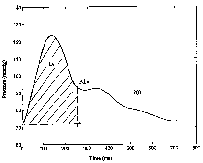
The fundamental relation used for calculating the stroke volume is SV=A/ZO
where
A, expressed in [mmHg t], is the area under the pressure curve p(t) (see
figure
?s Al), and ZO, expressed in [mmHg/cm/t], is the hydraulic impedance which
depends upon the dynamic resistances and upon the compliance of the artery
wall. LSV is measured in [cm3], hence Q = LSV*HR is the cardiac output
expressed in litres per minute if the heart rate is measured in beats per
minute. In
this connection we recall that the plot of the arterial pressure with respect
to time is
3o determined by the magnitude of LSV and by the vascular impedance.
Consequently, the Pulse Contour Method endeavours to separate and analyse
these two contributions; however, the method is unable to determine the
AMENDED SHEET
16-05-2001 CA 02368241 2001-10-18 EP 000003697
2
two contributions as independent variables over time.
Applying the Windkassel theory, many studies have attempted to determine LSV
only from the pressure waveform and from the characteristics linked to
transmission of the wave in the aorta or in the pulmonary artery [Remington
J.W.
et al., 1948; Warner H.R. et al., 1953; Herd J.A. et al., 1966; Kouchoukos
N.T. et
al., 1970].
The original idea of Franck has subsequently been applied over the years and
has
made it possible to estimate LSV in a continuous way from the measurement of
the pressure signal in the aorta or in the pulmonary artery [McDonald D.A. et
al.,
io 1974; Wesseling K.H. et al., 1976; Tajimi T. et al., 1983; Wesseling K.H.
et al.,
1993].
However, in the concrete application to the various possible clinical
situations, the
Pulse Contour Method currently requires a "calibration" for calculation of the
hydraulic impedance. For the calibration, generally one of the other two
methods
referred to above, i.e., the thermodilution method and Fick's method, is used,
or
else linear regressions of aortic parameters, such as the diameter of the
aorta, and
the age, sex, height and weight of the patient are used.
Unfortunately, the calibration and regression factors are subject to error,
given that
the methods from which they are obtained are in turn imprecise and that the
2o regressions are in any case obtained on a(imited number of subjects, and
are
hence acceptable only as a mean and not as a true measurement of the quantity
investigated.
in fact, cardiac output estimated using the thermodilution method and Fick's
method are not always in agreement with the clinical parameters obtained using
other diagnostic techniques, and this mainly occurs in patients suffering from
certain forms of heart disease, such as disease involving dilatation of the
heart,
valvular cardiopathy, and cardiac fibrillation.
To provide an example, consider two possible signals in the aorta studied
between
the points of opening and closing of the ventricle. These signals will in
general
present the same area but different forms, with different times of attainment
of the
systolic point.
The traditional Pulse Contour Method will therefore yield the same exact
AMENDED SHEET
16-05-2001 CA 02368241 2001-10-18 EP 000003697
3
measurement (same integral) evaluated on the basis of the calibration
impedance.
However, it is evident that from a different form of the signal there must
derive a
different impedance, which cannot be evaluated.
Hence, the limits of the invasive techniques currently in use are: a) the poor
precision achievable in the estimation of cardiac output on account of
clinical
illnesses; b) the non-applicability in general on account of the pathological
condition of the patient; and c) the impossibility of applying the said
invasive
techniques, for example during an ergometric test.
From US5647369 a method for the measuring of the cardiac output is described.
io In the patent the Cardiac Output Cois computed as:
CO=HR''PP*C*0.13 where C=X*10-6 Farads and X is precalculated as a function
of anthropometric parameters like age, weight, sex and the like, which are not
related directly to the measure which is performed. Therefore the method
involves
a level of inaccuracy when applied to a subject.
Furthermore the method of US5647369 does not compute the contribution to the
CO of the continuous phase of the blood pressure signal being limited to the
analysis of the pulse signal.
Scope of the invention
A first scope of the invention is to obtain measurements in a continuous way
that
zo are more reliable than the ones obtained using the invasive and non-
invasive
techniques currently applied.
A second scope is to render the measurement substantially independent of the
point of application of the sensor by introducing variations in the specific
formulas,
without any need for prior calibration of the measurement.
2 5 Summary of the invention
The above purposes have been achieved according to the invention using a
method which directly obtain the cardiac output from the pressure signal
measured
in an invasive way, in the ascending aorta, in the pulmonary artery and in
femoral
brachial and radial, or measured in a non-invasive way, for example from the
30 arteriole of the finger using a cuff meter. According to the method, the
impedance
of the pressure signal is calculated on the basis of the points of resonance
of the
signal by assimilating the signal to that of a flow in an elastic tube and
assuming
AMENDED SHEET
16-05-2001 CA 02368241 2001-10-18 EP 000003697
.3a
constancy of Young's modulus, which is in particular taken to have a value of
unity. In this way, it is possible to calculate the cardiac output without any
longer
having to use various calibrations, but exclusively from an analysis of the
pressure
wave and its characteristics.
Preferably, the hydraulic impedance is calculated by means of an analysis of
the
first and second derivatives with respect to time of the pressure signal
recorded.
According to a further aspect of the invention, a correction is moreover made
of
the mean pressure value to be used for the calculation of LSV in order to take
into
account the attenuations of the said value in the various points of possible
20
30
AMENDED SHEET
CA 02368241 2001-10-18
WO 00/64339 PCT/EP00/03697
4
recording of the signal.
According to a further aspect of the invention, from the signal recorded in
the
finger (or from some other point in a non-invasive way), the method makes
possible a direct reconstruction of the signal in the aorta and in the
pulmonary
artery, and from the latter signal a reconstruction of the cardiac flow.
More specifically, according to the invention In order to obtain the SV
estimation
we have taken into consideration the wave pressure in the ascending aorta
and/or
in pulmonary artery, the artery compliance (E) and the periferical resistance
(R).
Therefore we have taken into consideration that 1) the SV depends on the
io pressure variation obtained at the opening of the ventricular valve (that
is the
difference between systolic and diastolic pressure divided by the time
necessary to
pass between the systolic and diastolic) and 2) the SV is conditioned by E and
R.
In order to obtain these contributions we have taken into account the value of
the
dicrotic pressure and the other characteristic points between the systolic
dicrotic
pressure (this pressure values must be divided by time. This time is the
difference
between the end of the cardiac beat and the time of the event being taken into
consideration).
Therefore we have considered SV as being determined by three points: 1) the
bolus of blood ejected by the ventricular; 2) the reaction due to the aortic
wall; 3)
2o resistance due to periferic arterial cycle. As the value of the pressure at
the point
where it is taken is the result of these three components at the same time we
have
studied our system in a perturbative way. Therefore we have considered the
principle contribution of the ventricular and of the system E and R, the first
being
given by 1) as described above and the second, the E and R system principally
contributes to the closure of the valve ( point of diacrotic pressure) This
last event
point is conditioned by a series of perturbations on the pressure signal after
the
cardiac valve, according to the vase being crossed and the length of the
course.
That is it is necessary to take into consideration not only the contribution
due to
the systolic and to the diacrotic above described, and when present those due
to
secondary perturbations.
In conclusion all the event points which have been taken into consideration
are the
moments in which there is a state of balance between the various points (blood
CA 02368241 2007-05-16
= WO 00/64339 PGT/EP00/03697
ejected from the ventricular-E-R): the "principle" balancing points ( systolic
and
dicrotic points) can be or not "accompanied" by other balancing points (how
and if
to analyse them will be described later). All this information can be found in
the
wave pressure which flows after having been generated by the ventricular
(right or
5 left).
Advantageously, with the method according to the invention it is possible to
establish a relationship between the hydraulic impedance and the usable time,
also in combination with known methods (e.g., the thermodilution method) which
involve a phase of calibration of the signal recorded, where the contribution
of the
io area under the pressure curve is considered variable over time, whilst the
contribution of the impedance can only be considered as constant.
In particular, by the method of the present invention (hereinafter called
pulse
analytical method (PAM)) it has been possible: a) to find the SV from the
signal
pressure recorded invasively in ascending aorta and in pulmonary artery; b) to
find
the SV from the arterial signal pressure invasively recorded (brachial,
radial, and
femoral artery) and non invasively recorded (e.g. from the pressure obtained
with
the oscillometric method from the arterial finger).
In this way we estimated the LSV and RSV and so we determined the true Q in a
way that is completely free from any calibration. Therefore these results are
obtained only by the analysis of the wave pressure ( depending only on where
the
wave pressure was taken).
According to the invention an apparatus able to perform the method is
provided.
The apparatus comprises a microprocessor unit able to receive the blood
pressure
signal and to analyse it over the time in order to determine the above
identified
parameters and to calculate the cardiac output Q therefrom.
In a preferred embodiment the apparatus further comprises a sensor in the
shape
of a cuff meter able to be applied to a finger and to acquire the blood
pressure
signal.
Brief description of the figures
- Figure 1A shows a cardiac pressure signal diagram as analysed by the prior
art
method;
- Figures 1-19 illustrates pressure signal profiles as they are sensed at
various
points in or on a patient's body and as they are used in the invention; in
particular, these figures illustrate that the signals used in the invention
include
CA 02368241 2007-05-16
6
information about both first and second derivatives of the pressure.
- Figure 20 shows a reconstruction, according to the present method, of the
signal
in the aorta starting from the pressure signal recorded at the arteriole of
the finger.
Detailed description of the Invention
With reference to the annexed figures, various examples of application of the
method are desc(bed in what follows.
Example I
A) Relation between LSV and oressure taken in the ascending aorta (Pulse
Anal)tical Method. Aortic: PAMA) (Fig.1-6)
io i) The PAMA determines the heart flow Q in litres per minute using the
following
general relation (the pressure signal is acquired in the ascending aorta at
1000
Hz):
LSV = [K[A/((Za1+Za2)*1000)+A/((Za1+Za2)*1000)*(Pm-K1)/K11]/1000
Eq. 11]
is where:
K = I and has the dimensions [(km*sqrt(2p/(p)"Vm], expressed in [I3/ t2];
Xm is the mean wavelength, approximately equal to 10 m
Vm is the mean velocity, approximately equal to 10 rn/s
p is the blood density;
zo A is the integral between t1 (time at the diastolic pulse in [ms]) and tdic
(time at the
dicrotic pressure in [ms]) under the pressure curve p(t), expressed in
[mmHg*ms]
(Figure 1);
Ki = 100, expressed in [mmHg], and represents the correction factor of the
mean
pressure;
25 Zal = (psys-p(1))/tsys, expressed in [mmHg/ms];
Za2 =(pdic/tfinal - tdic), expressed in [mmHg/ms]; and
Pm =(psys+2p(1))/3. see the following Notel
tfinal= time of the beat being taken into consideration (time origin in t1 and
final in
tfinal)
3o The cardiac output is thus
Q = LSV*HR
where Q is expressed in [lit/min];
AMENDED SHEET
16-05-2001 CA 02368241 2001-10-18 EP 000003697
7
HR = 60000/T; and
T is the cardiac period expressed in [ms].
This relation was applied in cases in which the pressure curve and the
corresponding mean of the tangent at 21 points (i.e., the first derivative d')
and the
mean of the tangent at 21 points of the mean tangent (i.e., the second
derivative
d") were those shown in Figures 2 and 3 and could be associated to the
recording
point.
ii) With - Za3
In the cases where the pressure curves in the ascending aorta were of the type
io shown in Figure 4, and the corresponding first and second derivatives, d'
and d",
were as those shown in Figures 5 and 6 and revealed the point of resonance at
time t3, then the relation became:
LSV = [K[A/((Za1+Za2-Za3)*1000)+A/((Za1+Za2-Za3)*1000)"(Pm-K1)/K1]] /1000
Eq.[2]
where the symbols have the same meaning as in Eq. [1], and where t3 is the
time,
in [ms], at the minimum value of d" between the time tsys and the time tdic,
and p3
is the corresponding pressure expressed in [mmHg] at time t3 (see Figure 6)
and
Za3 = (P3/(tfinal - t3) mmHg/ms
In a similar way it is possible to calculate Q = LSV*HR.
.2o Note 1
The mean pressure for the pressure measured in the ascending aorta must be
considered as such for the interval (90 - 110] mmHg; for the mean pressure
between (110 - 120] and (80 - 90] mmHg it must be considered at 50% (for
example for a Pm= 118 mmHg for our method is = 114 mmHg); for mean pressure
values between (120 - 130] and (70 - 80] mmHg it must be considered at 25%,
for mean pressure values>130 and <=70 mmHg it must be considered 13%
Example iI
B) Relation between RSV and the pressure taken in the pulmonary artery (Pulse
Analytical Method, Pulmonary: PAMP)
'o Relationship between the volume of blood expelled from the right ventricle
RSV
and the pressure measured in the pulmonary artery. The corresponding signal
pressure is similar to the one represented for aortic pressure, but for
variations in
AMENDED SHEET
16-05-2001 CA 02368241 2001-10-18 EP 000003697
8
scale (see Figure 7).
The PAMP determines the heart flow Q in litres per minute using the following
general relation (the pressure is acquired in the pulmonary artery at 1000
Hz):
i) Case with mean pressure in pulmonary artery >= 19mmHg
RSV = [K[A/((Za1+Za2)*1000)+A/((Za1+Za2)*1000)*(Prn-K1)/K1]]/1000 [Eq.3]
where:
K = 1 and has the dimensions [(Xn,*sqrt(2p/(p)*Vmj, expressed in [I3/ t2], p
being the
density of the blood;
A is the integral between t1 (time at the diastoiic pulse in [ms]) and tdic
(time at the
io second dilatation of the artery in a dicrotic pulse in [ms]) under the
pressure curve
p(t), expressed in [mmHg*ms];
K1 = 12, expressed in [mmHg];
Zal = (psys)/tsys, expressed in [mmHg/ms];
Za2 = (pdic/tfinal-tdic), expressed in [mmHg/ms]; and
Pm = (psys+2p(1)/3); see following Note 2
Q = RSV*HR,
where Q is expressed in [lit/min];
HR = 60000/T; and
T is the cardiac period expressed in [ms].
-20. In Figure 7 a signal acquisition of the _pressure in the- pulmonary
artery.is shown.
For the pressure in pulmonary artery we have variations for d' and d" similar
to
those of the aorta. In consequence the determination of the point of dicrotic
pressure (Pdic), the systolic pressure(Psys), diastolic pressure (P(1)) and
the
relative times is as described above.
ii) Case with mean pressure in pulmonary artery < 19mmHg
In the cases in which Pm is < 19 mmHg the relation becomes:
RSV=[K [ A / ((Zal+Za2)*1000)+A / ((Za1+Za2)*1000]] / 1000 Eq.[4]
With the same meaning for the symbols as in the preceding cases. In the same
way it is possible to calculate Q=RSV*HR
3o Note 2
The mean pressure in the case of pressure taken in the pulmonart artery must
be
taken as such for the interval of pressure between (19 - 28] mmHg; for values
of
AMENDED SHEET
16-05-2001 CA 02368241 2001-10-18 EP 000003697
9
mean pressure between (28 - 33] mmHg it must be considered at 50%, for values
of mean pressure > 33mmHg it must be considered at 25% (for example a
pm=43mmHg for our method is equal to 33 mmHg); for values < 19mmHg we are
in case ii) and therefore we do not use the mean pressure.
Example III
C) Relation between LSV and the pressure non invasively recorded from the
arterial finger (Pulse Analysis Method. Finger: PAMF)
Direct relationship
i) The PAMF determines the cardiac output Q in Iitres per minute using the
io following general relation (the pressure is acquired at the finger of the
left hand at
1000 Hz):
LSV = [k[A/((Zfl +Zf2)*1000)+A/((Zf1 +Zf2)""1000)*(Pm-K1)/K1 ]]/1000 Eq. [5]
where (Figure 8):
K = 1 and has the dimensions [(a.m`sqrt(2p/(p)*Vrn], expressed in [I3/ t2];
A is the integral between t1 (time at the diastolic pulse in [ms]) and tdic
(time at the
dicrotic pressure in [ms]) under the pressure curve p(t), expressed
in [mmHg*ms];
KI = 90, expressed in [mmHg];
Zfl = (psys-p(1))/tsys, expressed in [mmHg/ms];
20'- Zf2 = pdict(tfinal- tdic); expressed -in [mmHg/ms]; and Pm
=(psys+2p(1))/3. see following Note 3
The corrected volume of blood expelled from the left ventricle (LSVC) is
LSVC = [LSV+LSV*abs(delta(Pd1-pdic))/(psys-pdias)] [6]
where:
(Pdl-pdic) = variation of pressure of the dicrotic point (Pdic) at its maximum
(Pd1)=[mmHg]. This correction exists only when there is an increase in the
pressure after the dicrotic pressure ((Pdl - Pdic)>0). In the cases in which
the
increase in pressure is not present ((Pdl - Pdic)<=0) we have LSV=LSVC.
Psys is the systolic pressure, expressed in [mmHg];
Pdias is the diastolic pressure, expressed in [mmHg]; and
the term Pd1 is calculated immediately after the dicrotic point and is the
maximum
value of the curve after (Pdic).
Q = LSVC*HR
AMENDED SHEET
CA 02368241 2001-10-18
WO 00/64339 PCT/EP00/03697
where Q is expressed in [lit/min];
HR = 60000/T; and
T is the cardiac period expressed in [ms].
The above relation was applied in the cases where the pressure curve and the
5 corresponding first and second derivatives, d' and d", were those shown in
Figures
9 and 10.
ii) With -Zf3
In the cases where the pressure curves were of the type shown in Figure 11 and
the corresponding first and second derivatives, d' and d", were as those shown
in
1o Figures 12 and 13, the relation became:
LSV = [k[A/((Zf1+Zf2-Zf3)*1000)+A/((Zf1+Zf2-Zf3)*1000)*(Pm-K1)/K1]]/1000
Eq.[7]
where:
Zf3 = P3/(tfinal - t3); and
1s the symbols have the same meanings as specified previously, and t3 is the
time,
in [ms], of the minimum value of d" between the time tsys and the time tdic,
and P3
is the corresponding pressure, expressed in [mmHg] at time t3 (see Figure 11).
LSVC = LSV+LSV*abs ((Pd1-Pdic))/(psys-P(1)) Eq. [8]
In a similar way it is possible to calculate Q = LSVC*HR.
iii) With -2Zf3
In the cases where the pressure curves were of the type shown in Figure 14 and
the corresponding first and second derivatives, d' and d", were as those shown
in
Figures 15 and 16, the relation became:
LSV = [k[A/((Zf1 +Zf2-2Zf3)* 1000)+A/((Zf1 +Zf2-2Zf3)* 1000)*(Pm-K1)/K1
]]/1000
Eq.[9]
where Zf3 = P3/(tfinal - t3); and
the symbols have the same meanings as specified previously, and t3 is the
time,
expressed in [ms], of the minimum of d" between the time tsys and the time
tdic,
and P3 is the corresponding pressure, expressed in [mmHg] at time t3 (see
Figure
14).
LSVC = LSV+LSV*abs ((Pd1-Pdic))/(Psys-P1) [10]
In a similar way it is possible to calculate Q = LSVC*HR, expressed in litres
per
16-05-2001 CA 02368241 2001-10-18 EP 000003697
11
minute.
iv) With -2Zf3-Zf5
In the cases where the pressure curves were of the type shown in Figure 17 and
the corresponding first and second derivatives, d' and d", were as those shown
in
Figures 18 and 19, the relation became:
LSV = [k[A/((Zf1 +Zf2-2Zf3-Zf5)*1000)+A!((Zf1 +Zf2-2Zf3-
Zf5)*1000)*(Pm-K1)/K1 ]]/1000
where Zf3 = P3/(tfinal - t3)
where Zf5 = P5/(ttinale-t5)
io the symbols have the same meanings as specified previously, and t5 is the
time,
expressed in [rns], of the minimum of d" between the time tsys and the time
tdic,
and P5 is the corresponding pressure, expressed in [mmHg] at time t5 (see
Figure
17).
LSVC = LSV+LSV*abs ((P1-Pdic))1(Psys-P1)[11] a
In a similar way it is possible to calculate Q = LSVC*HR, expressed in litres
per
minute.
Note 3
The mean pressure in the case of the pressure taken at the arterial finger non
invasively must be considered as such for the interval of mean pressure
between
- 20 70 - 110, for the values of mean pressure between (110 - 150] and- (40 -
70]
mmHg it must be considered at 50% (for example a pm=128 for our method is =
119 mmHg); for mean values of pressure >150 and < 40 mmHg it must be
considered at 25%
v) Reconstruction of the pressure signal in the ascending aorta by means of
linear
multiple regression in the time domain, by use of Zfl-Zf5
For these reconstruction, basically linear multiple regressions were used. In
order
to reconstruct the signal recorded in the ascending aorta (or in the pulmonary
artery) using a cardiac catheter from the arterial signal recorded in a
continuous
way by means of a small cuff wrapped around the middle finger of the left
hand, a
linear multiple regression was used in which the reconstructed pressure signal
was obtained in two successive steps:
1) An estimate was made of the mean pressure during*the cardiac cycle in the
ascending aorta (or in the pulmonary artery) from the signal taken at the
finger,
AMENDED SHEET
16-05-2001 CA 02368241 2001-10-18 EP 000003697
12
deriving the value Pmf (mean pressure in the aorta estimated from the
recording
taken at the finger) from the formulas used in the various cases of analysis
of the
arterial signal referred to in the previous points:
Pmf = LSV*Ztot/(k*A) [11] b
2) The waveform in the ascending aorta (or in the pulmonary artery) was
reconstructed from a fit that used the following parameters:
y = aO*Pmf + al*fin + a2*abs(derfin) + a3*abs(der2fin) + a4*abs(der3fin) +
a5*(intfin) + a6*slope*abs(derfin) + a7*slope*zZfl + a8*slope
+ a9*maxfin + alO*minfin + a11*HR*(intfin(up to the point considered))+
1o a12*areaf + a13*zf1 + a14*zf2 + a15*z3f + a16*z4f + a17*Zf5
areaf = cof*(Zfl +Zf2) / (Zfl +Zf2-z3f- Zf5) [12]
where
Zf5 and n = 0, 1 and 2 according to the criteria described previously;
zz4f = Pd1/(tfinale-td1) (Figure 14);
- fin is the pressure at the finger;
- abs(derfin) is the absolute value of the first derivative in the pressure
point
considered;
- abs(der2fin) is the absolute value of the second derivative in the pressure
point
considered;
- abs(der3fin) is the absolute value of the third derivative in the pressure
point
considered;
- infin is the integral up to the point considered of the signal at the
finger;
- slope is the angle between the horizontal axis and the straight line passing
through the minimum points on the left and on the right of the cardiac cycle;
- maxfin and minfin coincide with the systolic pressure and the diastolic
pressure;
- areaf is the total area of the pressure signal;
and the remaining symbols have the same meanings as specified previously.
A number of reconstructions obtained are iliustrated in Figure 20.
The errors in the comparison between the reconstructed curve of the signal
3o registered non invasively and that taken directly near the ascending aortic
are
SD(mmHg) Max(mmHg) Min(mmHg)
1.16=5.67 2.38=16.40 -2.82 = -16.41
AMENDED SHEET
CA 02368241 2001-10-18
WO 00/64339 PCT/EPOO/03697
13
mean:3.41 9.37 -9.32
With SD= Standard Deviation: the minimum of the interval is obtained for the
riconstrruction of the points around the diastolic pressure, the maximum of
the
difference is obtained near the point of the systolic pressure.
Max= interval of over estimation of the pressure in the point taken into
consideration reconstructed and that actually measured with the catheter
during
the cardiac beat: the minimum of this interval is obtained for the
reconstruction of
the points around the diastolic pressure, the maximum of the difference is
obtained
near the points of systolic pressure.
1o Max= interval of over estimation of the pressure in the point taken into
consideration reconstructed and that actually measured with the catheter
during
the cardiac beat: the minimum of this interval is obtained for the
reconstruction of
the points around the diastolic pressure, the maximum of the difference is
obtained
near the points of systolic pressure.
Min= interval of under estimation of the pressure in the point taken into
consideration reconstructed and that actually measured with the catheter
during
the cardiac beat: the minimum of this interval is obtained for the
reconstruction of
the points around the diastolic pressure, the maximum of the difference is
obtained
near the points of systolic pressure.
What is important in this calculation are Zfl, Zf2,Zf3, Zf5. which we
considered in
point C): these are necessary to have satisfactory results.
D) Relation between LSV and the pressure recorded invasively from femoral
artery
or from another periferical point such as brachial or radial artery (Pulse
Analytical
Method , Brachial, Radial and Femoral, PAM(BRF) )....
For these case we have seen that the formula to use are of the type used in
the
case C) non invasively with the following precisions: i) K1 for these invasive
signals must be considered =100; ii) note 3 remains unchanged.
According to the invention, the method can be applied in combination with
known
methods (such as the thermodilution method) comprising a phase of calibration
of
the signal recorded, in which the contribution of the area under the pressure
curve
is considered variable over time, whereas the contribution of the impedance
can
only be considered constant.
16-05-2001 CA 02368241 2001-10-18 EP 000003697
14
In this case, the proposed method also makes it possible to take into account
even
major variations in the heart rate, in the pressure values and in the pressure
waveform for purposes of calculation of the impedance.
It may therefore be concluded that, both in the case of normal subjects and in
the
case of patients affected by various pathological conditions, the method
proposed
represents an effective and advantageous diagnostic tool in the invasive and
non-
invasive evaluation of cardiac output.
In addition, the method can be applied both in healthy subjects and in
subjects
presenting cardiocirculatory alterations who are undergoing ergometric tests
that
lo are aimed at establishing the level of haemodynamic response to the tests.
It should be emphasized that the present method is based only on the study of
the
pressure signal (recorded invasively in the pulmonary artery and in the aortic
arch,
or in any other major arterial vessel, or else non-invasively at the finger),
and is
independent of the anthropometric data and of the age of the subject examined.
The present invention further comprises an apparatus for measuring cardiac
output, comprising at least one sensor for detecting a blood pressure signal
and a
computer unit connected to the said sensor for the execution of a measurement
according to the above described method and provided with at least one output
device of the measured value.
Preferably, the apparatus comprises a storage medium containing a computer
program to execute a method according to at least one of claims 1 to12.
The invention further relates to a computer program loadable in a computer
unit in
order to execute the method.
AMENDED SHEET