Note: Descriptions are shown in the official language in which they were submitted.
WO 00/45706 PCT/US00/02644
INTRABOD~' HIFU APPLICATOR
TECI~NICAL FIELD
The present invention relates to application of sonic energy, such as focused
ultra
sound energy v~~ithin the body of a living subject such a human or other
mammalian
subject.
BACKGROUND ART
Various forms of therapy can be applied within the body of a human or other
mammalian subject by applying energy from outside of the subject. In
hyperthermia,
I O ultrasonic or .radio frequency energy is applied from outside of the
subject's body to
heat the tissues. The applied. energy can be focused to a small spot within
the body
so as to heat the tissues at such spot to a temperature sufficient to create a
desired
therapeutic effect. This technique can be used to selectively destroy unwanted
tissue
within the body. Fox example, tumors or other unwanted tissues can be
destroyed by
applying heat to heat the tissue to a temperature sufficient to kill the
tissue,
commonly to about b0° to 80°C, without destroying adjacent
normal tissues. Such a
process is commonly referred to as °'thermal ablation'". Other
hyperthermia
treatments include selectively heating tissues so as to selectively activate
a' drug or
promote some other physiologic change in a selected portion of the subject's
body.
Other therapies use the applied energy to destroy foreign objects or deposits
within
the body as, for example, in ultrasonic lithotripsy.
In most cases, the focused ultrasound energy , used in said procedures is
applied by an ultrasonic energy source disposed outside of the body. For
example,
certain ernbodiments taught in co-pending, commonly assigned U.S. P atent
No. 6,128,522 and in the corresponding International Application WO 98/62465,
also filed May 22, I998; describe systems for applying focused ultrasound
energy in conjunction with a magnetic resonance device. An external
CA 02368707 2001-08-O1
WO 00/45706 PCT/US00/02644
2
ultrasonic energy applicator is also taught for example, in Fig. 1 of Aida et
al., U.S.
Pat. 5,590,653 and in Fig. 1 of Oppelt et al., U.S. Pat. 5,624,382. These
external
ultrasonic energy sources transmit ultrasonic energy to the desired treatment
location
through the tissues of the body. Various proposals have been advanced for
inserting
ultrasonic energy sources into the body and focusing energy from such
intrabody
sources on the desired treatment regions. For example, Fig. 5 of the
aforementioned
Oppelt et al. '382 patent illustrates a therapeutic ultrasound transducer
which may be
inserted into the rectum so as to direct ultrasonic energy onto the prostate
gland
through the wall of the rectum. Aida et al. '653 discloses various forms of
intrabody
transducer arrays (Figs. 9-12). Diederich, Transuretheral Ultrasound Array For
Prostate Thermal Therapy: Initial Studies, IEEE Transactions On Ultrasonics,
Ferroelectrics and Frequency Control, Vol. 43, No. 6, pp. 1011-1022 (Nov.
1996)
discloses a rod-like ultrasound transducer housed within a catheter. Such a
rod-like
transducer does not focus the ultrasound but instead provides a sound pressure
distribution which is at a maximum adjacent the transducer and which
diminishes
with distance. In use, the transducer is inserted into the urethra and the
catheter is
cooled by a flow of water. The cooling water limits the temperature rise of
the
urethra wall. Prostate tissue remote from the urethra is heated by the applied
energy.
Despite these and other attempts to utilize intrabody ultrasonic transducers,
still further improvement would be desirable.
SUMMARY OF THE INVENTION
One aspect of the present invention provides a probe for applying sonic
energy within the body of the subject. The probe according to this aspect of
the
invention includes a probe body having a proximal and having a distal end
adapted
for insertion into the body of the subject. The probe also includes a
spatially
distributed ultrasonic transducer disposed adjacent the distal end of the
probe body.
As used in this disclosure, the term "spatially distributed sonic transducer"
refers to a
sonic transducer which is capable of emitting sound from a plurality of
locations
CA 02368707 2001-08-O1
WO 00/45706 PCT/LJS00/02644
3
spaced apart from one another. One form of a spatially distributed transducer
includes a plurality of discrete transducer elements mounted at spaced apart
locations. Another form of spatially distributed transducer includes a
continuous
sheet of transducer material. In such a continuous-sheet transducer, various
regions
of the sheet are spaced apart from one another and hence can emit sound at
spaced
apart locations. The probe according to this aspect of the invention further
includes
means for moving one portion of the distributed transducer relative to another
portion
of the distributed transducer while distal end of the probe and hence the
distributed
transducer is disposed within the body of the subject. Such movement changes
the
configuration of the distributed transducer so as to focus the sound emitted
from the
distributed transducer onto a focal spot at a selected location relative to
the probe.
The distributed transducer may include a deformable element, which may be
separate from the active elements of the transducer. Alternatively, the
deformable
element may be integral with a continuous transducer sheet. In the simplest
embodiment, the entire distributed transducer includes only a continuous sheet
element, such as an elongated strip formed from a piezoelectric material and
the
electrodes used to actuate those portions of the material. Alternatively, the
distributed transducer may include plural separate transducer elements mounted
to
the deformable element at spaced-apart locations. The deformable element may
incorporate an elongated beam having a fixed end mounted to the probe body and
a
fixed end. The means for controlling deformation may include a control element
moveable mounted to the probe body. The control element desirably is a
flexible
cable having a distal end connected to the free end of the beam and having a
proximal end extending to the proximal end of the probe body. Thus, the
deformable
element may be bent to the desired degree of curvature by pulling the flexible
cable.
Alternatively, the deformable element may include a disc like element having a
central region and a peripheral region surrounding the central region. The
means for
CA 02368707 2001-08-O1
WO 00/45706 PCT/US00/02644
4
controlling deformation may include means from moving the peripheral and
central
regions relative to one another.
In yet another alternative, the probe may include a plurality of supports
movably mounted to the probe body adjacent to distal end thereof and the
distributed
transducer may include a plurality of transducer elements mounted to the
supports.
The means for moving one part of the transducer relative to the other may
include
means for moving one or more of the supports relative to the probe body. For
example, the plurality of supports may include a plurality of elongated
supports
arranged generally in the manner of the radial ribs of an umbrella. Thus, the
elongated supports may have central ends pivotally connected to a common
member
and may have peripheral ends remote from the central ends. The transducer
elements
are mounted to the elongated supports adjacent the peripheral ends thereof.
The
supports can be pivoted relative to the common member between a collapsed
condition in which the peripheral ends are close to a central axis and an
expanded
commission in which the peripheral ends are remote from the central axis. The
pivoting means may include a control member and a plurality of struts. Each
strut
has a first end pivotally connected to the control member and a second end
connected
to one of the elongated supports remote from the central end of such support.
The
means for pivoting the supports may include means for moving the control
member
and common member relative to one another. For example the probe body may
include an elongated tubular element and a flexible cable may be provided in
the
tubular element. The cable may be attached to the control member and the
distal end
of the tubular element may be connected to the common member or vice versa.
In yet another arrangement, the distal end of the probe body itself may be
deformable and the distributed transducer may be arranged along the distal end
of the
probe body so that deformation of the probe body distal end will move one part
of
the transducer relative to another part. For example, the probe body may be
elongated and the distributed transducer may include separate transducers or
portions
CA 02368707 2001-08-O1
WO 00/45706 PCT/US00/02644
of a continuous sheet spaced apart from one another in the lengthwise
direction along
the probe body. The means for deforming the distal end of the probe body may
include means for bending the distal end of the probe body transverse to its
lengthwise direction so as to vary the curvature of the distributed
transducer. The
S distal end of the probe body may be advanced into an intrabody space and the
probe
body may be deformed while the distal end is disposed in the intrabody space.
For
example, the probe body may be advanced through the urethra into the urinary
bladder and the distal end of the probe body may be bent while the distal end
of the
probe body is in the urinary bladder.
A further aspect of the present invention provides probe for applying sonic
energy within the body of the subject which includes an elongated probe body
having
a distal end and a spatially distributed sonic transducer disposed adjacent to
the distal
end of the probe body. In a probe according to this aspect of the present
invention,
the distributed transducer is moveable between a collapsed condition in which
the
1 S distributed transducer has relatively small dimensions in directions
transverse to the
direction of elongation of the probe body and an expanded condition in which
the
distributed transducer has relatively large transverse dimensions and hence
extends
outwardly from the probe body in one or more directions transverse to the
direction
of elongation of the probe body. A probe according to this aspect of the
invention
desirably includes means for controlling movement of the distributed
transducer
between the collapsed condition and the expanded condition. In a probe
according to
this aspect of the invention, the movement control means optionally may be
adapted
to vary the configuration of the distributed transducer while the transducer
is in the
expanded condition so as to vary the focus of sound waves emitted by the
transducer.
Still further aspects of the present invention provide methods of ultrasonic
treatment.
CA 02368707 2001-08-O1
WO 00/45706 PCT/US00/02644
6
BRIEF DESCRIPTION OF THE DRAWINGS
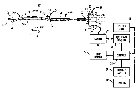
Figure 1 is a diagrammatic view depicting a probe in accordance with one
embodiment of the invention in conjunction with other apparatus.
Figure 2 is a fragmentary, diagrammatic sectional view depicting a portion of
the probe of Fig. 1.
Figure 3 is a fragmentary electrical schematic of the probe of Figs. 1-2.
Figure 4 is a fragmentary, perspective view depicting the probe of Figs. 1-3
in
one condition.
Figure 5 is a view similar to Fig. 4 but depicting the probe in a different
condition.
Figures 6, 7 and 8 are diagrammatic views of probe in accordance with further
embodiments of the invention.
Figure 9 is a fragmentary, diagrammatic sectional view depicting a probe in
accordance with yet another embodiment of the invention.
1 S Figure 10 is a fragmentary, perspective view depicting portions of a probe
in
accordance with another embodiment of the invention.
Figure 11 is a view similar to Fig. 10 but depicting the a probe of Fig. 10 in
a
different condition during operation.
Figure 12 is a fragmentary diagrammatic elevational view depicting portions
of a probe in accordance with another embodiment of the invention in one
condition
during operation.
Figure 13 is a view similar to Fig. 10 but depicting the probe of Fig. 12 in a
different condition during operation.
Figure 14 is a diagrammatic sectional view taken along line 14-14 in Fig. 12.
Figure 15 is a fragmentary diagrammatic sectional view depicting a probe in
accordance with yet another embodiment of the invention.
Figure 16 is a fragmentary diagrammatic view depicting a probe in
accordance with yet another embodiment of the invention.
CA 02368707 2001-08-O1
WO 00/45706 PCTNS00/02644
7
Figure 17 is a fragmentary diagrammatic sectional view depicting a probe in
accordance with yet another embodiment of the invention.
Figure 18 is a sectional view along line 18-18 in Fig. 17.
BEST MODE FOR CARRYING OUT THE INVENTION
S A probe in accordance with one embodiment of the present invention includes
a probe body 20 having a proximal end 22 and a distal end 24 adapted for
insertion
into the body of a subject. Probe body 20 may be a conventional catheter,
endoscope
or other conventional medical device. The particular probe body illustrated is
in the
form of an elongated tube having an interior bore 26 extending between the
proximal
and distal ends. A deformable distributed sonic transducer 30 is mounted to
the
distal end 24 of the probe body. As best seen in Fig. 2, transducer 30
includes a
continuous sheet 32 of a piezoelectric polymeric material such as a
polyvinyledene
fluoride piezoelectric material. Materials of this type are described in U.S.
Patents
Nos.4,830,795, 4,268,653 and 4,577,132. Particularly preferred piezoelectric
polymers are available from Measurement Specialties, Inc. of Norristown,
Pennsylvania. The transducer further includes a backing layer 34 and
electrodes 36
and 38 disposed on opposite sides of piezoelectric layer 32. Layer 34 may be
formed, for example, from a flexible dielectric polymer, a flexible metal
strip, or the
like. The electrodes are formed in pairs. Each pair includes a first electrode
36
disposed on one side of the piezoelectric layer 32 and a second electrode 38
disposed
on the opposite side of the piezoelectric layer, in alignment with the first
electrode.
For example, electrodes 36a and 38a (Figs. 2 and 3) form one such pair whereas
electrodes 36b and 38b (Fig. 3) form another such pair. The electrodes are
connected
to conductors 40 extending along layers 32 and 34. These conductors may be
fabricated, for example, by techniques such as those used in formation of
flexible
printed circuits.
The thicknesses of the various elements are greatly exaggerated for clarity of
illustration in Fig. 2. In practice, the entire transducer is formed as an
integral,
CA 02368707 2001-08-O1
WO 00/45706 PCT/US00/02644
8
strip-like structure. Thus, the electrodes may be provided as thin,
electrically-conductive coatings on opposite sides of layer 32.
Transducer 30 is generally in the shape of an elongated, flexible beam having
a fixed end 42 attached to the distal end 24 of probe body 20 and having a
free
S end 44 remote from the fixed end. The electrode pairs 36, 38 are arranged
along the
lengthwise extent of the beam. Conductors 40 are connected to further
conductors 46, which a few are seen in Fig. 2. Conductors 46 extend to the
proximal
end 22 of the probe body, and to an electrical connector 48 (Fig. 1 ) at the
proximal
end of the probe body.
A control element in the form of a flexible cable 50 is attached to the free
end 44 of the beam or transducer 30. Cable 50 is slideably received within the
bore 26 of the probe body and extends to a proximal end element 52. End
element 52 in turn is connected through a linkage 54 to the proximal end 22 of
the
probe body. Linkage 54 includes a mechanical device for controlling the
position of
proximal end element 52 relative to the proximal end of the probe body, and
hence
controlling the position of the control element 50 relative to probe body 20.
The
particular linkage illustrated includes a manually adjustable wheel 56, a
threaded
rod 58 and a nut 60 threadedly engaged on rod 58. Wheel 56 and screw 58 are
rotatably mounted to one element of the linkage, whereas nut 60 is pivotally
mounted
to another element of the linkage, so that by rotating knob 56 and screw 58,
the
linkage can be expanded or collapsed, thereby driving proximal end element 52
forwardly and rearwardly relative to the probe body. The particular linkage
depicted
in Fig. 2 is merely exemplary. Any other conventional positioning device
capable of
moving one element to a desired position relative to the other can be
employed. For
example, cams, levers, electromechanical actuators and hydraulic actuators may
be
employed. Also, the linkage may be omitted, so that the proximal end element
52
can be moved manually relative to the proximal end of the probe body. The
probe
CA 02368707 2001-08-O1
WO 00/45706 PCT/US00/02644
9
may also be provided with a separate device (not shown) for selectively
locking the
control element or cable 50 in position relative to the probe body.
Beam or transducer 30, in its free undeformed condition is nearly flat, as
indicated in broken lines at 30' in Fig. I. By moving control element or cable
50 in
the retracting direction, toward the proximal end 22 of the probe body, the
free
end 44 of the beam can be brought closer to the fixed end 42, thereby
deforming the
transducer or beam into configurations having a greater curvature, including
the fully
bowed condition illustrated in broken lines in Fig. I at 30" and also
illustrated in
Fig. 5. In the fully elongated or collapsed condition 30', the beam lies close
to the
axis 62 of the probe body distal end. In the fully bowed or expanded condition
30",
the probe projects laterally from axis 62.
As further discussed below, the transducer can be actuated as a multielement
array to provide ultrasonic emissions focused on a focal within a focal region
65 near
the center of curvature 64 of the beam. The focal spot can be moved within the
focal
region by altering the phasing of the electrical signal supplied to the array.
However,
bending the transducer moves the center of curvature and moves the focal
region.
With the transducer in the fully collapsed or flat condition, the focal spot
will lie at a
large distance from axis 62. As the transducer becomes progressively more
bowed,
the center of curvature 64 and hence the focal region and focal spot move
closer to
axis 62. With the transducer in the slightly bowed position illustrated in
solid lines in
Fig. 1 and illustrated in Fig. 4, the center of curvature 64 is at the
position indicated.
With the transducer in a more bowed position, as indicated in broken lines at
30" in
Fig. 1 and as shown in Fig. S, the center of curvature is at position 64".
The probe is used in conjunction with monitoring and driving elements
(Fig. 1 ). A switch 70 is connected by a multiconductor cable to a connector
72
matable with connector 48. An impedance measuring device 74 is provided. The
impedance measuring device can be connected by switch 70 to a pair of
electrodes 38a and 38g disposed at opposite ends of transducer 30, so that the
CA 02368707 2001-08-O1
WO 00/45706 PCT/US00/02644
impedance measuring device can measure the electrical impedance within
piezoelectric layer 32, from one end of the piezoelectric layer to the other.
Thus,
electrodes 38a and 38g serve as impedance measuring electrodes. The impedance
monitoring device may include a conventional bridge circuit, with the
impedance
5 between electrodes 38a and 38g on one leg of the bridge circuit. The
impedance
monitor may also include temperature compensation elements (not shown) mounted
at the distal end of the probe and connected in the bridge circuit so as to
compensate
for effects of temperature on the impedance on layer 32. The impedance monitor
may also include conventional components such as operational amplifiers and
10 analog-to-digital converters for providing a digital readout of the
impedance between
electrodes 38a and 38g. Desirably, the impedance monitoring device is arranged
to
monitor AC impedance rather than DC resistance.
The electrical impedance within piezoelectric layer 32 varies with mechanical
strain on the layer. As the beam is bent from undeformed, fully collapsed
condition 30' toward the fully expanded bowed condition 30", layer 32 is
placed
under progressively increasing compression and the electrical impedance within
the
layer. Thus, the electrical impedance between electrodes 38a and 38g through
layer 32 varies with the degree of curvature in the beam.
During operation of the impedance monitoring device, electrodes 36 and 38
which are not connected to the resistance monitoring device are inactive.
Depending
on the configuration and placement of the electrodes, a significant portion of
the
impedance along the piezoelectric layer may be shorted by conductivity along
the
inactive electrodes. To avoid such shorting, and increase the change in
resistance
between electrodes 38a and 38g, the intermediate electrodes 38b, 38c . . . and
36b,
36c . . . may be isolated from the piezoelectric layer by a very thin
dielectric layer
(not shown) disposed between the electrodes and the surface of the
piezoelectric
layer. Switch 70 is also arranged to disconnect electrodes 38a and 38g from
resistance monitor 74 and to connect all of the electrodes 36 and 38 to a HIFU
CA 02368707 2001-08-O1
WO 00/45706 PCT/US00/02644
11
driver 76. HIFU driver 76 includes conventional phased array driver components
for
applying electrical potentials between the electrodes 36 and 38 of each
electrode pair.
These electrical potentials vary at ultrasonic frequencies. The varying
potential is
applied across the region of piezoelectric film 32 between each pair of
electrodes and
causes mechanical vibration of each such region.
HIFU driver 76 is controlled by a computer 78. The computer controls the
frequency and phase of the excitations applied to the various electrode pairs
in
accordance with the known principles governing operation of phased array
ultrasonic
emitters so that the ultrasonic emissions from the various parts of the
piezoelectric
layer reinforce one another at the desired focal spot. Computer 78 stores a
value of
the curvature of the transducer or beam 30 based upon the resistance
measurement
from resistance monitor 74. This value is incorporated into the parameters
defining
the geometry of the emitter array, and such parameters are used in the normal
manner
to calculate the appropriate signals to be applied to each element of the
array. As
such calculations are well within the skill of the art and employ known
methods, they
are not described in detail herein.
Computer 78 is linked to conventional display and input/output devices 80
such as a CRT or other pictorial display and a mouse, joystick or other
control
elements. An imaging system 82 such as a magnetic resonance imaging, x-ray or
CAT scan imaging system 82 is also connected to the computer. The imaging
system is arranged to provide data in substantially real time constituting an
image of
the internal structures within the patient's body in the vicinity of probe
distal end 24.
This representation includes a depiction of the probe distal end and the
transducer 30.
A sensor 51 such as a sensor for detecting magnetic field components is also
mounted to the distal end of a probe. Sensor 51 is connected by additional
conductors (not shown) extending through the probe body to the proximal end
thereof to a position sensing unit 53. Position sensing unit 53 may be
arranged to
detect the position and/or orientation of sensor 51 based upon magnetic or
WO 00145706 PCTlUS00102b44
17
electromagnetic fields transmitted to or from sensor S 1. As described for
example, in
international patent publication WO 95/09562, sensor 51 may be arranged to
receive or
transmit magnetic field components, and may be used in conjunction with
additional sensors
(not shown) mounted in a fixed frame of reference or in a frame of reference
fixed to
the appropriate portion of the patient's body. As described in these
publications,
position sensing unit 53 is arranged to determine the position andror
orientation of
the probe distal end in such frame of reference. As also described in tfese
patents
and publications, computer 78 can combine the position and orientation of the
probe
distal end with the imaging data from imaging system 82 so that the position
and
orientation data and the imaging data are in a common frame of reference.
Display
82 can display a representation of the probe distal end and transducer in ahe
correct
position relative to the displayed anatomical structures. Such a
representation may
be displayed in multiple views.
I5 In operation, the probe distal end is advanced into the patient until the
probe
distal end is disposed adjacent the region of the patient to be treated. The
probe may
be advanced into naturally occurring body cavities as, for example, the
gastrointestinal tract circulatory system, respiratory tract or urinary tract:
While the
probe is being advanced, the transducer 30 desirably is in its fully collapsed
or flat
position 30' (Fig. I ) so.that the extent of the transducer in the directions
transverse to
the axis 62 of the probe distal end is small. . This facilitates advancement
of the probe
through confined spaces within the patient's body.
Once the probe distal end is near the anatofnical structure to be treated, the
physician adjusts .tire curvat~ire of the transducer by operating knob 56 and
linkage 54
?5 so as to move the control element or cable 50 arid thereby pull the free
end 44 of the
transducer towards the fixed end 42 and the distal end of the probe: As the
linkage is'
adjusted, switch 70 and resistance monitor 74 detect, the curvature of the
transducer.
Compc3ter 78 displays' a mark on the displ2~y unit 80 a.t ~a location
~canresponding to
3.
WO 00/45706 ~ FCTIUS00102644
13
the location of the center of cun~ature 64 of the transducer. This location
and
orientation is computed from the location of the probe distal end, as detected
by
transducer 51 and the curvature of the transducer, as measured by resistance
monitor
74.
As the physician adjusts linkage 60; resistance. monitor 74 registers the
changed curvature of transducer 30. The computer displays the new location of -
center of curvature 64 superposed on the depiction of anatomical structures
derived
from imaging unit 82. The computer may also display ,a representation of focal
region 6~ superposed on the anatomical features. When the physician is
satisfied that
the center of curvature is in the appropriate location relative to the
anatomical
features to be treated, he then actuates the computer and HIFU drifter to
apply
focused ultrasonic energy at one or more desired locations within the focal
regian 65.
The design of ultrasonic phased arrays, .and computer simulations of such
arrays are
disclosed in Ebbini, et al., Optimization of the intensity Gain of Multiple-
Focused
Phased Array Heating Patterns, Int: J. Hyperthermia, / 991, Vol. 7, #6, pp.
9S3-973;
Ebbini et al.; Multiple-Focused Ultrasound Phased-Array Pattern Synthesis:
Optimal
Driving Signal Distributions for Hyperthermia; IEEE Transactions on
Uitrasonics,
Ferro Electrics and Frequency Control, Vol. 36, pp. 540-548 (1989) and Fan et
al.,
Control Over the Necrosed Tissue Volume During Non-Invasive Ultrasound Surgery
2o Using a I6-Element Phased Array, Medical I?hysics, Vol. 22 {#3), pp: 297-
305
( 1995).
The curvature of the transducer can be adjusted after application of some
ultrasonic
treatments so as to move the center of curvature and the beam steering region.
Also,
the prol:e m2.y .be repositioned as desired so as to shift the center of
curvature and
~5 beam stePriiig region relative to the patient.
In a variant o:F the system discussed above, the curvature of the transducer
is
monitored by ~~o:2itorir..g the position of the control element or cable 50'
relative to
the prolrP body ~:(3': l~or exarrtple, a potentiometer 4~9 (Fig. 6), an
optical encoder or
CA 02368707 2001-08-O1
WO 00/45706 PCT/US00/02644
14
other conventional position monitoring devices may be connected between the
proximal end element 52' on the control cable and the proximal end 22' of the
probe
body. Measurements of the relative position of the control cable or control
element
50' relative to the probe body 20' can be translated directly into curvature
of
transducer 30. In a further variant, two or more position sensors 151 (Fig.
7), similar
to the position sensor 51 discussed above with reference to Figs. 1 and 2 may
be
provided on the deformable transducer itself. The location and orientation of
these
sensors can be translated into curvature of the transducer, as well as the
position and
orientation of the transducer in the patient's frame of reference.
In a probe according to a further variant (Fig. 8), the transducer is provided
as
a set of transducer elements 230 disposed along the length of the probe body
220
itself adjacent the distal end thereof. At least the distal region of the
probe body
having the transducers 230 thereon is arranged for flex in a controlled
fashion. The
probe body may be provided with conventional devices (not shown) for bending
the
probe body in a controlled fashion. Transducer elements 230 may be individual,
discrete transducers or else may be regions of a unitary piezoelectric sheet
as
discussed above with reference to Fig. 2. The transducer elements or sheet
constitute
a spatially-distributed transducer extending along the catheter tip. Bending
of the
probe body curves the array of transducer elements so that energy from the
transducer elements can be focused onto a focal region 264. A flexible
transducer of
this type may be provided with elements such as position sensors disposed
along the
length of the probe or devices for detecting the degree of curvature of the
probe
directly. In a further variant, flexible distributed transducers as discussed
above can
be provided with strain gauges formed separately from the piezoelectric
elements.
For example, a flexible beam-like transducer may include a strain-sensitive
layer
forming part or all of backing layer 34, with appropriate electrodes connected
to such
layer. Also, a discrete strain gauge such as a strain-sensitive wire may be
adhered to
CA 02368707 2001-08-O1
WO 00/45706 PCT/US00/02644
the beam element or embedded therein. Such strain gauges can be used to
monitor
the curvature of the beam or other distributed transducer.
Alternatively or additionally, curvature of the probe can be monitored by
imaging the probe and detecting the curvature based upon such imaging.
Detection
5 can be accomplished visually, as by a human operator observing the displayed
image
of the probe and measuring the curvature on the display. Curvature also can be
detected by using conventional pattern-recognition programs to detect the
curved line
of the probe in the data representing the image, with or without display of
the image
in a human perceptible form. These techniques can also be used to monitor the
10 curvature of a separate flexible transducer such as the transducer 30
discussed above.
In further variants, individual, discrete transducer elements, rather than a
single continuous piezoelectric layer, may be mounted on a flexible beam as
illustrated in Fig. 2 to form a spatially-distributed transducer. In yet
another variant,
a spatially-distributed transducer having a continuous piezoelectric layer as
discussed
15 above with reference to Fig. 2 may be provided with only two thin, flexible
electrodes, one electrode being disposed on each surface. Such a distributed
transducer would not be capable of acting as a phased array. However,
ultrasonic
energy emitted from such a transducer can be focused by changing the curvature
of
the transducer.
Apparatus according to a further embodiment of the invention (Fig. 9) has a
flexible transducer array 330 in the form of a diaphragm. The diaphragm is
mounted
in a housing 332 so that a chamber 331 defined by the housing is closed by the
diaphragm. By increasing or decreasing the pressure within chamber 331,
diaphragm
330 can be adjusted to a condition 330' of greater curvature or to a position
of lesser
curvature (not shown). Diaphragm 330 may have a structure similar to the
structure
of the beam-type transducer element discussed above, and desirably includes a
continuous layer of a piezoelectric film with electrodes 336 and 338 disposed
on
opposite sides of the piezoelectric film. However, the electrodes desirably
are
CA 02368707 2001-08-O1
WO 00/45706 PCT/US00/02644
16
disposed in a two-dimensional array on the surface of the diaphragm. In a
variant of
this arrangement, a control element may be connected to the diaphragm at its
center
for bending the diaphragm to a more curved or less curved condition. Curvature
of
such a diaphragm may be detected by impedance monitoring or other techniques
as
discussed above.
Apparatus according to a further embodiment of the invention (Figs. 10 and
11 ) includes a set of supports 402. Each support has a central end 404 and a
peripheral end 406. The central ends of these supports are pivotally connected
to a
common member 408, which in turn is connected to the control element or cable
450.
A set of struts 410 is also provided. Each strut is pivotally connected to one
of the
supports 402 between its central end 404 and peripheral end 406. Each strut is
also
pivotally connected to a control member 412. Control member 412 is mounted to
the
distal end 424 of the probe body 420. Individual transducer elements 430 are
mounted to the supports 402 adjacent the peripheral ends thereof. The
transducer
may be moved between the collapsed or closed configuration illustrated in Fig.
10 to
the expanded condition illustrated in solid lines in Fig. 11, and to the
further
expanded, over-center condition partially illustrated in broken lines in Fig.
11 by
moving the control cable or control element 450 relative to the probe body 420
so as
to move the common member 408 relative to control member 412. In the collapsed
or closed configuration (Fig. 10), supports 402 lie close to the axis 462 of
the probe
body. In the expanded condition, the supports project outwardly away from axis
462.
In the expanded, over center position depicted in broken lines in Fig. 1 l,
the various
individual transducers 430' will tend to focus their ultrasonic energy on a
common
focal location. The position of such focal location can be adjusted by moving
the
common member 408 relative to control member 412 so as to pivot the supports
402.
Alternatively or additionally, transducers 432 may be provided on the
opposite sides of the support. Transducers 432 are directed towards a common
focus
when the supports are in the condition illustrated in solid lines in Fig. 11.
In still
CA 02368707 2001-08-O1
WO 00/45706 PCT/US00/02644
17
further variants, the connection of the control member 412 and of common
member
408 may be reversed. Thus, control member 412 may be connected to cable or
control element 450 whereas common member 408 may be mounted to the probe
body. Also, the initial positions of the elements may be reversed so that in
the
collapsed condition, the supports 402 and struts 410 extend rearwardly along
the
probe body rather than forwardly from the distal tip of the probe body. Of
course,
the number of supports and struts may be varied. Also, the measures discussed
above for monitoring the curvature of a continuous curved transducer may be
used in
the case of a transducer having discrete transducer elements and separate
supports.
Thus, position sensors may be provided on supports 402. Alternatively, the
position
of the control element 450 relative to the probe body 420 may be monitored.
A transducer assembly as shown in Figs. 10 and 11 can be used by advancing
it in the closed or collapsed condition into a natural body cavity as, for
example, the
urinary bladder and then expanding the transducer assembly and bringing the
transducer elements to the appropriate locations to focus energy on a lesion
as, for
example, a lesion within the prostate gland. After therapy, the assembly
desirably is
returned to the closed or collapsed configuration and extracted from the
patient.
Probes as discussed above may be provided with balloons or other flexible
shields (not shown) covering the ultrasonic transducer. In use, such a shield
is filled
with a liquid such as water or saline solution, so that the shield bears
against the
surrounding tissues. Ultrasonic energy from the transducer is transmitted
through the
liquid and the shield to the patient's body. Liquid may be circulated through
the
probe body, into and out of the shield, to cool the transducer.
A probe according to a further embodiment of the invention (Figs. 12-14)
includes a spatially-distributed collapsible transducer 500 mounted to the
distal end
of an elongated probe 502. Transducer 500 incorporates a plurality of leaves
504.
As seen in plan view (Figs. 12 and 13) each leaf is generally wedge-shaped,
having a
narrow end and a broad end. As seen in section (Fig. 14) each leaf is curved.
Each
CA 02368707 2001-08-O1
WO 00/45706 PCT/US00/02644
18
leaf has one or more transducer elements thereon. For example, each leaf may
include a continuous piezoelectric layer with one or more electrodes as
discussed
above, or with a set of discrete transducers. The narrow ends of the leaves
are
pivotally connected to one another and to the probe body 502 for movement
about a
common pivot axis 506 transverse to the direction of elongation of the probe.
The
leaves are movable between the collapsed condition of Fig 13 the expanded
condition
of Fig. 12. In the expanded condition, the leaves wholly or partially overlie
one
another, whereas in the expanded condition at least a portion of each leaf is
exposed
and is not covered by another leaf. As the transducer expands or collapses,
the
leaves slide over one another. The collapsing and expanding action is similar
to the
action of a traditional Japanese fan. The collapsing and expanding action can
be
controlled by control cables or other elements (not shown) extending through
the
probe. Alternatively or additionally, the collapsing or expanding action can
be
driven by spring mechanisms, electrical, hydraulic or pneumatic mechanisms, or
even by a small electric motor disposed adjacent the distal end of the probe.
Thermally-responsive elements such as bimetallic or shape-memory metals can be
employed.
In the collapsed condition, the distributed transducer is small; all of the
leaves
lie close to the axis of probe body 502. Therefore, the transducer can be
advanced
readily into a body cavity. For example, the probe may be inserted vaginally,
rectally or orally and expanded inside the body of the patient. Desirably, the
radius
of curvature of each leaf is selected so that sonic energy emitted from all of
the
leaves when the leaves are in the expanded condition is focused to a common
point,
line or region. The leaves may be rigid or flexible. If the leaves are
flexible, control
elements (not shown) similar to those discussed above may be provided for
deforming the individual leaves or deforming the leaves together, and devices
for
monitoring the deformation of the leaves may be provided as discussed above
for
monitoring individual deformable elements.
CA 02368707 2001-08-O1
WO 00/45706 PCT/US00/02644
19
The embodiment depicted in Fig. 15 illustrates one way of implementing a
bendable catheter 700 or other probe with a transducer 702 distributed
lengthwise
along its distal end, as discussed above with reference to Fig. 8. The
interior of the
probe distal end desirably is filled with a liquid, gel or other energy-
transmissive
S medium so that sound can be transmitted from the transducers 702 through the
accordion-pleated wall 704 of the probe. A cable 710 is provided with one end
attached to the distal end of the probe so that the probe 700 can be bent. The
side
opposite the accordion-pleated wall 704 of the probe may have expandable sides
712
to accommodate the bending of the probe. As shown in Fig. 16, movable
transducer
in the form of a rigid emitting dish 706 of suitable diameter is housed inside
a liquid
or gel filled probe body having a balloon-like transmission window 708. The
emitting dish is movably mounted to the probe body, so that the location of
the focal
spot can be moved by moving the dish.
The probe depicted in Figs. 17 and 18 has a hollow body 600 having a
noncircular cross-sectional shape adjacent its distal end, so that the probe
body
defines dimension w in a widthwise direction larger than its dimension t in a
thickness direction, both such directions being transverse to the axis of
elongation
602 of the probe. The distal end of the probe body desirably is formed from a
rigid
polymer such as polycarbonate, whereas the remainder of the probe may be
flexible
or rigid. The cross-sectional shape may be uniform throughout the length of
the
probe, or may gradually merge into a circular or other shape adjacent the
proximal
end of the probe body (not shown). The probe body has a window 604 extending
lengthwise along the probe and extending in the widthwise direction of the
probe.
The window is covered by a thin energy-transmissive membrane such as a film or
shrink band formed from a polymer such as polyimide or glycol-modified
polyethelyene terephtalate ("PETG"). A spatially distributed transducer 606 is
mounted in the probe body. Transducer 606 has an emitting surface facing
towards
the window, generally in the thickness direction t. Transducer 606 also
extends in
CA 02368707 2001-08-O1
WO 00/45706 PCT/US00/02644
the widthwise direction and lengthwise directions of the probe body. The
projected
area of transducer 606 is greater than the projected area of a transducer
which could
fit within a probe body of circular cross section having the same cross-
sectional area.
All else being equal, this provides greater sonic energy emission in a probe
which
S can be threaded into a given bodily cavity or orifice.
Transducer 606 is deformable. The transducer may include a unitary
piezoelectric layer or a set of plural piezoelectric devices mounted to a
deformable
element. The transducer may include a beam-like element as discussed above,
curved about an axis of curvature 608 which extends in the widthwise direction
of
10 the probe body. One end of the beam, desirably the proximal end 610, is
fixed to the
probe body, whereas the opposite end 612 is free to slide within the probe
body. One
or more slide elements 614 are disposed within the probe body. The slide
elements
are connected to control devices (not shown) allowing the user to selectively
slide
one or more of the slide elements from the disengaged positions illustrated in
Fig. 17
15 to engaged positions in which the slide elements are disposed between
transducer
606 and the wall of the probe body. The control devices may include portions
of the
slide elements 614, or cables connected thereto, extending to the proximal end
of the
probe body so that the user can selectively manipulate the slide elements.
Other
devices such as hydraulic, pneumatic or electromechanical actuators can be
used. In
20 a rest condition, with all of the slide elements in their disengaged
positions,
transducer 606 rests against the rear wall of the probe body opposite from
window
604. In this condition, transducer 606 has a minimum radius of curvature. The
user
can change the curvature of the transducer by advancing one or more of the
slide
elements to engaged positions as indicated at 614' in Fig. 17. As the slide
elements
are engaged, the transducer is deformed to less-curved positions 606', 606",
etc.
With each combination of engaged and disengaged slide elements, the transducer
has
a known curvature. Therefore, there is no need for measurement devices to
monitor
the degree of curvature of the transducer.
CA 02368707 2001-08-O1
WO 00/45706 PCT/US00/02644
21
The probe further includes cooling fluid passages 616 for conducting a
coolant such as water or other energy-transmissive liquid into and out of the
probe
distal end. These passages may be formed integrally with the probe body, or
may be
formed integrally with one or more of the slide elements.
S In a variant of the probe shown in Figs. 17 and 18, the transducer may be
generally dome shaped, so that the transducer is curved about a first axis
transverse
to the axis of elongation of the probe body and along a second axis parallel
to the
axis of elongation of the probe body. One spot on the transducer is secured to
the
probe body. Here again, moving the slide elements into or out of engaged
positions
serves to flatten the dome to some degree or to allow the dome to return to a
more
curved condition. Also, although the term "slide element" is used in the above
discussion for ease of reference, the slide elements can be brought into and
out of
their respective engaged positions by rotary or other movement rather than
sliding
motion.
In the embodiments discussed above, the ultrasonic transducers include
piezoelectric elements. However, the invention can also be applied with other
types
of ultrasonic transducers as, for example, magnetostrictive elements.
As these and other variations and combinations of the features discussed
above can be utilized, the foregoing description of the preferred embodiment
should
be taken by way of illustration rather than by way of limitation of the
invention.