Note: Descriptions are shown in the official language in which they were submitted.
CA 02368791 2002-O1-22
01101406.51A11 Dr. Meyer-Dulheuer
Medicament for the Protection against Thrombotic Diseases
Subject of the invention is a medicament for the protection against thrombotic
diseases which comprises an antibody directed against the platelet collagen
receptor
glycoprotein (GP)VI
Platelet aggregation is a key mechanism for normal hemostasis limiting blood
loss
after tissue trauma (1;2), but may lead to arterial occlusion in the setting
of
atherosclerosis and precipitate diseases such as myocardial infarction (3;4).
Arterial
thrombosis is often initiated by abrupt disruption of the atherosclerotic
plaque and
deposition and activation of platelets on the subendothelial layers (4;5).
Although
several of the macromolecular components of the subendothelial layer such as
laminin, fibronectin, and von Willebrand factor (vWf), all provide a suitable
substrate
for platelet adhesion, fibrillar collagen is considered the most thrombogenic
constituent of the vascular subendothelium since it not only supports platelet
adhesion but is also a strong activator of platelets (6;7). The interaction
between
platelets and collagen involves firstly adhesion and, subsequently, activation
leading
to second phase adhesion, secretion, and ultimately aggregation (8;9). Besides
GP(b-IX-V, which indirectly interacts with collagen via von Willebrand factor
(10),
several collagen receptors have been identified on platelets, including
integrin a2a1
(11 ), and the nonintegrin GPVI (12). It is presently accepted that integrin
a2~i1 is the
major receptor supporting platelet adhesion to collagen, whilst GPVI mediates
13. Dezember 2001
CA 02368791 2002-O1-22
C9P52EP/A11 Dr. Meyer-Dulheuer
activation (13-15). The very recent cloning of human and mouse GPVI showed
that this receptor is a 60-65 kDa type I transmembrane glycoprotein belonging
to the immunoglobulin superfamily (16;17) that forms a complex with the FcR y-
chain at the cell surface in human and mouse platelets (14;15;18). Signaling
through GPVI occurs via a similar pathway to that used by immunoreceptors
(19) as revealed by the tyrosine phosphorylation of the FcR y-chain
immunoreceptor tyrosine-based activation motif (ITAM) by a src-like kinase
(20;21 ). GPVI-deficient patients suffer from a mild bleeding diathesis and
their
platelets show severely impaired responses to collagen (12;22). Furthermore,
platelets from FcR y-chain deficient mice, which lack GPVI (15), also fail to
aggregate in response to collagen (14;19) but major bleeding has not been
reported to occur in these mice .
It has, therefore, been proposed that GPVI may have a central function as
collagen receptor for activation of human platelets. In mice, similar
mechanisms
seem to exist as platelets from FcRy chain-deficient mice do not aggregate in
response to collagen. In the International patent application WO 01!00810
antibodies against GPVI are already described which, however, are not known
to irreversibly eliminate GPVI.
The function of GPVI has now been further investigated in control and FcRy
chain-deficient mice with an unique monoclonal antibody (mAb) against GPVI
(JAQ 1 ).
The results of these investigations can be summarized as follows:
On wild type platelets, JAQ1 inhibited platelet aggregation induced by
collagen,
but not platelet aggregation induced by PMA (phorbol-12-myristate-13-
acetate)or thrombin. Crosslinking of bound JAQ1, on the other hand, induced
aggregation of wild type, but not FcRy-chain-deficient platelets. JAQ1 stained
platelets and megakaryocytes from wild-type but not FcRy-chain-deficient mice.
Furthermore, JAQ1 recognized GPVI (approximately 60 kD) in
immunoprecipitation and Western blot experiments with wild-type, but not FcRy-
27. Dezember 2001 2
CA 02368791 2002-O1-22
C 9 P 52 EP ! A 11 Dr. Meyer-Dulheuer
chain-deficient platelets. These results strongly suggest that GPVI is the
collagen receptor responsible for platelet activation in mice and demonstrate
that the association with the FcRy-chain is critical for its expression and
function.
Further studies clearly showed that JAQ1 cross-reacts with human GPVI
(Nieswandt B, unpublished results).
Based on these results, it has now been found that .a medicament is effective
against thrombotic diseases if it comprises an active principle that induces
an
irreversible inactivation or degradation of a collagen receptor on
thrombocytes.
This active principle may be a chemical compound or a monoclonal or
polyclonal antibody. A preferred monoclonal antibody is JAQ1 and the preferred
collagen receptor is platelet GPVI. )f the monoclonal antibody JAQ1 is used it
should be a humanized monoclonal antibody JAQ1. The hybridoma cell fine
secreting JAQ1 has been deposited under DSM ACC 2487 at the Deutsche
Sammlung von Mikroorganismen and Zellkulturen GmbH in Braunschweig in
accordance with the Budapest Treaty.
In a further embodiment, the invention provides a diagnostic agent containing
2b the labelled monoclonal or polyclonal antibody directed against the GPVI
epitope, preferably as defined by JAQ1, for the determination of the
expression
rate of the collagen receptor GPVI. Patients with higher than normal levels
will
thus be recognized as being threatened by fhrombotic complications, whereas
patients with lower than normal GPVI levels are jeopardized by bleeding
events.
The antibody JAQ1 may be labelled by any conventional method all of which
are available to the expert in the field of diagnostics. ft may be radio-
labelled,
fluorescence-labelled, enzyme-labelled or may contain any other marker which
allows the detection of the antibody on cells or in tissue. The diagnostic
procedure may be performed as follows:
27. Dezember 2001
CA 02368791 2002-O1-22
c9P52EP1A11 Dr. Meyer-Duiheuer
a) a sample of diluted blood of the patient is incubated with fiuorescence-
labelled JAQ1 for 15 minutes at room temperature and the platelets are
directly analysed by flow cytometry.
b) a sample of blood of the patient is fixed on a solid carrier and
subsequently
treated with the labelled antibody JAQ1 alone or in mixture with unlabeled
antibody JAQ1 followed by the detection of the labelled antibody by
conventional methods.
These and other known alternative diagnostic procedures may be performed.
Most suitable is the use of fluorescence-labelled monoclonal antibody JAQ1 in
flow cytometry.
The following experiments have been performed:
Materials and Methods
Animals. Specific-pathogen-free mice (NMRI) 6 to 10 weeks of age were
obtained from Charles River (Sulzfeld, Germany) and kept in our animal
facilities.
Chemicals. Anesthetic drugs xylazine (Rompun°) and ketamine
(Imalgene
1000°) were delivered from Bayer (Leverkusen, Germany) and Merial
(Lyon,
France), respectively. Immobilized papain (Pierce, Rockford, IL, USA), high
molecular weight heparin, ADP, phorbol-12-myristate-13-acetate (PMA), (all
from Sigma, Deisenhofen, Germany), FITC-labeled Annexin V (Boehringer
Mannheim, Germany), and collagen (Kollagenreagent Horm, Nycomed, Munich,
Germany) were purchased. CRP (GKO-(GPO)~o-GKOG) (single letter amino
acid code where 0=hydroxyproline) and convufxin were kindly provided by S.P.
Watson (Oxford, U.K). FITC-labeled convulxin was a generous gift from M.
Jandrot-Perrus (Paris, France).
27. Dezember 2001
CA 02368791 2002-O1-22
C9P52EPIA11 Dr. Meyer-Dulheuer
Anfibodies. The rat anti-mouse P-selectin mAb RB40:34 was kindly provided by
D. Vestweber (Munster, Germany). Polyclonal rabbit antibodies to human
fibrinogen and vWF were purchased from DAKO (Glostrup, Denmark) and were
modified in our laboratories. Rabbit-anti fluorescein isothiocyanate (FITC)-
HRP
was from DAKO. Monoclonal antibodies against the integrin a2 and (31 subunits
were from Pharmingen. All other antibodies were generated, produced, and
modified in our laboratories and have been described (23;24). Modification of
antibodies: Fab fragments from JAQ1 were generated by 12-hour incubation of
10 mglml mAb with immobilized papain (Pierce), and the preparations were
then applied to an immobilized protein A column followed by an immobilized
protein G column {Pharmacia) to remove Fc frag-ments and any undigested :IgG.
The purity of the Fab fragments was checked by sodium dodecyl sulphate-
polyacrylamide gel electrophoresis (SDS-PAGE) and silver staining of the gel.
F(ab)2 fragments from JONIA (anti-mouse GPllblllla) were generated as
described (24).
Platelet preparation and counting. Mice were bled under ether anesthesia from
the retroorbital plexus. Blood was collected in a tube containing 10% (v/v)
0.1 M
sodium citrate or 7.5 Ulml heparin and platelet rich plasma was obtained by
centrifugation at 300g for 10 minutes at room temperature (RT). For
determination of platelet counts, blood (20 p1) was obtained from the
retroorbital
plexus of anesthetized mice using siliconized microcapillaries and immediately
diluted 1:100 in Unopette kits (Becton Dickinson, Heidelberg, Germany). The
diluted blood sample was allowed to settle for 20 minutes in an Improved
Neubauer haemocytometer (Superior, Bad Mergentheim, Germany), and
platelets were counted under a phase contrast microscope at x 400
magnification.
lmmunoblotting. Platelets (3 x 10⁸) were washed three times with PBS and
subsequently solubilized in 0.3 ml lysis buffer (Tris-buffered saline
containing 20
mM TrislHCl, pH 8, 150 mM NaCI, 1 mM EDTA, 1 mM phenylmethylsulfonyl
fluoride, 2 ~,glml aprotinin, 0.5 Ng/ml leupeptin, and 0.5% Nonidet P-4.0, all
from
27. Dezember 2001 5
CA 02368791 2002-O1-22
c 9 P 52 EP I A 11 Dr. Meyer-Dulheuer
Boehringer Mannheim) for 30 min at 4°C. Cell debris was removed by
centrifugation (15,000g, 10 minutes) and the whole-cell extract was run on a
SDS-PAGE gel under non-reducing conditions and transferred onto a PVDF
membrane. The membrane was first incubated with 5 pglml FITC-labeled
primary antibody followed by rabbit anti-FITC-horseradish peroxidase (1
~glml),
Proteins were visualized by ECL.
2D-electrophoresis. Washed platelets were peletted and resuspended in Tris 20
mM, pH 7.5, EDTA 2mM, sucrose 0.25 M. Platelets were solubilized by addition
of 4 vol of Urea 8.75 M, Thiourea 2.5 M, DTT 25 mM, Triton X100 1.25 % and
ampholytes 3-10, 0.75%. Two-dimensional gel electrophoresis (2D-E) was
carried out as described (25): Briefly, lEF was carried out with commercially
available immobilized pH gradient (linear pH gradient 3-10, 7 cm length),
using
the Protean IEF Cell apparatus (Biorad, Marnes-la-Coquette, France). The gels
were rehydrated in the presence of the samples (platelet lysates corresponding
to 5x10⁷ platelets) for 16 h and focused for 20.000 Vh. After IEFthe gel
strips
were incubated at room temperature in solutions containing DTT and then
iodoacetamide as described (26). The gels were then subjected to the second-
dimensionaf run and silver stained.
Aggregometry. To determine platelet aggregation, light transmission was
measured using prp (200 p1 with 0.5 x 10⁶ plateletslpl). Transmission was
recorded on a Fibrintimer 4 channel aggregometer (APACT Laborgerate and
Analysensysteme, Hamburg, Germany) over ten minutes and was expressed as
arbitrary units with 100% transmission adjusted with plasma. Platelet
aggregation was induced by addition of collagen (5 - 50 pg/ml}, PMA (50
nglml),
or ADP {10 NM).
Flow cytometry. Heparinized whole blood was diluted 1:30 with modified
Tyrodes-HEPES buffer (134 mM NaCI, 3.04 mM Na2HP04, 2.9 mM KCI, 12 mM
NaHC03, 20 mM HEPES, 5 mM glucose, 1 mM MgCl2, pH 6.6) and left for
30 min at 37°C prior to stimulation. Samples were stimulated with the
indicated
concentrations of ADP or CRP for 2 min at RT, stained with fluorophore-labeled
27. Dezember 2001
CA 02368791 2002-O1-22
C9P52EPIA11 Dr. Meyer-Dulheuer
mAbs for 10 min at RT, and directly analyzed on a FACScan (Beckton
Dickinson, Heidelberg). Flow cytometric analysis of Annexin V-FITC binding to
resting and activated (combination of 50 pglml collagen and 0.01 Ulml
thrombin) platelets was measured according to the instructions of the
manufacturer.
In vivo experiments. Antibodies (in 200 p1 PBS) were injected
intraperitoneally.
Thromboembolism indaced by collagen and epinephrine: Mice were
anesthetized by intraperitoneal injection of 150 p1 of a mixture of
Q.08°!° xylazine
base (Rompun, Bayer, Germany) and 1.6% ketamine (Imalgen 1000, Merial,
France). Anesthetized mice received a mixture of collagen (0.8 mglkg) and
epinephrine (60 pglkg) injected into the jugular vein (27). The incisions of
surviving mice were stitched, and they were allowed to recover. Necroscopy
and histological studies were performed on lungs fixed in 4% formaldehyde and
paraffin sections were stained with hematoxylinleosin. Bleeding time
experiments: Mice were anesthetized and 3 mm of tail tip was amputated with a
scalpel. The tail was then blotted with filter paper every 15s until the paper
was
no longer blood-stained (28). Where necessary, bleeding was manually
stopped at the 10 min-time point to prevent death. Experiments were conducted
in accordance to the regulations of the local authorities.
Immunohistochemistry. Acetone-fixed cryosections (6 pm) were blocked
(5°l°
normal goat serum, 5 mglml bovine serum albumin, BSA in PBS) for 30 min at
RT. HRP-conjugated pOp1 (anti-mouse GPIb-1X (23)) was added at a final
concentration of 2 pg/ml for 90 min and the AEC substrate was added after the
three washing steps. The sections were then counterstained with hematoxilin.
Platelet adhesion. Collagen (2 pg) in 100 girl PBS was immobilized on F96-
MaxiSorp plates (Nuns, Wiesbaden, Germany) at 4°C overnight. The
plates
were then saturated with 1 mglml BSA in PBS for 3 h at 37°C and washed
with
PBS. Washed platelets in Tyrode's-albumin buffer (106 l well) were incubated
in
the wells for up to 45 min. The plates Were washed three times with PBS and
then incubated with HRP-labeled anti-GPIb-IX (pOp1 ) for 30 min at room
27. Dezember 2001 7
CA 02368791 2002-O1-22
C9P52EPIA11 Dr. Meyer-Dulheuer
temperature, and TMB was added to each well after 3 washing steps. The
reaction was stopped by addition of 2 N H2S04 after 10 min. Absorbance at 450
nm was recorded on a Multiskan MCCI340 (Labsystems, Lugano, Switzerland).
The results can be summarized as follows:
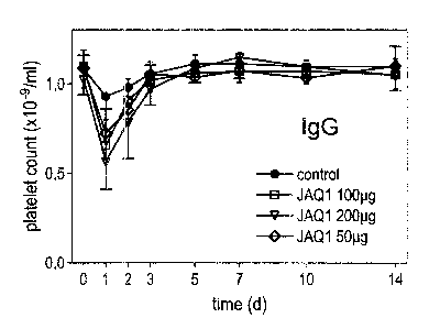
In the current study, we investigated the antithrombotic effects of JAQ1 in
vivo.
Injection of JAQ1 (100 pg) caused mild and transient thrombocytopenia with a
maximum drop of platelet counts of approximately 34 ~ 7.4 % on day 1 and a
return to normal after 72h where they remained for at least 11 more days
(Fig.1 a). Injection of higher (200 pg) or lower (50 pg) doses had comparable
effects on platelet counts (Fig.1a). The transient drop of platelet counts was
not
Fc-dependent as Fab fragments of JAQ1 had similar effects (Fig.1 b). JAQ1-
treated mice did not show any signs of anaphylactic reactions as known to be
induced by anti-GPllblllla mAbs (30) and did not develop spontaneous bleeding
far at least three weeks. JAQ1 was immunohistochemically detectable on
splenic and bone marrow-derived megakaryocytes 3 h after antibody injection,
demonstrating that the mAb reached these cells in both organs.
JAQ1 treatment abolishes platelet responses fo collagen and collagen related
peptides ex vivo for at least two weeks
The effect of JAQ1 on circulating platelets was studied ex vivo at different
time
points after antibody injection. The basal surface expression of the major
glycoprotein (GP) receptors GPllblllla and GPIb-IX-V, CD9, and integrin a2(3~
was unchanged as compared to control platelets at 3, 7, and 14 days after
antibody injection (Table 1 ). At no time after antibody injection did
circulating
platelets show any signs of activation, as demonstrated by the lack of surface
bound fibrinogen and surface expressed P-selectin (Table 1 ). On days 3, 7,
and
14, platelets from JAQ1-treated mice were resistant towards activation with
the
collagen related peptide (CRP up to 30 uglml), which is known to be a strong
GPVI-specific platelet agonist (31 ) (Fig.2a). In contrast, ADP induced normal
activation (fibrinogen binding) of these platelets. Furthermore, platelets
from
JAQ1-treated mice were completely resistant to activation with collagen at
concentrations of up to 50 pg / ml ex vivo and this profound inhibitory effect
also
2J. Dezember 2009
CA 02368791 2002-O1-22
c9P52EPlA11 Dr. Meyer-Dulheuer
lasted for at least 14 days upon a single injection of 100 pg JAQ1 (Fig.2b).
In
contrast to collagen, ADP and PMA induced normal aggregation of these
platelets, indicating that JAQ1 specifically blocked GPVI-dependent platelet
activation pathways whereas other functions were not affected. In vitro,
saturating concentrations of JAQ1 (20 pg/ml) only displayed a limited
inhibitory
effect on collagen-induced platelet aggregation which could, be overcome when
collagen concentrations higher than ~7 ~rglml were used (Fig. 2c), confirming
earlier results (29).
JAQ1 induces the loss of GPVI on circulating platelets in vivo
The discrepancy between the inhibitory effect of JAQ1 on collagen-induced
aggregation in vitro and ex vivo was surprising and suggested that mechanisms
other than pure blockage of an epitope on GPVI must be involved. Therefore,
the next step was to test platelets from JAQ1-treated mice for the presence of
GPVI in a Western blot analysis of whole cell lysates. As shown in Fig. 3a,
GPVI was not detectable in platelets from JAQ1-treated mice for at least 14
days upon a single injection of JAQ1, whereas GPllla was present in normal
amounts at any time point. In contrast, in all mice tested, new platelets
expressing functional GPVI were detectable after 28 days. To further assess
the
absence of GPVI on platelets from JAQ1-treated mice, we used the GPVI-
specific snake venom toxin convulxin (32): As shown in Fig. 3b, convulxin did
not induce aggregation of platelets from JAQ1-treated mice on day 3; 7, and
14,
whereas it induced aggregation of control platelets in the presence or absence
of saturating amounts of JAQ1. Furthermore, flow cytometric analysis
demonstrated that FITC-labeled convulxin did not bind to platelets from JAQ1-
treated mice (Fig. 3c). Finally, the absence of a -- 60 kD protein with an
isoelectric point of 5.6 in the platelets from JAQ1-treated mice was confirmed
by
2-D gel electrophoresis. Together, these results strongly suggested that GPVI
had been irreversibly inactivated and removed from these platelets in vivo.
JAQ1-induced GPVI loss occurs rapidly in vivo and is Fc-independent
To examine the mechanisms underlying the loss of GPVI, mice were injected
with biotinylated JAQ1 and the amount of surface-bound mAb was determined
27. Dezember 2001
CA 02368791 2002-O1-22
C9P52EPIA11 Dr. Meyer-Dulheuer
by flow cytometry ex vivo at early time points after injection. Interestingly,
as
soon as 6 hours after injection only very low levels of surface-bound JAQ1
were
detectable arid the signals further decreased to control after 24 and 48 h
while
JAQ1 FTC and CvxF~TC bound to the platelets at no time point. These data
suggested that the JAQ1-GPVI complex had been cleared from the surface of
those platelets within 6h. In contrast, platelets from mice injected with a
biotinylated mAb against GPV (24) constantly yielded positiv staining with
FITC-
labeled streptavidin. In the next step, we tested whole cell fysates from
platelets
of JAQ1-treated mice for the presence of GPVI and the biotinylated mAb. JAQ1
was strongly detectable in platelets 6 h after injection whereas signals
markedly
decreased at 24 h and even more at 48 h. A similar picture was found for GPVI,
strongly suggesting that the JAQ1-GPVI complex had become internalized and
was degraded within two days. In contrast to its in vivo effects, JAQ1 did not
induce any detectable downregulation of surface GPVI within 6 h incubation at
37°C on washed platelets or in whole blood (heparinized or citrated),
indicating
that a second signal may be required to induce this effect and that this
signal is
absent in vitro.
To determine whether the Fc part of JAQ1 or its divalent form is required for
internalizationl degradation of GPVI, mice received 100 pg Fab fragments of
the
mAb and the platelets were tested for the presence of GPVI after 48 h. The Fab
fragments, like the intact IgG, induced the complete loss of GPVI from
cirulating
platelets and the cells were completely resistant towards activation with CRP,
collagen, or convulxin.
GPVI-depleted platelets display reduced adhesion to collagen and abolished
collagen-dependent procoagulant activity
It is currently thought that GPVI is the platelet collagen receptor for
activation,
whilst integrin a2a1 and GPIb-V-IX (via vWF) mediate adhesion. As shown
before (Table 1 ), the basal surface expression of both receptors was not
influenced by the JAQ1 treatment. Further experiments demonstrated that
platelets from JAQ1-treated mice bound normal levels vWF in the presence of
botrocetin and thrombin induced normal activation of (31-integrins as assessed
27. Dezember 2001 1 p
CA 02368791 2002-O1-22
C9P52EP/A11 Dr. Meyer-Dulheuer
with the mAb 9EG7, which specifically recognizes the activated form of the ~i1
subunit (33) (Fig. 4a). In the next step, the adhesion of platelets from JAQ1-
treated mice to collagen was tested in a static assay. As shown in Fig. 4b,
the
adhesion of platelets from JAQ1-treated mice was -strongly reduced as
compared to control platelets and was abolished in the absence of
extracellular
free magnesium J calcium, strongly suggesting it to be mediated predominantly
by integrin a2~i1 (34). It is well known that GPVI is critically involved in
the
procoagulant response of platelets where stimulated platelets expose
negatively
charged phosphatidylserine (PS) at the plasma membrane which facilitates
thrombin generation (35). Indeed, platelets from JAQ1-treated mice did not
expose PS in response to a combination of collagen and thrombin on day 3, 7,
and 14 after antibody injection as demonstrated by the lack of Annexin V
binding (Fig. 4c).
Anti-GPVI treatment induces long-term antifhrombotic protection but only
moderately increased bleeding Times
The results of the previous experiments suggested that JAQ1 specifically
induced complete depletion of GPVI in platelets in vivo. To examine to which
extent this specific defect influenced normal hemostasis, we determined the
tail
bleeding times on day 7 after a bolus injection of JAQ1 (100 pg). As shown in
Fig. 5, the bleeding times were significantly increased in GPVI-depleted mice
compared to control mice (330 ~ 103 vs. 158 ~ 89 sec., respectively), but
consitently lower than in mice pre-treated with 100 pg blocking F(ab)2
fragments
against GPIIbJllla (24) (>600 sec.). In the next step, we examined the
protective
effect of JAQ1 in a model of lethal pulmonary thromboembolisrn induced by
infusion of a mixture of collagen (0.8 mglkg body weight) and epinephrine (60
Nglkg body weight) (27). Among control mice pre-treated with irrelevant rat
IgG2a, 95% (19 of 20) died within 5 min from widespread pulmonary thrombosis
and cardiac arrest. In contrast, all mice pre-treated with JAQ1 (100 Ng)
survived, irrespective of whether they had received the mAb 3, 7, or 14 days
before challenge (n=8 per group) (Fig. 6a). While the platelet counts in JAQ1
pre-treated mice had not been influenced significantly by the infusion of
collagen/epinephrine, there was a sharp decrease detectable in control mice
27. Dezembe~ 2001 11
CA 02368791 2002-O1-22
C 9 P 52 EP / A 11 Dr. Meyer-Dulheuer
(n=8) which was determined 3 min after induction of thromboembolism in a
separate group (Fig. 6b). For histological examination, control and JAQ1 pre-
treated (3, 7, and 14 days) mice received the same treatment in parallel
experiments but the lungs were removed after 3 min. While the vast majority of
large and small vessels were obstructed by platelet rich thrombi in the lungs
of
control mice, there were only very few thrombi detectable in the lungs of JAQ1
pre-treated mice (Fig. 6c).
Thus, it could be demonstrated that treatment of mice with a monoclonal
antibody against GPVI results in profound long-term antithrombotic protection
against collagen-dependent thromboembolism. These results confirm the
proposed critical role of GPV1 in collagen-induced activation of platelets in
vivo
and indicate that anti-GPVI agents might be effective in preventing arterial
thrombosis induced by atherosclerotic plaque rupture, where platelets are
thought to become activated mainly by the subendothelium under conditions of
high shear stress (4;5;36). Among the matrix proteins which support platelet
adhesion and subsequent activation, collagen has a critical role, at least in
normal hemostasis as patients with defects in collagen receptors display mild
bleeding disorders (12;37;38). Although the role of GPIb, GPllbllla and their
respective ligands von Willebrand factor (vWF) and fibrinogen in thrombosis
are
well documented (as reviewed by Z.M. Ruggeri (39)), the finding that vWF and
fibrinogen double knockout mice are still able to form occlusive thrombi
suggests that collagen and its platelet receptors might also have a critical
role in
thrombosis (40).
The profound inhibitory effect of JAQ1 in vivo was unexpected since it was
based on clearing of GPV1 from circulating platelets and no such specific
depletion of a platelet receptor has been described to date. The complete loss
of functional GPVI on circulating platelets in JAQ1-treated mice was confirmed
by different approaches. Firstly, the protein was not detectable in a Western
blot
analysis of platelet lysates for at feast two weeks (which exceeds the normal
fife-span of platelets (41 )). Secondly, the GPVI-specific snake venom toxin
convulxin, which binds to a different epitope than JAQ1 (Fig.3b, c), did not
bind
27. Dezember 2001 12
CA 02368791 2002-O1-22
C 9 P 52~P I A 11 Dr. Meyer-Dufheuer
to platelets from JAQ1-treated mice strongly suggesting the absence of GPVI
from the platelet membrane. Thirdly, a ~60 kD protein with an isoelectric
point
of ~5.fi (which is similar to that described for human GPVI (42)) is absent in
the
lysate of platelets from JAQ1-treated mice (Fig.4) and the same protein is
absent in platelets from FcRy chain-deficient mice (not shown) which are known
to lack GPVI (15). Most importantly, the functional platelef responses to
coNagen were completely abolished by JAQ1 in vivo, whereas the mAb only has
limited inhibitory effects in vitro (29), (Fig:2c). These results demonstrate
that
JAQ1 induced the clearing of GPVI from the surface of circulating platelets in
vivo. This finding is also supported by the observation that biotinylated JAQ1
was detectable in the lysates, but not on the surface, of platelets 6 h after
injection and the same was found for GPVI. Furthermore, the decreasing
signals for both GPVI and JAQ1 after 24 and 48 h strongly suggest that the
internalized complex was degraded in the intracellular compartments. GPVI
belongs to the immunoglobulin superfamily and is closely related to
immunoreceptors, some of which may become internalized when stimulated
appropriately (43;44). In the case of JAQ1-GPVI it was difficult to define
what
the appropriate stimulus is, but it seems clear that the Fc part of the mAb is
not
required to induce internalization as Fab fragments produced the same effect,
thereby also excluding the requirement for GPVI clustering. In vitro, JAQ1 did
not induce the downregulation of GPVI from the platelet membrane (Fig.Sa)
suggesting that a second signal may be required for the induction of this
process that is provided by other cells in vivo. This assumption may be
supported by the observation that JAQ1 and Fab fragments of the mAb induced
transient thrombocytopenia. The reason for this is not clear, but it might be
due
to weak activation of GPllblllla leading to formation of loose aggregates and
their temporary sequestration to the spleen where the actual loss of GPVI may
occur. Recent evidence indicates that JAQ1 recognizes an epitope identical
with or in close vicinity to the CRP binding site on GPVI (29) which is
regarded
as the major binding site for collagen on the receptor. So far, very little is
known
about the cellular regulation of GPVI but in the light of the current data it
seems
possible that occupancy of this epitope provides a signal that finally results
in
downregulation of the receptor.
27, Dezember 2001 13
CA 02368791 2002-O1-22
C9P52EPIA11 Dr. Meyer-Dulheuer
Irrespective of the underlying mechanism, platelets from JAQ1-treated mice
were completely unresponsive towards activation with high concentrations of
CRP or collagen whereas they were normally activatable with ADP or PMA.
This strongly suggests that JAQ1 selectively induced a transient GPVI
deficiency in mice while other membrane glycoproteins, including GPllblllla,
GPIb-IX-V, CD9, and integrin a2~i~ were not affected in expression andlor
function. JAQ1-treated mice had prolonged bleeding times which confirms the
important role of GPVI in normal hemostasis and correlates well with the
bleeding diathesis in GPVI deficient patients (12;22). Very interestingly, one
GPVI deficient patient developed highly specific antibodies against the absent
receptor (45) which may be difficult to explain. Based on the results
presented
here, however, it is feasible to speculate that this patient may suffer from
an
acquired GPVI deficiency, based on autoantibody-induced clearing of GPVI
from her circulating platelets.
Besides its pivotal role in collagen-induced platelet activation, GPVI is also
critically involved in the procoagulant reaction of platelets (46) which was
confirmed by the abolished collagen-dependent procoaguiant activity of
platelets from JAQ1-treated mice. This result strongly suggests that anti-GPVI
treatment also modulates coagulation at sites of vascular injury. Such an
anticoagulant activity has been demonstrated for GPllblllla antagonists (47),
which are currently considered the most powerful inhibitors of platelet
participation in thrombosis (48), as they inhibit the final common pathway of
platelet aggregation, irrespective of the agonist that stimulates the cells.
It has
been suggested that this more or less complete inhibition of platelet function
may come with a potential safety risk as platelet aggregation is also required
for
normal hemostasis (49). We found that JAQ1 induced significantly shorter
bleeding times than blocking antibodies against GPllb/llla in mice, indicating
that GPVI-depleted platelets still contributed significantly to normal
hemostasis
in vivo. Although there is no clear correlation between the bleeding time and
bleeding risk (50) it is tempting to speculate on the grounds of these results
that
27. Dezember 2001 14
CA 02368791 2002-O1-22
C 9 P 52 EP / A 11 Dr. Meyer-Dulheuer
anti-GPVI therapy might be associated with a relatively low risk of clinical
hemorrhage.
The mechanisms of collagen-platelet interactions are complex and involve
direct or indirect binding of collagen to several platelet receptors,
including the
GPIb-IX-V complex, integrin a2~il, GP1V, GPVI, and 65- and 85-kD proteins
(51 ). Despite its essential role in collagen-induced activation of platelets,
there
has been only very limited evidence for a role of GPVI in adhesion to collagen
(17) which is mainly thought to be mediated by GPIb-IX-V (via von Willebrand
factor, vWf) and integrin a2(i1. In mice, GPVI-depleted platelets expressed
normal amounts of integrin a2(3~ and (3~-integrins were normally activatable
which has been reported to be a prerequisite for effective binding of collagen
(Fig. 4b) (52). Indeed, GPVI-depleted platelets adhered to collagen through
a2~i~, but the extent of adhesion was strongly reduced as compared to control
platelets. A similar observation has been reported with platelets from GPVI
deficient patients {12;45), indicating that GPVI may be required for normal
adhesion to collagen probably by supporting the activation of a2~3~ {53). The
expression of GPIb-IX-V was not affected by the JAQ1 treatment and the
receptor bound normal levels of vWF in the presence of botrocetin (Fig. 4a).
Together, these results suggest that platelet adhesion to collagen at sites of
vascular injury may be reduced, but not blocked, by anti-GPVI treatment.
Very recent evidence suggests that GPVI is exclusively expressed in platelets
and mature megakaryocytes (17;54) and this is confirmed by
immunohistochemical studies with JAQ1. Therefore, the effects of anti-GPVI
agents (like JAQ1 ) should be restricted to platelets and, very importantly,
megakaryocytes. JAQ1 was detectable on megakaryocytes in spleen and bone
marrow 3h after antibody injection, suggesting that the next generation of
platelets was also affected by the mAb. This assumption may be confirmed by
the fact that GPVI was not detectable in platelets for at least' two weeks,
although the normal life-span of mouse platelets is only approximately 4-5
days
(41 ). Based on the estimated number of approximately 2 x 10⁹ platelets /
mouse (109 / ml blood) and a life span of the cells of 5 days, the GPVI
27. Dezember 2001 15
CA 02368791 2002-O1-22
c s P s2 EP r A" Dr. Meyer-Dulheuer
molecules of 6 x 10⁹ platelets must be depleted to result in the absence of
the
receptor for 15 days. The amount of 100 erg JAQ1 (MW: 150 kd) represents
~6.7 x 10'3 antibody molecules. Therefore, ~ 1.1 x 10⁴ antibody molecules per
platelet are available to bind and deplete GPVI. Since the estimated
expression
rate of GPVI is only 1-2 x 10³ copiesiplatelet (55) 100 Ng JAQ1 is sufficient
to
induce the observed effect.
Preliminary results show that a second injection of JAQ1 two weeks after the
first injection has no influence on platelet counts, but prolongs the absence
of
GPVI on circulating platelets. This indicates that the second dose of the mAb
affects newly differentiated megakaryocytes, but has no effect on circulating
(GPVI-depleted) platelets. Thus, JAQ1 can be used to induce a GPVI knock
out-like phenotype in mice for several weeks, allowing studies on platelet
function in the absence of this critical activating receptor in vitro and in
vivo.
The Thrombin response in JAQ~-treated mice is transiently reduced
After activation of the platelets of JAQ1-treated mice with the coagulation
protease a-thrombin on day 3 after JAQ1 injection a significantly reduced
thrombin response was detectable by measuring of GPllbllla activation and P-
selectin expression in response to increasing concentrations of a-thrombin. In
contrast, this inhibition was not detectable in JAQ1-treated mice on days 7
and
14. This finding suggests that a selective inhibition of the thrombin response
occurs during the early phase in platelets on anti-GPVI treatment.
To define this inhibition in more detail, the platelets from JAQ1-treated mice
were analyzed on days 1, 2, 3, 4 and 5 after antibody injection. In order to
minimize the transient drop of platelets counts, these mice received three
injections of 33 pg JAQ1 within 2 hours. This treatment resulted in a mild
decrease of platelet counts on day 1 and a return to normal on day 2 where
they remained for at feast 12 more days. The platelets from these mice showed
a marked reduction in the thrombin response on days 1 and 2 which
progressively returned to normal between days 3 and 5. In contrast, the
platelets were fully activatable with ADP and PMA at any time point.
27. Dezember 2001 16
CA 02368791 2002-O1-22
C 9 P 52 EP I A 11 Dr. Meyer-Dulheuer
These results strongly suggested that the JAQ1-induced GPVI internalisation
transiently affected the function of one ore more thrombin receptors in
circulating platelets. This effect was identified to be related to a reduced
activity
of the PAR4 thrombin receptor as shown by flow cytometric analysis of
platelets
from JAQ1-treated mice stimulated with an PAR4-activating peptide.
Next, JAQ1-treated mice were subjected to a model of thrombin-dependent
thromboembolism to determine the relevance of the observed effect for
thrombotic processes in vivo. Anesthetized male NMRI mice (28-30 g body
weight) received recombinant human thromboplastin (Thromborel, Dade
Behring) i.v.. This treatment is known to initiate intravascular thrombin
formation
which leads to platelet activation. These thrombin-activated platelets then
facilitate further thrombin generation and finally intravascular
thromboembolism.
The i.v. injection of 150 pl/kg body weight thromboplastin resulted in 60%
(12120) mortality in control mice pre-treated with irrelevant rat IgG, whereas
non
(0/20) of the JAQ1-treated mice-died on day 1 and 2 after antibody injection.
The mortality increased to 35% (7120) on day 3, further to 40% (8120) on day
4,
and finally reached the level of the control group on day 5 with 65% (13120).
These findings correlated well with the reduced thrombin response seen in
JAQ1 treated mice ex vivo. The following parameters were determined two
minutes after injection of 150 pl/kg body weight thromboplastin in control and
JAQ1-treated mice in separate groups. Platelet consumption and plasmatic TAT
concentrations were significantily reduced in JAQ1-treated mice as compared to
controls. Again, this effect was strongest on days 1 and 2 after JAQ1
injection
and progressively decreased between days 3 and 5.
Together, these findings demonstrate the treatment of mice with the anti-GPVI
mAb JAQ1 results in two distinct phases of platelet inhibition: During the
first 3 -
4 days, the platelets show a complete inhibition of collagen responses and
partial inhibition. of thrombin responses and therefore a profound
antithrombotic
protection. After this period, the thrombin response returns to normal whereas
27. Dezember 2001 17
CA 02368791 2002-O1-22
C 9 P 52 EP t A 11 Dr. Meyer-Dulheuer
the collagen response remains absent, resulting in a more moderate
antithrombotic protection.
Taken together, these results suggest that GPVI might become an interesting
target for long-term prophylaxis of ischemic cardiovascular diseases and
provide the first evidence that it is possible to specifically deplete an
activating
glycoprotein receptor from circulating platelets in vivo. These findings may
open
the way for the development of a new generation of powerful, yet safe,
antithrombotics.
Thus, it is an object of the present invention to provide a medicament for the
protection against thrombotic diseases which comprises an active principle,
preferably an antibody, against a platelet collagen receptor that not only
blocks,
but irreversibly depletes the target receptor. Such a monoclonal antibody is
defined by its binding to the same or a similar epitop of the collagen
receptor for
thrombocytes as the monoclonal antibody JAQ1. Preferably, as antibody the
monoclonal antibody JAQ1 should be used. The preferred collagen receptor is
platelet GPVI. Most preferred is a medicament which contains the respective
humanized monoclonal antibody for protection againts thrombotic diseases.
The monoclonal antibody JAQ1 can be humanized by standard methods which
are well known to the experts in the field: Said humanized monoclonal antibody
is usually administered to a patient who is jeopardized by thrombotic diseases
in the form of a physiologically acceptable aqueous injection. Other forms of
administration are not excluded. The monoclonal antibody will be administered
in a quantity which is subject to the physical condition of the patient. The
experienced medical doctor will have no difficulty to find out the optimum
quantity of the monoclonal antibody for the intended purpose.
27. Dezember 2001 1
CA 02368791 2002-O1-22
c9P52EPtA11 Dr. Meyer-Duiheuer
References
1. Weiss, H.J. 1975. Platelet physiology and abnormalities of platelet
function
(first of two parts). N Engl J Med 293:531.
2. Weiss, H.J. 1975. Platelet physiology and abnormalities of platelet
function
(second of two parts). N Engl J Med 293:580.
3. Fuster, V., L. Badimon, J.J. Badimon, and J.H. Chesebi-o. 1992. The
pathogenesis of coronary artery disease and the acute coronary
syndromes (1 ). N Engl J Med 326:242.
4. Fuster, V., L. Badimon, J.J. Badimon, and J.H. Chesebro. 1992. The
pathogenesis of coronary artery disease and the acute coronary
syndromes (2). N Engl J Med 326:310.
5. Conti, C.R. and J.L. Mehta. 1987. Acute myocardial ischemia: role of
atherosclerosis, thrombosis, platelet activation, coronary vasospasm, and
altered arachidonic acid metabolism. Circulation 75:V84.
6. Baumgartner, H.R. 1977. Platelet interaction with collagen fibrils in
flowing
blood. I. Reaction of human platelets with alpha chymotrypsin-digested
subendothelium. Thromb Haemost 37:1.
7. Hawiger, J. 1987. Macromolecules that link platelets following vessel wall
injury. Ann N YAcad Sci 509:131.
8. Morton, L.F., A.R. Peachey, and M.J. Barnes. 1989. Platelet-reactive sites
in collagens type I and type III. Evidence for separate adhesion and
aggregatory sites. Biochem J 258:157.
27. Dezember 2001
CA 02368791 2002-O1-22
C9P52EPIA11 Dr. Meyer-Dulheuer
9. Santoro, S.A., J.J. Walsh, W.D. Staatz; and K.J. Baranski. 1991. Distinct
determinants on collagen support alpha 2 beta 1 integrin-mediated platelet
adhesion and platelet activation. Cell Regul2:905.
10. Savage, B., F. Almus-Jacobs, and Z.M. Ruggeri: 1998. Specific synergy of
multiple substrate-receptor interactions in platelet thrombus formation
under flow. Cell 94:657.
11. Santoro, S.A. and M.M. Zutter. 1995. The alpha 2 beta 1 integrin: a
collagen receptor on platelets and other cells. Thromb Haernosl 74:813.
12. Moroi, M., S.M. ,lung, M. Okuma, and K. Shinmyozu. 1989. A patient with
platelets deficient in glycoprotein VI that lack both collagen-induced
aggregation and adhesion. J Clin Invest 84:1440.
13. Kehrel, B., S. Wierwille, K:J. Clemetson, O. Anders, M. Steiner, C.G.
Knight, R.W. Farndale, M. Okuma, and M.J. Barnes. 1998. Glycoprotein VI
is a major collagen receptor for platelet activation: it recognizes the
platelet-activating quaternary structure of collagen, whereas CD36,
glycoprotein Ilb/llla, and von Willebrand factor do not. Blood 91:491.
14. Gibbins, J.M., M: Okuma; R. Famdale, M. Barnes, and S.P: Watson. 1997.
Glycoprotein VI is the collagen receptor in platelets which underlies
tyrosine phosphorylation of the Fc receptor gamma-chain: FEBS Leif
413:255.
15. Nieswandt, B., W. Bergmeier, V. Schulte, K. Rackebrandt, ,I.E. Gessner,
and H. Zirngibl. 2000. Expression and function of the mouse collagen
receptor glycoprotein VI is strictly dependent on its association with the
FcRgamma chain. J Biol Chem 275:23998.
16. Clemetson, J.M., J. Polgar, E. Magnenat, T.N. Wells, and K.J. Clemetson.
1999. The platelet collagen receptor glycoprotein VI is a member of the
immunoglobulin superFamily closely related to FcaIphaR and the natural
killer receptors. J 8iol Chem 274:29019.
27. Dezember 2001 20
CA 02368791 2002-O1-22
C 9 P 52 EP /A 11 Dr. Meyer-Dulheuer
17. Jandrot-Perrus, M., S. Busfield, A.H. Lagrue, X. Xiong, N. Debili, T.
Chickering, J.P. Couedic, A. Goodearl, B. Dussault, C. Fraser, W.
Vainchenker, and J.L. Villeval. 2000. Cloning, characterization, and
functional studies of human and mouse glycoprotein VI: a platelet-specific
collagen receptor from the immunoglobulin superfamily [In Process
Citationj. Blood 96:1798.
18. Tsuji, M., Y. Ezumi, M. Arai, and H. Takayama. 1997. A novel association
of Fc receptor gamma-chain with glycoprotein VI and their co-expression
as a collagen receptor in human platelets. J Blol Chem 272:23528.
19. Poole, A., J.M. Gibbins, M. Tumer, M.J. van Vugt, J.G. van de Winkel, T.
Saito, V.L. Tybulewicz, and S.P. Watson. 1997. The Fc receptor gamma-
chain and the tyrosine kinase Syk are essential for activation of mouse
platelets by collagen. EMBO J 16:2333.
20. Ezumi, Y., K. Shindoh, M. Tsuji, and H. Takayama. 1998. Physical and
functional association of the Src family kinases Fyn and Lyn with the
collagen receptor glycoprotein VI-Fc receptor gamma chain complex on
human platelets. J Exp Med 188:267.
21. Briddon, S.J. and S.P. Watson. 1999. Evidence for the involvement of
p59fyn and p53/56fyn in collagen receptor signalling in human platelets.
Biochem J 338 ( Pt 1 ):203.
22. Arai, M., N. Yamamoto, M. Moroi, N. Akamatsu, K. Fukutake, and K.
Tanoue. 1995. Platelets with 10% of the normal amount of glycoprotein VI
have an impaired response to collagen that results in a mild bleeding
tendency [published erratum appears in Br J Haematol 1995
Apr;89(4):952]. Br J Haematol 89:124.
23. Bergmeier, W., K. Rackebrandt, W. Schroder, H. Zirngibl, and B.
Nieswandt. 200Ø Structural and functional characterization of the mouse
von Willebrand factor receptor GPIb-IX with novel monoclonal antibodies.
Blood 95:886.
21. Dezember 2001 21
CA 02368791 2002-O1-22
c9P52EPIA11 Dr. Meyer-Dulheuer
24. Nieswandt, B., W. Bergmeier, K. Rackebrandt, J.E. Gessner, and H,
Zirngibl. 2000. Identification of critical antigen-specific mechanisms in the
development of immune thrombocytopenic purpura in mice [In Process
Citation]. Blood 96:2520.
25. Rabilloud, T. 2000. Detecting proteins separated by 2-D gel
electrophoresis. Ana! Chem 72:48A.
26. Chevallet, M., V. Santoni, A. Poinas, D. Rouquie, A. Fuchs, S. Kieffer, M.
Rossignol, J. Lunardi, J. Garin, and T. Rabilloud. 1998. New zwitterionic
detergents improve the analysis of membrane proteins by two-dimensional
electrophoresis. Electrophoresis 19:1901.
27. DiMinno, G. and M.J. Silver. 1983. Mouse antithrombotic assay: a simple
method for the evaluation of antithrombotic agents in vivo. Potentiation of
antithrornbotic activity by ethyl alcohol. J Pharmacol Exp Ther 225:57.
28. Carmeliet, P.; J.M. Stassen, L. Schoonjans, B. Ream, J.J. van den Oord,
M. De Mol, R.C. Mulligan, and D. Collen. 1993. Plasminogen activator
inhibitor-9 gene-deficient mice. II. Effects on hemostasis, thrombosis, and
thrombolysis. J Clin Invesf 92:2756.
29. Schulte, V., D. Snell, W.: Bergmeier, H. Zirngibl, S.P. Watson, and B.
Nieswandt. 2000. Evidence for two distinct epitopes within, collagen for
activation of murine platelets. J Biol Chem
30. Nieswandt, B., B. Echtenacher, F.P. Wachs, J. Schroder, J.E. Gessner,
R.E. Schmidt, G.E. Grau, and D.N. Mannel. 1999. Acute systemic reaction
and lung alterations induced by an antiplatelet integrin gpffblllla antibody
in mice. Blood 94:684.
31. Asselin, J., J.M. Gibbins, M. Achison, Y.H: Lee, L.F. Morton, R.W.
Farndale, M.J. Barnes, and S.P. Watson. 1997. A collagen-like peptide
stimulates tyrosine phosphorylation of syk and phospholipase C gamma2
in platelets independent of the integrin alpha2beta 1. Blood 89:1235.
27. Dezember 2001 22
CA 02368791 2002-O1-22
c 9 P 52 FP l A 11 Dr. Meyer-Dutheuer
32. Polgar, J., J.M. Clemetson, B.E. Kehrel, M. Wiedemann, E:M. Magnenat,
T.N.C. Wells, and K.J. Clemetson. 1997. Platelet activation and signal
transduction by convulxin, a C-type lectin from Crotalus durissus terrificus
(tropical rattlesnake) venom via the p62IGPVl collagen receptor. J Biol
Chem 272:13576.
33. Lenter, M., H. Uhlig, A. Hamann, P. Jeno, B. Imhof, and D. Vestweber.
1993. A monoclonal antibody against an activation epitope on mouse
integrin chain beta 1 blocks adhesion of lymphocytes to the endothelial
integrin alpha 6 beta 1. Proc Natl Acad Sci U S A 90:9051.
34. Onley, D.J., C.G. Knight, D:S. Tuckwell, M.J. Barnes, and R.W. Farndale.
2000. Micromolar Ca2+ concentrations are essential for Mg2+-dependent
binding of collagen by the, integrin alpha 2beta 1 in human platelets. J Biol
Chem 275:24560.
35. Bevers, E.M., P. Comfurius, J.L. van Rijn, H.C. Hemker, and R.F. Zwaal.
1982. Generation of prothrombin-converting activity and the exposure of
phosphatidylserine at the outer surface of platelets. Eur J Biochem
122:429.
36. Lusis, A.J. 2000. Atherosclerosis. Nature 407:233.
37. Nieuwenhuis, H.K., J.W. Akkerman, W.P. Houdijk, and J.J. Sixma. 1985.
Human blood platelets showing no response to collagen fail to express
surface glycoprotein la. Nature 318:470.
38. Ryo, R., A: Yoshida, W. Sugano, M. Yasunaga, K. Nakayama, K: Saigo,
M. Adachi, N. Yamaguchi, and M. Okuma. 1992. Deficiency of P62, a
putative collagen receptor, in platelets from a patient with defective
collagen-induced platelet aggregation. Am J Hematol 39:25.
39. Ruggeri, Z.M. 1997. Mechanisms initiating platelet thrombus formation
[published erratum appears in Thromb Haemost 1997 Oct;78(4):1304]
[see comments]. Thromb Haemost 78:611.
27. Dezember 2001 23
CA 02368791 2002-O1-22
C 9 P 52 EP /A 11 Dr. Meyer-Dulheuer
40: Ni, H., C.V. Denis, S. Subbarao, J.L. Degen, T.N. Sato, R:O. Hynes, and
D.D. Wagner. 2000. Persistence of platelet thrombus formation in
arterioles of mice lacking both von Willebrand factor and fibrinogen. J Clip
Invesf 106:385.
41. Ault, K.A. and C. Knowles. 1995. In vivo biotinylation demonstrates that
reticulated platelets are the youngest platelets in circulation. Exp Hematol
23:996.
42. Clemetson, K.J., J.f_. McGregor, E. James, M. Dechavanne, and E.F.
Luscher. 1982. Characterization of the platelet membrane glycoprotein
abnormalities in Bernard-Soulier syndrome and comparison with normal
by surface-labeling techniques and high-resolution two-dimensional gel
electrophoresis. J Chn Invest 70:304.
43. Daeron, M. 1997. Fc receptor biology. Annu Rev Immunol 15:203.
44. Ravetch, J.V. 1997. Fc receptors. Curr Opin Immunol 9:121.
45. Sugiyama, T., M. Okuma, F. Ushikubi, S. Sensaki, K. Kanaji, and H.
Uchino. 1987. A novel platelet aggregating factor found in a patient with
defective collagen-induced platelet aggregation and autoimmune
thrombocytopenia. Blood 69:1712.
46. Heemskerk, J.W., P. Siljander, W.M. Vuist, G. Breikers, C.P.
Reutelingsperger, M.J. Barnes, C.G. Knight, R. Lassila, and R.W.
Farndale. 1999. Function of glycoprotein VI and integrin alpha2beta1 in
the procoagulant response of single, collagen-adherent platelets. Thromb
Haemost 81:782:
47. Reverter, J.C., S. Beguin, H. Kessels, R. Kumar, H.C. Hemker, and B.S.
Coller. 1996. Inhibition of platelet-mediated, tissue factor-induced thrombin
generation by the mouselhuman chimeric 7E3 antibody. Potential
implications for the effect of c7E3 Fab treatment on acute thrombosis and
"clinical restenosis". J Clip Invest 98:863.
2T. Dezer~ber 2001 24
CA 02368791 2002-O1-22
c 9 P 52 EP t A 11 Dr. Meyer-Dulheuer
48. Topol, E.J., T.V. Byzova, and E.F. Plow. 1999. Platelet GPllb-Ills
blockers.
Lancet 353:227.
49. Scarborough, R.M., N.S. Kleiman, and D.R. Phillips. 1999. Platelet
giycoprotein ilb/ltla antagonists. What are-the relevant issues concerning
their pharmacology and clinical use? Circuiafion 100:437.
50. Rodgers, R.P. and J. Levin. 1990. A critical reappraisal of the bleeding
time. Semin Thromb Hemost 16:1.
51. Barnes, M.J., C.G. Knight, and R.W. Farndale. 1998. The collagen-platelet
interaction. Curs Opin Hemafol 5:314.
52. Moroi, M., 1. Onitsuka, T. Imaizumi, and S.M. Jung. 2000. Involvement of
activated integrin alpha2beta1 in the firm adhesion of platelets onto a
surface of immobilized collagen under flow conditions. Thromb Haemost
83:769.
53. Watson, S., O. Berlanga, D. Best, and J. Frampton. 2000. Update on
collagen receptor interactions in platelets: is the two-state model still
valid?
[In Process Citation]. Plafefets 11:252.
54. Berlanga, O., R. Bobe, M. Becker, G. Murphy, M. Leduc, C. Bon, F.A.
Barry, J.M. Gibbins, P. Garcia, J. Frampton, and S.P. Watson. 2000.
Expression of the collagen receptor glycoprotein VI during megakaryocyte
differentiation [fn Process Citation]. Blood 96:2740.
55. Niedergang, F., A. Alcoves, C.G. Knight, R.W. Farndale, M.J: Barnes, I.M.
Francischetti, C. Bon, and M. Leduc. 2000. Convulxin binding to platelet
receptor GPVI: competition with collagen related peptides. Biochem
Biophys Res Common 273:246.
27. Dezember 2001 25
CA 02368791 2002-O1-22
C 9 P 52 EP I A 11 Dr. Meyer-Dulheuer
Table 1: Expression of glycoproteins and surface-bound fibrinogen on
platelets from JAQ1-treated mice. Diluted whole blood from the indicated
mice was incubated with F1TC-labeled antibodies at saturating concentrations
for 15 min at RT and platelets were analyzed directly. Results are expressed
as
mean log fluorescence ~ S.D. for 6 mice per group.
control JAQ 1 3d JAQ 1 7d JAQ 1 14d
GPllblllla321.3 9.7 318.1 9.4 328.7 9.1 325.3 9.8
GPIb-lX 278.9 16.8 275.4 18.0 269.5 15.9 273.1 11.4
GPV 165.4 10.9 163.3 14.1 169.1 15.3 158.1 10.5
CD9 543:8 15.8 554.3 14.6 549.5 19.6 557.0 13.0
GPIs 38.2 6.7 40.3 6.5 35.217.8 36.7 6.2
(a2)
fibrinogen14.1 1.7 15:0 1.4 14.3 1.5 14.4 1.5
P-selectin6.20.8 6.50.8 6.70.8 6.01.1
27. Dezember 2001 26
CA 02368791 2002-O1-22
C 9 P 52 EP! A 11 Dr. Meyer-Dulheuer
Legends to figures
Figure 1: JAQ1 induces transient thrombocytopenia
Mice received purified IgG (a) or Fab fragments (b) of the indicated mAb i.p,
in
200 ~I sterile PBS. Platelet counts were determined at the indicated times
using
an improved Neubauer hemocytometer. Results are expressed as the mean
platelet count ~ SD for groups of each 6 mice.
Figure 2: Platelets from JAQ1-treated miice do not respond to CRP and
collagen
(a) Two color flow cytometric analysis of platelets from JAQ1-treated or
control
mice 3 days after antibody injection. Diluted whole blood was stimulated with
10
pM ADP or 10 pglml CRP for 2 min and subsequently incubated with anti-
fibrinogenF~rc and anti-P-selectinPE antibodies for 10 min at RT and analyzed
directly. Platelets were gated by FSC/SSC characteristics and FI3 intensity
(anti-mouse GPlbaPS'cy5). The data shown are representative of 6 mice per
group. Similar results were obtained on days 7 and 14 after antibody
injection.
(b) Heparinized prp from the indicated mice was stimulated with collagen (50
pg/ml), ADP (10 pM) or PMA (50 ng/ml). Light transmission was recorded on a
Fibrintimer 4 channel aggregometer. (c) Heparinized prp from control mice was
incubated with stirring in the presence of irrelevant rat IgG2a (20 pg/ml -
circles) or JAQ1 (20 Irglml - triangles) for 5 min before the addition of the
indicated concentrations of collagen. In parallel, prp from JAQ1-treated mice
was tested (squares). Results are expressed as the max. platelet aggregation ~
S.D. for groups of each 6 mice.
Figure 3: GPVI is not detectable in platelets from JAQ1-treated mice for at
least two weeks
(a) Whole platelet proteins were separated by SDS-PAGE under non-reducing
conditions and immunoblotted with FITC-labeled JAQ1 (anti-GPVI) or EDL1
{anti-GPllla). Bound mAb was detected by HRP-labeled rabbit anti-FITC and
ECL. (b) Washed platelets from control, FcRy chain-deficient (FcRy -I-) and
27. Dezember 2001 27
CA 02368791 2002-O1-22
C9P52EPIA11 Dr. Meyer-Dulheuer
JAQ1-treated (day 7) mice were stimulated with 10 pglml convulxin (Gvx).
Control platelets were pre-incubated with irrelevant rat IgG2a or JAQ1 (20
Nglml) for 5 min before the addition of Cvx. (c) Washed platelets from the
indicated mice were incubated with FITC-labeled convulxin (5 pg/ml) for 15 min
at room temperature and then analyzed on a FACScan (Becton Dickinson). The
data shown are representative of 6 mice per group.
Figure 4: Reduced adhesion to collagen and abolished procoagulant
response of GPVI-depleted platelets
(a) Platelets from JAQ1-treated mice (d 7) bind normal amounts of plasma vWF
in the presence of botrocetin {2 pg/ml - solid line). Bound vWf was detected
by
FITC-labeled anti-vWF antibodies (10 pglml). No binding was detected in the
absence of botrocetin (shaded area). Norms! activation of ,~1-integrins on
platelets from JAQ1-treated mice in response to thrombin (0.1 U/ml). Resting
(shaded area) or thrombin activated (solid line) platelets were incubated with
FITC-labeled 9EG7 (5 pglml) for 15 min at RT and analyzed directly. (b)
Washed platelets from control or JAQ1-treated mice (d 7) were incubated in
collagen-coated microtiter plates in the presence or absence of MgCl2 (1 mM) I
CaCl2 (1 mM) for the indicated times and adherent platelets were quantitated
fluorimetrically. The data shown are from a single experiment, representative
of
five identical experiments and expressed as the mean of triplicate readings ~
SD. (c) Flow cytometric analysis of Annexin V-FITC binding to platelets from
control and JAQ1-treated (d 7); mice activated with a combination of collagen
(50 pg/ml) and thrombin (0.01 Ulml).
Figure 5: Bleeding time of JAQ1-treated mice
Bleeding times were determined i,n mice 7 days after injection of 100 pg non-
immune IgG2a or JAQ1 (n=15 per group). As a control, mice received 100 pg
F(ab)2 fragments of JONIA (anti-GPllblllfa) 24 h before the experiment (n=6).
Where necessary, bleeding was manually stopped at the 10 min-time point to
prevent death. Each point represents one individual.
27. Dezember 2001 28
CA 02368791 2002-O1-22
C9P52EPIA11 Dr. Meyer-Dulheuer
Figure 6: JAQ1 induces long-term protection from intravascular
thrombosis
Thromboembolism in response to a bolus injection of a mixture of collagen {0.8
mg/kg body weight) and epinephrine (60 pglkg body weight). (a) Mortality in
control mice and mice treated with 100 Irg JAQ1 at the indicated times before
challenge. (b) Platelet counts in control and JAQ1-treated mice 3 min after
infusion of coilagenlepinephrine (n=8 per group). (c) Upper panel:
representative histology of the lungs (original x 100); obstructed vessels are
indicated by arrows. Lower panel: immunohistochemical detection of platelets
in
the thrombi (original x 400). Acetone fixed frozen sections were reacted with
a
platelet-specific antibody (anti-GPIb-fX) and counterstained with hematoxylin.
The red horseradish peroxidase reaction product shows high density of
platelets
in the thrombus.
2T. Dezember 2001