Note: Descriptions are shown in the official language in which they were submitted.
CA 02372089 2001-11-29
WO 01/01877 PCT/US00/01886
MEDICAL DEVICE HAVING AN INCREMENTALLY
DISPLACEABLE ELECTRODE
Field of the Invention
This invention relates to medical devices for performing diagnostic,
mapping, ablation, and other procedures and, more particularly, to a medical
device for
incrementally moving an electrode a predetermined distance.
Background of the Invention
Cardiac arrhythmias (commonly known as irregular heart beats or racing
hearts) are the result of various physical defects in the heart itself. One
such defect
comprises an extraneous strand of muscle fiber in the heart that provides an
abnormal
short-circuit pathway for electric impulses normally existing in the heart.
This accessory
pathway often causes the electric impulses that normally travel from the upper
to the
lower chamber of the heart to be fed back to the upper chamber, causing the
heart to
beat irregularly and therefore inefficiently pump blood.
Another common type of cardiac arrhythmia is ventricular tachycardia
(VT), which may be a complication resulting from a heart attack or from a
temporary
reduction of blood supply to an area of heart muscle. VT is often caused by a
tiny
lesion, typically on the order of one to two millimeters, that is located
close to the inner
surface of the heart chamber. That lesion is often referred to as an "active
site", because
it does not fire in sequence with the rest of the heart muscle. VT causes the
heart's
normal rhythmic contraction to be altered, thereby affecting heart function. A
typical
symptom is rapid, inefficient heart beats.
Other common cardiac arrhythmias include atrial flutter and atrial
fibrillation, which originate in the atria and cause the atria to beat so
rapidly that they
quiver (i.e., fibrillate). This in turn causes the ventricles to beat too fast
(up to 200
beats per minute), which results in an inefficient pumping of blood.
Non-surgical procedures, such as management with drugs, have been
CA 02372089 2001-11-29
WO 01/01877 PCT/US00/01886
-2-
proposed for treating cardiac arrhythmias. However, some arrhythmias are not
treatable
with drugs. For example, drug therapy to combat VT is typically successful in
only 30
to 50 percent of patients. Because of this low success rate, another
conventional remedy
is to perform a surgical procedure in which various incisions are made in the
heart to
block conduction pathways, and thereby divide the atrial area available for
multiple
wavelet reentry in an effort to abolish the arrhythmia. Alternatively, an
automatic
implantable cardioverter/defibrillator (AICD) can be surgically implanted into
the
patient, as described in U.S. Patent No. 4,817,608 to Shapland et al. While
these
surgical procedures can be curative, they are associated with increased
morbidity and
mortality rates, and are extremely expensive. Even the use of an AICD requires
major
surgical intervention. Moreover, patients of advanced age or illness often
cannot
tolerate invasive surgery to excise the tachycardia focus which causes the
arrhythmia.
Thus, this type of treatment is unavailable to many.
Non-surgical, minimally invasive techniques have been developed which
are used to locate cardiac regions responsible for the cardiac arrhythmia, and
to disable
the short-circuit function of these areas. According to these techniques,
electrical energy
shocks are applied to a portion of the heart tissue to ablate that tissue and
produce scars
which interrupt the reentrant conduction pathways. The regions to be ablated
are usually
first determined by endocardial mapping techniques. Mapping typically involves
percutaneously introducing a diagnostic catheter, having one or more
electrodes, into the
patient, passing the diagnostic catheter through a blood vessel (e.g., the
femoral vein or
aorta) and into an endocardial site (e.g., the atrium or ventricle of the
heart), and
inducing a tachycardia so that a continuous, simultaneous recording can be
made with a
multichannel recorder at each of several different endocardial positions. When
a
tachycardia focus is located, as indicated in the electrocardiogram recording,
it is
marked by means of a fluoroscopic image so that the site can be ablated. A
conventional
electrode catheter, having electrodes with a greater surface area than the
diagnostic
catheter's electrodes, can then provide electrical energy to the tissue
adjacent the
electrode to create a lesion in the tissue. One or more suitably positioned
lesions will
create a region of necrotic tissue to disable the malfunction caused by the
tachycardia
focus .
Conventional catheter ablation techniques have used catheters each having
CA 02372089 2001-11-29
WO 01/01877 PCT/US00/01886
-3-
a single electrode fitted at its tip as one electrical pole. The other
electrical pole is
conventionally provided by a backplate in contact with a patient's external
body part to
form a capacitive coupling of the ablation energy source (DC, laser, RF,
etc.). Other
ablation catheters are known in which multiple electrodes are provided.
Ablation is carried out by applying energy to the catheter electrodes once
the electrodes are in contact with the cardiac tissue. The energy can be, for
example,
RF, DC, ultrasound, microwave, or laser radiation. When RF energy is delivered
between the distal tip of a standard electrode catheter and a backplate, there
is a
localized RF heating effect. This creates a well-defined, discrete lesion
slightly larger
than the tip electrode (i.e., the "damage range" for the electrode), and also
causes the
temperature of the tissue in contact with the electrode to rise.
To overcome certain types of cardiac arrhythmia, such as atrial flutter and
atrial fibrillation, it is often necessary to create a long, continuous lesion
(i.e., a linear
lesion) to block the aberrant pathway(s). One conventional ablation procedure
for
creating linear lesions is commonly referred to as a "drag" method. According
to that
method, an ablation catheter carrying one or more ablation electrodes is
manipulated
through a patient's blood vessels to a desired location within the patient's
heart. One or
more of the electrodes is manipulated into contact with the heart tissue.
Ablation energy
is then delivered to the electrode(s), causing them to heat up and scar the
adjacent tissue
to create a lesion which is typically slightly larger than the surface area of
the electrode
contacting the tissue (the electrode's damage range). After the electrode has
been
disposed in that location for a sufficient time to ablate the adjacent tissue,
the clinician
then manually moves the catheter a selected amount by pulling on the catheter
shaft, and
ablation energy is again delivered to the electrodes) to ablate the tissue
that is then
adjacent to the electrode. By continuing this procedure, the clinician
attempts to create a
continuous, linear lesion to block an aberrant pathway.
However, to create a continuous lesion, the clinician must be careful not
to move the catheter too far between successive ablations. If the clinician
should
accidentally move the catheter too far, then the lesion created will not be
continuous,
and the aberrant pathway may not be destroyed, requiring that the patient
undergo yet
another procedure, which is inefficient and undesirable.
Accordingly, it will be apparent that there continues to be a need for a
CA 02372089 2001-11-29
WO 01/01877 PCT/US00/01886
-4-
device for performing ablations which ensures the creation of linear lesions,
by
automatically displacing an ablation electrode in successive, incremental
movements of a
predetermined distance. In addition, the need exists for a device which moves
an
electrode in known increments to perform other medical procedures. The instant
invention addresses these needs.
Summary of the Invention
According to one aspect of the invention, an electrode is connected to a
movable member, such as a catheter shaft or an outer sheath, which is slidably
extended
over a guide wire, flexible shaft, or other tubular member. A displacement
mechanism
is connected to the movable member, and may be actuated one or more times to
displace
the movable member in successive, predetermined increments. In this manner,
the
electrode is reliably moved in constant increments, and is suitable for
creating a linear
lesion or for performing diagnostic functions, without forcing the clinician
to estimate
the distance the electrode has been moved.
Thus, in one illustrative embodiment, the present invention is directed to
a medical device comprising an elongated shaft, an electrode mounted on the
shaft, and an
electrode displacement mechanism connected to the shaft and operative to
displace the
shaft in predetermined increments.
In another illustrative embodiment, the invention is directed to a method
for creating continuous lesions, comprising: (a) positioning an ablation
electrode at a
selected site within a patient, the ablation electrode having predetermined
dimensions; (b)
delivering ablation energy to the electrode to ablate the patient's tissue
disposed adjacent
to the tissue; (c) displacing the electrode in a predetermined increment,
wherein the
predetermined increment is determined based upon one or more of the dimensions
of the
electrode; and (d) repeating steps (b) and (c) one or more times to create a
continuous
lesion.
Description of the Drawings
Other objects, features and advantages of the invention discussed in the
above summary of the invention will be more clearly understood from the
following
detailed description of preferred embodiments, which are illustrative only,
when taken
CA 02372089 2001-11-29
WO 01/01877 PCT/US00/01886
-5-
together with the accompanying drawings in which:
Fig. 1 is a side elevation view of a mechanism for incrementally
displacing an electrode according to one illustrative embodiment of the
present
invention;
Fig. 2 is a fragmented top plan view of the mechanism shown in Fig. 1;
Fig. 3 is a sectional side view, in enlarged scale, of the mechanism shown
in Fig. 1;
Fig. 4 is a fragmented side view of a ratchet mechanism included in the
mechanism shown in Fig. l;
Fig. 5 is a bottom view of the ratchet mechanism shown in Fig. 4;
Fig. 6 is a cross-sectional view taken along the line 6-6 of Fig. 3 and
looking in the direction of the arrows;
Fig. 7 is a fragmented side view of another illustrative embodiment of the
mechanism for incrementally displacing an electrode according to the present
invention;
and
Fig. 8 is a cross-sectional view taken along the line 8-8 of Fig. 7 and
looking in the direction of the arrows.
Detailed Description of the Preferred Embodiments
Referring now to FIGS. 1 through 3, there is shown a mechanism 10 for
incrementally displacing one or more electrodes 12 according to one
illustrative
embodiment of the invention. In one illustrative embodiment, the electrodes)
12 is
carried on a movable member 14, for example, a catheter shaft or slidable
sheath 16
which is slidably extended and retracted over an inner tubular member 18, for
example,
a guide wire, catheter shaft, or the like. An incremental displacement
mechanism,
generally designated 20, is connected to the movable member 14 and is
operative, upon
each actuation thereof, to displace the movable member, and thus the
electrode, a
predetermined distance relative to the inner member 18 and the mechanism 10.
Thus,
for example, in an ablation procedure, the device 10 may be manipulated
through a
patient's blood vessels by means of the guide wire or catheter shaft until the
movable
member 14 and electrode 12 are disposed in a desired location, such as in
contact with
an "active site" in the heart. Ablation energy is delivered to the electrode
to destroy the
CA 02372089 2001-11-29
WO 01/01877 PCT/US00/01886
-6-
adjacent tissue. The clinician then actuates the displacement mechanism 20 to
incrementally move the movable member 14 and thus the electrode 12 a
predetermined,
known distance. The process is repeated one or more times to create a
continuous
lesion.
While two electrodes 12 are shown on the movable member 14, it will be
apparent to those skilled in the art that the movable member may carry
virtually any
number of electrodes, for example, one or more. Preferably, one electrode will
be
disposed at the distal end of the member 14, with the other electrodes being
disposed at
spaced apart locations along the member.
Referring to FIG. l, the movable member 14 is preferably in the form of
a tubular shaft, which is flexible for manipulation through a patient's blood
vessels and
to a site of interest. The tubular shaft defines an interior lumen 22 which is
sized to
slidably receive the inner member 18 therethrough. The inner member may take
many
different forms, such as a guide wire having a preformed curve, and can be
slidably
inserted through the lumen 22 to impart a desired bend to a portion of the
movable
member, with the movable member and guide wire then being advanced together
through the patient's blood vessels to a desired site, as is well known to
those skilled in
the art.
The medical device 10 further comprises a housing 24 which houses a
portion of the inner member 18 and movable member 14 therein, and also houses
the
displacement mechanism 20 therein. The displacement mechanism is movable
relative to
the housing, as is described in greater detail below. The housing includes a
handle
portion 25 which may be gripped by a user's hand, and an elongated barrel
portion 26
connected to the upper end of the handle and which is open at either
longitudinal end
thereof for extension of the movable and tubular members 14 and 18
therethrough.
The housing 24 includes an interior side wall 27 with a row of teeth 28
formed along the longitudinal length of the wall (shown in fragment in Fig.
3), which
cooperate with the displacement mechanism to incrementally displace the
displacement
mechanism relative to the housing, as is described in greater detail below.
The teeth 28
include angled leading edges 30 which face toward the front of the housing 24,
and
stepped trailing edges 31 which face toward the rear of the housing.
Referring to Figs. 3 through 6, one illustrative embodiment of the
CA 02372089 2001-11-29
WO 01/01877 PCT/US00/01886
_7_
displacement mechanism 20 will be described. The displacement mechanism
generally
comprises a trigger mechanism 32 and ratchet mechanism 33. The trigger
mechanism
comprises an elongated bar 34 which, on an upper side thereof, defines a
plurality of
upwardly projecting ratchet teeth 36. The teeth include forwardly facing,
angled edges
30, and rearwardly facing, stepped edges 31, similar to the teeth 28 formed in
the side
wall 26 of the housing 24.
The bar 34 is connected on its lower side to a circular ring 38 defining a
trigger, which extends downwardly through an opening 37 formed in the lower
end of
the housing 24. Extending rearwardly from the back side of the trigger 38 is a
cylindrical rod 40, which is housed inside of the housing 24 for movement
relative to the
housing, and which slidably receives a compression spring 42 over it. The
rearward end
of the spring abuts against an internal stop 44 formed in the housing 24.
Thus, when the
trigger is squeezed (i.e., driven rearwardly relative to the housing), the
spring is
compressed. When the user then releases the trigger, the compression spring
urges the
trigger mechanism 32 back to its original position with the trigger 38
abutting against the
forward end of the opening 37 (shown in Fig. 3).
The ratchet mechanism 33 is slidably housed in the housing 24 and
engaged with the trigger mechanism 32 in such a manner that actuation of the
trigger
mechanism causes the ratchet mechanism to move rearwardly relative to the
housing.
However, when the trigger is released and driven forwardly relative to the
housing 24
by the spring 42, the ratchet mechanism does not move forwardly, but rather
remains in
place relative to the housing. The structure achieving such function is now
described in
detail. The ratchet mechanism includes a resilient, flexible bottom tab 46 and
a resilient,
flexible side tab 48, which are designed to ride along, respectively, the row
of teeth 36
formed on the trigger mechanism, and the row of side teeth 28 formed in the
side wall
27 of the housing 24. The bottom tab and side tab angle outwardly from the
ratchet
body and toward the front of the housing 24. Thus, as described above, when
the
trigger 38 is squeezed, the trigger mechanism 32 is driven rearwardly relative
to the
housing 24. One of the stepped trailing edges 31 of the trigger mechanism
teeth 36
engages the bottom tab 46 of the ratchet mechanism 33 and forces the ratchet
mechanism
33 rearwardly. At the same time, the side tab 48 is driven rearwardly over one
or more
of the angled forward edges 30 of the side wall teeth 28. When the trigger is
released,
CA 02372089 2001-11-29
WO 01/01877 PCT/US00/01886
_g_
the spring urges the trigger mechanism toward the front of the housing 24. The
bottom
tab 46 then slides over the angled edges 30 of the trigger mechanism teeth 36
as they are
driven forwardly relative to the housing, while the side tab 48 engages one of
the
stepped edges 31 of the side wall teeth 28 and is thereby prevented from
moving forward
relative to the housing 24. Thus, upon each actuation of the trigger 38, the
ratchet
mechanism 33 is displaced rearwardly a predetermined distance relative to the
housing,
but when the trigger is released, the side wall teeth act to keep the ratchet
mechanism in
place relative to the housing. The distance of the displacement is dependent
on the
travel path of the trigger 38. Preferably, the ratchet mechanism is displaced
the length
of one tooth 28 with each squeeze of the trigger 38. Therefore, in order to
alter the
length of the incremental displacement, the length of the teeth may be
adjusted.
Alternatively, the travel path of the trigger 38 can be altered so that the
ratchet
mechanism 33 is displaced a multiple number of teeth upon each squeeze of the
trigger,
such as two or more.
Referring now to Fig. 6, the internal structure of the housing 24 is shown
in detail. The housing includes an elongated slot 50 formed in the upper wall
of the
housing, and slidably receives an upwardly and rearwardly projecting arm 52 of
the
ratchet mechanism 33.
With continued reference to Fig. 6, the upper interior walls of the housing
24 have a stepped configuration (as indicated by reference numerals 55 and 56)
to
complement the configuration of the upper surface portion of the ratchet
mechanism 33,
and define a track to allow the ratchet mechanism to slide through the
housing, and
guide the ratchet mechanism along a linear travel path inside the housing.
The housing 24 further includes a pair of elongated, internal recesses
defining a pair of tracks 60 (FIG. 6), with one disposed on either lateral
side of the
housing. The trigger mechanism 32 includes a plurality of spaced apart,
laterally
outwardly projecting tabs or followers 62 which project outwardly from either
side of
the bar 34 and are formed having a complementary size to be slidably received
in the
respective tracks. Thus, the tracks and tabs cooperate to define a linear
travel path for
the trigger mechanism through the housing, and provide stability for the
trigger
mechanism as it is displaced relative to the housing 24.
As is well known to those skilled in the art, electrodes have different
CA 02372089 2001-11-29
WO 01/01877 PCT/US00/01886
-9-
"damage ranges", which depend on the design and dimensions of the electrode.
"Damage range" is defined herein to mean the area of tissue which is scarred
when
ablation energy is delivered to the electrode. Typically, the damage range is
slightly
larger than the surface area of the electrode contacting the tissue, and
depends on
electrode thickness, the electrode material, and the like. Thus, depending
upon the type
of electrode used, the length of the incremental displacement will vary.
Because the
damage range tends to approximate the length of the electrode, the length of
the
incremental displacement will preferably approximate the length of the
electrode itself.
It may even be preferably made slightly shorter than the length of the
electrode to ensure
overlapping damage ranges and therefore a continuous lesion. However, in the
case of
an electrode having a large damage range, the length of the incremental
displacement
can be longer than the length of the electrode.
In the embodiment shown in Figs. 1 through 6, the ratchet mechanism 33
is connected to an outer sheath 14 which is slidable over an inner member 18.
However, it will be apparent to those skilled in the art that the ratchet
mechanism could
alternatively be engaged directly to a catheter shaft with an electrode
mounted on the
catheter shaft.
It will also be apparent to those skilled in the art that the orientation of
the
teeth and springs could be reversed, to cause the ratchet mechanism 33 to be
advanced
toward the front of the housing 24 rather than be driven toward the rear of
the housing.
However, it is presently preferred to retract the ratchet mechanism and thus
the movable
member 14, for performing drag ablation procedures and the like.
In operation, the movable member 14 and inner member 18 are
manipulated through the patient's vasculature to an intended site, such as an
"active
site" . A power supply (not shown) is configured to energize the electrode 12
through an
electrical conductor (not shown) in either a constant voltage, power, or
temperature
mode, as is well known in the art. Radio-frequency energy is delivered to the
electrode
12 to ablate the tissue in close proximity to the electrode. Energy flows from
the
electrode 12 through the tissue to a return plate (not shown), which is
connected to the
ground potential of the power supply, to complete the circuit, as is well
known to those
skilled in the art. The flow of current through the tissue to the return plate
causes
heating which results in the destruction of the tissue near the electrode 12
(the
CA 02372089 2001-11-29
WO 01/01877 PCT/US00/01886
-10-
electrode's damage range).
As described above, in the case of a relatively long active site, a long,
continuous lesion must be formed. In order to create such a lesion, the
clinician simply
manipulates the medical device 10 until the ablation electrode 12 comes into
contact with
the patient's tissue and is located at one end of the active site. Ablation
energy, for
example, RF energy, is then delivered to the electrode 12, and the electrode
is
maintained in that location for an amount of time sufficient to ablate the
adjacent tissue,
as is known in the art. The clinician then squeezes and releases the trigger
38, so that
the ratchet mechanism 33, and thus the movable member 14 and electrode 12, is
displaced a predetermined distance. Once the electrode is in the new location,
ablation
energy is again delivered to the electrode 12 so that it ablates the adjacent
tissue. This
procedure is repeated one or more times to create the continuous lesion,
without
requiring the clinician to perform a drag procedure or to estimate the
distance the
electrode has been displaced.
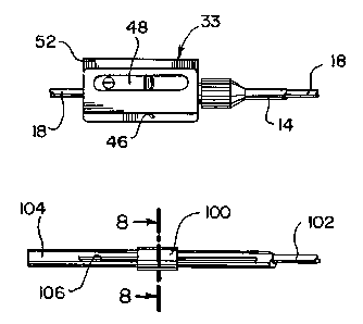
Referring now to FIGS. 7 and 8, there is shown another illustrative
embodiment of the mechanism 10 of the present invention. In that embodiment,
the
slidable electrode 100 is connected to an inner mandrel, shaft, or other
tubular member
102. The member 102 is slidably received inside of an outer tubular sheath 104
which is
formed having a longitudinal slot 106 formed in the side wall thereof. The
electrode 100
is connected to the member 102 through a laterally extending connector 108
which is
sized for passing through the slot 106. In all other respects, this embodiment
is similar
to the embodiment disclosed in FIGS. 1 through 6, with the ratchet mechanism
33
connected to the member 102 rather than to the outer member 104.
From the foregoing, it will be apparent to those skilled in the art that the
present invention provides a medical device which facilitates the creation of
continuous
lesions, without requiring an elongated electrode that hinders the flexibility
of the
medical device, and without requiring that the clinician perform a drag
procedure in
which the clinician would have to estimate the distance the electrode was
displaced. In
addition, the medical device of the present invention provides an easily
actuated
mechanism for displacing an electrode to facilitate creating continuous
lesions.
Having thus described preferred embodiments of the present invention, it
is to be understood that the above described arrangement and system is merely
CA 02372089 2001-11-29
WO 01/01877 PCT/US00/01886
-11-
illustrative of the principles of the present invention, and that other
arrangements and
systems may be devised by those skilled in the art without departing from the
spirit and
scope of the invention as claimed below.