Note: Descriptions are shown in the official language in which they were submitted.
CA 02373034 2007-10-18
WO 00/67647 PCT/US00/11274
INJECTION ARRAY APPARATUS AND METHOD
This application is related to U.S. Patent No. 6,319,230, entitled LATERAL
NEEDLE INJECTION APPARATUS AND METHOD; U.S. PatentNo. 6,613,026, entitled
LATERAL NEEDLE-LESS INJECTION APPARATUS AND METHOD; and U.S. Patent
No. 6,344,027, entitled NEEDLE-LESS INJECTION APPARATUS AND METHOD.
Field of the Invention
The present invention generally relates to delivering and injecting fluid into
heart tissue. More specifically, the present invention relates to delivering
and
injecting fluid into heart tissue utilizing an injection array.
Background of the Invention
Injection catheters may be used to inject therapeutic or diagnostic agents
into a
variety of organs, such as the heart. In the case of injecting a therapeutic
agent into
the heart, 27 or 28 gauge needles are generally used to inject solutions
carrying genes,
proteins, or drugs directly into the myocardium. A typical volume of an agent
delivered to an injection site is about 100 microliters. A limitation to this
method of
delivering therapeutic agents to the heart is that the injected fluid tends to
leak and/or
disperse from the site of the injection after the needle is disengaged from
the heart. In
fact, fluid may continue to leak over several seconds. In the case of dynamic
organs
such as the heart, there may be more pronounced leakage with each muscle
contraction.
Therapeutic and diagnostic agents may be delivered to a portion of the heart
as
part of a percutaneous myocardial revascularization (PMR) procedure. PMR is a
procedure which is aimed at assuring that the heart is properly oxygenated.
Assuring
that the heart muscle is adequately supplied wiin oxygen is critical to
sustaining the
life of a patient. To receive an adequate supply of oxygen, the heart muscle
must be
-1-
CA 02373034 2001-11-02
WO 00/67647 PCT/US00/11274
well perfused with blood. In a healthy heart, blood perfusion is accomplished
with a
system of blood vessels and capillaries. However, it is common for the blood
vessels
to become occluded (blocked) or stenotic (narrowed). A stenosis may be formed
by
an atheroma which is typically a harder, calcified substance which forms on
the walls
of a blood vessel.
Historically, individual stenotic lesions have been treated with a number of
medical procedures including coronary bypass surgery, angioplasty, and
atherectomy.
Coronary bypass surgery typically involves utilizing vascular tissue from
another part
of the patient's body to construct a shunt around the obstructed vessel.
Angioplasty
techniques such as percutaneous transluminal angioplasty (PTA) and
percutaneous
transluminal coronary angioplasty (PTCA) are relatively non-invasive methods
of
treating a stenotic lesion. These angioplasty techniques typically involve the
use of a
guide wire and a balloon catheter. In these procedures, a balloon catheter is
advanced
over a guide wire such that the balloon is positioned proximate a restriction
in a
diseased vessel. The balloon is then inflated and the restriction in the
vessel is
opened. A third technique which may be used to treat a stenotic lesion is
atherectomy. During an atherectomy procedure, the stenotic lesion is
mechanically
cut or abraded away from the blood vessel wall.
Coronary by-pass, angioplasty, and atherectomy procedures have all been
found effective in treating individual stenotic lesions in relatively large
blood vessels.
However, the heart muscle is perfused with blood through a network of small
vessels
and capillaries. In some cases, a large number of stenotic lesions may occur
in a large
number of locations throughout this network of small blood vessels and
capillaries.
The torturous path and small diameter of these blood vessels limit access to
the
stenotic lesions. The sheer number and small size of these stenotic lesions
make
techniques such as cardiovascular by-pass surgery, angioplasty, and
atherectomy
impractical.
When techniques which treat individual lesion are not practical, percutaneous
myocardial revascularization (PMR) may be used to improve the oxygenation of
the
myocardial tissue. A PMR procedure generally involves the creation of holes,
craters
or channels directly into the myocardium of the heart. In a typical PMR
procedure,
these holes are created using radio frequency energy delivered by a catheter
having
one or more electrodes near its distal end. After the wound has been created,
-2-
CA 02373034 2001-11-02
WO 00/67647 PCT/US00/11274
therapeutic agents are sometimes ejected into the heart chamber from the
distal end of
a catheter.
Positive clinical results have been demonstrated in human patients receiving
PMR treatments. These results are believed to be caused in part by blood
flowing
within the heart chamber through channels in myocardial tissue formed by PMR.
Increased blood flow to the myocardium is also believed to be caused in part
by the
healing response to wound formation. Specifically, the formation of new blood
vessels is believed to occur in response to the newly created wound. This
response is
sometimes referred to as angiogenesis. After the wound has been created,
therapeutic
to agents which are intended to promote angiogenesis are sometimes injected
into the
heart chamber. A limitation of this procedure is that the therapeutic agent
may be
quickly carried away by the flow of blood through the heart.
In addition to promoting increased blood flow, it is also believed that PMR
improves a patient's condition through denervation. Denervation is the
elimination of
nerves. The creation of wounds during a PMR procedure results in the
elimination of
nerve endings which were previously sending pain signals to the brain as a
result of
hibernating tissue.
Currently available injection catheters are not particularly suitable for
accurately delivering small volumes of therapeutic agents to heart tissue.
Improved
2o devices and methods are desired to address the problems associated with
retention of
the agent in the heart tissue as discussed above. This is particularly true
for agents
carrying genes, proteins, or other angiogenic drugs which may be very
expensive,
even in small doses.
Summary of the Invention
The present invention provides an improved apparatus and method for
delivering and injecting fluid into heart tissue, or other organ tissues such
as liver
tissue, bladder tissue, etc. The present invention addresses the problems
associated
with retention of the fluid in the tissue by utilizing an injection array,
such as a
plurality of microneedles or a plurality of injection lumens. The present
invention
may be used to deliver genes, proteins, or drugs directly into the myocardium
for
purposes of myocardial revascularization.
In an exemplary embodiment, the present invention provides a fluid delivery
system including an injection catheter disposed in an elongate sheath. A fluid
source
-3-
CA 02373034 2001-11-02
WO 00/67647 PCT/US00/11274
is connected to the proximal end of the injection catheter and is in fluid
communication with the lumen of the catheter. A nozzle is disposed adjacent
the
distal end of the injection catheter. In a first embodiment, the nozzle
includes a
plurality of microneedles each defining an injection lumen in fluid
communication
with the lumen of the catheter. In a second embodiment, the nozzle defines a
plurality
of injection lumens in fluid communication with the lumen of the catheter. The
first
embodiment may be referred to as the "microneedle" embodiment and the second
embodiment may be referred to as the "needle-less" embodiment.
The microneedles may each have a diameter in the range of approximately
to 0.005 to 0.05 inches, and a penetrating length in the range of
approximately 0.5 to 5
mm. The injection lumens in the microneedle embodiment may have a diameter in
the range of approximately 0.00005 to 0.005 inches. Similarly, the injection
lumens
in the needle-less embodiment may have a diameter in the range of
approximately
0.00005 to 0.005 inches.
In both embodiments, the injection lumens collectively form an injection array
terminating in a plurality of injection orifices. Fluid is transferred to the
injection
lumen array from the fluid source through the lumen in the catheter. The
injection
lumen array distributes the fluid at the injection site over a greater area
than would
otherwise be achieved with a single needle injection. Thus, the injection
lumen array
improves fluid retention in the tissue at the injection site.
Also in both embodiments, the catheter and/or sheath may be equipped with
an anchor disposed adjacent the distal end thereof. The anchor may comprise a
vacuum orifice in fluid communication with a vacuum source via a lumen in the
catheter and/or sheath. The vacuum orifice is adapted to stabilize the distal
end of the
injection catheter and/or the distal end of the sheath.
The sheath may include a hood portion disposed at its distal end. The distal
end of the injection catheter may be retracted within the hood of the sheath
to reduce
the probability that tissue damage will occur when the catheter is advanced
through
the vasculature of the patient.
The present invention also provides a method of delivering a fluid to heart
tissue comprising the following steps. An injection catheter substantially as
described
above is inserted into a patient's body and navigated to the desired target
site, for
example, heart tissue such as the myocardium. The injection catheter may be
-4-
CA 02373034 2007-10-18
WO 00/67647 PCT/US00/11274
navigated intravascularly or transthoracicly to the heart tissue. A sheath
substantially
as described above may also be advanced until its distal end is proximate the
target
site. The injection catheter is then advanced until the injection array is
proximate the
target tissue. Fluid is then urged out from the fluid source, through the
lumen of the
injecrion catheter, and into the heart tissue via the injection array. The
injection
lumen array distributes the fluid at the target site over a greater area
thereby
increasing retention of fluid in the heart tissue at the injection site.
Less than approximately 100 microliters of fluid may be injected into the
heart
tissue via the injection array. Approximately 0.1 to 20 microliters of fluid
may be
injected into the heart tissue via each injection lumen of the array. Due to
the
distribution of the injection array, a substantial amount if not all of the
injected fluid is
retained in the heart tissue.
According to one aspect of the invention, there is provided a catheter for
delivering a
therapeutic agent or a fluid into target tissue within the body of a patient,
comprising: an
elongate tubular member having a proximal end, a distal end, and a lumen
extending
therethrough; and, a nozzle member disposed proximate the distal end of the
tubular
member, the nozzle member including an injection array at a distal end of the
nozzle
member; wherein: the injection array is needle-less and comprises a plurality
of injection
lumens, each terminating with an injection orifice which is in fluid
communication with the
lumen of the elongate tubular member; the injection orifices are arranged at
the nozzle
member at its distal front face; and, the lumen is designed for connection at
the proximal end
of the catheter to a high-pressure fluid source. The catheter may further
include an outer
sheath having a proximal end, a distal end, and a lumen extending
therethrough, the tubular
member slidingly disposed in the lumen of the sheath. The catheter may further
include an
anchor disposed proximate the distal end of the sheath. The anchor may include
a vacuum
orifice defined by the lumen of the sheath adjacent the distal end thereof.
And, each of the
injection lumens may have a diameter in the range of approximately 0.000 127
cm to 0.0 127
cm (0.00005 inches to 0.005 inches).
-5a-
CA 02373034 2007-10-18
WO 00/67647 PCT/US00/11274
Brief Description of the Drawings
Figure 1 is a perspective view of a fluid delivery system in accordance with
the present invention;
Figure 2 is a perspective view of a first (microneedle) embodiment of the
distal end of the injection catheter for use with the fluid delivery system
illustrated in
Figure 1;
Figure 3 is a perspective view of a second (needle-less) embodiment of the
distal end of the injection catheter for use with the fluid delivery system
illustrated in
Figure 1;
Figure 4 is a schematic view of the fluid delivery system and a human patient;
Figure 5 is a perspective view of the distal end of the injection catheter
incorporating an anchor for stabilization;
Figure 6 is a schematic view of the fluid delivery system and a human patient
utilizing an anchor for stabilization;
Figure 7 is a cross sectional view of the distal portion of the fluid delivery
system incorporating a hood, shown in the extended position; and
Figure 8 is a cross sectional view of the fluid delivery system of Figure 7,
shown in the retracted position.
Detailed Description of the Invention
The following detailed description should be read with reference to the
drawings, in which like elements in different drawings are numbered
identically. The
-5b-
CA 02373034 2001-11-02
WO 00/67647 PCT/US00/11274
drawings which are not necessarily to scale, depict selected embodiments and
are not
intended to limit the scope of the invention. Examples of constructions,
materials,
dimensions, and manufacturing processes are provided for various elements.
Those
skilled in the art will recognize that many of the examples provided have
suitable
alternatives which may be utilized.
Figure 1 is a perspective view of a fluid delivery system 20 in accordance
with
the present invention. In the embodiment of Figure 1, fluid delivery system 20
includes a sheath 22 comprising an elongate tubular member 24 defining a
sheath
lumen 26. Sheath 22 also includes a distal end 28 and a proximal end 30. A hub
32 is
disposed at proximal end 30 of sheath 22.
Those of skill in the art will appreciate that sheath 22 may be comprised of
many materials without deviating from the spirit and scope of the present
invention.
Likewise, sheath 22 may be comprised of a single material, or a combination of
materials. For example, sheath 22 may include an inner tube 34. In a presently
preferred embodiment, inner tube 34 is comprised of PTFE
(polytetrafluoroethylene).
PTFE is a preferred material because it creates a smooth, low-friction surface
for the
passage of other devices through the sheath 22. Sheath 22 may also include a
support
member 36 wound or braided around inner tube 34. In a presently preferred
embodiment, support member 36 is comprised of a plurality of filaments 38.
Filaments 38 may be stainless steel wire. Those with skill in the art will
appreciate
that other embodiments of support member 36 are possible without deviating
from the
spirit and scope of the present invention. For example, support member 36 may
be
comprised of a woven polymer fabric. By way of a second example, support
member
36 may be comprised of polymer fibers arranged in a braided pattern.
Sheath 22 may be comprised of polyether block amide (PEBA) using an
extrusion process. Polyether block amide is commercially available from
Atochem
Polymers of Birdsboro, Pennsylvania under the trade name PEBAX. In the
extrusion
process, molten PEBA is extruded onto the combined layers of inner tube 34 and
support member 36. When this process is used, the extruded material fills
interstitial
spaces in support member 36.
It is to be understood that other manufacturing processes can be used without
departing from the spirit and scope of the present invention. Sheath 22 may
also be
comprised of other materials without departing from the spirit of scope of
this
-6-
CA 02373034 2007-10-18
WO 00/67647 PCT/US00/11274
invention. Examples of materials which may be suitable in some applications
include: polyethylene (PE), polypropylene (PP), polyvinylchloride (PVC),
polyurethane, and polytetrafluoroethylene (PTFE).
Fluid delivery system 20 also includes ?n injection catheter 40 which is
slidingly disposed in sheath iumeri 26 of sheath 22. Injection catheter 40
includes an
elongate tubular member 44 defining a lumen 46, a distal end 48, and a
proximal end
50. A fluid source 52 is releasably connected to the proximal end 50 of
injection
catheter 40. Fluid source 52 is in fluid communication with lumen 46 of
elongate
tubular member 44. Fluid source 52 is capable of injecting fluid 54 into lumen
46 of
elongate tubular member 44.
In the illustrated embodiment, fluid source 52 includes a variable volume
chamber 56 in fluid communication with lumen 46 of elongate tubular member 44.
Fluid source 52 further includes a plunger 58 slidingly disposed within
variable
volume chamber 56. Urging plunger 58 distally has the effect of urging fluid
54 into
lumen 46 of elongate tubular member 44. A number of energy sources may be
utilized to urge plunger 58 distally. Energy sources which may be suitable in
some
applications include springs, compressed gas, electricity, and manual forces.
Fluid
source 52 may alternatively comprise, for example, a conventional syringe or a
high
pressure injection system as disclosed in U.S. Patent No. 5,520,639 to
Peterson et al.
Elongate tubular member 44 is moveable between a retracted position and an
extended position wherein the distal end 48 of the injection catheter 40
extends
beyond the distal end 28 of sheath 22. A plurality of microneedles 60 are
disposed
proximate the distal end 48 of injection catheter 40. Each microneedle 60
defines an
injection lumen 62 in fluid communication with lumen 46 of elongate tubular
member
44. Injection lumens 62 collectively form an injection lumen array 64.
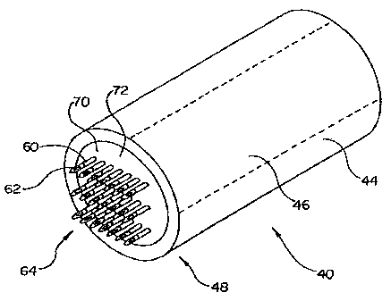
Figure 2 is a detailed perspective view of the distal end 48 of the injection
catheter 40 illustrating the mircroneedle embodiment in detail. A nozzle
member 70
is disposed within lumen 46 proximate the distal end 48 of injection catheter
40. The
microneedles 60 are disposed on a distal surface 72 of nozzle member 70.
Microneedles 60 may be separate members inserted into holes defined in nozzle
70 or
may be an integral part of nozzle 70. Nozzle member 70 and microneedles 60
define
a plurality of injection lumens 62. Injection lumens 62 collectively form an
injection
-7-
CA 02373034 2007-10-18
WO 00/67647 PCT/US00/11274
lumen array 64. Each injection lumen 62 is in fluid communication with lumen
46 of
elongate tubular member 44.
One embodiment of injection catheter 40 has been envisioned in which
microneedles 60 and nozzle member 70 are both comprised of silicon. An example
fabrication technique is described in U.S. Patent No. 5,697,901 to Eriksson.
The microneedles 60 may each have a diameter in
the range of approximately 0.005 to 0.05 inches, and a penetrating length in
the range
of approximately 0.5 to 5 mm. The injection lumens 62 in the microneedles 60
may
have a diameter in the range of approximately 0.00005 to 0.005 inches. The
microneedles 60 may be generally circular in cross section as shown. Those of
skill
in the art will appreciate that microneedles 60 may be other shapes without
departing
from the scope of the present invention. Examples of cross sectional shapes
which
may be suitable in some applications include oval, triangular, rectangular,
square, and
hexagonal.
Microneedles 60 and nozzle member 70 may be comprised of a variety of
metallic and non-metallic materials. Examples of metallic materials which may
be
suitable in some applications include stainless steel, and nickel-titanium
alloys,
although it is recognized that any suitable metal or alloy may be utilized.
Examples
of non-metallic materials which may be suitable in some applications include
silicon
as described above and rigid polymers. Examples of rigid polymers include:
polycarbonate, poly(L-lactide) (PLLA), poly(D,L-lactide) (PLA), polyglycolide
(PGA), poly(L-lactide-co-D,L-lactide) (PLLA/PLA), poly(L-lactide-co-glycolide)
(PLLA/PGA), poly(D, L-lactide-co-glycolide) (PLA/PGA), poly(glycolide-co-
trimethylene carbonate) (PGA/PTMC), polyethylene oxide (PEO), polydioxanone
(PDS), polycaprolactone (PCL), polyhydroxylbutyrate (PHBT), poly(phosphazene),
polyD,L-lactide-co-caprolactone) (PLAIPCL), poly(glycolide-co-caprolactone)
(PGA/PCL), polyanhydrides (PAN), poly(ortho esters), poly(phosphate ester),
poly(amino acid), poly(hydroxy butyrate), polyacrylate, polyacrylamid,
poly(hydroxyethyl methacrylate), polyurethane, polysiloxane and their
copolymers.
Figure 3 is a perspective view of the distal end 48 of an altemative
embodiment of an injection catheter 140 illustrating the needle-less
embodiment.
Injection catheter 140 is the same in form and function as catheter 40 and may
be used
in a similar manner, except as described below. As in the microneedle
embodiment
-8-
CA 02373034 2007-10-18
WO 00/67647 PCT/US00/11274
described previously, injection catheter 140 includes an elongate tubular
member 144
defining a lumen 146. A nozzle member 170 is disposed within lumen 146
proximate
distal end 148 of injection catheter 140. Nozzle member 170 defines a
plurality of
injection, lumens 1162. Injection lumens .162 collectively form an injection
lumen
array 164. Each injection lumen 162 is in fluid communication with !smen 146
of
elongate tubular member 144. The injection lumens 162 may each have a diameter
in
the range of approximately 0.00005 to 0.005 inches.
Injection catheter 140 may be used in conjunction with the fluid delivery
system 20 including the fluid source 52. The fluid source 52 is disposed
proximate
the proximal end of injection catheter 140 and is in fluid communication with
lumen
146 of elongate tubular member 144. The fluid source 52 is capable of
injecting fluid
into lumen 146 of elongate tubular member 144 at high pressure. The injection
of
fluid into lumen 146 of elongate tubular member 144 results in fluid 54 being
ejected
from injection lumens 162.
In the needle-less embodiment of Figure 3, fluid is ejected from injection
lumens 162 with a velocity which is sufficient to inject the fluid into tissue
disposed
proximate the distal end 148 of injection catheter 140. This technique is
commonly
referred to as needle-less injection. A number of energy sources may be
utilized to
urge fluid into lumen 146 of elongate tubular member 144. Energy sources which
may be suitable in some applications include springs and compressed gas.
Preferably,
a high pressure system is utilized as described in U.S. Patent No. 5,697,901
to
Eriksson,
Figure 4 is a schematic view of the fluid delivery system 20 and a patient 74.
Fluid delivery system 20 includes the injection catheter 40, the sheath 22,
and the
fluid source 52. Injection catheter 140 may be used in place of an injection
catheter
40. An access catheter 80 is positioned with a distal end 88 thereof
positioned within
a blood vessel 76. Access catheter 80 aids in the introduction of sheath 22
into blood
vessel 76. Injection catheter 40/140 is disposed within lumen 26 of sheath 22.
The
distal end 48 of injection catheter 40/140 is positioned within the heart 78
of patient
74.
A method of injecting a fluid into tissue of the heart 78 of patient 74 is
described with reference to Figure 4. The fluid delivery system 20 may be
navigated
intravascularly or transthoracicly to heart tissue, but is described with
reference to an
-9-
CA 02373034 2001-11-02
WO 00/67647 PCT/US00/11274
intravascular approach for purposes of illustration only. Those of skill in
the art will
appreciate that the methods and devices of the present invention may be used
to
deliver therapeutic and/or diagnostic agents to other areas of the body
without
departing from the spirit and scope of the invention. For example, devices and
methods in accordance with the present invention may be used to deliver fluid
agents
to esophageal varicies or to ulcers in the stomach lining.
The distal end of fluid delivery system 20 may enter the patient's vasculature
at a convenient location such as a blood vessel in the neck or near the groin.
Ideally,
the distal end of fluid delivery system 20 will be atraumatic to reduce the
probability
that vascular tissues will be damaged as the catheter is advanced through the
vascular
system. To prevent damage to the vasculature, the distal end of injection
catheter
40/140 may be retracted into lumen 26 of sheath 22.
Once the distal portion of fluid delivery system 20 has entered the patient's
vascular system, the physician may urge distal end 28 of sheath 22 forward by
applying longitudinal forces to hub 32 of sheath 22. Frequently, the path
taken by
sheath 22 through the vascular system is tortuous requiring sheath 22 to
change
direction frequently. While advancing sheath 22 through the torturous path of
the
patient's vasculature, the physician may apply torsional forces to the hub 32
to aid in
steering sheath 22. To facilitate the steering process, the distal portion of
sheath 22
may include a plurality of bends or curves. In some embodiments, it may be
desirable
to include a distal portion of sheath 22 which can be heated and bent to a
desired
shape, then allowed to cool.
To aid the physician in visualizing the vascular pathway, radiopaque contrast
solution may be dispensed from distal end 28 of sheath 22 to enhance
fluoroscopic
visualization. In one method in accordance with the present invention,
radiopaque
contrast solution is urged through lumen 26 of sheath 22. Sheath 22 and
injection
catheter 40/140 may also include radiopaque markers. One example of a
radiopaque
marker is a band of radiopaque material disposed proximate the distal end of
injection
catheter 40/140 and/or sheath 22. Radiopaque bands of this type aid the
physician in
determining the location of the distal end of the device relative to the
patient's
anatomy. The radiopaque band may be comprised of a number of materials.
Examples of materials which may be suitable in some applications include gold,
platinum, tungsten, iron, silver, and thermoplastic material loaded with a
radiopaque
-10-
CA 02373034 2001-11-02
WO 00/67647 PCT/US00/11274
filler. Examples of radiopaque filler which may be suitable in some
applications
include barium sulfate, bismuth subcarbonate, bismuth trioxide, bismuth
oxychloride,
bismuth subcarbonate, tungsten powder, and depleted uranium.
Once distal end 28 of sheath 22 is positioned proximate the target site,
injection catheter 40/140 may be advanced so that nozzle member 70 contacts
the
bodily tissue at the target site. If injection catheter 40 with mircroneedles
60 is used,
the microneedles 60 are advanced to penetrate the heart tissue. If injection
catheter
140 with injection lumens 162 is used, the distal end of the injection array
164 is
positioned adjacent the heart tissue surface. Force may then be applied to
plunger 58
urging fluid out of fluid source 52 and into lumen 46/146 of injection
catheter 40/140.
The addition of fluid from fluid source 52 results in the injection of fluid
into the
target tissue via the injection array 64/164. The total fluid injected into
the target
tissue may be referred to as a dose. The dose is more readily retained in the
heart
tissue by utilizing the injection array 64/164.
A portion of the dose is dispensed from each injection lumen 62/162. The
volume of fluid dispensed from each injection lumen 62/162 may be pre-
selected.
The pre-selected volume dispensed from each injection lumen may be a volume
which can be rapidly absorbed and/or retained by the target tissue. By way of
example, a dose of 100 microliters may be delivered via the injection array
64/164.
The volume of fluid injected by each microneedle or injection lumen may be 0.1
to 20
microliters.
In an embodiment of the present invention, a low volume (several microliters
but less than 100 microliters by a single injection) of solution is delivered
to the heart
such that it may absorb the delivered solution within the time frame of the
injection.
In contrast to higher volume injections, the heart is more capable of
absorbing these
low volumes. The effect of the low volume injection is to minimize expulsion
by the
tissue. In order to deliver the entire dose of virus, it may be desirable or
necessary to
concentrate the injection (i.e., deliver the same number of viral particles or
micrograms of protein, typically delivered in 100 1, in a volume of 10 1) or
keep the
concentration of virus the same as that typically used, but increase the
number of
injections from 10 (typical) to 20, 30, or more.
Each injectate may also be delivered in a prolonged manner such that the heart
can absorb the solution as it is being injected (rate of delivery < rate of
tissue
-11-
CA 02373034 2001-11-02
WO 00/67647 PCT/US00/11274
absorption). For instance, the injection can be delivered at a defined flow
rate using a
syringe pump. The time of injection will depend on the volume to be delivered.
For
example, low volumes (a few microliters) may be delivered in under a minute
while
higher volumes (10 to 100 l or more) may be delivered over several minutes. In
this
instance, it may be beneficial to include a method which gently attaches the
injection
catheter to the wall of the heart, for instance suction or vacuum.
If injection catheter 40 with mircroneedles 60 is used, the microneedles 60
may be left in the tissue for a period of time after the dose has been
delivered to allow
the fluid to be absorbed by the tissue. The amount of time required for the
fluid to be
to absorbed by the tissue will vary depending on the volume of fluid delivered
and the
absorption characteristics of the tissue. The time period may be relatively
short or
prolonged, ranging, for example, from about 5 seconds to 2 minutes.
Preferably, the
time period ranges from about 5 seconds to about 30 seconds. When the
mircroneedles 60 are subsequently withdrawn, leakage of the fluid from the
tissue is
further minimized or eliminated due to absorption thereof by the tissue.
The fluid injected into the tissue at the target area may include any number
of
therapeutic or diagnostic agents. Examples of therapeutic agents include
genes,
proteins, drugs, and caustic solutions. A radiopaque solution is an example of
a
diagnostic agent. Radiopaque solution may be used to mark an area. For
example,
when performing PMR, it may be desirable to mark the locations of wound
formation.
Those of skill in the art will appreciate that when the organ being treated is
the
heart, fluid may be injected into the myocardium either from the epicardial or
endocardial surface. In the exemplary embodiment of Figure 4, the epicardial
surface
was accessed intravascularly by advancing a catheter through the vascular
system.
Other methods of have been envisioned in which the endocardial surface of the
heart
is accessed using surgical techniques such as transthoracic minimally invasive
surgery.
Figure 5 is a perspective view of distal end 48 of an alternative embodiment
of an injection catheter 240. Injection catheter 240 is the same in form and
function
as injection catheter 40 and may be used in the same manner, except as
described
below. Injection catheter 240 includes an elongate tubular member 244 defining
a
lumen 246. A plurality of anchors 290 are disposed proximate distal end 248 of
injection catheter 240. During a procedure to inject a therapeutic agent into
body
-12-
CA 02373034 2001-11-02
WO 00/67647 PCTIUSOO/11274
tissue, anchors 290 may be utilized to retain distal end 248 of injection
catheter 240
proximate the targeted tissue. In the embodiment of Figure 5, each anchor 290
is
comprised of a vacuum orifice 292. Each vacuum orifice 292 is in fluid
communication with a vacuum lumen 294 defined by elongate tubular member 244.
Other embodiments of anchors 290 are possible without deviating from the
spirit or scope of the present invention. For example, each anchor 290 may be
comprised of an elongate wire with a helix disposed proximate its distal end.
The
helical end of the elongate wire may be "threaded" into the heart wall by
rotating the
wire. Additional examples, of anchors 290 which may be appropriate in some
applications include hooks and barbs.
In the embodiment of Figure 5, a nozzle member 270 is disposed within lumen
246 proximate distal end 248 of injection catheter 240. A plurality of
microneedles
260 are disposed on a distal surface 272 of nozzle member 270. Nozzle member
270
and microneedles 260 define a plurality of injection lumens 262. Injection
lumens
262 collectively form an injection lumen array 264. Each injection lumen 262
is in
fluid communication with lumen 246 of injection catheter 240.
Figure 6 is a schematic view of a fluid delivery system 220, including the
injection catheter 240 of Figure 5. Except as described below, fluid system
220 is the
same in form and function as fluid system 20 and may be used in a similar
manner.
Fluid delivery system 220 includes the injection catheter 240, a sheath 222, a
vacuum
source 100, and a fluid source 252. A hub 232 is disposed at proximal end 230
of
sheath 222, and a multi-port adapter 296 is disposed at the proximal end of
injection
catheter 240. Multi-port adapter 296 includes a plurality of ports 298. Fluid
source
252 is in fluid communication with one port 298 of multi-port adapter 296.
Vacuum
source 100 is also in fluid communication with one port 298 of multi-port
adapter
296. An access catheter 280 positioned with a distal end 288 positioned within
blood
vessel 276. Access catheter 280 may aid in the introduction of sheath 222 into
blood
vessel 276. Injection catheter 240 is disposed within lumen 226 of sheath 222.
The
distal end 248 of injection catheter 240 is positioned within the heart 278 of
patient
274.
A method of injecting a fluid into heart 278 of patient 274 is described with
reference to Figure 6. Sheath 222 is introduced into the vasculature of the
patient 274
and it is urged forward until it's distal tip is proximate the target tissue.
Once the
-13-
CA 02373034 2001-11-02
WO 00/67647 PCT/US00/11274
distal end of the sheath is positioned proximate the target site, injection
catheter 240
may be advanced so that nozzle member 270 contacts the bodily tissue at the
target
site. Anchors 290 may then be activated to stabilize distal end 248 of
injection
catheter 240 during injection. Each anchor 290 is comprised of a vacuum
orifice 292
in fluid communication with a vacuum lumen 294. In this embodiment, anchors
290
are activated by applying vacuum from vacuum source 100 to vacuum orifices 292
via vacuum lumens 294 and multi-port adapter 296. The vacuum orifaces apply
suction to the surface of the tissue to stabilize the catheter 240 relative to
the tissue.
With distal end 248 of injection catheter 240 anchored, force may be applied
to plunger 258 urging fluid out of fluid source 252 and into lumen 246 of
injection
catheter 240. The addition of fluid from fluid source 252 results in the
injection of
fluid into the target tissue via injection array 264.
Figure 7 is a cross sectional view of the distal portion of an alternative
embodiment of fluid delivery system 320. Except as described below, fluid
delivery
system 320 is the same in form and function as fluid delivery system 220, and
may be
used in a similar manner. Fluid delivery system 320 includes a sheath 322
comprising
an elongate tubular member 24 defining a lumen 326. Sheath 322 also includes a
distal end 328 and a proximal end 330. In the embodiment of Figure 7, sheath
322
includes a hood portion 102 disposed proximate its distal end 28.
Fluid delivery system 320 also includes an injection catheter 340 which is
slidingly disposed in lumen 326 of sheath 322. Injection catheter 340 includes
a distal
end 348, a proximal end 350, and an elongate tubular member 344 defining a
lumen
346. A fluid source (not shown) is connected to proximal end 350 of injection
catheter 340.
A nozzle 370 is disposed proximate the distal end 348 of injection catheter
340. Nozzle 370 includes a plurality or microneedles 360. Each microneedle 360
defines an injection lumen 362 in fluid communication with lumen 346 of
elongate
tubular member 344. Injection lumens 362 collectively form an injection lumen
array
364.
Sheath 322 and injection catheter 340 define an annular passage 108 disposed
about injection catheter 340. Annular passage 108 terminates at an annular
opening
110. Vacuum may be applied to annular passage 108 in order to anchor distal
end
348 of injection catheter 340 to the bodily tissue at a desired target site.
By doing so,
-14-
CA 02373034 2001-11-02
WO 00/67647 PCT/US00/11274
the distal end of the sheath 322 and thus the distal end of the catheter 340
is stabilized
relative to the heart tissue.
Figure 8 is a cross sectional view of the distal portion of the fluid delivery
system 320 illustrated in Figure 7, shown in the retracted position. The
distal end of
injection catheter 40 has been retracted within hood portion 102 of sheath
322. The
distal end 348 of injection catheter 340 is retracted within hood portion 102
of sheath
322 to reduce the probability that vascular damage will occur when fluid
delivery
system 320 is advanced through the vasculature of the patient. Upon
positioning the
system 320 at the target site, the catheter 340 may be advanced to the
extended
to position as shown in Figure 7.
With all embodiments described herein, the fluid injected into the target area
may include any therapeutic or diagnostic agents needed to treat the medical
condition
which the physician is treating. It is to be appreciated that methods in
accordance
with the present invention may be used in the treatment of a number of medical
conditions. For example, methods and devices of performing percutaneous
myocardial revascularization (PMR) in accordance with the present invention
have
been envisioned.
A PMR procedure involves creating a plurality of wounds in hibernating tissue
of the heart. These wounds are created by injecting a fluid into the tissue of
the heart.
As a result of these wounds, there will be increased blood flow to the
myocardium
caused in part by the body's healing response to the wounds. One healing
response of
the body is sometimes referred to as angiogenisis. In addition to promoting
increased
blood flow, it is also believed that PMR improves a patient's condition
through
denervation. Denervation is the elimination of nerves. The creation of wounds
during this procedure may result in the elimination of nerve endings which
were
previously sending pain signals to the brain as a result of hibernating
tissue.
Suitable wounds may be created by injecting a fluid such as water or saline
into the heart tissue. Wound formation and revascularization of myocardial
tissue
may enhanced by injecting a fluid including a therapeutic agent into the
tissue of the
heart. Examples, of therapeutic agents which may be suitable include growth
factors,
drugs and caustic agents. The fluid injected into the heart tissue may also
include a
radiopaque material. Injecting a radiopaque material into the wound
effectively
marks the locations which have been treated. This will aid the physician in
-15-
CA 02373034 2001-11-02
WO 00/67647 PCT/US00/11274
procedures which are being performed percutaneously using fluoroscopic
equipment.
As describe above, injection catheters 40/140/240/340 may be used in the
treatment of a number of medical conditions. By way of an additional example,
injection catheters 40/140/240/340 may be used in the treatment of esophageal
varicies, a condition where blood vessels of the esophagus are enlarged and
may
potentially burst. For such a procedure, the array of injection orifices is
disposed
proximate the enlarged varix and an appropriate agent is injected into the
varix.
When treating an esophageal varice, the agent may be a coagulant such as
sodium
morrhuate. When a coagulant is injected into a varix, it causes the occlusion
thereof.
Although treatment of the heart is used as an example herein, the medical
devices of the present invention are useful for treating any mammalian tissue
or
organ. Non-limiting examples include tumors; organs including but not limited
to the
heart, lung, brain, liver, kidney, bladder, urethra and ureters, eye,
intestines, stomach,
pancreas, ovary, prostate; skeletal muscle; smooth muscle; breast, cartilage
and bone.
The terms "therapeutic agents" and "drugs" are used interchangeably herein
and include pharmaceutically active compounds, cells, nucleic acids with and
without
carrier vectors such as lipids, compacting agents (such as histones), virus,
polymers,
proteins, and the like, with or without targeting sequences.
Specific examples of therapeutic agents used in conjunction with the present
invention include, for example, proteins, oligonucleotides, ribozymes, anti-
sense
genes, DNA compacting agents, gene/vector systems (i.e., anything that allows
for the
uptake and expression of nucleic acids), nucleic acids (including, for
example,
recombinant nucleic acids; naked DNA, cDNA, RNA; genomic DNA, cDNA or RNA
in a non-infectious vector or in a viral vector which may have attached
peptide
targeting sequences; antisense nucleic acid (RNA or DNA); and DNA chimeras
which
include gene sequences and encoding for ferry proteins such as membrane
translocating sequences ("MTS") and herpes simplex virus-1 ("VP22")), and
viral,
liposomes and cationic polymers that are selected from a number of types
depending
on the desired application. Other pharmaceutically active materials include
anti-
thrombogenic agents such as heparin, heparin derivatives, urokinase, and PPACK
(dextrophenylalanine proline arginine chloromethylketone); antioxidants such
as
probucol and retinoic acid; angiogenic and anti-angiogenic agents; agents
blocking
smooth muscle cell proliferation such as rapamycin, angiopeptin, and
monoclonal
-16-
CA 02373034 2001-11-02
WO 00/67647 PCT/US00/11274
antibodies capable of blocking smooth muscle cell proliferation; anti-
inflammatory
agents such as dexamethasone, prednisolone, corticosterone, budesonide,
estrogen,
sulfasalazine, acetyl salicylic acid, and mesalamine; calcium entry blockers
such as
verapamil, diltiazem and nifedipine; antineoplastic / antiproliferative / anti-
mitotic
agents such as paclitaxel, 5-fluorouracil, methotrexate, doxorubicin,
daunorubicin,
cyclosporine, cisplatin, vinblastine, vincristine, epothilones, endostatin,
angiostatin
and thymidine kinase inhibitors; antimicrobials such as triclosan,
cephalosporins,
aminoglycosides, and nitorfurantoin; anesthetic agents such as lidocaine,
bupivacaine,
and ropivacaine; nitric oxide (NO) donors such as lisidomine, molsidomine, L-
1 o arginine, NO-protein adducts, NO-carbohydrate adducts, polymeric or
oligomeric NO
adducts; anti-coagulants such as D-Phe-Pro-Arg chloromethyl ketone, an RGD
peptide-containing compound, heparin, antithrombin compounds, platelet
receptor
antagonists, anti-thrombin antibodies, anti-platelet receptor antibodies,
enoxaparin,
hirudin, Warafin sodium, Dicumarol, aspirin, prostaglandin inhibitors,
platelet
inhibitors and tick antiplatelet factors; vascular cell growth promotors such
as growth
factors, growth factor receptor antagonists, transcriptional activators, and
translational
promotors; vascular cell growth inhibitors such as growth factor inhibitors,
growth
factor receptor antagonists, transcriptional repressors, translational
repressors,
replication inhibitors, inhibitory antibodies, antibodies directed against
growth
factors, bifunctional molecules consisting of a growth factor and a cytotoxin,
bifunctional molecules consisting of an antibody and a cytotoxin; cholesterol-
lowering agents; vasodilating agents; agents which interfere with endogeneus
vascoactive mechanisms; survival genes which protect against cell death, such
as anti-
apoptotic Bcl-2 family factors and Akt kinase; and combinations thereof.
Examples of polynucleotide sequences useful in practice of the invention
include DNA or RNA sequences having a therapeutic effect after being taken up
by a
cell. Examples of therapeutic polynucleotides include anti-sense DNA and RNA;
DNA coding for an anti-sense RNA; or DNA coding for tRNA or rRNA to replace
defective or deficient endogenous molecules. The polynucleotides of the
invention
can also code for therapeutic proteins or polypeptides. A polypeptide is
understood to
be any translation product of a polynucleotide regardless of size, and whether
glycosylated or not. Therapeutic proteins and polypeptides include as a
primary
example, those proteins or polypeptides that can compensate for defective or
deficient
-17-
CA 02373034 2001-11-02
WO 00/67647 PCT/US00/11274
species in an animal, or those that act through toxic effects to limit or
remove harmful
cells from the body. In addition, the polypeptides or proteins useful in the
present
invention include, without limitation, angiogenic factors and other molecules
competent to induce angiogenesis, including acidic and basic fibroblast growth
factors, vascular endothelial growth factor, hif-1, epidermal growth factor,
transforming growth factor a and (3, platelet-derived endothelial growth
factor,
platelet-derived growth factor, tumor necrosis factor a, hepatocyte growth
factor and
insulin like growth factor; growth factors; cell cycle inhibitors including
CDK
inhibitors; anti-restenosis agents, including p15, p16, p18, p19, p21, p27,
p53, p57,
Rb, nFkB and E2F decoys, thymidine kinase ("TK") and combinations thereof and
other agents useful for interfering with cell proliferation, including agents
for treating
malignancies; and combinations thereof. Still other useful factors, which can
be
provided as polypeptides or as DNA encoding these polypeptides, include
monocyte
chemoattractant protein ("MCP-1"), and the family of bone morphogenic proteins
("BMP's"). The known proteins include BMP-2, BMP-3, BMP-4, BMP-5, BMP-6
(Vgr-1), BMP-7 (OP-1), BMP-8, BMP-9, BMP-10, BMP-11, BMP-12, BMP-13,
BMP-14, BMP-15, and BMP-16. Currently preferred BMP's are any of BMP-2,
BMP-3, BMP-4, BMP-5, BMP-6 and BMP-7. These dimeric proteins can be
provided as homodimers, heterodimers, or combinations thereof, alone or
together
with other molecules. Alternatively or, in addition, molecules capable of
inducing an
upstream or downstream effect of a BMP can be provided. Such molecules include
any of the "hedgehog" proteins, or the DNA's encoding them.
The present invention is also useful in delivering cells as the therapeutic
agent.
Cells can be of human origin (autologous or allogeneic) or from an animal
source
(xenogeneic), genetically engineered if desired to deliver proteins of
interest at a
delivery or transplant site. The delivery media is formulated as needed to
maintain
cell function and viability.
Having thus described the preferred embodiments of the present invention,
those of skill in the art will readily appreciate that yet other embodiments
may be
made and used within the scope of the claims hereto attached. Numerous
advantages
of the invention covered by this document have been set forth in the foregoing
description. It will be understood, however, that this disclosure is, in many
respects,
only illustrative. Changes may be made in details, particularly in matters of
shape,
-18-
CA 02373034 2001-11-02
WO 00/67647 PCT/US00/11274
size, and arrangement of parts without exceeding the scope of the invention.
The
invention's scope is, of course, defined in the language in which the appended
claims
are expressed.
-19-