Note: Descriptions are shown in the official language in which they were submitted.
CA 02374455 2001-11-19
WO 00/79277 PCT/CAOO/00723
1
NEW ATPASE ASSAY
FIELD OF THE INVENTION
The present invention relates to a new ATPase assay. This invention
particularly
relates to a new method for the detection and measurement of the amount of
orthophosphate released by hydrolysis of ATP or any other phosphate containing
molecule. More particularly, this invention relates to a new method for the
measurement of the ATPase activity of the El helicase enzyme from the human
papilloma virus (HPV).
BACKGROUND OF THE INVENTION
HPV-associated disease
The human papillomaviruses (HPVs) are small DNA viruses that infect cells
of the cutaneous and mucosal epidermis. Over 80 different HPV genotypes have
been characterized. Some types, such as HPV-1, -2, -3, -4 and -10, cause
cutaneous lesions known as warts or papillomas. These growths are benign and
self-limiting, and are found on the hands and feet of 7-10% of the general
population. Of greater medical concern are those HPV types that infect the
anogenital tract. These genotypes are designated as either "low-risk" or "high-
risk"
based on their correlation with malignant progression.
So-called low-risk HPVs are associated with genital warts, or condyloma
acuminata. For instance, HPV types 6 and 11 are found in more than 90% of
benign
genital lesions, and very rarely associated with malignant transformation.
However,
they nonetheless represent a serious public health problem. Approximately 1%
of
sexually active adults in the U.S.A. have visible genital warts, but in many
more
cases the infection is sub-clinical. In fact, an estimated further 15% of
people aged
15-49 display molecular evidence of HPV infection, in the form of viral DNA
detectable by polymerase chain reaction (PCR) assay. Indeed, HPV is ranked as
the
most common sexually-transmitted viral agent in the U.S.A. and U.K., and its
incidence is increasing steadily.
CA 02374455 2001-11-19
WO 00/79277 PCT/CAOO/00723
2
Infection with high-risk HPV types such as 16,18, 31 and 33, has been
strongly linked to the development of anogenital malignancies, most notably
cervical
cancer. In fact, HPV types 16 and 18, while rarely found in benign genital
lesions,
are detectable in about 70% of all invasive carcinomas of the cervix. The link
between HPV and anogenital cancer is well documented - recent studies have
found that almost 90% of cervical carcinomas contain HPV DNA.
Current therapies and the need for a virus-specific treatment
In spite of the pervasiveness of HPV infection and its possibly life-
threatening
consequences, no virus-specific inhibitor has yet been described. Antiviral
drug
discovery for HPV has proven quite difficult thus far as a result of
difficulties
encountered in propagating the virus in the laboratory.
All current therapies for HPV infection rely on the non-specific destruction
or
removal of infected tissue. Accepted surgical procedures include the use of
dry ice,
liquid nitrogen, CO2laser therapy, electrocautery or local excision. Various
cytotoxic
agents are also used to destroy tissue, such as salicylic acid,
tricholoroacetic acid,
podophyllin, colchicine, bleomycin and cantharidine.
While the risk of cancer makes these procedures the most prudent for the
treatment of high-risk HPVs, less invasive treatments are being sought to
manage
the low-risk genotypes. Compounds that stimulate the immune system have been
investigated with the goal of reproducing the spontaneous regression often
seen
with benign lesions. Imiquimod, such an immune response modifier, has recently
passed clinical trials and been approved for treatment of HPV-associated
genital
warts.
Patients with genital warts often experience high recurrence rates - usually
30-90% -following non-specific treatments such as surgery. Such poor
efficiency is a
result of the incomplete elimination of HPV DNA, or the presence of virus in
normal-
appearing tissue adjacent to the papilloma. Obviously, there is a substantial
need for
an effective, virus-specific therapy for HPV infection, which has thus far
gone unmet.
Viral DNA replication and El
Semi-conservative DNA replication is an intricate process mediated by many
enzymes and accessory proteins. Helicases are enzymes that function during DNA
replication, catalyzing the unwinding of duplex DNA ahead of the replication
fork.
CA 02374455 2001-11-19
WO 00/79277 PCT/CAOO/00723
3
They are very common in prokaryotic and eukaryotic cells, as well as most
viruses.
The exact mechanism by which helicases convert the binding and hydrolysis of
ATP
into mechanical energy to power the unwinding of DNA and their own
simultaneous
motion along the nucleic acid stand is still not completely understood.
The 72 kDa HPV El protein has been classified as a member of helicase
superfamily III along with the T antigen of Simian Virus 40 (SV40 TAg), with
which it
is structurally and functionally homologous. El and Tag belong to a noteworthy
subgroup of viral DNA helicases which have the ability to recognize and bind
specific
DNA sequences at the viral origin of replication (ori). Also, while most DNA
helicases
require a region of single-stranded DNA for entry, these proteins can initiate
unwinding from completely double-stranded DNA, provided it contains an ori.
Molecular events at the HPV origin of replication
Human papiliomaviruses contain approximately 8 kb of double-stranded
circular DNA. In the basal cells of the epidermis, the genome is replicated
and
maintained extra-chromosomally at a steady-state level of about 20-100 copies
per
cell. High-level amplification of the genome only occurs once the cell has
terminally
differentiated and migrated to the upper layers of the epithelium.
In a cell-free DNA replication system, the El protein can direct origin-
specific
DNA replication by itself at sufficient concentrations, when provided with the
full
complement of host replication proteins including the DNA polymerase a primase
enzyme. However, replication is greatly stimulated by the viral E2 protein,
and at
limiting concentrations of El the in vitro replication becomes completely E2-
dependent. This is a consequence of El having a relatively low affinity for
its DNA
binding site. E2 helps to localize El to the origin by acting as an accessory
protein .
The El and E2 binding sites at the viral ori are in close proximity, falling
within about
100 bp of each other. The carboxy terminus of E2 binds its palindromic site on
DNA,
while the amino terminus binds El, thus bringing El to its binding site.
El as a target for antiviral therapy
Recently, pharmaceutical companies have been able to substantially expand
and accelerate their antiviral compound screening programs as a consequence of
advances in molecular biology. Viruses are now routinely examined at the
molecular
level to find specific inhibitors of virus-encoded gene products.
CA 02374455 2006-09-01
4
For several viruses, enzymes such as polymerases, kinases and proteases
have been targets for inhibition. In contrast, of the approximately 8 distinct
proteins
encoded by the HPV genome, the El helicase is the only one with enzymatic
activity
(Fields et a., 1996, Fields Virology, 3d Ed. Lippincott-Raven, Philadelphia,
Chap. 65).
El displays or'r-specific DNA-binding activity, E2-binding activity, ATPase
activity,
and DNA helicase activity - all of which can be assayed independently for
potential
inhibitors. In addition, it is the most highly conserved of all papillomavirus
proteins,
so an inhibitor of El would likely be effective against multiple HPV types.
High throughput screens are known that allow the discovery of inhibitors of
the helicase activity of El (WO 99/57283, Nov. 11, 1999). Even though ATP is
needed to drive El helicase activity and is included in the reaction, this
helicase
assay cannot be used to identify competitive inhibitors of El ATPase function.
This
is a direct result of very low Km of the ATPase, for example approximately 10
M for
HPV-11 El, and the fact that the helicase assay is routinely run with 300pm-1
mM
ATP). A more sensitive assay must be developed if the ATP binding site of El
is to
be targeted for inhibition.
Existing ATPase assays
Helicase activity is virtually always associated with nucleoside
triphosphatase
activity (Matson et al., Ann. Rev. Biochem., 1990, 59, 289). Enzymatic ATP
hydrolysis has been measured by a variety of methods, including colorimetric
reactions; in all cases, enzymatic reactions are performed according to enzyme-
specific protocols where reaction conditions are not dependent on the
detection
procedure (except for the inclusion of radiolabeled ATP). The detection
procedure
differs for the different assays in the following ways:
TLC: The inclusion of [a-33P] or [y-33P] ATP in the substrate for an ATPase
reaction
results in the release of radiolabeled phosphate or ADP. Because of their
different
polarity, [33P]-labeled ATP, ADP and phosphate can also be separated by thin
layer
chromatography (Bronnikov et al., Anal. Biochem., 1983, 131, 69) in a running
solvent (e.g. lithium chloride/formic acid). The two species migrate at
different
distances on a TLC plate based on their relative affinities for the polar
mobile phase
and non-polar solid phase. Results are analyzed by scintillation counting or
CA 02374455 2006-09-01
Phosphorlmager analysis.
Although the TLC assay for quantification of released phosphate produces
accurate data for ATPase activity and inhibition, it is unsuited for the mass-
screening
of potential inhibitors. The spotting and running of large numbers of TLC
plates is
5 time-consuming and labor-intensive. A method that lends itself to 96-well
plate
format and rapid quantification is needed if an ATPase assay is to be
implemented
in HTS format.
Charcoal: ATP binds to charcoal but orthophosphate does not (Zimmerman et al.
J.
Biol. Chem. 1961, 236 (5), 1480). Thus if a reaction is run using y-labeled
ATP and
charcoal is added, the starting material is adsorbed, but the product remains
in
solution. One can run this as a 96-well plate assay by filtering solutions
through
charcoal-containing filter plates, and counting the flow-through. This is not
likely to
be highly reproducible, and is not amenable to robotic screening.
Coupled-enzyme assays: There are a number of related procedures in which
another reaction is carried out on the phosphate product by a second enzyme
(Rieger et al., 1997, Anal. Biochem. 246, 86). These assays are very useful
for
kinetic studies, because absorbance change is generated continuously over the
course of the assay, so that the reaction course can be monitored without
removing
aliquots as necessary for the other methods above (the distinction between
continuous and stop-time assays). These methods are not significantly more
sensitive than the molybdate assay (below) however, and screening results
would be
further complicated by the possibility of false positives being inhibitors of
the
coupling enzyme.
Molybdate: Ammonium molybdate forms a complex only with phosphate to form
phosphomolybdate. Pyrophosphate, nucleotide triphosphates, or other phosphate-
containing molecules resulting from the reaction do not interact with
molybdenum
oxides. Most of the colorimetric reactions are based the formation of a
complex
between phosphate and the molybdate ion in acid solution, followed by
reduction or
binding to dyes that form colored complexes. Many variations to these
techniques
have been introduced with the goal of increasing sensitivity and color
stability, and
decreasing the amount of spontaneous ATP hydrolysis that occurs during the
color-
CA 02374455 2006-09-01
6
developing incubation (Gonzalez-Romo et al., Anal. Biochem. 1992, 200, 235).
For
instance, the phosphomolybdate complex can be reduced by ascorbic acid to
generate a blue molybdenum chromogen with maximum absorbance at 700 nm
(Hergenrother et al., Anal. Biochem. 1997, 251, 45). Another method is based
on the
formation of a brilliant green complex with malachite green in an acid medium,
which
has a maximum absorbance at 650 nm (Moslen et al., Anal. Biochem. 1983, 131,
69).
In fact, the malachite green assay was previously evaluated as a potential
test for the ATPase activity of El, but was found to be unsuitabie because it
could
not accurately detect concentrations of phosphate lower than 25 M. This
presented
a problem, because detection of competitive inhibitors is optimal at substrate
concentrations below the Kn, of an enzyme. As previously mentioned, the Km
(ATP)
of the HPV-11 El ATPase has been shown to be about 10 M, so the El ATPase
reaction is routinely carried out at around 1-10 M ATP. In addition,
substrate
consumption in an inhibition experiment is kept below 30%, so that substrate
concentration remains essentially constant over the time of the reaction. The
result is
that 3 M is the maximum concentration of phosphate that is released - well
below
the 25 M detection limit of the malachite green assay.
All these colorimetric ATPase assays require a minimum ATP concentration
of several hundred micromolar. The value of Km(ATP) for HPV-11 El being
approximately 10 M (measured in the absence of DNA), thus to effectively
screen
for competitive inhibitors of El ATPase activity, one should perform assays
using
[ATP] < 10 M.
Adsorption of phosphomolybdate on solid support: Phosphomolybdate is a large
heteropolymolybdate, with a stoichiometry of [PM012040]3". Because of its
relatively
low charge, it can be extracted from aqueous solution into organic solvents or
adsorbed onto a hydrophobic surface such as Sephadex beads or nitrocellulose
filters. Ohnishi et al. (J. Solid-Phase Biochem. 1976, 1(4), 287) and Ohnishi
(Anal.
Biochem. 1978, 86, 201) disclose a method for isolating the phosphomolybdate
complex from solution by affinity chromatography on polyvinyl polypyrrolidone
(PVPP) column. PVPP acts as a catalyst for the complexing reaction between P04
and molybdenum and thereby selectively adsorbs the complex over other
CA 02374455 2006-09-01
7
phosphate-containing molecules. Phosphate may be radioactively labeled and
eluted
from the column for counting of radioactivity. This method is limited by the
fact that
the labeled phosphate needs to be separated from the reaction mixture before
counting. There remains a need for a robust method for phosphate determination
that is amenable to a high throughput format.
Yoshimura et al. (Anal. Biochem. 1986, 58, 591) disclose a colorimetric
micro-determination of molybdenum-blue by adsorbing the complex on Sephadex
gel-phase. This procedure requires reduction of the complex prior to the
adsorption
and measures the phosphate concentration by direct absortiometry of the
heteropoly
acid concentrated in the gel phase. This procedure requires separation of the
gel
beads from the supernatant prior to measurement by colorimetry. Although this
colorimetric method allows for detection of low concentrations of phosphate,
it
remains unsuitable for automation.
Scintillation Proximity Assay: Hart et al. (Molec. Immunol. 1979, 16, 265) and
Hart et
al. (J. Nucl. Med. 1979, 20, 1062) disclose a new method for immunoassay
called
"scintillation proximity assay". This technology used scintillant latex
particles coated
with a ligand that specifically binds an organic reactant being investigated.
All further
applications of this technology with hydrophobic beads has relied on providing
a
specific ligand coated on the beads to bind specifically to a molecule.
US patent 4,568,649 discloses such beads coated with a specific ligand and
specifies that the remaining active sites on the beads must be blocked prior
to the
assay to prevent the reactant of interest or others from binding directly to
the beads
rather than to the ligand. This disclosure leads away from the present
invention.
Despite the wide applications of this technology since its inception, there
has
not been the slightest suggestion that this same technology could be used
advantageously to detect radiolabeled phosphate through hydrophobic
interaction
with a phosphomolybdate complex. Applicant's use of the SPA concept in the
detection of ATPase activity is founded on the observation that the
hydrophobic
phosphomolybdate complex binds to hydrophobic surfaces, particularly to the
surface of polyvinyl toluene SPA beads, whereas the charged ATP molecule does
not. Applicant has used that property to separate the orthophosphate from ATP
or
ADP and takes advantage of the scintillant-coated beads for measurement of
radioactive orthophosphate. Applicant therefore provides a robust method for
CA 02374455 2001-11-19
WO 00/79277 PCT/CAOO/00723
8
detecting and measuring orthophosphate. This assay is amenable to large scale
and
provides reproducible results for detection of Pi in the low nanomolar range.
This
method is also suitable for kinetic analysis not easily performed by prior art
assays.
SUMMARY OF THE INVENTION
The present invention uses the principle that phosphomolybdate binds to
hydrophobic surfaces to isolate the phosphomolybdate complex from other
phosphate-containing molecules and further uses the SPA concept to bring a
radiolabeled phosphomolybdate complex in close contact with a scintillant for
measurement by scintillation counting.
In a first embodiment, the present invention provides a method for detecting
and
measuring radiolabeled orthophosphate (Pi) in an aqueous reaction mixture,
comprising the steps of:
a. adding a solution of molybdate to said reaction mixture under acidic
conditions to form a phosphomolybdate complex; and
b. contacting said phosphomolybdate complex with a scintillant hydrophobic
surface;
whereby binding of phosphomolybdate to the surface provides enough proximity
for
the radiolabeled phosphate to induce measurable scintillation of the
scintillant
correlating the amount of orthophosphate.
In a second embodiment, the present invention consists of a method for
determining
ATPase activity, comprising the steps of:
a. mixing radiolabeled [y-33P]ATP with an ATP hydrolyzing enzyme;
b. incubating reaction mixture a sufficient time to afford orthophosphate to
be
released from hydrolysis;
c. adding a solution of molybdate to said reaction mixture to form a
phosphomolybdate complex;
d. contacting said phosphomolybdate complex with a scintillant hydrophobic
surface; and
e. measuring scintillation of said scintillant as a means to calculate the
amount
of said orthophosphate.
CA 02374455 2001-11-19
WO 00/79277 PCT/CAOO/00723
9
Optionally, the method also comprises: step f) adding a solution of CsCI to
said
reaction mixture prior to counting. Further optionally, the method comprises
step g)
adding a solution of citric acid to said CsCl-containing mixture prior to
counting.
In a third embodiment, the present invention consists of an assay for
screening
inhibitors of a phosphate-hydrolyzing enzyme activity comprising the steps of:
carrying out steps a to e (and optionally steps f and g) according to the
method
above, in the presence and absence of a candidate inhibitor; and comparing the
amount of phosphate in each case to calculate the levels of inhibition.
Commercially available SPA beads are microscopic beads impregnated with
scintillant, and are available with a variety of molecules attached to their
surface (e.g.
streptavidin, glutathione, protein A). Polyvinyl toluene (PVT) SPA beads have
relatively hydrophobic surfaces, and are capable of selectively adsorbing
phosphomolybdate from reaction mixtures in the presence of excess ATP.
Although
SPA beads are treated to make them less hydrophobic, the hydrophobic
interaction
may be enhanced by high concentrations of cesium chloride commonly used to
float
beads in SPA protocols. In an aqueous medium, weak R-particle emitters such as
[33P] need to be in close physical proximity to scintillant molecules to cause
them to
emit light - otherwise their energy is dissipated and lost in the solvent.
Thus, [33P]-
labeled phosphate complexed with molybdate and bound to the bead surface
causes activation of the scintillant, whereas [33P]-labeled ATP free in
solution does
not. The light emitted by a sample is measured by a R scintillation counter
and is
proportional to the amount of phosphate present. SPA beads are commercially
available and are presently treated by the company with a polyhydroxy film to
be less
hydrophobic. It is however contemplated by the Applicant that non-treated SPA
beads may be particularly suitable for this particular assay.
The method and assays of the present invention are useful not only for the HPV
El
helicase, but for any ATPase, NTPase, or any enzyme which generates
orthophosphate as a product, especially if the substrate Km is in the
nanomolar to
low micromolar range.
Particularly, the assay of the present invention is useful for determining the
ATPase
CA 02374455 2001-11-19
WO 00/79277 PCT/CA00/00723
activity of various ATP hydrolyzing enzymes where it is desirable to run the
assay at
nM to low pM concentrations of substrate. Such enzymes include (without being
restricted thereto): helicases (e.g. from other viruses such as HPV, HSV, CMV,
HCV), other infectious agents (e.g. bacteria), or cellular helicases; other
energy
5 transducing ATPases (such as for example myosins, dyneins, kinesins), ion
transport ATPases, or chaperonins; other nucleotide phosphate-hydrolyzing
enzymes (e.g. G proteins); protein or small molecule phosphatases; or
inorganic
pyrophosphatases.
10 In a fourth embodiment, the present invention comprises a kit for measuring
radiolabeled orthophosphate in an aqueous solution, said kit comprising:
a. a solution of molybdate; and
b. a scintillant hydrophobic surface;
wherein said molybdate solution is added to said aqueous solution to form a
phosphomolybdate complex, said complex being captured by said hydrophobic
surface to induce measurable scintillation thereto.
Other objects, advantages and features of the present invention will become
more
apparent upon reading of the following non-restrictive description of the
preferred
embodiments with reference to the accompanying drawings which is exemplary and
should not be interpreted as limiting the scope of the invention.
BRIEF DESCRIPTION OF THE DRAWINGS
Figure 1: The effect on SPA signal of using various bead types. (a) cpm for El
reactions and blanks. (b) control to blank ratio.
Figure 2: The effect on SPA signal of varying the AmMo concentration.
Concentrations given are %AmMo (w/v) as dissolved in HCI prior to addition to
reactions. (a) cpm for El reactions (squares) and blanks (diamonds). (b)
control to
blank ratio.
Figure 3: The effect on SPA signal of varying the HCI concentration in the
AmMo
solution. (a) cpm for El reactions (squares) and blanks (diamonds). (b)
control to
blank ratio.
Figure 4: The effect on SPA signal of varying the CsCl concentration.
Concentrations given for the stock solution which was added to reactions as
CA 02374455 2006-09-01
11
described in Example 3. (a) cpm for El reactions (squares) and blanks
(diamonds).
(b) control to blank ratio.
Figure 5: The effect on SPA signal of varying the time the reaction is
incubated with
AmMo prior to additions of SPA bead suspension and CsCI. (a) cpm for El
reactions (squares) and blanks (diamonds). (b) control to blank ratio.
Figure 6: The effect on SPA signal of varying the time the complete reaction
mixture
is incubated after addition of CsCi and prior to counting. (a) cpm for El
reactions
(squares) and blanks (diamonds). (b) control to blank ratio.
Figure 7: The effect of CsCi volume and addition of citric acid to reaction on
the El
reaction: blank ratio. Results shown are for addition of 30 or 90 pL of CsCi
with or
without 0.2 M citric acid. The assay plate was read at times ranging from 1 to
74
hours after addition of CsCl.
Figure 8: The effect of HCI, AmMo, and citrate concentrations on the control:
blank
ratio. Results are shown for three different reading times after addition of
CsCI. The
marked results are for the preferred reagent concentrations. This marked
combination is repeated three times to facilitate comparisons within each
concentration series. As described in Example 3, the first two sets of results
represent absence of Igepal@-CA630 from reactions and addition of Tween-20 to
the AmMo solution, respectively.
Figure 9: Reaction time-course, as described at the end of Example 3. Shown
are
cpm for El reactions (squares) and blanks (diamonds).
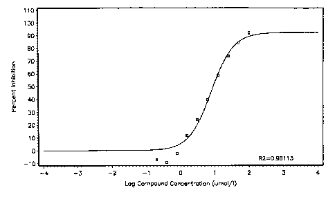
Figure 10: IC50 curve for inhibition of HPV-6a ATPase activity by ATP-y-S.
Nonlinear regression gave an IC50 value of 8.0 1.6 M.
Figure 11: Linearity and accuracy of phosphate detection. Lines shown are:
theoretical reference (observed value = expected value, diamonds); SPA result
using 1.2 M HCI, 2% AmMo, and 0.2 M citrate as described in example 5
(squares);
SPA result using 1.8 M HCI, 0.5% AmMo, and 0.1 M citrate (triangles); SPA
result
using 1.8 M HCI, 1% AmMo, and 0.1 M citrate (crosses); SPA result using 2.4 M
HCI, 1 k AmMo, and 0.1 M citrate (asterisks); and TLC result (circles).
Figure 12: Signal obtained for El reactions (squares) and blanks (diamonds)
run
on three separate plates. The x-axis is the well number, an arbitrary scale to
spread
the data for viewing.
Figure 13: SPA signal obtained for 0.2 to 200 M phosphate. Results are shown
for four different SPA bead concentrations. (a) linear scale (b) logarithmic
scale.
CA 02374455 2001-11-19
WO 00/79277 PCT/CAOO/00723
12
The theoretical line shown in (b) (asterisks) is simply a straight line for
reference.
Figure 14: SPA signal obtained for 0.5 to 10 M phosphate or ATP. Results are
shown for phosphate in the presence of AmMo (diamonds); phosphate in the
absence of AmMo (triangles); ATP in the presence of AmMo (squares); and ATP in
the absence of AmMo (crosses). (a) linear scale (b) logarithmic scale.
Figure 15: Estimation of the value of KR,(ATP) for HPV-1 1 El determined by
SPA
detection. Experimentally determined values are represented as circles, the
line
represents the calculated theoretical best-fit for the Michaelis-Menten
equation.
Figure 16: Estimation of the value of Km(ATP) for HPV-1 1 El determined by TLC
detection. Experimentally determined values are represented as circles, the
line
represents the calculated theoretical best-fit for the Michaelis-Menten
equation.
Figure 17: Lineweaver-Burke (double reciprocal) plots comparing experimental
inhibition data to theoretical results based on parameters obtained by
nonlinear
regression. (A) competitive inhibition model. (B) noncompetitive inhibition
model
(equal binding to E and ES). For both graphs, experimental values are given as
points with inhibitor concentrations of 24 M (diamonds); 12 M (asterisks); 6
M
(squares); 3 M (triangles); and 0 (circles). Lines are based on the best-fit
parameters for each inhibition model.
DETAILED DESCRIPTION OF THE PREFERRED EMBODIMENTS
Definitions
The term "scintillant hydrophobic surface" as used herein means a hydrophobic
surface that is impregnated, integrated, coated or otherwise contains a
scintillant.
The term "scintillant" as used herein means a fluorescent molecule (also
called
fluorescer) that, when placed in close proximity with radiation energy emitted
from a
radiolabeled reactant thereto, is activated to emit light energy detectable
and
measurable by a scintillation counter.
Preferred embodiments
According to a first embodiment of the present invention, there is provided a
method
for detecting and measuring radiolabeled orthophosphate (Pi) in an aqueous
reaction mixture, comprising the steps of:
CA 02374455 2001-11-19
WO 00/79277 PCT/CAOO/00723
13
a. adding a solution of molybdate to said reaction mixture under acidic
conditions
to form a phosphomolybdate complex; and
b. contacting said phosphomolybdate complex with a scintillant hydrophobic
surface;
whereby binding of phosphomolybdate to said surface provides enough proximity
for
the radiolabeled phosphate to induce measurable scintillation of the
scintillant
correlating the amount of said orthophosphate.
According to second embodiment of the invention, there is provided a method
for
determining phosphate-hydrolyzing enzyme activity, comprising the steps of:
a. mixing radiolabeled [y-33P]ATP with a said enzyme;
b. incubating reaction mixture a sufficient time to afford orthophosphate to
be
released from hydrolysis;
c. adding a solution of molybdate to said reaction mixture to form a
phosphomolybdate complex;
d. contacting said phosphomolybdate complex with a scintillant hydrophobic
surface; and
e. measuring scintillation of said scintillant as a means to calculate the
amount of
said orthophosphate.
Optionally, the method also comprises: step f) adding a solution of CsCl to
said
reaction mixture prior to counting. Further optionally, the method comprises
step g)
adding a solution of citric acid to said CsCI-containing mixture prior to
counting.
According to a third embodiment of the present invention, there is provided an
assay
of screening inhibitors of a phosphate-hydrolyzing enzyme activity comprising
the
steps of: carrying out steps a to e (optionally steps f and g) according to
the method
described above, in the presence and absence of a candidate inhibitor; and
comparing the levels of inhibition.
According to a fourth embodiment of the present invention, there is provided a
kit for
measuring radiolabeled orthophosphate in an aqueous solution, said kit
comprising:
a. a solution of molybdate; and
b. a scintillant hydrophobic surface;
CA 02374455 2006-09-01
14
wherein said molybdate solution is added to said aqueous solution to form a
phosphomolybdate complex, said complex being captured by said hydrophobic
surface to induce measurable scintillation thereto.
Preferably, according to the above embodiments of this invention, the
molybdate
solution and hydrophobic surface may be added simultaneously to the reaction
mixture.
Preferably, the embodiments of this invention further comprises the step of:
adding a solution of CsCI to the reaction mixture prior to counting. More
preferably,
the invention further comprises a step of: adding a solution of citric acid to
said CsCI-
containing mixture prior to counting. Most preferably, the CsCI and citric
acid are
added simultaneously to the reaction mixture.
Preferably, the reaction mixture containing CsCI and citric acid is incubated
for
longer than one hour prior to scintillation counting.
Preferably, the ammonium molybdate is at a final concentration of from 0.05%
to 0.3
%, more preferably from 0.1 % to 0.2% w/v. Most preferably, the ammonium
molybdate is at a final concentration of about 0.17% w/v.
Preferably, the hydrophobic surface is selected from the group consisting of:
polyvinyltoluene (PVT), Sephadex , latex, polystyrene, polyacrylamide,
acrylamide,
agarose, polypropylene, polycarbonate, and Sepharose . More preferably, the
hydrophobic surface is polyvinyl toluene beads such as SPA beads.
Preferably, the CsCl is at a final concentration higher than 1 M. More
preferably, the
CsCI is at a final concentration ranging from 2M and 4M. Most preferably, the
CsCl is
at a final concentration of about 3.5M.
Preferably, the citric acid is at a final concentration ranging from 0.05 and
0.2M.
More preferably, the citric acid is at a final concentration of about 0.1 M.
Preferably, the phosphate-hydrolyzing enzyme is selected from the group
consisting
CA 02374455 2006-09-01
of: helicase, ATPase, and phosphatase. More preferably, the enzyme is an
ATPase.
Most preferably, the ATPase is the El helicase from human papillomavirus.
EXAMPLES
5 Abbreviations used in the examples include:
AmMo ammonium molybdate
ATP-y-S adenosine-5'-O-(3-thiotriphosphate)
cpm counts per minute
DMSO dimethylsulfoxide
10 DTT dithiothreitol
EDTA ethylenediaminetetraacetic acid
HEPES N-[2-hydroxyethyl]piperazine-N'-[2-ethane sulfonic acid]
MES 2-[N-morpholino]ethane sulfonic acid
MgOAc magnesium acetate
15 PEI polyethyleneimine
Pi inorganic orthophosphate
PVPP polyvinylpolypyrrolidone
PVT polyvinyl toluene
SPA scintillation proximity assay
TLC thin-layer chromatography
Materials and methods
Polyhistidine-tagged HPV-11 El was expressed in baculovirus-infected insect
cells
and purified by Ni-affinity chromatography as described in WO 99/57283.
EXAMPLE 1
Protocol forATPase scintillation proximity assay (SPA) using HPV El
Radiolabeled [y-33P]ATP (NEN) was prepared upon receipt by diluting it 100-
fold in
the reaction buffer and storing it at -80 C. Material stored in this way was
good for
greater than one month. El ATPase reactions were run in a buffer consisting of
20
mM HEPES, pH 7.5, 0.05 mM EDTA and 1 mM DTT, and 0.05% IgepalO CA-630
(equivalent to Nonidet P40). Volumes and concentrations given below are
typical,
but these can be varied somewhat with minimal effect on results, as shown in
later
examples. One M ATP (Amersham Pharmacia) and 500 M MgOAc were mixed
CA 02374455 2006-09-01
16
with [y-33P]ATP at 100-fold dilution from the stored material (10,000 dilution
from the
stock), or approximately 1 nCi/ L when fresh. The actual ATP concentration
contributed by [y-33P]ATP was approximately 1 nM; this amount could be reduced
further if necessary. Sufficient enzyme was added to give the desired level of
conversion. For example, 4 nM HPV-6a El converted approximately 30% of the
substrate to ADP and phosphate in 2 hours. A typical reaction volume was 40
L;
reactions were run at room temperature in 96-well plates, typically Optiplates
(Packard).
At the desired time, 40 L reactions were stopped by adding 20 L of a SPA
bead-
AmMo mixture. This mixture consisted of one part 2% (w/v) AmMo in 2.4M HCI to
two parts streptavidin PVT SPA beads (Pharmacia Amersham #RPNQ0007)
suspended at 30 mg/mL in 50 mM HEPES plus 0.02% sodium azide. The
ammonium-molybdate solution was usually made fresh daily whereas the SPA bead
suspension was stable for greater than one month. The mixture can be made
several hours in advance, and can even be used for several days when stored at
4 C. Immediately after adding the ammonium molybdate-bead mixture, 80 L of 7M
cesium chloride plus 0.2 M citric acid were added. Plates were shaken briefly
and
then allowed to sit for greater than one hour. The extent of phosphate
production
was then determined by scintillation counting using the TopCount (Packard). If
desired, cpm can be converted to phosphate concentration by comparison of SPA
results to those determined by TLC (see below) for identical reactions.
Alternatively,
results can be compared to a"100% control", a reaction with a large excess of
enzyme previously confirmed by TLC to give complete conversion of ATP to
phosphate and ADP. Blanks containing no enzyme but otherwise the same were
run in parallel and subtracted.
EXAMPLE 2
E1 ATPase TLC assay
ATPase reactions were run just as in the SPA format. At the end of the planned
reaction time, reactions were stopped by adding one-half volume of ice-cold
500 mM
EDTA, pH 8Ø One to two L reaction samples were spotted onto
polyethyleneimine-coated cellulose TLC plates (Sigma) and eluted in a solution
of
1 M LiCI and 1 M formic acid. [y-33P] phosphate and ATP are determined using
the
CA 02374455 2006-09-01
17
Storm 860 phosphorimaging system (Molecular Dynamics). For each sample,
including blanks, the phosphate and ATP spot intensities were quantified and %
phosphate was calculated as:
100 X (phosphate intensity)
(phosphate intensity + ATP intensity).
Blank values were in the same range as for the SPA, approximately 2-5%, and
were
subtracted from the values for each reaction to give the value of %-phosphate
produced by the enzymatic reaction.
EXAMPLE 3
Effect of varying detection conditions on signal and blank
Many of the reactions in this section were run with slightly different
conditions using
a reaction buffer of 20 mM MES, pH 7.0, 10% glycerol, and 0.05 mM EDTA.
Reactions were run with 10 M ATP (approximately 1 nM [y-33P]ATP), 500 M
MgOAc, and sufficient El to give approximately 20% conversion of substrate in
90
minutes at room temperature. As for examples I and 2, reactions were run in
Packard Optiplates . In these experiments, detection was typically
accomplished by
mixing 20 L of ATPase reaction with 20 L 2% AmMo in 1.2 M HCI containing -
0.05% Tween-20 . After 10 minutes, 20 L of streptavidin SPA bead suspension
was added (10 mg/mL in 50 mM HEPES, pH 7.5 + 0.02% NaN3), after a brief mix
this was followed by 20 L of 7.0 M CsCI. After mixing, plates were allowed to
stand
for one hour before counting on the TopCount as described in Example 1.
SPA bead type: PVT-SPA beads are available with a variety of molecules
attached
to their surface. Types that were available for evaluation in the assay
included
streptavidin, wheat germ agglutinin, protein A, anti-mouse-IgG, and
glutathione.
Although it is the hydrophobic properties of PVT beads that has affinity for
phosphomolybdate, a variety of coatings were tested to see if the type of the
molecule on the surface had any effect on the SPA signal. Cpm data are shown
in
Figure 1A. The ratios of control signal and blank signal, or "signal-to-
background"
ratios, are shown in Figure 1 B. There is no significant effect of coating
type. Similar
results have also been obtained with copper (His-tag-binding) beads, and in
fact,
uncoated PVT beads, provided by Amersham-Pharmacia, also give an equivalent
signal. Yttrium silicate SPA beads do not work in this assay, as expected
since they
CA 02374455 2001-11-19
WO 00/79277 PCT/CAOO/00723
18
lack a hydrophobic surface.
Ammonium molybdate concentration: The function of ammonium molybdate in the
stop solution is to complex the released phosphate in the ATPase reaction. The
tested concentrations of ammonium molybdate in the stop solution ranged from 1
to
4%. The effects on cpm are shown in Figure 2A, and signal-to-background ratios
are
shown in Figure 2B. [Note: the blank is relatively unaffected by increasing
concentrations of AmMo. Thus, under these conditions, the background does not
seem to result from contaminating orthophosphate present in the ATP solution,
but
may rather be due to nonspecific sticking of ATP to the beads or capture of
some R-
particle radiation emitted by [r 33P]ATP in solution].
HCI concentration: The function of HCI in the stop solution is to provide an
acidic
medium in which the phosphomolybdate complex can form. The effects on cpm of
HCI concentrations ranging from 0 to 2.4 M in the stop solution are shown in
Figure
3A, and signal-to-background ratios are shown in Figure 3B. Under these
conditions, values greater than 1 M were determined to be optimal.
CsCI concentration: CsCI is added prior to scintillation counting for two
purposes.
The first is to produce a high-salt medium that enhances the hydrophobic
effect,
strengthening the binding of the phosphomolybdate complex to the SPA beads;
the
second is to increase the density of the fluid in the well. PVT SPA beads have
a
specific gravity of approximately 1.05 g/mL, and tend to stay dispersed in
aqueous
solution, settling only slowly to the bottom over several hours. The addition
of high-
molarity CsCI increases the density of the liquid, causing the SPA beads to
form a
thin layer floating at the surface, thus increasing the detectable signal. The
7.0 M
CsCi is essentially a saturated solution. The effect on cpm of adding 20 L of
CsCl
at different concentrations is shown in Figure 4A, and the effect on signal-to-
background ratios is shown in Figure 4B. Optimum signal to background was
obtained using a 7.0 M solution.
Incubation time in ammonium molybdate before addition of CsCI: The effect on
cpm
of varying the length of time between AmMo addition and CsCI addition is shown
in
Figure 5A, and the effect on signal-to-background ratios is shown in Figure
5B. It
CA 02374455 2006-09-01
19
appears that the reaction signal is approximately constant, but the blank
rises with
time; thus signal to background decreases with increased incubation times.
Time between CsCI addition and scintillation counting: In the standard
procedure,
plates are counted one hour following addition of CsCI solution. The cpm and
signal-
to-noise ratios obtained by counting the same plate at various times over a 48-
hour
period are shown in Figure 6A and 6B.
Stability of signal: The experiments in Figures 5 and 6 indicate that the
assay signal
is unstable. The blank increases steadily with time. It was shown by TLC
detection
that mixing ATP and HCI at the concentrations above results in a slow
degradation
of ATP to phosphate, and this almost certainly accounts for the increase in
signal
observed. The same problem occurs in other assays which rely on
phosphomolybdate formation to detect phosphate, for example by a change in
color
or the formation of a precipitate. It has been shown that citric acid, added
immediately after AmMo, will tightly bind to any free molybdate, preventing
the
incorporation of subsequently released phosphate into phosphomolybdate anions.
Exchange is extremely slow, so addition of citrate does not decrease the
concentration of preformed phosphomolybdate, even after several days.
The effect of adding 0.2 M citric acid to the 7M CsCI solution is shown in
Figure 7. In this experiment, 30 L El ATPase reactions were run as described
at
the beginning of Example 3. After this, 30 L of 2% AmMo in 1 M HCI was added
followed immediately by 30 L of 10 mg/mL SPA beads and then 30 or 90 L of 7M
CsCI 0.2 M citric acid. The signal is enhanced approximately two-fold after
a one
hour incubation, and is significantly stabilized by the addition of citric
acid, so that
little change is observed even after three days.
An additional experiment showing the effect of AmMo, CsCI, HCI, and citric
acid
concentrations on the signal and signal stability is shown in Figure 8.
Reactions were
run as described at the beginning of this example except that the detergent
Igepal -
CA630 (Sigma, equivalent to Nonidet-P40) was present at 0.005% in all but one
set
of reactions, and citric acid was included in the CsCI solution at 0.1 M, 0.2M
or 0.4M.
0.05% Tween-200 was included in the ammonium molybdate solution in one case.
Tween-20 is known to stabilize the phosphomolybdate-malachite green complex
in
the assay of Itaya & Ui (Clin Chim Acta, 1966, Sep;14(3):361-6), but has no
beneficial
CA 02374455 2006-09-01
effect in this assay. The assay plate was read at 1.5, 6.5, and 20 hours after
addition of CsCI/citric acid. The signal increased only slightly after 1.5
hours and
was stable up to at least 20 hours.
The control inhibitor ATP-y-S was added to some wells at 10 M (data not
5 shown, but see example 4), to verify the robustness of the data obtained at
the
different time points. Over all conditions and time-points tested, the level
of inhibition
o-nly varied from 63.9 to 70.0%. Thus the variations in detection under the
different
conditions of the assay may have an effect on observed cpm, but do not affect
the
relative signals between enzyme reactions, inhibited reactions, and blanks,
and thus
10 do not affect the fundamental accuracy of the assay.
An example of a time-course run using HPV-1 1 El is shown in Figure 9. This
experiment was run under the conditions described above for Figure 8, except
for
the presence of 0.005% detergent as described above. Data shown are the
averages for four 180 L reactions. At each time-point, 30 L was removed and
15 mixed with 30pL AmMo/SPA beads solution followed by 90 L of 7.0 M CsCI /
0.2 M
citric acid prior to scintillation counting.
EXAMPLE 4
Inhibition by ATP-y-S
20 The following solutions were used to run 45 L reactions for IC50 curves:
- ATP (15 L per reaction), consisting of 3.0 M ATP, 1.5 mM Mg acetate, and
0.06
Ci [y-33P]ATP;
- El (15 L per reaction), consisting of 18 nM HPV-6 El;
- inhibitor solution (15 L per well) consisting of y-S-ATP dissolved in
buffer plus
18% DMSO.
All solutions are made in the assay buffer described in Example 3 except that
the assay buffer also contained 0.005% Igepal CA-630. All components are
diluted 3-fold on mixing the reactions. The reactants were added in the
following
order: a) inhibitor, b) HPV-6 El, c) ATP. The concentration range for the
inhibitor in
the reactions was 0.2 to 100 M. After 75 minutes, reactions were quenched by
adding 45 L of a mixture consisting of two parts streptavidin SPA bead (15
mg/mL
suspension) in resuspension buffer (Example 2) and one part 2.4 M HCI
containing
2% ammonium molybdate. Then 90 L of 7 M CsCl containing 0.1 M citric acid was
CA 02374455 2006-09-01
21
added. After mixing briefly, plates were left for 90 minutes and then counted
on a
TopCount. Inhibition data (see Figure 10) were fit to a logistic using SAS
(Statistical Software System; SAS Institute, Inc. Cary, N.C.) with positive
controls
averaging 16,000 cpm and blanks averaging 1300 cpm. Similar results have also
been obtained in cases where detection was performed by TLC.
EXAMPLE 5
Linearity and accuracy of phosphate detection
. In this experiment, one large ATPase reaction was run with sufficient El to
give 100% ATP hydrolysis. The El was then heat-inactivated, and the reaction
mixture was mixed in various proportions with a reaction blank containing no
El.
Reaction buffer and incubation conditions were as described in example 1
except
that the reaction buffer was as described in example 3, though 0.005% Igepal -
CA630 was present. Reaction-blank mixtures were made at 2:3, 1:5, 1:10, and
1:20
to simulate a range from 40% to 5% hydrolysis. As for some of the experiments
above, SPA detection was performed using ammonium molybdate, HCI, and citric
acid at several different concentrations. Results for some conditions, along
with
those for TLC, are shown in Figure 11. In all cases, the signal detected
(expressed
as proportion of 100% for SPA and as the observed phosphate concentration for
TLC) are very similar. Although the absolute signal (cpm) varies with
conditions, the
relative signal and thus the accuracy of the assay is constant. In particular,
20%
conversion simulates a typical extent of reaction under screening conditions
and
10% conversion represents the signal which would be observed for test
compounds
giving 50% inhibition.
EXAMPLE 6
Variation for assay controls under screening conditions
To verify the well-to-well variability of this method, reactions were run as
described in example 1 in 80 wells of three separate 96-well plates. Reaction
blanks without enzyme were run in the other 16 wells. Results are shown in
Figure
12. Well-to-well variability can be measured through the z' statistic, which
takes into
account standard deviations of the signals and the separation between the
signal
from enzymatic reactions and blanks (J-H Zhang, et al, J. Biomol. Screening,
1999,
4, 67-73). Values can range from less than 0 to 1.0, with values greater than
0.5
CA 02374455 2001-11-19
WO 00/79277 PCT/CAOO/00723
22
deemed very acceptable for a screening assay. The z' value for this experiment
was
0.63 or 0.75 when two clear outliers were removed from the analysis.
EXAMPLE 7
Linearity of phosphate detection at higher phosphate concentrations
The Examples above demonstrate that SPA detection of phosphate works
well for low concentrations of phosphate, 0.1-1 M. However, in order to carry
out
mechanistic work, it is necessary to vary the ATP concentration more widely.
We
had observed that the procedure outlined in Example 1 did not quantitatively
capture
all orthophosphate, since increased signal was observed if larger amounts of
SPA
beads were added. Based on those initial results, an SPA bead titration
experiment
was carried out as described below.
Similarly to Example 5, for this experiment one larger reaction was run using
a high concentration of El. In this case, the ATP concentration was 200 M.
The
reaction was run for four hours at room temperature and then for two hours at
37 C
to ensure complete conversion. A sample was analyzed by TLC to verify
quantitative
conversion. The reaction was then serially diluted with reaction buffer plus
magnesium to give a range from 200 M to 0.2 M phosphate concentrations.
Samples were analyzed by combining 45 L of diluted reaction with 15 L of
ammonium molybdate (2% in 2.4 M HCI) and 30 L of SPA bead suspension at 7.5,
15, 30, or 60 mg/mL. Finally 90 L of 7M CsCl plus 0.2 M citric acid was added
to all
wells.
Results are shown in Figure 13A and as a log-log plot in Figure 13B. As
seen most clearly in Fig. 13B, for each SPA bead concentration the signal was
directly proportional to the phosphate concentration up to about 100 M.
However,
the absolute signal does depend on the bead concentration. Interestingly, the
lines
in Fig. 13B are parallei, i.e. the proportional increase in signal obtained by
increasing the SPA bead concentration is the same at all phosphate
concentrations,
up to about 100 M.
EXAMPLE 8
Titration of ATP and phosphate in the presence and absence of AmMo in order to
determine the source of signal in the absence of enzyme
In this experiment, a large reaction similar to the one above was run to
CA 02374455 2006-09-01
23
completely convert 10 M ATP to phosphate and ADP; complete conversion was
verified by TLC (Example 2). In parallel, an identical mixture lacking enzyme
was
produced. Each was diluted in buffer as above to give solutions with phosphate
or
ATP concentrations ranging from 10 M to 0.5 M. Duplicate 45 L samples of
each
were mixed with either 40 L of AmMo/SPA bead mixture as described in Example
1
or with a similar mixture lacking AmMo (but still containing HCI), followed by
80 L of
CsCI/citric acid. Results are graphed in Figure 14A or as a log-log plot in
Figure
14B. As expected, based on Example 7, the signal is linearly dependent on the
radioactivity concentration in all cases. In the presence of AmMo, the
reaction blank
(ATP solution) gives a signal equal to approximately 5% of the phosphate
solution
produced by total conversion. Unlike the experiment in Example 3 (Figure 2),
most
of the blank signal is dependent on AmMo, and thus under these conditions, the
blank is primarily due to contaminating orthophosphate already present in the
commercial ATP solution.
EXAMPLE 9
Value of Kn, (A TP) for HPV-11 as determined by SPA and TLC methods
ATPase time-courses were run at different concentrations of ATP in order to
determine the kinetic parameter Km(ATP) for HPV-1 1 El. Reactions were run
under
similar conditions in both experiments, using the procedures given in Examples
1
and 2, except that the concentration of Igepal -CA630 was 0.01 % rather than
0.005% and with experiment-specific changes noted below. The El concentration
was 2 nM and the ATP concentrations ranged from 3-75 M (TLC) or 2-50 M
(SPA), with the MgOAc concentration equal to the ATP concentration plus 0.5
mM.
Stock solutions of ATP at each concentration were obtained by diluting the
highest
concentration solution with buffer containing 0.5 mM MgOAc; thus a constant
ratio of
radiolabeled to unlabeled ATP was used for all reactions. Reaction rates were
measured by taking time-points from 10 or 20 to 120 minutes. To insure initial
velocity conditions, time-points giving greater than 20% conversion were not
used for
analysis.
Detection and data processing for the SPA Km experiment: Total reaction
volumes were 150 L. For each time-point, 20 L of reaction mixture was
removed
and combined with 40 L of ammonium molybdate/SPA bead mixture followed by 80
L of CsCI/citric acid. All reactions were run in triplicate. Plates were
counted after
CA 02374455 2006-09-01
24
overnight incubation. At the last time-point, an additional 20 L aliquot was
removed
and combined with 10 pL 0.5 M EDTA. In this case, the conversion of ATP to
phosphate was quantified by TLC, and the concentration of phosphate determined
by TLC was compared to the cpm from the SPA procedure, after subtraction of
blanks in both cases. The relationship between phosphate concentration and cpm
is
linear and the slope of the line was used to convert cpm values from SPA
detection
to concentration of phosphate produced. Rates of phosphate production at each
ATP concentration were fit by nonlinear regression to the Michaelis-Menten
equation
using the program GrafitO (V 3.01, R. Leatherbarrow). Results are shown in
Figure
15.
Detection and data processing for the TLC Km experiment: As for the SPA
experiment above, 150 L reactions were run, in duplicate. At each time-point,
20
L were removed and combined with 10 L of ice-cold 0.5 M EDTA. At the
completion of the time-course, reactions were diluted such that the total
radioactivity
concentration for each point was approximately equal. Thus reactions at 75 M
ATP
were diluted 25-fold whereas 3 M ATP reactions were not diluted. The dilution
buffer consisted of two parts reaction buffer containing 500 M magnesium
acetate
and one part 0.5 M EDTA. One L of each diluted reaction was spotted for TLC
detection. The values for percent conversion of ATP to phosphate were
determined
as described in Example 2 and these were used to determine reaction rates at
each
ATP concentration. These rates were then fit to the Michaelis-Menten equation
as
described above to give an estimate for the value of Km(ATP) (Figure 16).
Comparison of Figures 15 and 16 clearly shows that the two techniques give
similar results, but also that the quality of data is superior for the SPA
method.
Furthermore, the SPA method requires significantly less manipulation and
shorter
data processing time. Results shown are typical examples, each has been
reproduced multiple times.
EXAMPLE10
Value of Ki(ATP-y-S) for HPV-11 as determined by the SPA methods
The y-thio phosphate analog of ATP inhibits many ATPases by a competitive
mechanism. Experimental determination of the mechanism of inhibitor action
requires measuring initial velocities for a number of substrate and inhibitor
CA 02374455 2006-09-01
concentrations. The number of data points needed (several hundred) and the
precision required for this experiment mean that performing the experiment by
TLC
detection is much more difficult and tedious. The SPA procedure works well,
however. For this experiment, ATP concentrations of 5, 10, 20 and 40 M were
5 used, along with inhibitor concentrations of 0, 3, 6, 12 and 24 M.
Reactions were
run as described for Example 9 (SPA detection). Data were processed as above
and fit by nonlinear regression to an equation for competitive inhibition
using GraFit.
We obtained values for Ki(ATP-y-S) of 3.8 0.4 M and for Km(ATP) of 6.7
0.7 M.
A Lineweaver-Burke plot illustrating the fitness is shown in Figure 17A. The
plot
10 was generated in ExcelO using the fitted parameters for the lines and the
experimental values for points. For comparison, the data were also fit to an
equation for non-competitive inhibition (equal binding of inhibitor to enzyme
and
enzyme-substrate complexes). Values obtained were 16 2 M for K;(ATP-y-S)
and
12 2 M for Km(ATP). The corresponding double reciprocal plot is shown in
15 Figure 17B. Because of the quality of the data obtained it is possible to
observe a
systematic deviation between the experimental and fitted values for this
second
mechanism, especially at higher inhibitor concentrations. A similar conclusion
can
be drawn from the significantly lower reduced chi squared value for the
competitive
fit compared to the noncompetitive fit (62 and 347 respectively). Thus as
expected,
20 the competitive model is more appropriate for this inhibitor.
DISCUSSION
The procedure presented above is a sensitive, accurate, and robust method for
the
detection of orthophosphate produced by the cleavage of radiolabeled phosphate-
25 containing compounds. It is highly suited to the task of measuring the
activity of
enzymes for which orthophosphate is a reaction product, and to measuring the
inhibition of such activities. There are many such enzymes; common examples
are
helicases, ATPases and phosphatases. It is particularly appropriate for cases
in
which only low concentrations of orthophosphate (nM or low M) are produced.
Important cases will be those enzymes, such as the El helicase of HPV, which
bind
the phospho substrate tightly. This procedure allows assays to be run at
substrate
concentrations below the value of Km for maximum sensitivity to competitive
inhibitors. The method is simple and robust enough for large scale inhibitor
CA 02374455 2006-09-01
26
screening. In particular, the method is not sensitive to many common
artifacts, for
example apparent inhibition caused by colored or fluorescent compounds, and
the
signal produced is stable, reproducible, and relatively insensitive to small
fluctuations
in concentrations or volumes of assay components. The method is also accurate
enough to be applied to quantitative enzymology studies.
Other methods to detect orthophosphate have been discussed in the literature.
Two
widely reported methods use radiolabeled ATP to measure ATPase activity. Both
of
these methods involve the physical separation of products (e.g. ADP and Pi)
from
the starting material, using either TLC on PEI cellulose or selective
adsorption of
ATP onto charcoal. While sensitive enough to detect very low concentrations of
orthophosphate, these are classical methods which cannot be easily adapted to
modern screening applications. Other assay methods rely on coupling enzymes
which use orthophosphate (or another reaction product) as the substrate in a
second
reaction, producing an absorbance or fluorescence change. These can be quite
accurate, but are less sensitive than radioactivity-based assays. Furthermore,
the
addition of a coupling enzyme complicates the interpretation of results, since
coupled-enzyme assays are subject to additional artifacts. Several other
procedures
involve formation of phosphomolybdate followed by reduction or dye absorption
to
produce a color change, which can be correlated with phosphate concentration.
Some enzyme assays based on these procedures are accurate and robust enough
to be used in compound screening efforts or enzymology studies, but it is not
practical to use these methods to detect low M or nM concentrations of
orthophosphate. In some applications, it is possible to enhance the
sensitivity of
these methods by concentrating large volumes of dilute phosphomolybdate onto
Sephadex or related resins. This has not proved applicable to screening
applications, however, since only very small volumes are normally used in each
test
reaction. It has been shown that one can selectively adsorb radiolabeled
phosphate
onto the surface of polyvinylpolypyrrolidone (PVPP). Radiolabeled ATP or other
contaminants can then be washed away and the remaining phosphate detected by
elution at elevated pH. This specific procedure is not very practical for
enzymatic
studies, since it requires the physical separation of reactants and products,
and the
reproducibility, which is dependent on elution of multiple samples from PVPP
columns, would be relatively poor. The authors suggest that an important
component of the selectivity of their procedure is the ability of
polyvinylpyrrolidone to
CA 02374455 2001-11-19
WO 00/79277 PCT/CAOO/00723
27
catalyze the formation of phosphomolybdate, thereby implying that other
hydrophobic surfaces would be less suited to their method, thus leading away
from
the present invention.