Note: Descriptions are shown in the official language in which they were submitted.
CA 02376798 2001-10-23
WO 00/55373 PCT/US00/06883
NOVEL METHODS OF DIAGNOSING MACROPHAGE DEVELOPMENT RELATED
DISORDERS, COMPOSITIONS, AND METHODS OF SCREENING FOR
MACROPHAGE DEVELOPMENT MODULATORS
FIELD OF THE INVENTION
The invention relates to the identification of expression profiles and the
nucleic acids involved in
destructive macrophage development and disorders associated with destructive
macrophage
development, and to the use of such expression profiles and nucleic acids in
diagnosis and prognosis
of macrophage related disorders. The invention further relates to methods for
identifying and using
candidate agents and/or targets which modulate macrophage development.
BACKGROUND OF THE INVENTION
The mononuclear phagocytic system consists of circulating monocytes in the
blood and macrophages
in the tissues. During hematopoiesis in the bone marrow, myeloid progenitor
cells differentiate into
promonocytes, which leave the bone marrow and enter the blood, where they
differentiate further into
monocytes. After circulating in the blood stream for some period of time, the
monocytes enlarge and
then migrate into the tissues as they differentiate to become macrophages.
Macrophages play a central role in the immune response, and have three primary
important functions:
phagocytosis, antigen processing and presentation, and the secretion of
biologically important factors.
Phagocytosis allows the ingestion and digestion of erogenous antigens such as
whole pathogenic
organisms, insoluble particles, injured and dead cells, cellular debris, etc.
However, not all of the
2 0 antigen ingested by macrophages is digested; some phagocytosed antigen is
metabolically converted
within the endosomal processing pathway into peptides that associate with MHC-
II molecules. These
peptide-MHC II complexes are transported to the macrophage membrane, wherein
the antigenic
peptides are presented to T helper cells, resulting in T cell helper
activation. The activated T cells
then secrete a variety of cytokines that in turn activate the macrophages,
which exhibit increased
levels of phagocytosis and express increased levels of MHC II molecules and
cellular adhesion
CA 02376798 2001-10-23
WO 00/55373 PCT/LTS00/06883
-2-
molecules. Activated macrophages thus are more effective antigen-presenting
cells, and they also
migrate more vigorously in response to chemotactic factors.
In chronic inflammatory diseases such as rheumatoid arthritis (RA), monocyte-
derived macrophages
(MDMs) are presumed to damage host tissues by producing proteolytic enzymes
that can dissolve the
extracellular matrix. Recently, monocyte culture conditions were identified
that result in a highly
degradative macrophage population. See Reddy et al., Proc. Natl. Acad. Sci. US
92:3849 (1995).
While this cell population was shown to secrete fully processed and
enzymatically active cathepsins,
including cathepsin B, L and S, the gene expression profiles of these cells
were not evaluated.
Accordingly, it is an object of this invention to identify novel genes that
are differentially expressed in
macrophage development, and expression vectors, host cells, and biochips
comprising these novel
nucleic acids. In addition, it is an object of the invention to provide gene
expression profiles which are
unique to this destructive macrophage phenotype. It is further an object to
use the expression profiles
in assays to identify agents which can be used in the modulation of the
macrophage phenotype,
including the expression of destructive proteases, phagocytosis, the secretion
of other factors and
antigen presentation. It is further an object to use the expression profiles
as diagnostics to identify
diseases associated with these destructive macrophages. It is further an
object to provide assays to
identify agents for the treatment of macrophage- related disorders.
SUMMARY OF THE INVENTION
The present invention provides methods for screening for compositions which
modulate Destructive
2 0 Macrophage Disorders (DMD). Methods of treatment of DMD, as well as
compositions, are also
provided herein.
In one aspect, a method of screening drug candidates comprises providing a
cell that expresses an
expression profile gene or fragments thereof. Preferred embodiments of the
expression profile gene
are genes which are differentially expressed in macrophage, as compared to
other cells. Preferred
2 5 embodiments of expression profile genes used in the methods herein include
but are not limited to the
group consisting of the sequence of Figure 4, Figure 8, Figure 9, Figure 10
and Figure 19 and the
sequence represented by accession number X92521, X62466, J04130, X62078 and
X76534; the
proteins encoded this group and fragments thereof are also preferred. It is
understood that molecules
for use in the present invention may be from any figure or any subset of
listed molecules. Therefore,
3 0 for example, any one or more of the genes listed above can be used in the
methods herein. In
another embodiment, a nucleic acid is selected from Figures 1-76 or 78-81.
Preferred nucleic acids
are in Figure 81, more preferably in Figure 4, Figure 8, Figure 9, Figure 10
and Figure 19 and the
having the sequence represented by accession number X92521, X62466, J04130,
X62078 and
CA 02376798 2001-10-23
WO 00/55373 PCT/US00/06883
-3-
X76534, most preferably having the sequence represented by accession number
X92521. The
method further includes adding a drug candidate to the cell and determining
the effect of the drug
candidate on the expression of the expression profile gene.
In one embodiment, the method of screening drug candidates includes comparing
the level of
expression in the absence of the drug candidate to the level of expression in
the presence of the drug
candidate, wherein the concentration of the drug candidate can vary when
present, and wherein the
comparison can occur after addition or removal of the drug candidate. In a
preferred embodiment, the
cell expresses at least two expression profile genes. The profile genes may
show an increase or
decrease.
Also provided herein is a method of screening for a bioactive agent capable of
binding to a Destructive
Macrophage (DM) modulator protein, the method comprising combining the DM
modulator protein and
a candidate bioactive agent, and determining the binding of the candidate
agent to the DM modulator
protein. Preferably the DM modulator protein is a protein or fragment thereof
encoded by the
sequences selected from the group consisting of the sequence of Figure 4,
Figure 8, Figure 9, Figure
10 and Figure 19 and the sequence represented by accession number X92521,
X62466, J04130,
X62078 and X76534. In another embodiment, the protein is encoded by a nucleic
acid selected from
Figures 1-76 and 78-81. Preferred nucleic acids are in Figure 81, more
preferably Figure 4, Figure 8,
Figure 9, Figure 10 and Figure 19 and the sequence represented by accession
number X92521,
X62466, J04130, X62078 and X76534, and most preferably the sequence
represented by accession
2 0 number X92521
Further provided herein is a method for screening for a bioactive agent
capable of modulating the
activity of a DM modulator protein. In one embodiment, the method comprises
combining the DM
modulator protein and a candidate bioactive agent, and determining the effect
of the candidate agent
on the bioactivity of the DM modulator protein. Preferably the DM modulator
protein is a protein or
2 5 fragment thereof encoded by a sequence selected from the group consisting
of the sequence of
Figure 4, Figure 8, Figure 9, Figure 10 and Figure 19 and the sequence
represented by accession
number X92521, X62466, J04130, X62078 and X76534. In another embodiment, the
protein is
encoded by a nucleic acid selected from Figures 1-76 and 78-81. Preferred
nucleic acids are in
Figure 81, more preferably Figure 4, Figure 8, Figure 9, Figure 10 and Figure
19 and the sequence
3 0 represented by accession number X92521, X62466, J04130, X62078 and X76534,
and most
preferably the sequence represented by accession number X92521.
Also provided is a method of evaluating the effect of a candidate DMD drug
comprising administering
the drug to a transgenic animal expressing or over-expressing the DM modulator
protein, or an animal
lacking the DM modulator protein, for example as a result of a gene knockout.
CA 02376798 2001-10-23
WO 00/55373 PCT/US00/06883
-4-
Additionally, provided herein is a method of evaluating the effect of a
candidate DMD drug comprising
administering the drug to a patient and removing a cell sample from the
patient. The expression
profile of the cell is then determined. This method may further comprise
comparing the expression
profile to an expression profile of a healthy individual.
Moreover, provided herein is a biochip comprising a nucleic acid segment which
encodes a colorectal
cancer protein, preferably selected from the group consisting of the sequence
of Figure 4, Figure 8,
Figure 9, Figure 10 and Figure 19 and the sequence represented by accession
number X92521,
X62466, J04130, X62078 and X76534, or a fragment thereof, wherein the biochip
comprises fewer
than 1000 nucleic acid probes. Preferably at least two nucleic acid segments
are included. In another
embodiment, the nucleic acid selected from Figures 1-76 and 78-81. Preferred
nucleic acids are in
Figure 81, more preferably Figure 4, Figure 8, Figure 9, Figure 10 and Figure
19 and the sequence
represented by accession number X92521, X62466, J04130, X62078 and X76534, and
most
preferably the sequence represented by accession number X92521.
Furthermore, a method of diagnosing a DMD is provided. The method comprises
determining the
expression of a gene which encodes a DMD protein preferably encoded by a
nucleic acid selected
from the group consisting of the sequence of Figure 4, Figure 8, Figure 9,
Figure 10 and Figure 19
and the sequence represented by accession number X92521, X62466, J04130,
X62078 and X76534
or a fragment thereof in a first tissue type of a first individual, and
comparing the distribution to the
expression of the gene from a second normal tissue type from the first
individual or a second
2 0 unaffected individual. In another embodiment, the protein is encoded by a
nucleic acid selected from
Figures 1-76 and 78-81. Preferred nucleic acids are in Figure 81, more
preferably Figure 4, Figure 8,
Figure 9, Figure 10 and Figure 19 and the sequence represented by accession
number X92521,
X62466, J04130, X62078 and X76534, and most preferably the sequence
represented by accession
number X92521. A difference in the expression indicates that the first
individual has a DMD.
In another aspect, the present invention provides an antibody which
specifically binds to a DM protein,
preferably encoded by a nucleic acid selected from the group consisting of the
sequence of Figure 4,
Figure 8, Figure 9, Figure 10 and Figure 19 and the sequence represented by
accession number
X92521, X62466, J04130, X62078 and X76534 or a fragment thereof. In another
embodiment, the
protein is encoded by a nucleic acid selected from Figures 1-76 and 78-81.
Preferred nucleic acids
3 0 are in Figure 81, more preferably Figure 4, Figure 8, Figure 9, Figure 10
and Figure 19 and the
sequence represented by accession number X92521, X62466, J04130, X62078 and
X76534, and
most preferably the sequence represented by accession number. Preferably the
antibody is a
monoclonal antibody. The antibody can be a fragment of an antibody such as a
single stranded
antibody as further described herein, or can be conjugated to another
molecule. In one embodiment,
3 5 the antibody is a humanized antibody.
CA 02376798 2001-10-23
WO 00/55373 PCT/US00/06883
-5-
In one embodiment a method for screening for a bioactive agent capable of
interfering with the binding
of a DM modulator protein or a fragment thereof and an antibody which binds to
said DM modulator
protein or fragment thereof. In a preferred embodiment, the method comprises
combining a DM
modulator protein or fragment thereof, a candidate bioactive agent and an
antibody which binds to
said DM modulator protein or fragment thereof. The method further includes
determining the binding
of said DM modulator protein or fragment thereof and said antibody. Wherein
there is a change in
binding, an agent is identified as an interfering agent. The interfering agent
can be an agonist or an
antagonist. Preferably, the antibody as well as the agent inhibits DMD.
In a further aspect, a method for inhibiting DMD is provided. In one
embodiment, the method
comprises administering to a cell a composition comprising an antibody to a DM
modulating protein,
preferably encoded by the nucleic acids selected from the group consisting of
the sequence of Figure
4, Figure 8, Figure 9, Figure 10 and Figure 19 and the sequence represented by
accession number
X92521, X92521, J04130, X62078 and X76534, or a fragment thereof. In another
embodiment, the
protein is encoded by a nucleic acid selected from Figures 1-76 and 78-81.
Preferred nucleic acids
are in Figure 81, more preferably Figure 4, Figure 8, Figure 9, Figure 10 and
Figure 19 and the
sequence represented by accession number X92521, X62466, J04130, X62078 and
X76534, and
most preferably in the sequence represented by accession number X92521. The
method can be
performed in vitro or in vivo, preferably in vivo to an individual. In a
preferred embodiment the method
of inhibiting DMD is provided to an individual with arthritis. As described
herein, methods of inhibiting
2 0 DMD can be performed by administering an inhibitor of DM protein activity,
including antisense
molecules, and preferably small molecules.
Also provided herein are methods eliciting an immune response in an
individual. In one embodiment a
method provided herein comprises administering to an individual a composition
comprising a DM
modulating protein, preferably encoded by the nucleic acid selected from the
group consisting of the
2 5 sequence of Figure 4, Figure 8, Figure 9, Figure 10 and Figure 19 and the
sequence represented by
accession number X92521, X62466, J04130, X62078 and X76534, or a fragment
thereof. In another
embodiment, the protein is encoded by a nucleic acid selected from Figures 1-
76 and 78-81.
Preferred nucleic acids are in Figure 81, more preferably Figure 4, Figure 8,
Figure 9, Figure 10 and
Figure 19 and the sequence represented by accession number X92521, X62466,
J04130, X62078 and
3 0 X76534, and most preferably the sequence represented by accession number
X92521. In another
aspect, said composition comprises a nucleic acid comprising a sequence
encoding a DM modulating
protein, preferably encoded by the nucleic acid selected from the group
consisting of the sequence of
Figure 4, Figure 8, Figure 9, Figure 10 and Figure 19 and the sequence
represented by accession
number X92521, X62466, J04130, X62078 and X76534, or a fragment thereof. In
another
3 5 embodiment, the nucleic acid is selected from Figures 1-76 and 78-81.
Preferred nucleic acids are in
Figure 81, more preferably Figure 4, Figure 8, Figure 9, Figure 10 and Figure
19 and the sequence
CA 02376798 2001-10-23
WO 00/55373 PCT/US00/06883
-6-
represented by accession number X92521, X62466, J04130, X62078 and X76534, and
most
preferably the sequence represented by accession number X92521.
Further provided herein are compositions capable of eliciting an immune
response in an individual. In
one embodiment, a composition provided herein comprises a DM modulating
protein, preferably
encoded by a nucleic acid selected from the group consisting of the sequence
of Figure 4, Figure 8,
Figure 9, Figure 10 and Figure 19 and the sequence represented by accession
number X92521,
X62466, J04130, X62078 and X76534, or a fragment thereof, and a
pharmaceutically acceptable
carrier. In another embodiment, the protein is encoded by a nucleic acid
selected from Figures 1-76
and 78-81. Preferred nucleic acids are in Figure 81, more preferably Figure 4,
Figure 8, Figure 9,
Figure 10 and Figure 19 and the sequence represented by accession number
X92521, X62466,
J04130, X62078 and X76534, and most preferably the sequence represented by
accession number
X92521. In another embodiment, said composition comprises a nucleic acid
comprising a sequence
encoding a DM modulating protein, preferably selected from the group
consisting of the sequence of
Figure 4, Figure 8, Figure 9, Figure 10 and Figure 19 and the sequence
represented by accession
number X92521, X62466, J04130, X62078 and X76534, or a fragment thereof, and a
pharmaceutically acceptable carrier. In another embodiment, the nucleic acid
is selected from Figures
1-76 and 78-81. Preferred nucleic acids are in Figure 81, more preferably
Figure 4, Figure 8, Figure 9,
Figure 10 and Figure 19 and the sequence represented by accession number
X92521, X62466,
J04130, X62078 and X76534, and most preferably the sequence represented by
accession number
2 0 X92521
A method of neutralizing the effect of a DM protein, preferably encoded by the
nucleic acid selected
from the group consisting of the sequence of Figure 4, Figure 8, Figure 9,
Figure 10 and Figure 19
and the sequence represented by accession number X92521, X62466, J04130,
X62078 and X76534,
or a fragment thereof, comprising contacting an agent specific for said
protein with said protein in an
2 5 amount sufficient to effect neutralization. In another embodiment, the
protein is encoded by a nucleic
acid selected from Figures 1-76 and 78-81. Preferred nucleic acids are in
Figure 81, more preferably
Figure 4, Figure 8, Figure 9, Figure 10 and Figure 19 and the sequence
represented by accession
number X92521, X62466, J04130, X62078 and X76534, and most preferably the
sequence
represented by accession number X92521.
3 0 In another aspect of the invention, a method of treating an individual for
DMD is provided. In one
embodiment, the method comprises administering to said individual an inhibitor
of matrix
metalloproteinase 19 (MMP-19). In another embodiment, the method comprises
administering to a
patient having DMD an antibody to MMP-19 conjugated to a therapeutic moiety.
Such a therapeutic
moiety can be a cytotoxic agent or a radioisotope.
CA 02376798 2001-10-23
WO 00/55373 PCT/US00/06883
Also provided herein is a method for determining the prognosis of an
individual with DMD comprising
determining the level of MMP-19 in a sample, wherein a high level of MMP-19
indicates a poor
prognosis.
Novel sequences are also provided herein. Other aspects of the invention will
become apparent to the
skilled artisan by the following description of the invention.
DETAILED DESCRIPTION OF THE FIGURES
Figures 1-76 depict the sequences of the invention. DM sequence 1 (also
sometimes referred to
herein as DMS1 or Eos1) is depicted in Figure 1; DM sequence 2 (DMS2 or Eos2)
is depicted in
Figure 2, etc.
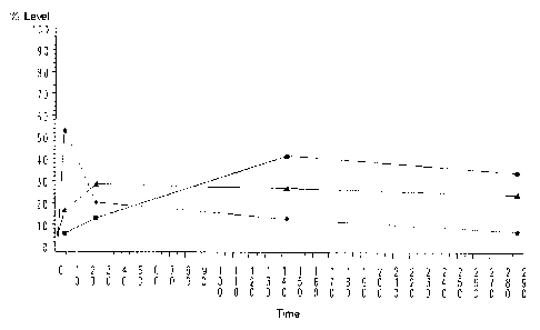
Figure 77 is a graph of expression levels of genes up-regulated in the
macrophage development
model whose sequences are identified in Figures 78-80. Expression profiles are
clustered into 3
groups (C1, C2, and C3) that define 3 different expression time courses. Group
C1 is identified by
triangles. Group C2 is identified by squares. Group C3 is identified by closed
circles.
Figure 78 provides accession numbers of 148 genes of group C1 identified in
the macrophage
development model (incorporated in their entirety here and throughout the
application where
Accession numbers are provided). A. depicts 148genes that are upregulated in a
similar time course.
B. depicts 69 genes that are upregulated in a similar time course. C. depicts
76 genes that are
upregulated in a similar time course.
Figure 79 provides accession numbers of 69 genes of group C2 identified in the
macrophage
2 0 development model.
Figure 80 provides accession numbers of 76 genes of group C1 identified in the
macrophage
development model.
Figure 81 provides accession numbers of a preferred subset of 35 genes
identified in Figures 780 that
were selected based on minimal normal tissue expression.
2 5 Figure 82 shows the nucleic acid sequence represented by accession number
X92521, encoding
matrix metalloproteinase 19.
Figure 83 shows the amino acid sequence of the protein (matrix
metalloproteinase 19) encoded by the
nucleic acid represented by accession number X92521
CA 02376798 2001-10-23
WO 00/55373 PCT/LTS00/06883
_g_
DETAILED DESCRIPTION OF THE INVENTION
The present invention provides *novel methods for diagnosis and prognosis
evaluation for destructive
macrophage disorders (DMD), as well as methods for screening for compositions
which modulate
DMDs. In one aspect, the expression levels of genes are determined in
different patient samples for
which either diagnosis or prognosis information is desired, to provide
expression profiles. An
expression profile of a particular sample is essentially a "fingerprint" of
the state of the sample; while
two states may have any particular gene similarly expressed, the evaluation of
a number of genes
simultaneously allows the generation of a gene expression profile that is
unique to the state of the cell.
That is, normal tissue may be distinguished from DMD tissue, and different
prognosis states (with
respect to severity of disease) may be determined. By comparing expression
profiles of DMD tissue in
different states, information regarding which genes are important (including
both up-and down-
regulation of genes) in each of these states is obtained. The identification
of sequences that are
differentially expressed in the destructive phenotype compared to a non-
destructive one allows the use
of this information in a number of ways. For example, the evaluation of a
particular treatment regime
may be evaluated; does a chemotherapeutic drug act to improve the prognosis of
a particular patient.
Similarly, the diagnosis is performed or confirmed by comparing patient
samples with the known
expression profiles. Furthermore these gene expression profiles (or individual
genes) allow screening
of drug candidates with an eye to mimicking or altering a particular
expression profile; for example,
screening can be done for drugs that suppress the expression profile gene or
convert a poor
2 0 prognosis profile to a better prognosis profile. This may be done by
making biochips comprising sets
of the important DMD genes, which can then be used in these screens. This can
also be done on a
protein basis; that is, protein expression levels of the DM proteins can be
evaluated for diagnostic *or
prognostic purposes or to screen candidate agents. In addition, the DM nucleic
acid sequences can
be administered for gene therapy purposes, including the administration of
antisense nucleic acids, or
the DM proteins (*including antibodies and other modulators thereof)
administered as therapeutic
drugs.
*The methods of screening, diagnosis, prognosis and treatment provided herein
relate to disorders
associated with destructive macrophages. By "disorder associated with
destructive macrophages",
"destructive macrophages disorder", "disease associated with destructive
macrophages" or
3 0 grammatical equivalents as used herein, is meant a disease state or
condition which is marked by
either an excess or a deficit of macrophage development. Destructive
macrophages disorders
include, but are not limited to, arthritis. Inhibition of the growth or
development of macrophages is
provided herein to provide a therapeutic benefit. Similarly, pathological
processes considered
disorders associated with macrophage development as defined herein include
inflammatory bowel
3 5 disease, chronic obstructive pulmonary disorder and vascular disease,
including atherosclerosis and
CA 02376798 2001-10-23
WO 00/55373 PCT/US00/06883
-9-
aneurysms, since each of these processes depend, to varying extents, on the
development of
destructive macrophages.
In the case of treating DMD, a DMD inhibitor is desired in order to keep
macrophages from
developing. In one embodiment herein an DMD inhibitor includes a molecule
which inhibits
macrophage cell division. In another embodiment, a DMD inhibitor includes a
molecule which inhibits
a DMD protein as defined herein, at the nucleic acid or protein level. In some
cases, however,
macrophage development is desired such as in the case of immune responses.
Methods of inhibiting
or enhancing macrophage development are further described below. It is
understood that wherein the
term "macrophage development" is used herein, in certain embodiments, the term
encompasses
macrophage development related conditions. Similarly, the methods are
applicable in alternative
embodiments to macrophage development related disorders including but not
limited to arthritis,
inflammatory bowel disease, chronic obstructive pulmonary disorder and
vascular disease, including
atherosclerosis and aneurysms.
Thus, the present invention provides novel nucleic acid and protein sequences
that are differentially
expressed in the development path of destructive macrophages (DMs), herein
termed "DM
sequences". Moreover in macrophage development models, the sequences provided
herein are
expressed in correspondence with the time frame of macrophage development. The
sequences
provided herein are termed "differentially expressed sequences". As outlined
below, DM sequences
include those that are up-regulated (i.e. expressed at a higher level) during
DM development, as well
2 0 as those that are down-regulated (i.e. expressed at a lower level) during
DM differentiation.
In a preferred embodiment, the differentially expressed sequences are from
human; however, as will
be appreciated by those in the art, differentially expressed sequences from
other organisms may be
useful n animal models of disease and drug evaluation; thus, other
differentially expressed sequences
are provided, from vertebrates, including mammals, including rodents (rats,
mice, hamsters, guinea
pigs, etc.), primates, farm animals (including sheep, goats, pigs, cows,
horses, etc). Using the
techniques outlined below, DM sequences from other organisms may also be
obtained.
DM sequences can include both nucleic acid and amino acid sequences. In a
preferred embodiment,
the DM sequences are recombinant nucleic acids. By the term "recombinant
nucleic acid" herein is
meant nucleic acid, originally formed in vitro, in general, by the
manipulation of nucleic acid by
3 0 endonucleases, in a form not normally found in nature. Thus an isolated
nucleic acid, in a linear form,
or an expression vector formed in vitro by ligating DNA molecules that are not
normally joined, are
both considered recombinant for the purposes of this invention. It is
understood that once a
recombinant nucleic acid is made and reintroduced into a host cell or
organism, it will replicate non-
recombinantly, i.e. using the in vivo cellular machinery of the host cell
rather than in vitro
CA 02376798 2001-10-23
WO 00/55373 PCT/US00/06883
-10-
manipulations; however, such nucleic acids, once produced recombinantly,
although subsequently
replicated non-recombinantly, are still considered recombinant for the
purposes of the invention.
Similarly, a "recombinant protein" is a protein made using recombinant
techniques, i.e. through the
expression of a recombinant nucleic acid as depicted above. A recombinant
protein is distinguished
from naturally occurring protein by at least one or more characteristics. For
example, the protein may
be isolated or purified away from some or all of the proteins and compounds
with which it is normally
associated in its wild type. host, and thus may be substantially pure. For
example, an isolated protein
is unaccompanied by at least some of the material with which it is normally
associated in its natural
state, preferably constituting at least about 0.5%, more preferably at least
about 5% by weight of the
total protein in a given sample. A substantially pure protein comprises at
least about 75% by weight of
the total protein, with at least about 80% being preferred, and at least about
90% being particularly
preferred. The definition includes the production of a DM protein from one
organism in a different
organism or host cell. Alternatively, the protein may be made at a
significantly higher concentration
than is normally seen, through the use of a inducible promoter or high
expression promoter, such that
the protein is made at increased concentration levels. Alternatively, the
protein may be in a form not
normally found in nature, as in the addition of an epitope tag or amino acid
substitutions, insertions
and deletions, as discussed below.
In a preferred embodiment, the DM sequences are nucleic acids. As will be
appreciated by those in
the art and is more fully outlined below, DM sequences are useful in a variety
of applications, including
2 0 diagnostic applications, which will detect naturally occurring nucleic
acids, as well as screening
applications; for example, biochips comprising nucleic acid probes to the DM
sequences can be
generated. In the broadest sense, then, by "nucleic acid" or "oligonucleotide"
or grammatical
equivalents herein means at least two nucleotides covalently linked together.
A nucleic acid of the
present invention will generally contain phosphodiester bonds, although in
some cases, as outlined
2 5 below, nucleic acid analogs are included that may have alternate
backbones, comprising, for
example, phosphoramide (Beaucage et al., Tetrahedron 49(10):1925 (1993) and
references therein;
Letsinger, J. Org. Chem. 35:3800 (1970); Sprinzl et al., Eur. J. Biochem.
81:579 (1977); Letsinger et
al., Nucl. Acids Res. 14:3487 (1986); Sawai et al, Chem. Lett. 805 (1984),
Letsinger et al., J. Am.
Chem. Soc. 110:4470 (1988); and Pauwels et al., Chemica Scripts 26:141
91986)), phosphorothioate
3 0 (Mag et al., Nucleic Acids Res. 19:1437 (1991 ); and U.S. Patent No.
5,644,048), phosphorodithioate
(Briu et al., J. Am. Chem. Soc. 111:2321 (1989), O-methylphophoroamidite
linkages (see Eckstein,
Oligonucleotides and Analogues: A Practical Approach, Oxford University
Press), and peptide nucleic
acid backbones and linkages (see Egholm, J. Am. Chem. Soc. 114:1895 (1992);
Meier et al., Chem.
Int. Ed. Engl. 31:1008 (1992); Nielsen, Nature, 365:566 (1993); Carlsson et
al., Nature 380:207
3 5 (1996), all of which are incorporated by reference). Other analog nucleic
acids include those with
positive backbones (Denpcy et al., Proc. Natl. Acad. Sci. USA 92:6097 (1995);
non-ionic backbones
CA 02376798 2001-10-23
WO 00/55373 PCT/US00/06883
-11-
(U.S. Patent Nos. 5,386,023, 5,637,684, 5,602,240, 5,216,141 and 4,469,863;
Kiedrowshi et al.,
Angew. Chem. Intl. Ed. English 30:423 (1991 ); Letsinger et al., J. Am. Chem.
Soc. 110:4470 (1988);
Letsinger et al., Nucleoside & Nucleotide 13:1597 (1994); Chapters 2 and 3,
ASC Symposium Series
580, "Carbohydrate Modifications in Antisense Research", Ed. Y.S. Sanghui and
P. Dan Cook;
Mesmaeker et al., Bioorganic & Medicinal Chem. Lett. 4:395 (1994); Jeffs et
al., J. Biomolecular NMR
34:17 (1994); Tetrahedron Lett. 37:743 (1996)) and non-ribose backbones,
including those described
in U.S. Patent Nos. 5,235,033 and 5,034,506, and Chapters 6 and 7, ASC
Symposium Series 580,
"Carbohydrate Modifications in Antisense Research", Ed. Y.S. Sanghui and P.
Dan Cook. Nucleic
acids containing one or more carbocyclic sugars are also included within the
definition of nucleic acids
(see Jenkins et al., Chem. Soc. Rev. (1995) pp169-176). Several nucleic acid
analogs are described
in Rawls, C & E News June 2, 1997 page 35. All of these references are hereby
expressly
incorporated by reference. These modifications of the ribose-phosphate
backbone may be done for a
variety of reasons, for example to increase the stability and half-life of
such molecules in physiological
environments.
As will be appreciated by those in the art, all of these nucleic acid analogs
may find use in the present
invention. In addition, mixtures of naturally occurring nucleic acids and
analogs can be made;
alternatively, mixtures of different nucleic acid analogs, and mixtures of
naturally occurring nucleic
acids and analogs may be made.
Particularly preferred are peptide nucleic acids (PNA) which includes peptide
nucleic acid analogs.
2 0 These backbones are substantially non-ionic under neutral conditions, in
contrast to the highly
charged phosphodiester backbone of naturally occurring nucleic acids. This
results in two
advantages. First, the PNA backbone exhibits improved hybridization kinetics.
PNAs have larger
changes in the melting temperature (Tm) for mismatched versus perfectly
matched basepairs. DNA
and RNA typically exhibit a 2-4°C drop in Tm for an internal mismatch.
With the non-ionic PNA
2 5 backbone, the drop is closer to 7-9°C. Similarly, due to their non-
ionic nature, hybridization of the
bases attached to these backbones is relatively insensitive to salt
concentration. In addition, PNAs
are not degraded by cellular enzymes, and thus can be more stable.
The nucleic acids may be single stranded or double stranded, as specified, or
contain portions of both
double stranded or single stranded sequence. As will be appreciated by those
in the art, the depiction
3 0 of a single strand ("Watson") also defines the sequence of the other
strand ("Crick"); thus the
sequences described herein also includes the complement of the sequence. The
nucleic acid may be
DNA, both genomic and cDNA, RNA or a hybrid, where the nucleic acid contains
any combination of
deoxyribo- and ribo-nucleotides, and any combination of bases, including
uracil, adenine, thymine,
cytosine, guanine, inosine, xathanine hypoxathanine, isocytosine, isoguanine,
etc. As used herein, the
3 5 term "nucleoside" includes nucleotides and nucleoside and nucleotide
analogs, and modified
CA 02376798 2001-10-23
WO 00/55373 PCT/US00/06883
-12-
nucleosides such as amino modified nucleosides. In addition, "nucleoside"
includes non-naturally
occurring analog structures. Thus for example the individual units of a
peptide nucleic acid, each
containing a base, are referred to herein as a nucleoside.
A DM sequence can be initially identified by substantial nucleic acid andlor
amino acid sequence
homology to the sequences outlined herein. Such homology can be based upon the
overall nucleic
acid or amino acid sequence, and is generally determined as outlined below,
using either homology
programs or hybridization conditions.
The differentially expressed sequences of the present invention can be
identified as follows. Samples
of normal and DM tissue or cells isolated from the DM model are applied to
biochips comprising
nucleic acid probes. The samples are first microdissected, if applicable, and
treated as is known in
the art for the preparation of mRNA . Suitable biochips are commercially
available, for example from
Affymetrix. Gene expression profiles as described herein are generated, and
the data analyzed.
In a preferred embodiment, the genes showing changes in expression as between
normal and
disease states are compared to genes expressed in other normal tissues,
including, but not limited to
lung, heart, brain, liver, breast, kidney, muscle, prostate, small intestine,
large intestine, spleen, bone,
and placenta. In a preferred embodiment, those genes identified during the DM
screen that are
expressed in any significant amount in other tissues are removed from the
profile, although in some
embodiments, this is not necessary. That is, when screening for drugs, it is
preferable that the target
be disease specific, to minimize possible side effects.
2 0 In a preferred embodiment, differentially expressed sequences are those
that are up-regulated in
macrophage development; that is, the expression of these genes is higher in DM
tissue as compared
to normal tissue, or higher during the initial period of macrophage
development than before or after
the macrophages have been formed. "Up-regulation" as used herein means at
least about a 50%
increase, preferably a two-fold change, more preferably at least about a three
fold change, with at
2 5 least about five-fold or higher being preferred. All accession numbers
herein are for the GenBank
sequence database and the sequences of the accession numbers are hereby
expressly incorporated
by reference. GenBank is known in the art, see, e.g., Benson, DA, et al.,
Nucleic Acids Research
26:1-7 (1998) and http://www.ncbi.nlm.nih.gov/. In addition, these genes were
found to be expressed
in a limited amount or not at all in heart, brain, lung, liver, kidney,
testes, small intestine and spleen.
3 0 In another embodiment, differentially expressed sequences are those that
are down-regulated in DM;
that is, the expression of these genes is lower in, for example, DM as
compared to normal tissue.
"Down-regulation" as used herein means at least about a two-fold change,
preferably at least about a
three fold change, with at least about five-fold or higher being preferred.
CA 02376798 2001-10-23
WO 00/55373 PCT/US00/06883
-13-
Differentially expressed proteins of the present invention may be classified
as secreted proteins,
transmembrane proteins or intracellular proteins. In a preferred embodiment
the differentially
expressed protein is an intracellular protein. Intracellular proteins are
involved in all aspects of cellular
function and replication (including, for example, signaling pathways);
aberrant expression of such
proteins results in unregulated or disregulated cellular processes. For
example, many intracellular
proteins have enzymatic activity such as protein kinase activity, protein
phosphatase activity, protease
activity, nucleotide cyclase activity, polymerise activity and the like.
Intracellular proteins also serve
as docking proteins that are involved in organizing complexes of proteins, or
targeting proteins to
various subcellular localizations, and are involved in maintaining the
structural integrity of organelles.
An increasingly appreciated concept in characterizing intracellular proteins
is the presence in the
proteins of one or more motifs for which defined functions have been
attributed. In addition to the
highly conserved sequences found in the enzymatic domain of proteins, highly
conserved sequences
have been identified in proteins that are involved in protein-protein
interaction. For example, Src-
homology-2 (SH2) domains bind tyrosine-phosphorylated targets in a sequence
dependent manner.
PTB domains, which are distinct from SH2 domains, also bind tyrosine
phosphorylated targets. SH3
domains bind to proline-rich targets. In addition, PH domains,
tetratricopeptide repeats and WD
domains to name only a few, have been shown to mediate protein-protein
interactions. Some of these
may also be involved in binding to phospholipids or other second messengers.
As will be appreciated
by one of ordinary skill in the art, these motifs can be identified on the
basis of primary sequence;
2 0 thus, an analysis of the sequence of proteins may provide insight into
both the enzymatic potential of
the molecule and/or molecules with which the protein may associate.
In a preferred embodiment, the differentially expressed sequences are
transmembrane proteins.
Transmembrane proteins are molecules that span the phospholipid bilayer of a
cell. They may have
an intracellular domain, an extracellular domain, or both. The intracellular
domains of such proteins
may have a number of functions including those already described for
intracellular proteins. For
example, the intracellular domain may have enzymatic activity and/or may serve
as a binding site for
additional proteins. Frequently the intracellular domain of transmembrane
proteins serves both roles.
For example certain receptor tyrosine kinases have both protein kinase
activity and SH2 domains. In
addition, autophosphorylation of tyrosines on the receptor molecule itself,
creates binding sites for
3 0 additional SH2 domain containing proteins.
Transmembrane proteins may contain from one to many transmembrane domains. For
example,
receptor tyrosine kinases, certain cytokine receptors, receptor guanylyl
cyclases and receptor
serine/threonine protein kinases contain a single transmembrane domain.
However, various other
proteins including channels and adenylyl cyclases contain numerous
transmembrane domains. Many
CA 02376798 2001-10-23
WO 00/55373 PCT/US00106883
-14-
important cell surface receptors are classified as "seven transmembrane
domain" proteins, as they
contain 7 membrane spanning regions. Important transmembrane protein receptors
include, but are
not limited to insulin receptor, insulin-like growth factor receptor, human
growth hormone receptor,
glucose transporters, transferrin receptor, epidermal growth factor receptor,
low density lipoprotein
receptor, epidermal growth factor receptor, leptin receptor, interleukin
receptors, e.g. IL-1 receptor,
IL-2 receptor, etc.
Characteristics of transmembrane domains include approximately 20 consecutive
hydrophobic amino
acids that may be followed by charged amino acids. Therefore, upon analysis of
the amino acid
sequence of a particular protein, the localization and number of transmembrane
domains within the
protein may be predicted.
The extracellular domains of transmembrane proteins are diverse; however,
conserved motifs are
found repeatedly among various extracellular domains. Conserved structure
and/or functions have
been ascribed to different extracellular motifs. For example, cytokine
receptors are characterized by a
cluster of cysteines and a WSXWS (W= tryptophan, S= serine, X=any amino acid)
motif.
Immunoglobulin-like domains are highly conserved. Mucin-like domains may be
involved in cell
adhesion and leucine-rich repeats participate in protein-protein interactions.
Many extracellular domains are involved in binding to other molecules. In one
aspect, extracellular
domains are receptors. Factors that bind the receptor domain include
circulating ligands, which may
be peptides, proteins, or small molecules such as adenosine and the like. For
example, growth
2 0 factors such as EGF, FGF and PDGF are circulating growth factors that bind
to their cognate
receptors to initiate a variety of cellular responses. Other factors include
cytokines, mitogenic factors,
neurotrophic factors and the like. Extracellular domains also bind to cell-
associated molecules. In this
respect, they mediate cell-cell interactions. Cell-associated ligands can be
tethered to the cell for
example via a glycosylphosphatidylinositol (GPI) anchor, or may themselves be
transmembrane
2 5 proteins. Extracellular domains also associate with the extracellular
matrix and contribute to the
maintenance of the cell structure.
Differentially expressed proteins that are transmembrane are particularly
preferred in the present
invention as they are good targets for immunotherapeutics, as are described
herein. In addition, as
outlined below, transmembrane proteins can be also useful in imaging
modalities.
3 0 In a preferred embodiment, the differentially expressed proteins are
secreted proteins; the secretion of
which can be either constitutive or regulated. These proteins have a signal
peptide or signal sequence
that targets the molecule to the secretory pathway. Secreted proteins are
involved in numerous
physiological events; by virtue of their circulating nature, they serve to
transmit signals to various other
CA 02376798 2001-10-23
WO 00/55373 PCT/CTS00/06883
-15-
cell types. The secreted protein may function in an autocrine manner (acting
on the cell that secreted
the factor), a paracrine manner (acting on cells in close proximity to the
cell that secreted the factor) or
an endocrine manner (acting on cells at a distance). Thus secreted molecules
find use in modulating
or altering numerous aspects of physiology. Differentially expressed proteins
that are secreted
proteins are particularly preferred in the present invention as they serve as
good targets for diagnostic
markers, for example for blood tests.
A differentially expressed sequence is initially identified by substantial
nucleic acid and/or amino acid
sequence homology to the differentially expressed sequences outlined herein.
Such homology can be
based upon the overall nucleic acid or amino acid sequence, and is generally
determined as outlined
below, using either homology programs or hybridization conditions.
As used herein, a nucleic acid is a "differentially expressed nucleic acid" if
the overall homology of the
nucleic acid sequence to the nucleic acid sequences encoding the amino acid
sequences of the
figures is preferably greater than about 75%, more preferably greater than
about 80%, even more
preferably greater than about 85% and most preferably greater than 90%. In
some embodiments the
homology will be as high as about 93 to 95 or 98%. Homology in this context
means sequence
similarity or identity, with identity being preferred. A preferred comparison
for homology purposes is to
compare the sequence containing sequencing errors to the correct sequence.
This homology will be
determined using standard techniques known in the art, including, but not
limited to, the local
homology algorithm of Smith & Waterman, Adv. Appl. Math. 2:482 (1981 ), by the
homology alignment
2 0 algorithm of Needleman & Wunsch, J. Mol. Biool. 48:443 (1970), by the
search for similarity method of
Pearson & Lipman, PNAS USA 85:2444 (1988), by computerized implementations of
these algorithms
(GAP, BESTFIT, FASTA, and TFASTA in the Wisconsin Genetics Software Package,
Genetics
Computer Group, 575 Science Drive, Madison, WI), the Best Fit sequence program
described by
Devereux et al., Nucl. Acid Res. 12:387-395 (1984), preferably using the
default settings, or by
2 5 inspection.
In a preferred embodiment, the sequences which are used to determine sequence
identity or similarity
are selected from the sequences set forth in the figures, preferably those
shown in Figures *4, 8, 9, 10
and 14 and those represented by accession numbers X76534, X92521, X62466,
J04130 and X62078,
most preferably that represented by accession number X92521 (encoding matrix
metalloproteinase
3 0 19), and fragments thereof. It is understood that any molecule of the
figures and any molecule of a
designated set of molecules or subset thereof can be used in the present
invention.
In one embodiment the sequences utilized herein are those set forth in the
figures. In another
embodiment, the sequences are naturally occurring allelic variants of the
sequences set forth in the
figures. In another embodiment, the sequences are sequence variants as further
described herein.
CA 02376798 2001-10-23
WO 00/55373 PCT/US00/06883
-16-
One example of a useful algorithm is PILEUP. PILEUP creates a multiple
sequence alignment from a
group of related sequences using progressive, pairwise alignments. It can also
plot a tree showing the
clustering relationships used to create the alignment. PILEUP uses a
simplification of the progressive
alignment method of Feng & Doolittle, J. Mol. Evol. 35:351-360 (1987); the
method is similar to that
described by Higgins & Sharp CABIOS 5:151-153 (1989). Useful PILEUP parameters
including a
default gap weight of 3.00, a default gap length weight of 0.10, and weighted
end gaps.
Another example of a useful algorithm is the BLAST algorithm, described in
Altschul et al., J. Mol. Biol.
215, 403-410, (1990) and Karlin et al., PNAS USA 90:5873-5787 (1993). A
particularly useful BLAST
program is the WU-BLAST-2 program which was obtained from Altschul et al.,
Methods in
Enzymology, 266: 460-480 (1996); http://blast.wustl/edu/blast/ READ.html]. WU-
BLAST-2 uses
several search parameters, most of which are set to the default values. The
adjustable parameters
are set with the following values: overlap span =1, overlap fraction = 0.125,
word threshold (T) = 11.
The HSP S and HSP S2 parameters are dynamic values and are established by the
program itself
depending upon the composition of the particular sequence and composition of
the particular
database against which the sequence of interest is being searched; however,
the values may be
adjusted to increase sensitivity. A % amino acid sequence identity value is
determined by the number
of matching identical residues divided by the total number of residues of the
"longer" sequence in the
aligned region. The "longer" sequence is the one having the most actual
residues in the aligned
region (gaps introduced by WU-Blast-2 to maximize the alignment score are
ignored).
2 0 Thus, "percent (%) nucleic acid sequence identity" is defined as the
percentage of nucleotide residues
in a candidate sequence that are identical with the nucleotide residues of the
sequences of the figures.
A preferred method utilizes the BLASTN module of WU-BLAST-2 set to the default
parameters, with
overlap span and overlap fraction set to 1 and 0.125, respectively.
The alignment may include the introduction of gaps in the sequences to be
aligned. In addition, for
2 5 sequences which contain either more or fewer nucleosides than those of the
figures, it is understood
that the percentage of homology will be determined based on the number of
homologous nucleosides
in relation to the total number of nucleosides. Thus, for example, homology of
sequences shorter than
those of the sequences identified herein and as discussed below, will be
determined using the number
of nucleosides in the shorter sequence.
3 0 In one embodiment, the nucleic acid homology is determined through
hybridization studies. Thus, for
example, nucleic acids which hybridize under high stringency to the nucleic
acid sequences which
encode the peptides identified in the Figures, or their complements, are
considered a DM sequence.
High stringency conditions are known in the art; see for example Maniatis et
al., Molecular Cloning: A
CA 02376798 2001-10-23
WO 00/55373 PCT/US00/06883
-17-
Laboratory Manual, 2d Edition, 1989, and Short Protocols in Molecular Biology,
ed. Ausubel, et al.,
both of which are hereby incorporated by reference. Stringent conditions are
sequence-dependent
and will be different in different circumstances. Longer sequences hybridize
specifically at higher
temperatures. An extensive guide to the hybridization of nucleic acids is
found in Tijssen, Techniques
in Biochemistry and Molecular Biology--Hybridization with Nucleic Acid Probes,
"Overview of principles
of hybridization and the strategy of nucleic acid assays" (1993). Generally,
stringent conditions are
selected to be about 5-10'C lower than the thermal melting point (Tm) for the
specific sequence at a
defined ionic strength pH. The Tm is the temperature (under defined ionic
strength, pH and nucleic
acid concentration) at which 50% of the probes complementary to the target
hybridize to the target
sequence at equilibrium (as the target sequences are present in excess, at Tm,
50% of the probes are
occupied at equilibrium). Stringent conditions will be those in which the salt
concentration is less than
about 1.0 M sodium ion, typically about 0.01 to 1.0 M sodium ion concentration
(or other salts) at pH
7.0 to 8.3 and the temperature is at least about 30'C for short probes (e.g.
10 to 50 nucleotides) and
at least about 60'C for long probes (e.g. greater than 50 nucleotides).
Stringent conditions may also
be achieved with the addition of destabilizing agents such as formamide.
In another embodiment, less stringent hybridization conditions are used; for
example, moderate or low
stringency conditions may be used, as are known in the art; see Maniatis and
Ausubel, supra, and
Tijssen, supra.
In addition, the DM nucleic acid sequences of the invention are fragments of
larger genes, i.e. they are
2 0 nucleic acid segments. "Genes" in this context includes coding regions,
non-coding regions, and
mixtures of coding and non-coding regions. Accordingly, as will be appreciated
by those in the art,
using the sequences provided herein, additional sequences of the DM genes can
be obtained, using
techniques well known in the art for cloning either longer sequences or the
full length sequences; see
Maniatis et al., and Ausubel, et al., supra, hereby expressly incorporated by
reference.
2 5 Once the DM nucleic acid is identified, it can be cloned and, if
necessary, its constituent parts
recombined to form the entire DM nucleic acid. Once isolated from its natural
source, e.g., contained
within a plasmid or other vector or excised therefrom as a linear nucleic acid
segment, the
recombinant DM nucleic acid can be further-used as a probe to identify and
isolate other DM nucleic
acids, such as additional coding regions. It can also be used as a "precursor"
nucleic acid to make
3 0 modified or variant DM nucleic acids and proteins.
The DM nucleic acids of the present invention are used in several ways. In a
first embodiment,
nucleic acid probes to the DM nucleic acids are made and attached to biochips
to be used in
screening and diagnostic methods, as outlined below, or for administration,
for example for gene
therapy and/or antisense applications. Alternatively, the DM nucleic acids
that include coding regions
CA 02376798 2001-10-23
WO 00/55373 PCT/US00/06883
-18-
of DM proteins can be put into expression vectors for the expression of DM
proteins, again either for
screening purposes or for administration to a patient.
In a preferred embodiment, nucleic acid probes to DM nucleic acids (both the
nucleic acid sequences
encoding peptides outlined in the figures and/or the complements thereof) are
made. The nucleic acid
probes attached to the biochip are designed to be substantially complementary
to the DM nucleic
acids, i.e. the target sequence (either the target sequence of the sample or
to other probe sequences,
for example in sandwich assays), such that hybridization of the target
sequence and the probes of the
present invention occurs. As outlined below, this complementarity need not be
perfect; there may be
any number of base pair mismatches which will interfere with hybridization
between the target
sequence and the single stranded nucleic acids of the present invention.
However, if the number of
mutations is so great that no hybridization can occur under even the least
stringent of hybridization
conditions, the sequence is not a complementary target sequence. Thus, by
"substantially
complementary" herein is meant that the probes are sufficiently complementary
to the target
sequences to hybridize under normal reaction conditions, particularly high
stringency conditions, as
outlined herein.
A nucleic acid probe is generally single stranded but can be partially single
and partially double
stranded. The strandedness of the probe is dictated by the structure,
composition, and properties of
the target sequence. In general, the nucleic acid probes range from about 8 to
about 100 bases long,
with from about 10 to about 80 bases being preferred, and from about 30 to
about 50 bases being
2 0 particularly preferred. That is, generally whole genes are not used. In
some embodiments, much
longer nucleic acids can be used, up to hundreds of bases.
In a preferred embodiment, more than one probe per sequence is used, with
either overlapping
probes or probes to different sections of the target being used. That is, two,
three, four or more
probes, with three being preferred, are used to build in a redundancy for a
particular target. The
probes can be overlapping (i.e. have some sequence in common), or separate.
As will be appreciated by those in the art, nucleic acids can be attached or
immobilized to a solid
support in a wide variety of ways. By "immobilized" and grammatical
equivalents herein is meant the
association or binding between the nucleic acid probe and the solid support is
sufficient to be stable
under the conditions of binding, washing, analysis, and removal as outlined
below. The binding can be
3 0 covalent or non-covalent. By "non-covalent binding" and grammatical
equivalents herein is meant one
or more of either electrostatic, hydrophilic, and hydrophobic interactions.
Included in non-covalent
binding is the covalent attachment of a molecule, such as, streptavidin to the
support and the non-
covalent binding of the biotinylated probe to the streptavidin. By "covalent
binding" and grammatical
equivalents herein is meant that the two moieties, the solid support and the
probe, are attached by at
CA 02376798 2001-10-23
WO 00/55373 PCT/US00/06883
-19-
least one bond, including sigma bonds, pi bonds and coordination bonds.
Covalent bonds can be
formed directly between the probe and the solid support or can be formed by a
cross linker or by
inclusion of a specific reactive group on either the solid support or the
probe or both molecules.
Immobilization may also involve a combination of covalent and non-covalent
interactions.
In general, the probes are attached to the biochip in a wide variety of ways,
as will be appreciated by
those in the art. As described herein, the nucleic acids can either be
synthesized first, with
subsequent attachment to the biochip, or can be directly synthesized on the
biochip.
The biochip comprises a suitable solid substrate. By "substrate" or "solid
support" or other
grammatical equivalents herein is meant any material that can be modified to
contain discrete
individual sites appropriate for the attachment or association of the nucleic
acid probes and is
amenable to at least one detection method. As will be appreciated by those in
the art, the number of
possible substrates are very large, and include, but are not limited to, glass
and modified'or
functionalized glass, plastics (including acrylics, polystyrene and copolymers
of styrene and other
materials, polypropylene, polyethylene, polybutylene, polyurethanes, TefIonJ,
etc.), polysaccharides,
nylon or nitrocellulose, resins, silica or silica-based materials including
silicon and modified silicon,
carbon, metals, inorganic glasses, plastics, etc. In general, the substrates
allow optical detection and
do not appreciably fluorescese. A preferred substrate is described in
copending application entitled
Reusable Low Fluorescent Plastic Biochip filed Amrch 15, 1999, herein
incorporated by reference in
its entirety.
2 0 Generally the substrate is planar, although as will be appreciated by
those in the art, other
configurations of substrates may be used as well. For example, the probes may
be placed on the
inside surface of a tube, for flow-through sample analysis to minimize sample
volume. Similarly, the
substrate may be flexible, such as a flexible foam, including closed cell
foams made of particular
plastics.
In a preferred embodiment, the surface of the biochip and the probe may be
derivatized with chemical
functional groups for subsequent attachment of the two. Thus, for example, the
biochip is derivatized
with a chemical function group including, but not limited to, amino groups,
carboxy groups, oxo groups
and thiol groups, with amino groups being particularly preferred. Using these
functional groups, the
probes can be attached using functional groups on the probes. For example,
nucleic acids containing
3 0 amino groups can be attached to surfaces comprising amino groups, for
example using linkers as are
known in the art; for example, homo-or hetero-bifunctional linkers as are well
known (see 1994 Pierce
Chemical Company catalog, technical section on cross-linkers, pages 155-200,
incorporated herein by
reference). In addition, in some cases, additional linkers, such as alkyl
groups (including substituted
and heteroalkyl groups) may be used.
CA 02376798 2001-10-23
WO 00/55373 PCT/US00/06883
-20-
In this embodiment, the oligonucleotides are synthesized as is known in the
art, and then attached to
the surface of the solid support. As will be appreciated by those skilled in
the art, either the 5' or 3'
terminus may be attached to the solid support, or attachment may be via an
internal nucleoside.
In an additional embodiment, the immobilization to the solid support may be
very strong, yet non-
covalent. For example, biotinylated oligonucleotides can be made, which bind
to surfaces covalently
coated with streptavidin, resulting in attachment. In another embodiment, the
probe is immobilized to
the solid support that is coated by an antibody.
Alternatively, the oligonucleotides may be synthesized on the surface, as is
known in the art. For
example, photoactivation techniques utilizing photopolymerization compounds
and techniques are
used. In a preferred embodiment, the nucleic acids can be synthesized in situ,
using well known
photolithographic techniques, such as those described in WO 95/25116; WO
95/35505; U.S. Patent
Nos. 5,700,637 and 5,445,934; and references cited within, all of which are
expressly incorporated by
reference; these methods of attachment form the basis of the Affimetrix
GeneChipT"" technology.
In a preferred embodiment, DM nucleic acids encoding DM proteins are used to
make a variety of
expression vectors to express DM proteins which can then be used in screening
assays, as described
below. The expression vectors may be either self-replicating extrachromosomal
vectors or vectors
which integrate into a host genome. Generally, these expression vectors
include transcriptional and
translational regulatory nucleic acid operably linked to the nucleic acid
encoding the DM protein. The
term "control sequences" refers to DNA sequences necessary for the expression
of an operably linked
2 0 coding sequence in a particular host organism. The control sequences that
are suitable for
prokaryotes, for example, include a promoter, optionally an operator sequence,
and a ribosome
binding site. Eukaryotic cells are known to utilize promoters, polyadenylation
signals, and enhancers.
Nucleic acid is "operably linked" when it is placed into a functional
relationship with another nucleic
2 5 acid sequence. For example, DNA for a presequence or secretory leader is
operably linked to DNA
for a polypeptide if it is expressed as a preprotein that participates in the
secretion of the polypeptide;
a promoter or enhancer is operably linked to a coding sequence if it affects
the transcription of the
sequence; or a ribosome binding site is operably linked to a coding sequence
if it is positioned so as to
facilitate translation. Generally, "operably linked" means that the DNA
sequences being linked are
3 0 contiguous, and, in the case of a secretory leader, contiguous and in
reading phase. However,
enhancers do not have to be contiguous. Linking is accomplished by ligation at
convenient restriction
sites. If such sites do not exist, the synthetic oligonucleotide adaptors or
linkers are used in
accordance with conventional practice. The transcriptional and translational
regulatory nucleic acid
will generally be appropriate to the host cell used to express the DM protein;
for example,
3 5 transcriptional and translational regulatory nucleic acid sequences from
Bacillus are preferably used to
CA 02376798 2001-10-23
WO 00/55373 PCT/US00/06883
-21-
express the DM protein in Bacillus. Numerous types of appropriate expression
vectors, and suitable
regulatory sequences are known in the art for a variety of host cells.
In general, the transcriptional and translational regulatory sequences may
include, but are not limited
to, promoter sequences, ribosomal binding sites, transcriptional start and
stop sequences,
translational start and stop sequences, and enhancer or activator sequences.
In a preferred
embodiment, the regulatory sequences include a promoter and transcriptional
start and stop
sequences.
Promoter sequences encode either constitutive or inducible promoters. The
promoters may be either
naturally occurring promoters or hybrid promoters. Hybrid promoters, which
combine elements of
more than one promoter, are also known in the art, and are useful in the
present invention.
In addition, the expression vector may comprise additional elements. For
example, the expression
vector may have two replication systems, thus allowing it to be maintained in
two organisms, for
example in mammalian or insect cells for expression and in a procaryotic host
for cloning and
amplification. Furthermore, for integrating expression vectors, the expression
vector contains at least
one sequence homologous to the host cell genome, and preferably two homologous
sequences which
flank the expression construct. The integrating vector may be directed to a
specific locus in the host
cell by selecting the appropriate homologous sequence for inclusion in the
vector. Constructs for
integrating vectors are well known in the art.
In addition, in a preferred embodiment, the expression vector contains a
selectable marker gene to
2 0 allow the selection of transformed host cells. Selection genes are well
known in the art and will vary
with the host cell used.
The DM proteins of the present invention are produced by culturing a host cell
transformed with an
expression vector containing nucleic acid encoding an DM protein, under the
appropriate conditions to
induce or cause expression of the DM protein. The conditions appropriate for
DM protein expression
2 5 will vary with the choice of the expression vector and the host cell, and
will be easily ascertained by
one skilled in the art through routine experimentation. For example, the use
of constitutive promoters
in the expression vector will require optimizing the growth and proliferation
of the host cell, while the
use of an inducible promoter requires the appropriate growth conditions for
induction. In addition, in
some embodiments, the timing of the harvest is important. For example, the
baculoviral systems used
3 0 in insect cell expression are lytic viruses, and thus harvest time
selection can be crucial for product
yield.
CA 02376798 2001-10-23
WO 00/55373 PCT/US00/06883
-22-
Appropriate host cells include yeast, bacteria, archebacteria, fungi, and
insect and animal cells,
including mammalian cells. Of particular interest are Drosophila melangaster
cells, Saccharomyces
cerevisiae and other yeasts, E. coli, Bacillus subtilis, SF9 cells, C129
cells, 293 cells, Neurospora,
BHK, CHO, COS, HeLa cells, THP1 cell line (a macrophage cell line) and human
cells and cell lines.
In a preferred embodiment, the DM proteins are expressed in mammalian cells.
Mammalian
expression systems are also known in the art, and include retroviral systems.
A preferred expression
vector system is a retroviral vector system such as is generally described in
PCT/US97/01019 and
PCT/US97i01048, both of which are hereby expressly incorporated by reference.
Of particular use as
mammalian promoters are the promoters from mammalian viral genes, since the
viral genes are often
highly expressed and have a broad host range. Examples include the SV40 early
promoter, mouse
mammary tumor virus LTR promoter, adenovirus major late promoter, herpes
simplex virus promoter,
and the CMV promoter.
Typically, transcription termination and polyadenylation sequences recognized
by mammalian cells are
regulatory regions located 3' to the translation stop codon and thus, together
with the promoter
elements, flank the coding sequence. Examples of transcription terminator and
polyadenlytion signals
include those derived form SV40.
The methods of introducing exogenous nucleic acid into mammalian hosts, as
well as other hosts, is
well known in the art, and will vary with the host cell used. Techniques
include dextran-mediated
transfection, calcium phosphate precipitation, polybrene mediated
transfection, protoplast fusion,
2 0 electroporation, viral infection, encapsulation of the polynucleotide(s)
in liposomes, and direct
microinjection of the DNA into nuclei.
In a preferred embodiment, DM proteins are expressed in bacterial systems.
Bacterial expression
systems are well known in the art.
Promoters from bacteriophage may also be used and are known in the art. In
addition, synthetic
2 5 promoters and hybrid promoters are also useful; for example, the tac
promoter is a hybrid of the trp
and lac promoter sequences. Furthermore, a bacterial promoter can include
naturally occurring
promoters of non-bacterial origin that have the ability to bind bacterial RNA
polymerase and initiate
transcription.
3 0 In addition to a functioning promoter sequence, an efficient ribosome
binding site is desirable.
The expression vector may also include a signal peptide sequence that provides
for secretion of the
DM protein in bacteria. The protein is either secreted into the growth media
(gram-positive bacteria)
CA 02376798 2001-10-23
WO 00/55373 PCT/US00/06883
-23-
or into the periplasmic space, located between the inner and outer membrane of
the cell (gram-
negative bacteria).
The bacterial expression vector may also include a selectable marker gene to
allow for the selection of
bacterial strains that have been transformed. Suitable selection genes include
genes which render the
bacteria resistant to drugs such as ampicillin, chloramphenicol, erythromycin,
kanamycin, neomycin
and tetracycline. Selectable markers also include biosynthetic genes, such as
those in the histidine,
tryptophan and leucine biosynthetic pathways.
These components are assembled into expression vectors. Expression vectors for
bacteria are well
known in the art, and include vectors for Bacillus subtilis, E. coli,
Streptococcus cremoris, and
Streptococcus lividans, among others.
The bacterial expression vectors are transformed into bacterial host cells
using techniques well known
in the art, such as calcium chloride treatment, electroporation, and others.
In one embodiment, DM proteins are produced in insect cells. Expression
vectors for the
transformation of insect cells, and in particular, baculovirus-based
expression vectors, are well known
in the art.
In a preferred embodiment, DM protein is produced in yeast cells. Yeast
expression systems are well
known in the art, and include expression vectors for Saccharomyces cerevisiae,
Candida albicans and
C. malfosa, Hansenula polymorpha, Kluyveromyces fragilis and K. lactis, Pichia
guillerimondii and P.
pastoris, Schizosaccharomyces pombe, and Yarrowia lipolytica.
2 0 Preferred promoter sequences for expression in yeast include the inducible
GAL1,10 promoter, the
promoters from alcohol dehydrogenase, enolase, glucokinase, glucose-6-
phosphate isomerase,
glyceraldehyde-3-phosphate-dehydrogenase, hexokinase, phosphofructokinase, 3-
phosphoglycerate
mutase, pyruvate kinase, and the acid phosphatase gene. Yeast selectable
markers include ADE2,
HIS4, LEU2, TRP1, and ALG7, which confers resistance to tunicamycin; the
neomycin
phosphotransferase gene, which confers resistance to 6418; and the CUP1 gene,
which allows yeast
to grow in the presence of copper ions.
The DM protein may also be made as a fusion protein, using techniques well
known in the art. Thus,
for example, for the creation of monoclonal antibodies, if the desired epitope
is small, the DM protein
may be fused to a carrier protein to form an immunogen. Alternatively, the DM
protein may be made
3 0 as a fusion protein to increase expression, or for other reasons. For
example, when the DM protein is
CA 02376798 2001-10-23
WO 00/55373 PCT/US00/06883
-24-
a DM peptide, the nucleic acid encoding the peptide may be linked to other
nucleic acid for expression
purposes.
In one embodiment, the DM nucleic acids, proteins and antibodies of the
invention are labeled. By
"labeled" herein is meant that a compound has at least one element, isotope or
chemical compound
attached to enable the detection of the compound. In general, labels fall into
three classes: a) isotopic
labels, which may be radioactive or heavy isotopes; b) immune labels, which
may be antibodies or
antigens; and c) colored or fluorescent dyes. The labels may be incorporated
into the differentially
expressed nucleic acids, proteins and antibodies at any position. For example,
the label should be
capable of producing, either directly or indirectly, a detectable signal. The
detectable moiety may be a
radioisotope, such as 3H,'4C, 32P, ssS, or'z51, a fluorescent or
chemiluminescent compound, such as
fluorescein isothiocyanate, rhodamine, or luciferin, or an enzyme, such as
alkaline phosphatase, beta-
galactosidase or horseradish peroxidase. Any method known in the art for
conjugating the antibody to
the label may be employed, including those methods described by Hunter et al.,
Nature, 144:945
(1962); David et al., Biochemistry, 13:1014 (1974); Pain et al., J. Immunol.
Meth., 40:219 (1981 ); and
Nygren, J. Histochem. and Cytochem., 30:407 (1982).
As is outlined in the examples, the majority of the DM sequences described
herein were identified
using DNA indexing, a procedure that can result in the sequencing of 3'
untranslated regions.
Accordingly, some of the DM sequences herein include protein coding region,
and some do not.
Accordingly, the present invention also provides DM protein sequences. A DM
protein of the present
2 0 invention may be identified in several ways. "Protein" in this sense
includes proteins, polypeptides,
and peptides. As will be appreciated by those in the art, the nucleic acid
sequences of the invention
can be used to generate protein sequences. There are a variety of ways to do
this, including cloning
the entire gene and verifying its frame and amino acid sequence, or by
comparing it to known
sequences to search for homology to provide a frame, assuming the
differentially expressed protein
2 5 has homology to some protein in the database being used. Generally, the
nucleic acid sequences are
input into a program that will seqrch all three frames for homology. This is
done in a preferred
embodiment using the following NCBI Advanced BLAST parameters. The program is
blastx or blastn.
The database is nr. The input data is as "Sequence in PASTA format". The
organism list is "none".
The "expect" is 10; the filter is default. The "descriptions" is 500, the
"alignments" is 500, and the
3 0 "alignment view" is pairwise. The "Query Genetic Codes" is standard (1 ).
The matrix is BLOSUM62;
gap existence cost is 11, per residue gap cost is 1; and the lambda ratio is
.85 default. This results
in the generation of a putative protein sequence.
Also included within one embodiment of differentially expressed proteins ar
amino acid variants of the
naturally occurring sequences, as determined herein. Preferably, the variants
are preferably greater
CA 02376798 2001-10-23
WO 00/55373 PCT/US00/06883
-25-
than about 75% homologous to the wild-type sequence, more preferably greater
than about 80%, even
more preferably greater than about 85% and most preferably greater than 90%.
In some
embodiments the homology will be as high as about 93 to 95 or 98%. As for
nucleic acids, homology
in this context means sequence similarity or identity, with identity being
preferred. This homology wilt
be determined using standard techniques known in the art as are outlined above
for the nucleic acid
homologies.
DM proteins of the present invention may be shorter or longer than the wild-
type amino acid
sequences shown. Thus, in a preferred embodiment, included within the
definition of DM proteins are
portions or fragments of the wild-type sequences herein. In addition, as
outlined above, the DM
nucleic acids of the invention may be used to obtain additional coding
regions, and thus additional
protein sequence, using techniques known in the art.
In a preferred embodiment, the DM proteins are derivative or variant DM
proteins as compared to the
wild-type sequence. That is, as outlined more fully below, the derivative DM
peptide will contain at
least one amino acid substitution, deletion or insertion, with amino acid
substitutions being particularly
preferred. The amino acid substitution, insertion or deletion may occur at any
residue within the DM
peptide.
Also included in an embodiment of DM proteins of the present invention are
amino acid sequence
variants. These variants fall into one or more of three classes:
substitutional, insertional or deletional
variants. These variants ordinarily are prepared by site specific mutagenesis
of nucleotides in the
2 0 DNA encoding the DM protein, using cassette or PCR mutagenesis or other
techniques well known in
the art, to produce DNA encoding the variant, and thereafter expressing the
DNA in recombinant cell
culture as outlined above. However, variant DM protein fragments having up to
about 100-150
residues may be prepared by in vitro synthesis using established techniques.
Amino acid sequence
variants are characterized by the predetermined nature of the variation, a
feature that sets them apart
2 5 from naturally occurring allelic or interspecies variation of the DM
protein amino acid sequence. The
variants typically exhibit the same qualitative biological activity as the
naturally occurring analogue,
although variants can also be selected which have modified characteristics as
will be more fully
outlined below.
While the site or region for introducing an amino acid sequence variation is
predetermined, the
3 0 mutation per se need not be predetermined. For example, in order to
optimize the performance of a
mutation at a given site, random mutagenesis may be conducted at the target
codon or region and the
expressed DM variants screened for the optimal combination of desired
activity. Techniques for
making substitution mutations at predetermined sites in DNA having a known
sequence are well
CA 02376798 2001-10-23
WO 00/55373 PCT/US00/06883
-26-
known, for example, M13 primer mutagenesis and PCR mutagenesis. Screening of
the mutants is
done using assays of DM protein activities.
Amino acid substitutions are typically of single residues; insertions usually
will be on the order of from
about 1 to 20 amino acids, although considerably larger insertions may be
tolerated. Deletions range
from about 1 to about 20 residues, although in some cases deletions may be
much larger.
Substitutions, deletions, insertions or any combination thereof may be used to
arrive at a final
derivative. Generally these changes are done on a few amino acids to minimize
the alteration of the
molecule. However, larger changes may be tolerated in certain circumstances.
When small
alterations in the characteristics of the DM protein are desired,
substitutions are generally made in
accordance with the following chart:
Chart I
Original Residue Exemplary Substitutions
Ala Ser
Arg Lys
Asn Gln, His
Asp Glu
Cys Ser
Gln Asn
Glu Asp
2 0 Gly Pro
His Asn, Gln
Ile Leu, Val
Leu Ile, Val
Lys Arg, Gln, Glu
2 5 Met Leu, Ile
Phe Met, Leu, Tyr
Ser Thr
Thr Ser
Trp Tyr
3 0 Tyr Trp, Phe
Val Ile, Leu
Substantial changes in function or immunological identity are made by
selecting substitutions that are
less conservative than those shown in Chart t. For example, substitutions may
be made which more
significantly affect: the structure of the polypeptide backbone in the area of
the alteration, for example
3 5 the alpha-helical or beta-sheet structure; the charge or hydrophobicity of
the molecule at the target
site; or the bulk of the side chain. The substitutions which in general are
expected to produce the
greatest changes in the polypeptide's properties are those in which (a) a
hydrophilic residue, e.g. seryl
or threonyl, is substituted for (or by) a hydrophobic residue, e.g. leucyl,
isoleucyl, phenylalanyl, valyl or
alanyl; (b) a cysteine or proline is substituted for (or by) any other
residue; (c) a residue having an
4 0 electropositive side chain, e.g. lysyl, arginyl, or histidyl, is
substituted for (or by) an electronegative
CA 02376798 2001-10-23
WO 00/55373 PCT/US00/06883
-27-
residue, e.g. glutamyl or aspartyl; or (d) a residue having a bulky side
chain, e.g. phenylalanine, is
substituted for (or by) one not having a side chain, e.g. glycine.
The variants typically exhibit the same qualitative biological activity and
will elicit the same immune
response as the naturally-occurring analogue, although variants also are
selected to modify the
characteristics of the DM proteins as needed. Alternatively, the variant may
be designed such that the
biological activity of the DM protein is altered. For example, glycosylation
sites may be altered or
removed.
Covalent modifications of DM polypeptides are included within the scope of
this invention. One type of
covalent modification includes reacting targeted amino acid residues of a DM
polypeptide with an
organic derivatizing agent that is capable of reacting with selected side
chains or the N-or C-terminal
residues of a DM polypeptide. Derivatization with bifunctional agents is
useful, for instance, for
crosslinking DM to a water-insoluble support matrix or surface for use in the
method for purifying anti-
DM antibodies or screening assays, as is more fully described below. Commonly
used crosslinking
agents include, e.g., 1,1-bis(diazoacetyl)-2-phenylethane, glutaraldehyde, N-
hydroxysuccinimide
esters, for example, esters with 4-azidosalicylic acid, homobifunctional
imidoesters, including
disuccinimidyl esters such as 3,3'-dithiobis(succinimidylpropionate),
bifunctional maleimides such as
bis-N-maleimido-1,8-octane and agents such as methyl-3-[(p-
azidophenyl)dithio]propioimidate.
Other modifications include deamidation of glutaminyl and asparaginyl residues
to the corresponding
2 0 glutamyl and aspartyl residues, respectively, hydroxylation of proline and
lysine, phosphorylation of
hydroxyl groups of seryl, threonyl or tyrosyl residues, methylation of the a-
amino groups of lysine,
arginine, and histidine side chains [T.E. Creighton, Proteins: Structure and
Molecular Properties, W.H.
Freeman & Co., San Francisco, pp. 79-86 (1983)], acetylation of the N-terminal
amine, and amidation
of any C-terminal carboxyl group.
Another type of covalent modification of the DM polypeptide included within
the scope of this invention
comprises altering the native glycosylation pattern of the polypeptide.
"Altering the native glycosylation
pattern" is intended for purposes herein to mean deleting one or more
carbohydrate moieties found in
native sequence DM polypeptide, and/or adding one or more glycosylation sites
that are not present in
3 0 the native sequence DM polypeptide.
Addition of glycosylation sites to DM polypeptides may be accomplished by
altering the amino acid
sequence thereof. The alteration may be made, for example, by the addition of,
or substitution by, one
or more serine or threonine residues to the native sequence DM polypeptide
(for O-linked
3 5 glycosylation sites). The DM amino acid sequence may optionally be altered
through changes at the
CA 02376798 2001-10-23
WO 00/55373 PCT/US00/06883
-28-
DNA level, particularly by mutating the DNA encoding the DM polypeptide at
preselected bases such
that codons are generated that will translate into the desired amino acids.
Another means of increasing the number of carbohydrate moieties on the DM
polypeptide is by
chemical or enzymatic coupling of glycosides to the polypeptide. Such methods
are described in the
art, e.g., in WO 87/05330 published 11 September 1987, and in Aplin and
Wriston, CRC Crit. Rev.
Biochem., pp. 259-306 (1981 ).
Removal of carbohydrate moieties present on the DM polypeptide may be
accomplished chemically or
enzymatically or by mutational substitution of codons encoding for amino acid
residues that serve as
targets for glycosylation. Chemical deglycosylation techniques are known in
the art and described, for
instance, by Hakimuddin, et al., Arch. Biochem. Biophys., 259:52 (1987) and by
Edge et al., Anal.
Biochem., 118:131 (1981 ). Enzymatic cleavage of carbohydrate moieties on
polypeptides can be
achieved by the use of a variety of endo-and exo-glycosidases as described by
Thotakura et al., Meth.
Enzymol., 138:350 (1987).
Another type of covalent modification of DM comprises linking the DM
polypeptide to one of a variety
of nonproteinaceous polymers, e.g., polyethylene glycol, polypropylene glycol,
or polyoxyalkylenes, in
the manner set forth in U.S. Patent Nos. 4,640,835; 4,496,689; 4,301,144;
4,670,417; 4,791,192 or
4,179,337.
DM polypeptides of the present invention may also be modified in a way to form
chimeric molecules
comprising an DM polypeptide fused to another, heterologous polypeptide or
amino acid sequence. In
2 0 one embodiment, such a chimeric molecule comprises a fusion of an DM
polypeptide with a tag
polypeptide which provides an epitope to which an anti-tag antibody can
selectively bind. The epitope
tag is generally placed at the amino-or carboxyl-terminus of the DM
polypeptide. The presence of
such epitope-tagged forms of an DM polypeptide can be detected using an
antibody against the tag
polypeptide. Also, provision of the epitope tag enables the DM polypeptide to
be readily purified by
2 5 affinity purification using an anti-tag antibody or another type of
affinity matrix that binds to the epitope
tag. In an alternative embodiment, the chimeric molecule may comprise a fusion
of an DM
polypeptide with an immunoglobulin or a particular region of an
immunoglobulin. For a bivalent form
of the chimeric molecule, such a fusion could be to the Fc region of an IgG
molecule.
Various tag polypeptides and their respective antibodies are well known in the
art. Examples include
3 0 poly-histidine (poly-his) or poly-histidine-glycine (poly-his-gly) tags;
the flu HA tag polypeptide and its
antibody 12CA5 (Field et al., Mol. Cell. Biol., 8:2159-2165 (1988)]; the c-myc
tag and the 8F9, 3C7,
6E10, G4, B7 and 9E10 antibodies thereto [Evan et al., Molecular and Cellular
Biology, 5:3610-3616
(1985)]; and the Herpes Simplex virus glycoprotein D (gD) tag and its antibody
[Paborsky et al.,
CA 02376798 2001-10-23
WO 00/55373 PCT/US00/06883
-29-
Protein Engineering, 3(6):547-553 (1990)]. Other tag polypeptides include the
Flag-peptide [Hopp et
al., BioTechnology, 6:1204-1210 (1988)]; the KT3 epitope peptide [Martin et
al., Science, 255:192-194
(1992)]; tubulin epitope peptide [Skinner et al., J. Biol. Chem., 266:15163-
15166 (1991 )]; and the T7
gene 10 protein peptide tag [Lutz-Freyermuth et al., Proc. Natl. Acad. Sci.
USA, 87:6393-6397 (1990)].
Also included with the definition of DM protein in one embodiment are other DM
proteins of the DM
family, and DM proteins from other organisms, which are cloned and expressed
as outlined below.
Thus, probe or degenerate polymerase chain reaction (PCR) primer sequences may
be used to find
other related DM proteins from humans or other organisms. As will be
appreciated by those in the art,
particularly useful probe and/or PCR primer sequences include the unique areas
of the DM nucleic
acid sequence. As is generally known in the art, preferred PCR primers are
from about 15 to about 35
nucleotides in length, with from about 20 to about 30 being preferred, and may
contain inosine as
needed. The conditions for the PCR reaction are well known in the art.
In addition, as is outlined herein, DM proteins can be made that are longer
than those depicted in the
figures, for example, by the elucidation of additional sequences, the addition
of epitope or purification
tags, the addition of other fusion sequences, etc.
DM proteins may also be identified as being encoded by DM nucleic acids. Thus,
DM proteins are
encoded by nucleic acids that will hybridize to the sequences of the sequence
listings, or their
complements, as outlined herein.
In a preferred embodiment, when the DM protein is to be used to generate
antibodies, for example for
2 0 immunotherapy, the DM protein should share at least one epitope or
determinant with the full length
protein. By "epitope" or "determinant" herein is meant a portion of a protein
which will generate and/or
bind an antibody or T-cell receptor in the context of MHC. Thus, in most
instances, antibodies made
to a smaller DM protein will be able to bind to the full length protein. In a
preferred embodiment, the
epitope is unique; that is, antibodies generated to a unique epitope show
little or no cross-reactivity. In
a preferred embodiment, the epitope is selected from an epitope of a protein
encoded by the
sequence of Figure 4, 8, 9, 10 or 19, or the protein encoded by the sequence
represented by
accession number X76534, X92521, X62466, X62708 or J04130.
In one embodiment, the term "antibody" includes antibody fragments, as are
known in the art,
including Fab Fab2, single chain antibodies (Fv for example), chimeric
antibodies, etc., either produced
3 0 by the modification of whole antibodies or those synthesized de novo using
recombinant DNA
technologies.
CA 02376798 2001-10-23
WO 00/55373 PCT/US00/06883
-30-
Methods of preparing polyclonal antibodies are known to the skilled artisan.
Polyclonal antibodies can
be raised in a mammal, for example, by one or more injections of an immunizing
agent and, if desired,
an adjuvant. Typically, the immunizing agent and/or adjuvant will be injected
in the mammal by
multiple subcutaneous or intraperitoneal injections. The immunizing agent may
include the protein
encoded by the sequence represented by accession number X92521 or fragment
thereof or a fusion
protein thereof. It may be useful to conjugate the immunizing agent to a
protein known to be
immunogenic in the mammal being immunized. Examples of such immunogenic
proteins include but
are not limited to keyhole limpet hemocyanin, serum albumin, bovine
thyroglobulin, and soybean
trypsin inhibitor. Examples of adjuvants which may be employed include
Freund's complete adjuvant
and MPL-TDM adjuvant (monophosphoryl Lipid A, synthetic trehalose
dicorynomycolate). The
immunization protocol may be selected by one skilled in the art without undue
experimentation.
The antibodies may, alternatively, be.monoclonal antibodies. Monoclonal
antibodies may be prepared
using hybridoma methods, such as those described by Kohler and Milstein,
Nature, 256:495 (1975).
In a hybridoma method, a mouse, hamster, or other appropriate host animal, is
typically immunized
with an immunizing agent to elicit lymphocytes that produce or are capable of
producing antibodies
that will specifically bind to the immunizing agent. Alternatively, the
lymphocytes may be immunized in
vitro. The immunizing agent will typically include the *protein encoded by a
sequence disclosed in the
figures, preferably the polypeptide encoded by the sequence of Figure 4, 8, 9,
10 or 19 or by the
sequence represented by accession number X92521, X62466, J04130, X62078 or
X76534, most
2 0 preferably by the protein encoded by the sequence represented by accession
number X92521, or
fragment thereof or a fusion protein thereof. Generally, either peripheral
blood lymphocytes ("PBLs")
are used if cells of human origin are desired, or spleen cells or lymph node
cells are used if non-
human mammalian sources are desired. The lymphocytes are then fused with an
immortalized cell
line using a suitable fusing agent, such as polyethylene glycol, to form a
hybridoma cell [coding,
Monoclonal Antibodies: Principles and Practice, Academic Press, (1986) pp. 59-
103]. Immortalized
cell lines are usually transformed mammalian cells, particularly myeloma cells
of rodent, bovine and
human origin. Usually, rat or mouse myeloma cell lines are employed. The
hybridoma cells may be
cultured in a suitable culture medium that preferably contains one or more
substances that inhibit the
growth or survival of the unfused, immortalized cells. For example, if the
parental cells lack the
3 0 enzyme hypoxanthine guanine phosphoribosyl transferase (HGPRT or HPRT),
the culture medium for
the hybridomas typically will include hypoxanthine, aminopterin, and thymidine
("HAT medium"), which
substances prevent the growth of HGPRT-deficient cells.
In a preferred embodiment, the antibodies to differentially expressed are
capable of reducing or
eliminating the biological function of DM proteins, as is described below.
That is, the addition of anti-
3 5 DM protein antibodies (either polyclonal or preferably monoclonal) to
differentially expressed (or cells
containing differentially expressed) may reduce or eliminate the
differentially expressed activity.
CA 02376798 2001-10-23
WO 00/55373 PCT/US00/06883
-31-
Generally, at least a 25% decrease in activity is preferred, with at least
about 50% being particularly
preferred and about a 95-100% decrease being especially preferred.
In a preferred embodiment the antibodies to the DM proteins are humanized
antibodies. Humanized
forms of non-human (e.g., murine) antibodies are chimeric molecules of
immunoglobulins,
immunoglobulin chains or fragments thereof (such as Fv, Fab, Fab', F(ab')2 or
other antigen-binding
subsequences of antibodies) which contain minimal sequence derived from non-
human
immunoglobulin. Humanized antibodies include human immunoglobulins (recipient
antibody) in which
residues form a complementary determining region (CDR) of the recipient are
replaced by residues
from a CDR of a non-human species (donor antibody) such as mouse, rat or
rabbit having the desired
specificity, affinity and capacity. In some instances, Fv framework residues
of the human
immunoglobulin are replaced by corresponding non-human residues. Humanized
antibodies may also
comprise residues. which are found neither in the recipient antibody nor in
the imported CDR or
framework sequences. In general, the humanized antibody will comprise
substantially all of at least
one, and typically two, variable domains, in which all or substantially all of
the CDR regions correspond
to those of a non-human immunoglobulin and all or substantially all of the FR
regions are those of a
human immunoglobulin consensus sequence. The humanized antibody optimally also
will comprise at
least a portion of an immunoglobulin constant region (Fc), typically that of a
human immunoglobulin
[Jones et al., Nature, 321:522-525 (1986); Riechmann et al., Nature, 332:323-
329 (1988); and Presta,
Curr. Op. Struct. Biol., 2:593-596 (1992)].
2 0 Methods for humanizing non-human antibodies are well known in the art.
Generally, a humanized
antibody has one or more amino acid residues introduced into it from a source
which is non-human.
These non-human amino acid residues are often referred to as import residues,
which are typically
taken from an import variable domain. Humanization can be essentially
performed following the
method of Winter and co-workers [Jones et al., Nature, 321:522-525 (1986);
Riechmann et al., Nature,
332:323-327 (1988); Verhoeyen et al., Science, 239:1534-1536 (1988)], by
substituting rodent CDRs
or CDR sequences for the corresponding sequences of a human antibody.
Accordingly, such
humanized antibodies are chimeric antibodies (U.S. Patent No. 4,816,567),
wherein substantially less
than an intact human variable domain has been substituted by the corresponding
sequence from a
non-human species. In practice, humanized antibodies are typically human
antibodies in which some
3 0 CDR residues and possibly some FR residues are substituted by residues
from analogous sites in
rodent antibodies.
Human antibodies can also be produced using various techniques known in the
art, including phage
display libraries [Hoogenboom and Winter, J. Mol. Biol., 227:381 (1991 );
Marks et al., J. Mol. Biol.,
222:581 (1991 )]. The techniques of Cole et al. and Boerner et al. are also
available for the preparation
3 5 of human monoclonal antibodies (Cole et al., Monoclonal Antibodies and
Cancer Therapy, Alan R.
CA 02376798 2001-10-23
WO 00/55373 PCT/US00/06883
-32-
Liss, p. 77 (1985) and Boerner et al., J. Immunol., 147 1 :86-95 (1991 )].
Similarly, human antibodies
can be made by introducing of human immunoglobulin loci into transgenic
animals, e.g., mice in which
the endogenous immunoglobulin genes have been partially or completely
inactivated. Upon
challenge, human antibody production is observed, which closely resembles that
seen in humans in all
respects, including gene rearrangement, assembly, and antibody repertoire.
This approach is
described, for example, in U.S. Patent Nos. 5,545,807; 5,545,806; 5,569,825;
5,625,126; 5,633,425;
5,661,016, and in the following scientific publications: Marks et al.,
Bio/Technology 10, 779-783
(1992); Lonberg et al., Nature 368 856-859 (1994); Morrison, Nature 368, 812-
13 (1994); Fishwild et
al., Nature Biotechnoloay 14, 845-51 (1996); Neuberger, Nature Biotechnology
14, 826 (1996);
Lonberg and Huszar, Intern. Rev. Immunol. 13 65-93 (1995).
By immunotherapy is meant treatment of DMD or a DMD related disorder with an
antibody raised
against differentially expressed proteins. As used herein, immunotherapy can
be passive or active.
Passive immunotherapy as defined herein is the passive transfer of antibody to
a recipient (patient).
Active immunization is the induction of antibody and/or T-cell responses in a
recipient (patient).
Induction of an immune response is the result of providing the recipient with
an antigen to which
antibodies are raised. As appreciated by one of ordinary skill in the art, the
antigen may be provided
by injecting a polypeptide against which antibodies are desired to be raised
into a recipient, or
contacting the recipient with a nucleic acid capable of expressing the antigen
and under conditions for
expression of the antigen.
2 0 In a preferred embodiment the differentially expressed proteins against
which antibodies are raised
are secreted proteins as described above. Without being bound by theory,
antibodies used for
treatment, bind and prevent the secreted protein from binding to its receptor,
thereby inactivating the
secreted differentially expressed protein.
In another preferred embodiment, the differentially expressed protein to which
antibodies are raised is
2 5 a transmembrane protein. Without being bound by theory, antibodies used
for treatment, bind the
extracellular domain of the differentially expressed protein and prevent it
from binding to other
proteins, such as circulating ligands or cell-associated molecules. The
antibody may cause down-
regulation of the transmembrane differentially expressed protein. As will be
appreciated by one of
ordinary skill in the art, the antibody may be a competitive, non-competitive
or uncompetitive inhibitor
3 0 of protein binding to the extracellular domain of the differentially
expressed protein. The antibody is
also an antagonist of the differentially expressed protein. Further, the
antibody prevents activation of
the transmembrane differentially expressed protein. In one aspect, when the
antibody prevents the
binding of other molecules to the differentially expressed protein, the
antibody prevents growth of the
cell. The antibody also sensitizes the cell to cytotoxic agents, including,
but not limited to TNF-a, TNF-
CA 02376798 2001-10-23
WO 00/55373 PCT/US00/06883
-33-
(3, IL-1, INF-y and IL-2, or chemotherapeutic agents including 5FU,
vinblastine, actinomycin D,
cisplatin, methotrexate, and the like. In some instances the antibody belongs
to a sub-type that
activates serum complement when complexed with the transmembrane protein
thereby mediating
cytotoxicity. Thus, differentially expressed is treated by administering to a
patient antibodies directed
against the transmembrane differentially expressed protein.
In another preferred embodiment, the antibody is conjugated to a therapeutic
moiety. In one aspect
the therapeutic moiety is a small molecule that modulates the activity of the
differentially expressed
protein. In another aspect the therapeutic moiety modulates the activity of
molecules associated with
or in close proximity to the differentially expressed protein. The therapeutic
moiety may inhibit
enzymatic activity such as protease or protein kinase activity.
In a preferred embodiment, the therapeutic moiety may also be a cytotoxic
agent. In this method,
targeting the cytotoxic agent to tumor tissue or cells, results in a reduction
in the number of afflicted
cells, thereby reducing symptoms associated with DMD. Cytotoxic agents are
numerous and varied
and include, but are not limited to, cytotoxic drugs or toxins or active
fragments of such toxins.
Suitable toxins and their corresponding fragments include diptheria A chain,
exotoxin A chain, ricin A
chain, abrin A chain, curcin, crotin, phenomycin, enomycin and the like.
Cytotoxic agents also include
radiochemicals made by conjugating radioisotopes to antibodies raised against
differentially
expressed proteins, or binding of a radionuciide to a chelating agent that has
been covalently attached
to the antibody. Targeting the therapeutic moiety to transmembrane
differentially expressed proteins
2 0 not only serves to increase the local concentration of therapeutic moiety
in the differentially expressed
afflicted area, but also serves to reduce deleterious side effects that may be
associated with the
therapeutic moiety.
The differentially expressed antibodies of the invention specifically bind to
differentially expressed
proteins. By "specifically bind" herein is meant that the antibodies bind to
the protein with a binding
2 5 constant in the range of at least 10~'- 10-6 M'', with a preferred range
being 10-' - 10'9 M-'.
In a preferred embodiment, the differentially expressed protein is purified or
isolated after expression.
Differentially expressed proteins may be isolated or purified in a variety of
ways known to those skilled
in the art depending on what other components are present in the sample.
Standard purification
methods include electrophoretic, molecular, immunological and chromatographic
techniques, including
3 0 ion exchange, hydrophobic, affinity, and reverse-phase HPLC
chromatography, and
chromatofocusing. For example, the differentially expressed protein may be
purified using a standard
anti-differentially expressed antibody column. Ultrafiltration and
diafiltration techniques, in conjunction
with protein concentration, are also useful. For general guidance in suitable
purification techniques,
see Scopes, R., Protein Purification, Springer-Verlag, NY (1982). The degree
of purification
CA 02376798 2001-10-23
WO 00/55373 PCT/US00/06883
-34-
necessary will vary depending on the use of the differentially expressed
protein. In some instances no
purification will be necessary.
Once expressed and purified if necessary, the differentially expressed
proteins and nucleic acids are
useful in a number of applications.
In one aspect, the expression levels of genes are determined for different
cellular states in the DMD
phenotype; that is, the expression levels of genes in normal tissue and DMD
tissue or cells undergoing
macrophage development (and in some cases, for varying severities of DMD that
relate to prognosis,
as outlined below) are evaluated to provide expression profiles. An expression
profile of a particular
cell state or point of development is essentially a "fingerprint" of the
state; while two states may have
any particular gene similarly expressed, the evaluation of a number of genes
simultaneously allows
the generation of a gene expression profile that is unique to the state of the
cell. By comparing
expression profiles of cells in different states, information regarding which
genes are important
(including both up- and down-regulation of genes) in each of these states is
obtained. Then, diagnosis
may be done or confirmed: does tissue from a particular patient have the gene
expression profile of
normal or DMD cells.
"Differential expression," or grammatical equivalents as used herein, refers
to both qualitative as well
as quantitative differences in the genes' temporal and/or cellular expression
patterns within and
among the cells. Thus, a differentially expressed gene can qualitatively have
its expression altered,
including an activation or inactivation, in, for example, monocytes versus
destructive macrophages, or
2 0 in a healthy macrophage response versus an abnormal macrophage response.
That is, genes may
be turned on or turned off in a particular state, relative to another state.
As is apparent to the skilled
artisan, any comparison of two or more states can be made. Such a
qualitatively regulated gene will
exhibit an expression pattern within a state or cell type which is detectable
by standard techniques in
one such state or cell type, but is not detectable in both. Alternatively, the
determination is quantitative
2 5 in that expression is increased or decreased; that is, the expression of
the gene is either upregulated,
resulting in an increased amount of transcript, or downregulated, resulting in
a decreased amount of
transcript. The degree to which expression differs need only be large enough
to quantify via standard
characterization techniques as outlined below, such as by use of Affymetrix
GeneChipT"' expression
arrays, Lockhart, Nature Biotechnology, 14:1675-1680 (1996), hereby expressly
incorporated by
3 0 reference. Other techniques include, but are not limited to, quantitative
reverse transcriptase PCR,
Northern analysis and RNase protection. As outlined above, preferably the
change in expression (i.e.
upregulation or downregulation) is at least about 50%, more preferable at
least about 100%, more
preferably at least about 150%, more preferably, at least about 200%, with
from 300 to at least 1000%
being especially preferred.
CA 02376798 2001-10-23
WO 00/55373 PCT/US00/06883
-35-
As will be appreciated by those in the art, this may be done by evaluation at
either the gene transcript,
or the protein level; that is, the amount of gene expression may be monitored
using nucleic acid
probes to the DNA or RNA equivalent of the gene transcript, and the
quantification of gene expression
levels, or, alternatively, the final gene product itself (protein) can be
monitored, for example through
the use of antibodies to the differentially expressed protein and standard
immunoassays (ELISAs,e
tc.) or other techniques, including mass spectroscopy assays, 2D gel
electrophoresis assays, etc.
Thus, the proteins corresponding to DMD genes, i.e. those identified as being
important in a DMD
phenotype, can be evaluated in a DMD diagnostic test.
In a preferred embodiment, gene expression monitoring is done and a number of
genes, i.e. an
expression profile, is monitored simultaneously, although multiple protein
expression monitoring can
be done as well. Similarly, these assays may be done on an individual basis as
well.
In this embodiment, the differentially expressed nucleic acid probes are
attached to biochips as
outlined herein for the detection and quantification of differentially
expressed sequences in a particular
cell. The assays are further described below in the example.
In a preferred embodiment nucleic acids encoding the differentially expressed
protein are detected.
Although DNA or RNA encoding the differentially expressed protein may be
detected, of particular
interest are methods wherein the mRNA encoding a differentially expressed
protein is detected. The
presence of mRNA in a sample is an indication that the differentially
expressed gene has been
transcribed to form the mRNA, and suggests that the protein is expressed.
Probes to detect the
2 0 mRNA can be any nucleotide/deoxynucleotide probe that is complementary to
and base pairs with the
mRNA and includes but is not limited to oligonucleotides, cDNA or RNA. Probes
also should contain a
detectable label, as defined herein. In one method the mRNA is detected after
immobilizing the
nucleic acid to be examined on a solid support such as nylon membranes and
hybridizing the probe
with the sample. Following washing to remove the non-specifically bound probe,
the label is detected.
2 5 In another method detection of the mRNA is performed in situ. In this
method permeabilized cells or
tissue samples are contacted with a detectably labeled nucleic acid probe for
sufficient time to allow
the probe to hybridize with the target mRNA. Following washing to remove the
non-specifically bound
probe, the label is detected. For example a digoxygenin labeled riboprobe (RNA
probe) that is
complementary to the mRNA encoding a differentially expressed protein is
detected by binding the
3 0 digoxygenin with an anti-digoxygenin secondary antibody and developed with
nitro blue tetrazolium
and 5-bromo-4-chloro-3-indoyl phosphate.
In a preferred embodiment, any of the three classes of proteins as described
herein (secreted,
transmembrane or intracellular proteins) are used in diagnostic assays. The
differentially expressed
proteins, antibodies, nucleic acids, modified proteins and cells containing
differentially expressed
CA 02376798 2001-10-23
WO 00/55373 PCT/US00/06883
-36-
sequences are used in diagnostic assays. This can be done on an individual
gene or corresponding
polypeptide level. In a preferred embodiment, the expression profiles are
used, preferably in
conjunction with high throughput screening techniques to allow monitoring for
expression profile genes
and/or corresponding polypeptides.
As described and defined herein, differentially expressed proteins, including
intracellular,
transmembrane or secreted proteins, find use as markers of macrophages.
Detection of these
proteins in tissue of DMD related disorders of patients allows for a
determination or diagnosis of DMD
or DMD related disorders. Numerous methods known to those of ordinary skill in
the art find use in
detecting DMD. In one embodiment, antibodies are used to detect DMD proteins.
A preferred method
separates proteins from a sample or patient by electrophoresis on a gel
(typically a denaturing and
reducing protein gel, but may be any other type of gel including isoelectric
focusing gels and the like).
Following separation of proteins, the DMD protein is detected by
immunoblotting with antibodies raised
against the DMD protein. Methods of immunoblotting are well known to those of
ordinary skill in the
art.
In another preferred method, antibodies to the differentially expressed
protein find use in in situ
imaging techniques. In this method cells are contacted with from one to many
antibodies to the
differentially expressed protein(s). Following washing to remove non-specific
antibody binding, the
presence of the antibody or antibodies is detected. In one embodiment the
antibody is detected by
incubating with a secondary antibody that contains a detectable label. In
another method the primary
2 0 antibody to the differentially expressed proteins) contains a detectable
label. In another preferred
embodiment each one of multiple primary antibodies contains a distinct and
detectable label. This
method finds particular use in simultaneous screening for a pluralilty of
differentially expressed
proteins. As will be appreciated by one of ordinary skill in the art, numerous
other histological imaging
techniques are useful in the invention.
2 5 In a preferred embodiment the label is detected in a fluorometer which has
the ability to detect and
distinguish emissions of different wavelengths. In addition, a fluorescence
activated cell sorter (FACS)
can be used in the method.
In another preferred embodiment, antibodies find use in diagnosing
differentially expressed from blood
samples. As previously described, certain differentially expressed proteins
are secreted/circulating
3 0 molecules. Blood samples, therefore, are useful as samples to be probed or
tested for the presence
of secreted differentially expressed proteins. Antibodies can be used to
detect the differentially
expressed by any of the previously described immunoassay techniques including
ELISA,
immunoblotting (Western blotting), immunoprecipitation, BIACORE technology and
the like, as will be
appreciated by one of ordinary skill in the art.
CA 02376798 2001-10-23
WO 00/55373 PCT/US00/06883
-37-
In a preferred embodiment, in situ hybridization of labeled differentially
expressed nucleic acid probes
to tissue arrays is done. For example, arrays of tissue samples, including DMD
tissue and/or normal
tissue, are made. In situ hybridization as is known in the art can then be
done.
It is understood that when comparing the fingerprints between an individual
and a standard, the skilled
artisan can make a diagnosis as well as a prognosis. It is further understood
that the genes which
indicate the diagnosis may differ from those which indicate the prognosis.
In a preferred embodiment, the differentially expressed proteins, antibodies,
nucleic acids, modified
proteins and cells containing differentially expressed sequences are used in
prognosis assays. As
above, gene expression profiles can be generated that correlate to DMD and/or
DMD related disorder
severity, in terms of prognosis. Again, this may be done on either a protein
or gene level, with the use
of genes being preferred. As above, the differentially expressed probes are
attached to biochips for
the detection and quantification of differentially expressed sequences in a
tissue or patient. The
assays proceed as outlined for diagnosis.
In a preferred embodiment, any of the three classes of proteins as described
herein are used in drug
screening assays. The differentially expressed proteins, antibodies, nucleic
acids, modified proteins
and cells containing differentially expressed sequences are used in drug
screening assays or by
evaluating the effect of drug candidates on a "gene expression profile" or
expression profile of
polypeptides. In a preferred embodiment, the expression profiles are used,
preferably in conjunction
with high throughput screening techniques to allow monitoring for expression
profile genes after
2 0 treatment with a candidate agent, Zlokarnik, et al., Science 279, 84-8
(1998), Heid, 1996 #69.
In a preferred embodiment, the differentially expressed proteins, antibodies,
nucleic acids, modified
proteins and cells containing the native or modified differentially expressed
proteins are used in
screening assays. That is, the present invention provides novel methods for
screening for
compositions which modulate the DMD phenotype. As above, this can be done on
an individual gene
level or by evaluating the effect of drug candidates on a "gene expression
profile". In a preferred
embodiment, the expression profiles are used, preferably in conjunction with
high throughput
screening techniques to allow monitoring for expression profile genes after
treatment with a candidate
agent, see Zlokarnik, supra.
Having identified the differentially expressed genes herein, a variety of
assays may be executed. In a
3 0 preferred embodiment, assays may be run on an individual gene or protein
level. That is, having
identified a particular gene as up regulated during destructive macrophage
development, candidate
bioactive agents may be screened to modulate this gene's response; preferably
to down regulate the
CA 02376798 2001-10-23
WO 00/55373 PCT/US00/06883
-38-
gene, although in some circumstances to up regulate the gene. "Modulation"
thus includes both an
increase and a decrease in gene expression. The preferred amount of modulation
will depend on the
original change of the gene expression in normal versus DMD tissue, with
changes of at least 10%,
preferably 50%, more preferably 100-300%, and in some embodiments 300-1000% or
greater. Thus,
if a gene exhibits a 4 fold increase in DMD tissue compared to normal tissue,
a decrease of about four
fold is desired; a 10 fold decrease in DMD compared to normal tissue gives a
10 fold increase in
expression for a candidate agent is desired.
As will be appreciated by those in the art, this may be done by evaluation at
either the gene or the
protein level; that is, the amount of gene expression may be monitored using
nucleic acid probes and
the quantification of gene expression levels, or, alternatively, the gene
product itself can be monitored,
for example through the use of antibodies to the differentially expressed
protein and standard
immunoassays.
In a preferred embodiment, gene expression monitoring is done and a number of
genes, i.e. an
expression profile, is monitored simultaneously, although multiple protein
expression monitoring can
be done as well.
In this embodiment, the DM nucleic acid probes are attached to biochips as
outlined herein for the
detection and quantification of DM sequences in a particular cell. The assays
are further described
below.
Generally, in a preferred embodiment, a candidate bioactive agent is added to
the cells prior to
2 0 analysis. Moreover, screens are provided to identify a candidate bioactive
agent whtich modulates
DMD, modulates DMD proteins, binds to a DMD protein, or interferes between the
binding of a DMD
protein and an antibody.
The term "candidate bioactive agent" or "drug candidate" or grammatical
equivalents as used herein
describes any molecule, e.g., protein, oligopeptide, small organic molecule,
polysaccharide,
polynucleotide, etc., to be tested for bioactive agents that are capable of
directly or indirectly altering
either the destructive macrophage phenotype or the expression of a DM
sequence, including both
nucleic acid sequences and protein sequences. In preferred embodiments, the
bioactive agents
modulate the expression profiles, or expression profile nucleic acids or
proteins provided herein. In a
particularly preferred embodiment, the candidate agent suppresses a DM
phenotype, for example to a
3 0 monocyte fingerprint or non-destructive macrophage. Generally a plurality
of assay mixtures are run in
parallel with different agent concentrations to obtain a differential response
to the various
concentrations. Typically, one of these concentrations serves as a negative
control, i.e., at zero
concentration or below the level of detection.
CA 02376798 2001-10-23
WO 00/55373 PCT/US00/06883
-39-
Candidate agents encompass numerous chemical classes, though typically they
are organic
molecules, preferably small organic compounds having a molecular weight of
more than 100 and less
than about 2,500 daltons. Preferred small molecules are less than 2000, or
less than 1500 or less
than 1000 or less than 500 D. Candidate agents comprise functional groups
necessary for structural
interaction with proteins, particularly hydrogen bonding, and typically
include at least an amine,
carbonyl, hydroxyl or carboxyl group, preferably at least two of the
functional chemical groups. The
candidate agents often comprise cyclical carbon or heterocyclic structures
and/or aromatic or
polyaromatic structures substituted with one or more of the above functional
groups. Candidate
agents are also found among biomolecules including peptides, saccharides,
fatty acids, steroids,
purines, pyrimidines, derivatives, structural analogs or combinations thereof.
Particularly preferred are
peptides.
Candidate agents are obtained from a wide variety of sources including
libraries of synthetic or natural
compounds. For example, numerous means are available for random and directed
synthesis of a
wide variety of organic compounds and biomolecules, including expression of
randomized
oligonucleotides. Alternatively, libraries of natural compounds in the form of
bacterial, fungal, plant
and animal extracts are available or readily produced. Additionally, natural
or synthetically produced
libraries and compounds are readily modified through conventional chemical,
physical and
biochemical means. Known pharmacological agents may be subjected to directed
or random
chemical modifications, such as acylation, alkylation, esterification,
amidification to produce structural
2 0 analogs.
In a preferred embodiment, the candidate bioactive agents are proteins. By
"protein" herein is meant
at least two covalently attached amino acids, which includes proteins,
polypeptides, oligopeptides and
peptides. The protein may be made up of naturally occurring amino acids and
peptide bonds, or
synthetic peptidomimetic structures. Thus "amino acid", or "peptide residue",
as used herein means
both naturally occurring and synthetic amino acids. For example, homo-
phenylalanine, citrulline and
noreleucine are considered amino acids for the purposes of the invention.
"Amino acid" also includes
imino acid residues such as proline and hydroxyproline. The side chains may be
in either the (R) or
the (S) configuration. In the preferred embodiment, the amino acids are in the
(S) or L-configuration.
If non-naturally occurring side chains are used, non-amino acid substituents
may be used, for example
3 0 to prevent or retard in vivo degradations.
In a preferred embodiment, the candidate bioactive agents are naturally
occurring proteins or
fragments of naturally occurring proteins. Thus, for example, cellular
extracts containing proteins, or
random or directed digests of proteinaceous cellular extracts, may be used. In
this way libraries of
procaryotic and eucaryotic proteins may be made for screening in the methods
of the invention.
CA 02376798 2001-10-23
WO 00/55373 PCT/US00/06883
-40-
Particularly preferred in this embodiment are libraries of bacterial, fungal,
viral, and mammalian
proteins, with the latter being preferred, and human proteins being especially
preferred.
In a preferred embodiment, the candidate bioactive agents are peptides of from
about 5 to about 30
amino acids, with from about 5 to about 20 amino acids being preferred, and
from about 7 to about 15
being particularly preferred. The peptides may be digests of naturally
occurring proteins as is outlined
above, random peptides, or "biased" random peptides. By "randomized" or
grammatical equivalents
herein is meant that each nucleic acid and peptide consists of essentially
random nucleotides and
amino acids, respectively. Since generally these random peptides (or nucleic
acids, discussed below)
are chemically synthesized, they may incorporate any nucleotide or amino acid
at any position. The
synthetic process can be designed to generate randomized proteins or nucleic
acids, to allow the
formation of all or most of the possible combinations over the length of the
sequence, thus forming a
library of randomized candidate bioactive proteinaceous agents.
In one embodiment, the library is fully randomized, with no sequence
preferences or constants at any
position. In a preferred embodiment, the library is biased. That is, some
positions within the
sequence are either held constant, or are selected from a limited number of
possibilities. For
example, in a preferred embodiment, the nucleotides or amino acid residues are
randomized within a
defined class, for example, of hydrophobic amino acids, hydrophilic residues,
sterically biased (either
small or large) residues, towards the creation of nucleic acid binding
domains, the creation of
cysteines, for cross-linking, prolines for SH-3 domains, serines, threonines,
tyrosines or histidines for
2 0 phosphorylation sites, etc., or to purines, etc.
In a preferred embodiment, the candidate bioactive agents are nucleic acids,
as defined above.
As described above generally for proteins, nucleic acid candidate bioactive
agents may be naturally
occurring nucleic acids, random nucleic acids, or "biased" random nucleic
acids. For example, digests
of procaryotic or eucaryotic genomes may be used as is outlined above for
proteins.
2 5 In a preferred embodiment, the candidate bioactive agents are organic
chemical moieties, a wide
variety of which are available in the literature.
After the candidate agent has been added and the cells allowed to incubate for
some period of time,
the sample containing the target sequences to be analyzed is added to the
biochip. If required, the
target sequence is prepared using known techniques. For example, the sample
may be treated to
3 0 lyse the cells, using known lysis buffers, electroporation, etc., with
purification and/or amplification
such as PCR occurring as needed, as will be appreciated by those in the art.
For example, an in vitro
CA 02376798 2001-10-23
WO 00/55373 PCT/US00/06883
-41-
transcription with labels covalently attached to the nucleosides is done.
Generally, the nucleic acids
are labeled with biotin-FITC or PE, or with cy3 or cy5.
In a preferred embodiment, the target sequence is labeled with, for example, a
fluorescent, a
chemiluminescent, a chemical, or a radioactive signal, to provide a means of
detecting the target
sequence's specific binding to a probe. The label also can be an enzyme, such
as, alkaline
phosphatase or horseradish peroxidase, which when provided with an appropriate
substrate produces
a product that can be detected. Alternatively, the label can be a labeled
compound or small molecule,
such as an enzyme inhibitor, that binds but is not catalyzed or altered by the
enzyme. The label also
can be a moiety or compound, such as, an epitope tag or biotin which
specifically binds to streptavidin.
For the example of biotin, the streptavidin is labeled as described above,
thereby, providing a
detectable signal for the bound target sequence. As known in the art, unbound
labeled streptavidin is
removed prior to analysis.
As will be appreciated by those in the art, these assays can be direct
hybridization assays or can
comprise "sandwich assays", which include the use of multiple probes, as is
generally outlined in U.S.
Patent Nos. 5,681,702, 5,597,909, 5,545,730, 5,594,117, 5,591,584, 5,571,670,
5,580,731, 5,571,670,
5,591,584, 5,624,802, 5,635,352, 5,594,118, 5,359,100, 5,124,246 and
5,681,697, all of which are
hereby incorporated by reference. In this embodiment, in general, the target
nucleic acid is prepared
as outlined above, and then added to the biochip comprising a plurality of
nucleic acid probes, under
conditions that allow the formation of a hybridization complex.
2 0 A variety of hybridization conditions may be used in the present
invention, including high, moderate
and low stringency conditions as outlined above. The assays are generally run
under stringency
conditions which allows formation of the label probe hybridization complex
only in the presence of
target. Stringency can be controlled by altering a step parameter that is a
thermodynamic variable,
including, but not limited to, temperature, formamide concentration, salt
concentration, chaotropic salt
2 5 concentration pH, organic solvent concentration, etc.
These parameters may also be used to control non-specific binding, as is
generally outlined in U.S.
Patent No. 5,681,697. Thus it may be desirable to perform certain steps at
higher stringency
conditions to reduce non-specific binding.
The reactions outlined herein may be accomplished in a variety of ways, as
will be appreciated by
3 0 those in the art. Components of the reaction may be added simultaneously,
or sequentially, in any
order, with preferred embodiments outlined below. In addition, the reaction
may include a variety of
other reagents may be included in the assays. These include reagents like
salts, buffers, neutral
proteins, e.g. albumin, detergents, etc which may be used to facilitate
optimal hybridization and
CA 02376798 2001-10-23
WO 00/55373 PCT/US00/06883
-42-
detection, and/or reduce non-specific or background interactions. Also
reagents that otherwise
improve the efficiency of the assay, such as protease inhibitors, nuclease
inhibitors, anti-microbial
agents, etc., may be used, depending on the sample preparation methods and
purity of the target.
Once the assay is run, the data is analyzed to determine the expression
levels, and changes in
expression levels as between states, of individual genes, forming a gene
expression profile.
The screens are done to identify drugs or bioactive agents that modulate the
DM phenotype.
Specifically, there are several types of screens that can be run. A preferred
embodiment is in the
screening of candidate agents that can induce or suppress a particular
expression profile, thus
preferably generating the associated phenotype. That is, candidate agents that
can mimic or produce
an expression profile in macrophages similar to the expression profile of
monocytes is expected to
result in a suppression of the DM phenotype. Thus, in this embodiment,
mimicking an expression
profile, or changing one profile to another, is the goal.
In a preferred embodiment, having identified the differentially expressed
genes important in any one
state, screens can be run to alter the expression of the genes individually.
That is, screening for
modulation of regulation of expression of a single gene can be done; that is,
rather than try to mimic all
or part of an expression profile, screening for regulation of individual genes
can be done. Thus, for
example, particularly in the case of target genes whose presence or absence is
unique between two
states, screening is done for modulators of the target gene expression.
In a preferred embodiment, screening is done to alter the biological function
of the expression product
2 0 of the differentially expressed gene. Again, having identified the
importance of a gene in a particular
state, screening for agents that bind and/or modulate the biological activity
of the gene product can be
run as is more fully outlined below.
Thus, screening of candidate agents that modulate the DM phenotype either at
the gene expression
level or the protein level can be done.
2 5 In addition screens can be done for novel genes that are induced in
response to a candidate agent.
After identifying a candidate agent based upon its ability to suppress a DMD
expression pattern
leading to a normal expression pattern, or modulate a single differentially
expressed gene expression
profile so as to mimic the expression of the gene from normal tissue, a screen
as described above can
be performed to identify genes that are specifically modulated in response to
the agent. Comparing
3 0 expression profiles between normal tissue and agent treated DMD tissue
reveals genes that are not
expressed in normal tissue or DMD, but are expressed in agent treated tissue.
These agent specific
sequences can be identified and used by any of the methods described herein
for differentially
CA 02376798 2001-10-23
WO 00/55373 PCT/US00/06883
-43-
expressed genes or proteins. In particular these sequences and the proteins
they encode find use in
marking or identifying agent treated cells. In addition, antibodies can be
raised against the agent
induced proteins and used to target novel therapeutics to the treated DMD
tissue sample.
Thus, in one embodiment, a candidate agent is administered to a population of
destructive
macrophages, that thus has an associated DM expression profile. By
"administration" or "contacting"
herein is meant that the candidate agent is added to the cells in such a
manner as to allow the agent
to act upon the cell, whether by uptake and intracellular action, or by action
at the cell surface. In
some embodiments, nucleic acid encoding a proteinaceous candidate agent (i.e.
a peptide) may be
put into a viral construct such as a retroviral construct and added to the
cell, such that expression of
the peptide agent is accomplished; see PCT US97/01019, hereby expressly
incorporated by
reference.
Once the candidate agent has been administered to the cells, the cells can be
washed if desired and
are allowed to incubate under preferably physiological conditions for some
period of time. The cells
are then harvested and a new gene expression profile is generated, as outlined
herein.
Thus, for example, destructive macrophages may be screened for agents that
reduce or suppress the
DM phenotype. A change in at least one gene of the expression profile
indicates that the agent has an
effect on DM activity. By defining such a signature for the DM phenotype,
screens for new drugs that
alter the phenotype can be devised. With this approach, the drug target need
not be known and need
not be represented in the original expression screening platform, nor does the
level of transcript for
2 0 the target protein need to change.
In a preferred embodiment, as outlined above, screens may be done on
individual genes and gene
products. That is, having identified a particular differentially expressed
gene as important in a
particular state, screening of modulators of either the expression of the gene
or the gene product itself
can be done. The gene products of differentially expressed genes are sometimes
referred to herein
2 5 as "differentially expressed proteins" or "DM proteins", or "DMD
modulating proteins". Additionally,
"modulator" and "modulating" proteins are sometimes used interchangeably
herein. In one
embodiment, the differentially expressed protein is the polypeptide encoded by
the sequence of Figure
4, 8, 9, 10 or 19 or by the sequence represented by accession number X92521,
X62466, J04130,
X62078 or X76534, preferably by the protein encoded by the sequence
represented by accession
3 0 number X92521, or fragment thereof. Modulator protein sequences can be
identified as described
herein for differentially expressed sequences. In one embodiment, modulator
protein sequences are
encoded by the sequences depicted in Figures 4, 8, 9, 10 or 19, or by the
sequence represented by
accession number X92521, X62466, J04130, X62078 or X76534. The differentially
expressed protein
may be a fragment, or alternatively, be the full length protein to the
fragment shown herein.
CA 02376798 2001-10-23
WO 00/55373 PCT/US00/06883
-44-
Preferably, the fragment of approximately 14 to 24 amino acids long. More
preferably the fragment is
a soluble fragment. In another embodiment a modulator protein fragment has at
least one bioactivity
as defined below.
In one embodiment the differentially expressed proteins are conjugated to an
immunogenic agent as
discussed herein. In one embodiment the differentially expressed protein is
conjugated to BSA.
Thus, in a preferred embodiment, screening for modulators of expression of
specific genes can be
done. This will be done as outlined above, but in general the expression of
only one or a few genes
are evaluated.
In a preferred embodiment, screens are designed to first find candidate agents
that can bind to
differentially expressed proteins, and then these agents may be used in assays
that evaluate the
ability of the candidate agent to modulate differentially expressed activity.
Thus, as will be appreciated
by those in the art, there are a number of different assays which may be run;
binding assays and
activity assays.
In a preferred embodiment binding assays are done. In general, purified or
isolated gene product is
used; that is, the gene products of one or more differentially expressed
nucleic acids are made. In
general, this is done as is known in the art. For example, antibodies are
generated to the protein gene
products, and standard immunoassays are run to determine the amount of protein
present.
Alternatively, cells comprising the differentially expressed proteins can be
used in the assays.
Thus, in a preferred embodiment, the methods comprise combining a
differentially expressed protein
2 0 and a candidate bioactive agent,and determining the binding of the
candidate agent to the differentially
expressed protein. Preferred embodiments utilize the human differentially
expressed protein, although
other mammalian proteins may also be used, for example for the development of
animal models of
human disease. In some embodiments, as outlined herein, variant or derivative
differentially
expressed proteins may be used.
Generally, in a preferred embodiment of the methods herein, the differentially
expressed protein or the
candidate agent is non-diffusably bound to an insoluble support having
isolated sample receiving
areas (e.g. a microtiter plate, an array, etc.). It is understood that
alternatively, soluble assays known
in the art maybe performed. The insoluble supports may be made of any
composition to which the
3 0 compositions can be bound, is readily separated from soluble material, and
is otherwise compatible
with the overall method of screening. The surface of such supports may be
solid or porous and of any
convenient shape. Examples of suitable insoluble supports include microtiter
plates, arrays,
membranes and beads. These are typically made of glass, plastic (e.g.,
polystyrene),
CA 02376798 2001-10-23
WO 00/55373 PCT/US00/06883
-45-
polysaccharides, nylon or nitrocellulose, teflonT"", etc. Microtiter plates
and arrays are especially
convenient because a large number of assays can be carried out simultaneously,
using small amounts
of reagents and samples. The particular manner of binding of the composition
is not crucial so long
as it is compatible with the reagents and overall methods of the invention,
maintains the activity of the
composition and is nondiffusable. Preferred methods of binding include the use
of antibodies (which
do not sterically block either the ligand binding site or activation sequence
when the protein is bound to
the support), direct binding to "sticky" or ionic supports, chemical
crosslinking, the synthesis of the
protein or agent on the surface, etc. Following binding of the protein or
agent, excess unbound
material is removed by washing. The sample receiving areas may then be blocked
through incubation
with bovine serum albumin (BSA), casein or other innocuous protein or other
moiety.
In a preferred embodiment, the differentially expressed protein is bound to
the support, and a
candidate bioactive agent is added to the assay. Alternatively, the candidate
agent is bound to the
support and the difFerentially expressed protein is added. Novel binding
agents include specific
antibodies, non-natural binding agents identified in screens of chemical
libraries, peptide analogs, etc.
Of particular interest are screening assays for agents that have a low
toxicity for human cells. A wide
variety of assays may be used for this purpose, including labeled in vitro
protein-protein binding
assays, electrophoretic mobility shift assays, immunoassays for protein
binding, functional assays
(phosphorylation assays, etc.) and the like.
The determination of the binding of the candidate bioactive agent to the
differentially expressed protein
2 0 may be done in a number of ways. In a preferred embodiment, the candidate
bioactive agent is
labelled, and binding determined directly. For example, this may be done by
attaching all or a portion
of the differentially expressed protein to a solid support, adding a labelled
candidate agent (for
example a fluorescent label), washing off excess reagent, and determining
whether the label is
present on the solid support. Various blocking and washing steps may be
utilized as is known in the
2 5 a rt.
By "labeled" herein is meant that the compound is either directly or
indirectly labeled with a label which
provides a detectable signal, e.g. radioisotope, fluorescers, enzyme,
antibodies, particles such as
magnetic particles, chemiluminescers, or specific binding molecules, etc.
Specific binding molecules
include pairs, such as biotin and streptavidin, digoxin and antidigoxin etc.
For the specific binding
3 0 members, the complementary member would normally be labeled with a
molecule which provides for
detection, in accordance with known procedures, as outlined above. The label
can directly or indirectly
provide a detectable signal.
In some embodiments, only one of the components is labeled. For example, the
proteins (or
proteinaceous candidate agents) may be labeled at tyrosine positions using
'z51, or with fluorophores.
CA 02376798 2001-10-23
WO 00/55373 PCT/US00/06883
-46-
Alternatively, more than one component may be labeled with different labels;
using '251 for the proteins,
for example, and a fluorophor for the candidate agents.
In a preferred embodiment, the binding of the candidate bioactive agent is
determined through the use
of competitive binding assays. In this embodiment, the competitor is a binding
moiety known to bind
to the target molecule (i.e. DM), such as an antibody, peptide, binding
partner, ligand, etc. Under
certain circumstances, there may be competitive binding as between the
bioactive agent and the
binding moiety, with the binding moiety displacing the bioactive agent.
In one embodiment, the candidate bioactive agent is labeled. Either the
candidate bioactive agent, or
the competitor, or both, is added first to the protein for a time sufficient
to allow binding, if present.
Incubations may be performed at any temperature which facilitates optimal
activity, typically between 4
and 40°C. Incubation periods are selected for optimum activity, but may
also be optimized to facilitate
rapid high through put screening. Typically between 0.1 and 1 hour will be
sufficient. Excess reagent
is generally removed or washed away. The second component is then added, and
the presence or
absence of the labeled component is followed, to indicate binding.
In a preferred embodiment, the competitor is added first, followed by the
candidate bioactive agent.
Displacement of the competitor is an indication that the candidate bioactive
agent is binding to the
differentially expressed protein and thus is capable of binding to, and
potentially modulating, the
activity of the differentially expressed protein. In this embodiment, either
component can be labeled.
Thus, for example, if the competitor is labeled, the presence of label in the
wash solution indicates
2 0 displacement by the agent. Alternatively, if the candidate bioactive agent
is labeled, the presence of
the label on the support indicates displacement.
In an alternative embodiment, the candidate bioactive agent is added first,
with incubation and
washing, followed by the competitor. The absence of binding by the competitor
may indicate that the
bioactive agent is bound to the differentially expressed protein with a higher
affinity. Thus, if the
2 5 candidate bioactive agent is labeled, the presence of the label on the
support, coupled with a lack of
competitor binding, may indicate that the candidate agent is capable of
binding to the differentially
expressed protein.
In a preferred embodiment, the methods comprise differential screening to
identity bioactive agents
that are capable of modulating the activity of the differentially expressed
proteins. In this embodiment,
3 0 the methods comprise combining a differentially expressed protein and a
competitor in a first sample.
A second sample comprises a candidate bioactive agent, a differentially
expressed protein and a
competitor. The binding of the competitor is determined for both samples, and
a change, or difference
in binding between the two samples indicates the presence of an agent capable
of binding to the
CA 02376798 2001-10-23
WO 00/55373 PCT/US00/06883
-47-
differentially expressed protein and potentially modulating its activity. That
is, if the binding of the
competitor is different in the second sample relative to the first sample, the
agent is capable of binding
to the differentially expressed protein.
Alternatively, a preferred embodiment utilizes differential screening to
identify drug candidates that
bind to the native differentially expressed protein, but cannot bind to
modified differentially expressed
proteins. The structure of the differentially expressed protein may be
modeled, and used in rational
drug design to synthesize agents that interact with that site. Drug candidates
that affect DMD
bioactivity are also identified by screening drugs for the ability to either
enhance or reduce the activity
of the protein.
Positive controls and negative controls may be used in the assays. Preferably
all control and test
samples are performed in at least triplicate to obtain statistically
significant results. Incubation of all
samples is for a time sufficient for the binding of the agent to the protein.
Following incubation, all
samples are washed free of non-specifically bound material and the amount of
bound, generally
labeled agent determined. For example, where a radiolabel is employed, the
samples may be
counted in a scintillation counter to determine the amount of bound compound.
A variety of other reagents may be included in the screening assays. These
include reagents like
salts, neutral proteins, e.g. albumin, detergents, etc which may be used to
facilitate optimal
protein-protein binding and/or reduce non-specific or background interactions.
Also reagents that
otherwise improve the efficiency of the assay, such as protease inhibitors,
nuclease inhibitors,
2 0 anti-microbial agents, etc., may be used. The mixture of components may be
added in any order that
provides for the requisite binding.
Screening for agents that modulate the activity of differentially expressed
proteins may also be done.
In a preferred embodiment, methods for screening for a bioactive agent capable
of modulating the
activity of differentially expressed proteins comprise the steps of adding a
candidate bioactive agent to
2 5 a sample of differentially expressed proteins, as above, and determining
an alteration in the biological
activity of differentially expressed proteins. "Modulating the activity" of
DMD includes an increase in
activity, a decrease in activity, or a change in the type or kind of activity
present. Thus, in this
embodiment, the candidate agent should both bind to DMD proteins (although
this may not be
necessary), and alter its biological or biochemical activity as defined
herein. The methods include
3 0 both in vitro screening methods, as are generally outlined above, and in
vivo screening of cells for
alterations in the presence, distribution, activity or amount of
differentially expressed proteins.
Thus, in this embodiment, the methods comprise combining a DMD sample and a
candidate bioactive
agent, and evaluating the effect on DMD activity, respectively. By "DMD
activity" or grammatical
CA 02376798 2001-10-23
WO 00/55373 PCT/US00/06883
-48-
equivalents herein is meant at least one biological activity of a macrophage.
In one embodiment,
DMD activity includes activation of the matrix metalloproteinase 19 (MMP-19)
or a substrate thereof by
the MMP-19 . An inhibitor of DMD activity is an agent which inhibits any one
or more DMD activities.
In a preferred embodiment, the activity of the differentially expressed
protein is increased; in another
preferred embodiment, the activity of the differentially expressed protein is
decreased. Thus, bioactive
agents that are antagonists are preferred in some embodiments, and bioactive
agents that are
agonists may be preferred in other embodiments.
In a preferred embodiment, the invention provides methods for screening for
bioactive agents capable
of modulating the activity of a differentially expressed protein. The methods
comprise adding a
candidate bioactive agent, as defined above, to a cell comprising
differentially expressed proteins.
Preferred cell types include almost any cell. The cells contain a recombinant
nucleic acid that encodes
a differentially expressed protein. In a preferred embodiment, a library of
candidate agents are tested
on a plurality of cells.
In one aspect, the assays are evaluated in the presence or absence or previous
or subsequent
exposure of physiological signals, for example hormones, antibodies, peptides,
antigens, cytokines,
growth factors, action potentials, pharmacological agents including
chemotherapeutics, radiation,
carcinogenics, or other cells (i.e. cell-cell contacts). In another example,
the determinations are
determined at different stages of the cell cycle process.
In this way, bioactive agents are identified. Compounds with pharmacological
activity are able to
2 0 enhance or interfere with the activity of the differentially expressed
protein. In one embodiment, "the
MMP-19 protein activity" as used herein includes at least one of the
following: DMD activity, binding to
the MMP-19 , activation of the MMP-19 or activation of substrates of the MMP-
19 by the MMP-19 .
An inhibitor of the MMP-19 inhibits at least one of the MMP-19 's
bioactivities.
In one embodiment, a method of inhibiting macrophage cell division is
provided. The method
2 5 comprises administration of a macrophage inhibitor.
In another embodiment, a method of inhibiting macrophage development is
provided. The method
comprises administration of a macrophage development inhibitor. In a preferred
embodiment, the
inhibitor is an inhibitor of the MMP-19 .
In one aspect, a candidate agent will neutralize the effect of a CRC protein.
By "neutralize" is meant
3 0 that activity of a protein is either inhibited or counter acted against so
as to have substantially no effect
on a cell or individual.
CA 02376798 2001-10-23
WO 00/55373 PCT/US00/06883
-49-
In a further embodiment, methods of treating cells or individuals with DMD are
provided. The
method comprises administration of a macrophage development inhibitor. In a
preferred
embodiment, the inhibitor is an inhibitor of the MMP-19 .
In one embodiment, a method of inhibiting monocyte cell division is provided.
The method comprises
administration of an macrophage development inhibitor. In another embodiment,
a method of
inhibiting DMD is provided. The method comprises administration of a
macrophage development
inhibitor. In yet another embodiment, methods of treating arthritis,
inflammatory bowel disease,
chronic obstructive pulmonary disorder or vascular disease, including
atherosclerosis and aneurysms
are provided. Each method comprises administration of a macrophage development
inhibitor.
In one embodiment, a differentially expressed protein inhibitor is an antibody
as discussed above. In
another embodiment, the inhibitor is an antisense molecule. Antisense
molecules as used herein
include antisense or sense oligonucleotides comprising a singe-stranded
nucleic acid sequence
(either RNA or DNA) capable of binding to target mRNA (sense) or DNA
(antisense) sequences for
differentially expressed molecules. A preferred antisense molecule is for the
MMP-19 or for a ligand
or activator thereof. Antisense or sense oligonucleotides, according to the
present invention, comprise
a fragment generally at least about 14 nucleotides, preferably from about 14
to 30 nucleotides. The
ability to derive an antisense or a sense oligonucleotide, based upon a cDNA
sequence encoding a
given protein is described in, for example, Stein and Cohen (Cancer Res.
48:2659, 1988) and van der
2 0 Krol et al. (BioTechnicrues 6:958, 1988).
Antisense molecules may be introduced into a cell containing the target
nucleotide sequence by
formation of a conjugate with a ligand binding molecule, as described in WO
91/04753. Suitable
ligand binding molecules include, but are not limited to, cell surface
receptors, growth factors, other
cytokines, or other ligands that bind to cell surface receptors. Preferably,
conjugation of the ligand
2 5 binding molecule does not substantially interfere with the ability of the
ligand binding molecule to bind
to its corresponding molecule or receptor, or block entry of the sense or
antisense oligonucleotide or
its conjugated version into the cell. Alternatively, a sense or an antisense
oligonucleotide may be
introduced into a cell containing the target nucleic acid sequence by
formation of an oligonucleotide-
lipid complex, as described in WO 90/10448. It is understood that the use of
antisense molecules or
3 0 knock out and knock in models may also be used in screening assays as
discussed above, in addition
to methods of treatment.
The compounds having the desired pharmacological activity may be administered
in a physiologically
acceptable carrier to a host, as previously described. The agents may be
administered in a variety of
ways, orally, parenterally e.g., subcutaneously, intraperitoneally,
intravascularly, etc. Depending upon
CA 02376798 2001-10-23
WO 00/55373 PCT/US00/06883
-50-
the manner of introduction, the compounds may be formulated in a variety of
ways. The concentration
of therapeutically active compound in the formulation may vary from about 0.1-
100 wt.%. The agents
may be administered alone or in combination with other treatments, i.e.,
radiation.
The pharmaceutical compositions can be prepared in various forms, such as
granules, tablets, pills,
suppositories, capsules, suspensions, salves, lotions and the like.
Pharmaceutical grade organic or
inorganic carriers and/or diluents suitable for oral and topical use can be
used to make up
compositions containing the therapeutically-active compounds. Diluents known
to the art include
aqueous media, vegetable and animal oils and fats. Stabilizing agents, wetting
and emulsifying
agents, salts for varying the osmotic pressure or buffers for securing an
adequate pH value, and skin
penetration enhancers can be used as auxiliary agents.
Without being bound by theory, it appears that the various differentially
expressed sequences are
important in macrophage development. Accordingly, disorders based on mutant or
variant DM genes
may be determined. In one embodiment, the invention provides methods for
identifying cells
containing variant DM genes comprising determining all or part of the sequence
of at least one
endogeneous DM gene in a cell. As will be appreciated by those in the art,
this may be done using
any number of sequencing techniques. In a preferred embodiment, the invention
provides methods of
identifying the DM genotype of an individual comprising determining all or
part of the sequence of at
least one DM gene of the individual. This is generally done in at least one
tissue of the individual, and
may include the evaluation of a number of tissues or different samples of the
same tissue. The
2 0 method may include comparing the sequence of the sequenced gene to a known
gene, i.e. a wild-type
gene.
The sequence of all or part of the differentially expressed gene can then be
compared to the
sequence of a known differentially expressed gene to determine if any
differences exist. This can be
done using any number of known homology programs, such as Bestfit, etc. In a
preferred
embodiment, the presence of a difference in the sequence between the
differentially expressed gene
of the patient and the known differentially expressed gene is indicative of a
disease state or a
propensity for a disease state, as outlined herein.
In a preferred embodiment, the differentially expressed genes are used as
probes to determine the
number of copies of the differentially expressed gene in the genome.
3 0 In another preferred embodiment differentially expressed genes are used as
probed to determine the
chromosomal localization of the differentially expressed genes. Information
such as chromosomal
localization finds use in providing a diagnosis or prognosis in particular
when chromosomal
abnormalities such as translocations, and the like are identified in
differentially expressed gene loci.
CA 02376798 2001-10-23
WO 00/55373 PCT/US00/06883
-51-
Thus, in one embodiment, methods of modulating DM in cells or organisms are
provided. In one
embodiment, the methods comprise administering to a cell an antibody that
reduces or eliminates the
biological activity of an endogenous differentially expressed protein.
Alternatively, the methods
comprise administering to a cell or organism a recombinant nucleic acid
encoding a differentially
expressed protein. As will be appreciated by those in the art, this may be
accomplished in any
number of ways. In a preferred embodiment; for example when the differentially
expressed sequence
is down-regulated in DM, the activity of the differentially expressed gene is
increased by increasing the
amount in the cell, for example by overexpressing the endogenous protein or by
administering a gene
encoding the sequence, using known gene-therapy techniques, for example. In a
preferred
embodiment, the gene therapy techniques include the incorporation of the
exogenous gene using
enhanced homologous recombination (EHR), for example as described in
PCT/US93/03868, hereby
incorporated by reference in its entirety. Alternatively, for example when the
differentially expressed
sequence is up-regulated in DM, the activity of the endogeneous gene is
decreased, for example by
the administration of an inhibitor of DM, such as an antisense nucleic acid.
In one embodiment, the differentially expressed proteins of the present
invention may be used to
generate polyclonal and monoclonal antibodies to differentially expressed
proteins, which are useful
as described herein. Similarly, the differentially expressed proteins can be
coupled, using standard
technology, to affinity chromatography columns. These columns may then be used
to purify
differentially expressed antibodies. In a preferred embodiment, the antibodies
are generated to
2 0 epitopes unique to a differentially expressed protein; that is, the
antibodies show little or no cross-
reactivity to other proteins. These antibodies find use in a number of
applications. For example, the
differentially expressed antibodies may be coupled to standard affinity
chromatography columns and
used to purify differentially expressed proteins. The antibodies may also be
used as blocking
polypeptides, as outlined above, since they will specifically bind to the
differentially expressed protein.
2 5 In one embodiment, a therapeutically effective dose of a differentially
expressed or modulator thereof
is administered to a patient. By "therapeutically effective dose" herein is
meant a dose that produces
the effects for which it is administered. The exact dose will depend on the
purpose of the treatment,
and will be ascertainable by one skilled in the art using known techniques. As
is known in the art,
adjustments for degradation, systemic versus localized delivery, and rate of
new protease synthesis,
3 0 as well as the age, body weight, general health, sex, diet, time of
administration, drug interaction and
the severity of the condition may be necessary, and will be ascertainable with
routine experimentation
by those skilled in the art.
A "patient" for the purposes of the present invention includes both humans and
other animals,
particularly mammals, and organisms. Thus the methods are applicable to both
human therapy and
CA 02376798 2001-10-23
WO 00/55373 PCT/US00/06883
-52-
veterinary applications. In the preferred embodiment the patient is a mammal,
and in the most
preferred embodiment the patient is human.
The administration of the differentially expressed proteins and modulators of
the present invention can
be done in a variety of ways as discussed above, including, but not limited
to, orally, subcutaneously,
intravenously, intranasally, transdermally, intraperitoneally,
intramuscularly, intrapulmonary, vaginally,
rectally, or intraocularly. In some instances, for example, in the treatment
of wounds and
inflammation, the differentially expressed proteins and modulators may be
directly applied as a
solution or spray.
The pharmaceutical compositions of the present invention comprise a
differentially expressed protein
in a form suitable for administration to a patient. In the preferred
embodiment, the pharmaceutical
compositions are in a water soluble form, such as being present as
pharmaceutically acceptable salts,
which is meant to include both acid and base addition salts. "Pharmaceutically
acceptable acid
addition salt" refers to those salts that retain the biological effectiveness
of the free bases and that are
not biologically or otherwise undesirable, formed with inorganic acids such as
hydrochloric acid,
hydrobromic acid, sulfuric acid, nitric acid, phosphoric acid and the like,
and organic acids such as
acetic acid, propionic acid, glycolic acid, pyruvic acid, oxalic acid, malefic
acid, malonic acid, succinic
acid, fumaric acid, tartaric acid, citric acid, benzoic acid, cinnamic acid,
mandelic acid,
methanesulfonic acid, ethanesulfonic acid, p-toluenesulfonic acid, salicylic
acid and the like.
"Pharmaceutically acceptable base addition salts" include those derived from
inorganic bases such as
2 0 sodium, potassium, lithium, ammonium, calcium, magnesium, iron, zinc,
copper, manganese,
aluminum salts and the like. Particularly preferred are the ammonium,
potassium, sodium, calcium,
and magnesium salts. Salts derived from pharmaceutically acceptable organic
non-toxic bases
include salts of primary, secondary, and tertiary amines, substituted amines
including naturally
occurring substituted amines, cyclic amines and basic ion exchange resins,
such as isopropylamine,
trimethylamine, diethylamine, triethylamine, tripropylamine, and ethanolamine.
The pharmaceutical compositions may also include one or more of the following:
carrier proteins such
as serum albumin; buffers; fillers such as microcrystalline cellulose,
lactose, corn and other starches;
binding agents; sweeteners and other flavoring agents; coloring agents; and
polyethylene glycol.
Additives are well known in the art, and are used in a variety of
formulations.
3 0 In a preferred embodiment, differentially expressed proteins and
modulators are administered as
therapeutic agents, and can be formulated as outlined above. Similarly,
differentially expressed genes
(including both the full-length sequence, partial sequences, or regulatory
sequences of the
differentially expressed coding regions) can be administered in gene therapy
applications, as is known
in the art. These differentially expressed genes can include antisense
applications, either as gene
CA 02376798 2001-10-23
WO 00/55373 PCT/US00/06883
-53-
therapy (i.e. for incorporation into the genome) or as antisense compositions,
as will be appreciated by
those in the art.
In a preferred embodiment, differentially expressed genes are administered as
DNA vaccines, either
single genes or combinations of differentially expressed genes. Naked DNA
vaccines are generally
known in the art. Brower, Nature Biotechnology, 16:1304-1305 (1998).
In one embodiment, differentially expressed genes of the present invention are
used as DNA
vaccines. Methods for the use of genes as DNA vaccines are well known to one
of ordinary skill in the
art, and include placing a differentially expressed gene or portion of a
differentially expressed gene
under the control of a promoter for expression in a patient with DMD or a DMD
related disorder. The
differentially expressed gene used for DNA vaccines can encode full-length
differentially expressed
proteins, but more preferably encodes portions of the differentially expressed
proteins including
peptides derived from the differentially expressed protein. In a preferred
embodiment a patient is
immunized with a DNA vaccine comprising a plurality of nucleotide sequences
derived from a
differentially expressed gene. Similarly, it is possible to immunize a patient
with a plurality of
differentially expressed genes or portions thereof as defined herein. Without
being bound by theory,
expression of the polypeptide encoded by the DNA vaccine, cytotoxic T-cells,
helper T-cells and
antibodies are induced which recognize and destroy or eliminate cells
expressing differentially
expressed proteins.
In a preferred embodiment, the DNA vaccines include a gene encoding an
adjuvant molecule with the
2 0 DNA vaccine. Such adjuvant molecules include cytokines that increase the
immunogenic response to
the differentially expressed polypeptide encoded by the DNA vaccine.
Additional or alternative
adjuvants are known to those of ordinary skill in the art and find use in the
invention.
In another preferred embodiment differentially expressed genes find use in
generating animal models
of DMD. For example, as is appreciated by one of ordinary skill in the art,
when the DM gene
2 5 identified is repressed or diminished in DM tissue, gene therapy
technology using antisense RNA
directed to the DM gene will also diminish or repress expression of the gene.
An animal generated as
such serves as an animal model of DMD that finds use in screening bioactive
drug candidates.
Similarly, gene knockout technology, for example as a result of homologous
recombination with an
appropriate gene targeting vector, will result in the absence of the DM
protein. When desired, tissue-
3 0 specific expression or knockout of the DM protein may be necessary.
It is also possible that the differentially expressed protein is overexpressed
in DMD. As such,
transgenic animals can be generated that overexpress the differentially
expressed protein. Depending
on the desired expression level, promoters of various strengths can be
employed to express the
CA 02376798 2001-10-23
WO 00/55373 PCT/US00/06883
-54-
transgene. Also, the number of copies of the integrated transgene can be
determined and compared
for a determination of the expression level of the transgene. Animals
generated by such methods find
use as animal models of differentially expressed and are additionally useful
in screening for bioactive
molecules to treat disorders related to the differentially expressed protein.
The following examples serve to more fully describe the manner of using the
above-described
invention, as well as to set forth the best modes contemplated for carrying
out various aspects of the
invention. It is understood that these examples in no way serve to limit the
true scope of this invention,
but rather are presented for illustrative purposes. All references cited
herein are incorporated by
reference.
EXAMPLES
Example 1
Identification of differentially expressed genes
Differentially expressed DNA sequences from human monocyte-derived macrophages
versus
peripheral blood monocytes were enriched by three standard methods: 1 ) cDNA
library subtraction
(Hara et al., Blood 84:189-199 (1994); 2) PCR-SelectT"' subtraction (Clontech
Laboratories;
Diatchenko et al., PNAS USA 93:6025 (1996); and 3) a differential display
technique known as
NdexingT"' (Unrau et al., Gene 145:163-169 (1994)), all of which are expressly
incorporated by
reference. The DNA from individual cDNA/PCR clones was isolated using standard
miniprep
protocols (AIAGEN, Inc.) and subjected to dye-terminator dideoxy sequencing on
an ABI 377 machine
2 0 (Perkin Elmer).
DNA nucleotide sequences were analyzed using the BLAST program, and sequences
which did not
have significant homology to the GenBank nr or dbEST databases were labelled
as "novel" genes.
Sequences with homology to the dbEST but not to the nr GenBank database were
labeled as "EST"
genes.
2 5 Example 2
Tissue Preparation, Labeling Chips and Fingerprints
Purify total RNA from tissue using TRlzol Reagent
Estimate tissue weight. Homogenize tissue samples in 1 ml of TRlzol per 50mg
of tissue using a
Polytron 3100 homogenizer. The generator/probe used depends upon the tissue
size. A
3 0 generator that is too large for the amount of tissue to be homogenized
will cause a loss of sample
and lower RNA yield. Use the 20mm generator for tissue weighing more than
0.6g. If the working
CA 02376798 2001-10-23
WO 00/55373 PCT/US00/06883
-55-
volume is greater than 2m1, then homogenize tissue in a 15m1 polypropylene
tube (Falcon 2059).
Fill tube no greater than 10m1.
HOMOGENIZATION
Before using generator, it should have been cleaned after last usage by
running it through soapy
H20 and rinsing thoroughly. Run through with EtOH to sterilize. Keep tissue
frozen until ready.
Add TRlzol directly to frozen tissue then homogenize.
Following homogenization, remove insoluble material from the homogenate by
centrifugation at
7500 x g for 15 min. in a Sorvall superspeed or 12,000 x g for 10 min. in an
Eppendorf centrifuge
at 4°C. Transfer the cleared homogenate to a new tube(s). The samples
may be frozen now at -
60 to -70°C (and kept for at least one month) or you may continue with
the purification.
PHASE SEPARATION
Incubate the homogenized samples for 5 minutes at room temperature.
Add 0.2m1 of chloroform per 1 ml of TRlzol reagent used in the original
homogenization.
Cap tubes securely and shake tubes vigorously by hand (do not vortex) for 15
seconds.
Incubate samples at room temp. for 2-3 minutes. Centrifuge samples at 6500rpm
in a Sorvall
superspeed for 30 min. at 4°C. (You may spin at up to 12,000 x g for 10
min. but you risk
breaking your tubes in the centrifuge.)
RNA PRECIPITATION
Transfer the aqueous phase to a fresh tube. Save the organic phase if
isolation of DNA or protein
2 0 is desired. Add 0.5m1 of isopropyl alcohol per 1 ml of TRlzol reagent used
in the original
homogenization. Cap tubes securely and invert to mix. Incubate samples at room
temp. for 10
minutes. Centrifuge samples at 6500rpm in Sorvall for 20min. at 4°C.
RNA WASH
Pour off the supernate. Wash pellet with cold 75% ethanol. Use 1 ml of 75%
ethanol per 1 ml of
TRlzol reagent used in the initial homogenization. Cap tubes securely and
invert several times to
loosen pellet. (Do not vortex). Centrifuge at <8000rpm (<7500 x g) for 5
minutes at 4°C.
Pour off the wash. Carefully transfer pellet to an eppendorf tube (let it
slide down the tube into the
new tube and use a pipet tip to help guide it in if necessary). Depending on
the volumes you are
working with, you can decide what size tubes) you want to precipitate the RNA
in. When I tried
3 0 leaving the RNA in the large 15ml tube, it took so long to dry (i.e. it
did not dry) that I eventually
had to transfer it to a smaller tube. Let pellet dry in hood. Resuspend RNA in
an appropriate
volume of DEPC H20. Try for 2-5ug/ul. Take absorbance readings.
CA 02376798 2001-10-23
WO 00/55373 PCT/US00/06883
-56-
Purify poly A+ mRNA from total RNA or clean u~~ total RNA with Qiagen' s
RNeasy kit
Purification of poly A+ mRNA from total RNA. Heat oligotex suspension to
37°C and mix
immediately before adding to RNA. Incubate Elution Buffer at 70°C. Warm
up 2 x Binding Buffer
at 65°C if there is precipitate in the buffer. Mix total RNA with DEPC-
treated water, 2 x Binding
Buffer, and Oligotex according to Table 2 on page 16 of the Oligotex Handbook.
Incubate for 3
minutes at 65°C. Incubate for 10 minutes at room temperature.
Centrifuge for 2 minutes at 14,000 to 18,000 g. If centrifuge has a "soft
setting," then use it.
Remove supernatant without disturbing Oligotex pellet. A little bit of
solution can be left behind to
reduce the loss of Oligotex. Save sup until certain that satisfactory binding
and elution of poly A+
mRNA has occurred.
Gently resuspend in Wash Buffer OW2 and pipet onto spin column. Centrifuge the
spin column
at full speed (soft setting if possible) for 1 minute.
Transfer spin column to a new collection tube and gently resuspend in Wash
Buffer OW2 and
centrifuge as describe herein.
Transfer spin column to a new tube and elute with 20 to 100 of of preheated
(70°C) Elution Buffer.
Gently resuspend Oligotex resin by pipetting up and down. Centrifuge as above.
Repeat elution
with fresh elution buffer or use first eluate to keep the elution volume low.
Read absorbance, using diluted Elution Buffer as the blank.
2 0 Before proceeding with cDNA synthesis, the mRNA must be precipitated.
Some component leftover or in the Elution Buffer from the Oligotex
purification procedure will
inhibit downstream enzymatic reactions of the mRNA.
Ethanol Precipitation
Add 0.4 vol. of 7.5 M NH40Ac + 2.5 vol. of cold 100% ethanol. Precipitate at -
20°C 1 hour to
2 5 overnight (or 20-30 min. at -70°C). Centrifuge at 14,000-16,000 x g
for 30 minutes at 4°C. Wash
pellet with 0.5m1 of 80%ethanol (-20°C) then centrifuge at 14,000-
16,000 x g for 5 minutes at room
temperature. Repeat 80% ethanol wash. Dry the last bit of ethanol from the
pellet in the hood.
(Do not speed vacuum). Suspend pellet in DEPC H20 at 1 ug/ul concentration.
Clean up total RNA using Qiaaen's RNeasy kit
CA 02376798 2001-10-23
WO 00/55373 PCT/US00/06883
-57-
Add no more than 100ug to an RNeasy column. Adjust sample to a volume of 100u1
with RNase-
free water. Add 350u1 Buffer RLT then 250u1 ethanol (100%) to the sample. Mix
by pipetting (do
not centrifuge) then apply sample to an RNeasy mini spin column. Centrifuge
for 15 sec at
>10,OOOrpm. If concerned about yield, re-apply flowthrough to column and
centrifuge again.
Transfer column to a new 2-ml collection tube. Add 500u1 Buffer RPE and
centrifuge for 15 sec
at >10,OOOrpm. Discard flowthrough. Add 500u1 Buffer RPE and centrifuge for 15
sec at
>10,OOOrpm. Discard flowthrough then centrifuge for 2 min at maximum speed to
dry column
membrane. Transfer column to a new 1.5-ml collection tube and apply 30-50u1 of
RNase-free
water directly onto column membrane. Centrifuge 1 min at >10,OOOrpm. Repeat
elution.
Take absorbance reading. If necessary, ethanol precipitate with ammonium
acetate and 2.5X
volume 100% ethanol.
Make cDNA using Gibco's "Superscript Choice System for cDNA S~rnthesis" kit
First Strand cDNA Synthesis
Use 5ug of total RNA or 1 ug of polyA+ mRNA as starting material. For total
RNA, use 2ul of
Superscript RT. For polyA+ mRNA, use 1 ul of Superscript RT. Final volume of
first strand
synthesis mix is 20u1. RNA must be in a volume no greater than 10u1. Incubate
RNA with 1ul of
100pmol T7-T24 oligo for 10 min at 70C. On ice, add 7 ul of: 4ul 5X 1St Strand
Buffer, 2ul of
0.1M DTT, and 1 ul of 10mM dNTP mix. Incubate at 37C for 2 min then add
Superscript RT
Incubate at 37C for 1 hour.
2 0 Second Strand Synthesis
Place 1 S' strand reactions on ice.
Add: 91 ul DEPC H20
30u1 5X 2"d Strand Buffer
3ul 10mM dNTP mix
1ul 10U/ul E.coli DNA Ligase
4ul 10U/ul E.coli DNA Polymerise
1 ul 2U/ul RNase H
Make the above into a mix if there are more than 2 samples. Mix and incubate 2
hours at 16C.
3 0 Add 2ul T4 DNA Polymerise. Incubate 5 min at 16C. Add 10u1 of 0.5M EDTA
Clean up cDNA
PhenoI:Chloroform:lsoamyl Alcohol (25:24:1 ) purification using Phase-Lock gel
tubes:
Centrifuge PLG tubes for 30 sec at maximum speed. Transfer cDNA mix to PLG
tube. Add equal
volume of phenol:chloroform:isamyl alcohol and shake vigorously (do not
vortex). Centrifuge 5
3 5 minutes at maximum speed. Transfer top aqueous solution to a new tube.
Ethanol precipitate:
CA 02376798 2001-10-23
WO 00/55373 PCT/US00/06883
-58-
add 7.5X 5M NH4Oac and 2.5X volume of 100% ethanol. Centrifuge immediately at
room temp.
for 20 min, maximum speed. Remove sup then wash pellet 2X with cold 80%
ethanol. Remove
as much ethanol wash as possible then let pellet air dry. Resuspend pellet in
3ul RNase-free
water.
In vitro Transcription (IVT) and labeling with biotin
Pipet 1.5u1 of cDNA into a thin-wall PCR tube.
Make NTP labeling mix:
Combine at room temperature: 2ul T7 10xATP (75mM) (Ambion)
2ul T7 10xGTP (75mM) (Ambion)
1.5u1 T7 10xCTP (75mM) (Ambion)
1.5u1 T7 10xUTP (75mM) (Ambion)
3.75u1 10mM Bio-11-UTP (Boehringer-Mannheim/Roche or
Enzo)
3.75u1 10mM Bio-16-CTP (Enzo)
2ul 10x T7 transcription buffer (Ambion)
2ul 10x T7 enzyme mix (Ambion)
Final volume of total reaction is 20u1. Incubate 6 hours at 37C in a PCR
machine.
RNeasy clean-up of IVT product
Follow previous instructions for RNeasy columns or refer to Qiagen's RNeasy
protocol handbook.
2 0 cRNA will most likely need to be ethanol precipitated. Resuspend in a
volume compatible with
the fragmentation step.
Fragmentation
15 ug of labeled RNA is usually fragmented. Try to minimize the fragmentation
reaction volume;
a 10 ul volume is recommended but 20 ul is all right. Do not go higher than 20
ul because the
magnesium in the fragmentation buffer contributes to precipitation in the
hybridization buffer.
Fragment RNA by incubation at 94 C for 35 minutes in 1 x Fragmentation buffer.
5 x Fragmentation buffer:
200 mM Tris-acetate, pH 8.1
500 mM KOAc
3 0 150 mM MgOAc
CA 02376798 2001-10-23
WO 00/55373 PCT/US00/06883
-59-
The labeled RNA transcript can be analyzed before and after fragmentation.
Samples can be
heated to 65C for 15 minutes and electrophoresed on 1 % agarose/TBE gels to
get an
approximate idea of the transcript size range
Hybridization
200 ul (10ug cRNA) of a hybridization mix is put on the chip. If multiple
hybridizations are to be
done (such as cycling through a 5 chip set), then it is recommended that an
initial hybridization
mix of 300 ul or more be made.
Hybrization Mix: fragment labeled RNA (50ng/ul final cone)
50 pM 948-b control oligo
1.5 pM BioB
5 pM BioC
25 pM BioD
100 pM CRE
0.1 mg/ml herring sperm DNA
0.5mg/ml acetylated BSA
to 300 ul with 1xMES hyb. buffer
The instruction manuals for the products used herein are incorporated herein
in their entirety.
Labeling Protocol Provided Herein
Hybridization reaction:
2 0 Start with non-biotinylated IVT (purified by RNeasy columns)
(see example 1 for steps from tissue to IVT)
IVT antisense RNA; 4 fig: ~I
Random Hexamers (1 ~g/~I): 4 ul
HzO: ~I
14 pl
- Incubate 70°C, 10 min. Put on ice.
Reverse transcription:
SX First Strand (BRL) buffer: 6 p,l
3 0 0.1 M DTT: 3 ~I
50X dNTP mix: 0.6 ~I
H20: 2.4 NI
CA 02376798 2001-10-23
WO 00/55373 PCT/US00/06883
-60-
Cy3 or Cy5 dUTP (1mM): 3 ~I
SS RT II (BRL): 1 ~I
16 ~I
- Add to hybridization reaction.
- Incubate 30 min., 42°C.
- Add 1 ~I SSII and let go for another hour.
Put on ice.
- 50X dNTP mix (25mM of cold dATP, dCTP, and dGTP, 10mM of dTTP: 25 ~I each of
100mM
dATP, dCTP, and dGTP; 10 ~I of 100mM dTTP to 15 ~I H20. dNTPs from Pharmacia)
RNA degradation:
86 ~I H20
- Add 1.5 ~I 1 M NaOH/ 2mM EDTA, incubate at 65°C, 10 min. 10 ~I 10N
NaOH
4 ~i 50mM EDTA
U-Con 30
500 NI TE/sample spin at 7000g for 10 min, save flow through for purification
Qiagen purification:
-suspend u-con recovered material in 5001 buffer PB
-proceed w/ normal Qiagen protocol
2 0 DNAse digest:
-Add 1 ~I of 1/100 dil of DNAse/30~1 Rx and incubate at 37°C for 15
min.
-5 min 95°C to denature enzyme
Sample preparation:
- Add:
Cot-1 DNA: 10 ~I
50X dNTPs: 1 ~I
20X SSC: 2.3 ~1
Na pyro phosphate: 7.5 ~I
10mg/ml Herring sperm DNA 1ul of 1/10 dilution
3 0 21.8 final vol.
- Dry down in speed vac.
- Resuspend in 15 ~I H20.
- Add 0.38 pl 10% SDS.
- Heat 95°C, 2 min.
3 5 - Slow cool at room temp. for 20 min.
CA 02376798 2001-10-23
WO 00/55373 PCT/US00/06883
-61-
Put on slide and hybridize overnight at 64°C.
Washing after the hybridization:
3X SSC/0.03% SDS: 2 min. 37.5 mls 20X SSC+0.75m1s 10% SDS in 250m1s H20
1X SSC: 5 min. 12.5 mls 20X SSC in 250m1s H20
0.2X SSC: 5 min. 2.5 mls 20X SSC in 250m1s H20
Dry slides in centrifuge, 1000 RPM, 1 min.
Scan at appropiate PMT's and channels.
The results are shown in Figures 1 through 76 and 78-81 . The lists of genes
come from the
macrophage development model. The genes that are up regulated in the
macrophage
development model (overall) were also found to be expressed at a limited
amount or not at all in
the body map. The body map for the macrophage development model project
encompasses
several tissues, which may included Heart, Brain, Lung, Liver, Breast, Kidney,
Prostate, Small
Intestine, Spleen, Stomach, Skin, Bladder, Bone Marrow, Muscle, Pancreas and
Colon. The down
regulated genes in macrophage development model (overall) versus monocytes
were not
selected for their expression or lack of expression in the body map. As
indicated, some of the
Accession numbers include expression sequence tags (ESTs). Thus, in one
embodiment herein,
genes within an expression profile, also termed expression profile genes,
include ESTs and are
not necessarily full length. Figures 1 to 76 show differentially regulated
genes. Figures 78-81
show accession number representing the sequences of differentially regulated
genes.