Note: Descriptions are shown in the official language in which they were submitted.
CA 02377852 2006-05-23
SPATIAL FILTER FOR ENHANCING HARTMANN-SHACK
IMAGES AND ASSOCIATED METHODS
BACKGROUND OF THE INVENTION
Field of the Invention
The present invention relates to systems and methods for performing
objective measurements of a visual system, and, more particularly, to such a
system
and method for enhancing data from a Hartmann-Shack image.
Description of Related Art
Optical systems having a real image focus can receive collimated light and
focus it at a point. Such optical systems can be found in nature, e.g., human
and
animal eyes, or can be manmade, e.g., laboratory systems, guidance systems;
and
the like. In either case, aberrations in the optical system can affect the
system's
performance. By way of example, the human eye will be used to explain this
problem.
A perfect or ideal eye diffusely reflects an impinging light beam from its
retina
through the optics of the eye, which includes a lens and a comea. For such an
ideal
eye in a relaxed state, i.e., not accommodating to provide near-field focus,
reflected
light exits the eye as a sequence of plane waves. . However, an eye typically
has
aberrations that cause deformation or distortion of reflected light waves
exiting the
eye. An aberrated eye diffusely reflects an impinging light beam from its
retina
through its lens and cornea as a sequence of distorted wavefronts.
There are a number of technologies that attempt to provide the patient with
improved visual acuity. Examples of such technologies include remodeling of
the
cornea using refractive laser surgery or intra-corneal implants, adding
synthetic
lenses to the optical system using intra-ocular lens implants, and precision-
ground
spectacles. In each case, the amount of corrective treatment is typically
determined
by placing spherical and/or cylindrical lenses of known refractive power at
the
CA 02377852 2001-12-21
WO 01/82228 PCT/1B01/00829
2
spectacle plane (approximately 1.0-1.5 cm anteriorto the cornea) and literally
asking
the patient which lens or lens combination provides the clearest vision. This
is an
imprecise measurement of true distortions in the reflected wavefront because
(1) a
single spherocylindrical compensation is applied across the entire wavefront;
(2)
vision is tested at discrete intervals (i.e., diopter units) of refractive
correction; and
(3) subjective determination by the patient is made in order to determine the
optical
correction. Thus conventional methodology for determining refractive errors in
the
eye is substantially less accurate than the techniques now available for
correcting
ocular aberrations.
One method of measuring ocular refractive errors is disclosed in U.S. Patent
No. 5,258,791 to Penney et al. for "Spatially Resolved Objective
Autorefractometer,"
which teaches the use of an autorefractometer to measure the refraction of the
eye
at numerous discrete locations across the corneal surface. Penney '791 further
teaches the use of autorefractometer measurements in determining an
appropriate
corneal surface reshaping to provide emmetropia, a condition of a normal eye
when
parallel beams or rays of light are focused exactly on the retina and vision
is perfect.
By way of example, one method and system known in the art are disclosed
by Junzhong Liang et al. in "Objective Measurement Of Wave Aberrations of the
Human Eye with the Use of a Hartmann-Shack Wave-Front Sensor" [J. Opt. Soc.
Am. 11(7), July 1994, pp 1949-57]. Liang et al. teach the use of a Hartmann-
Shack
wavefront sensor to measure ocular aberrations by measuring the wavefront
emerging from the eye by the retinal reflection of a focused laser light spot
on the
retina's fovea. The actual wavefront is reconstructed using wavefront
estimation with
Zernike polynomials. A parallel beam of laser light passes through beam
splitters
and a lens pair, which brings the beam to a focus point on the retina by the
optics of
the eye. Possible myopia or hyperopia of the tested eye is corrected by
movement
of a lens within the lens pair. The focused light on the fovea is then assumed
to be
diffusely reflected and acts as a point source located on the retina. The
reflected
light passes through the eye and forms a distorted wavefront in front of the
eye that
results from the ocular aberrations. The aberrated wavefront is then directed
to the
wavefront sensor.
CA 02377852 2006-05-23
3
The Hartmann-Shack wavefront sensor disclosed by Liang et al. includes two
identical layers of cylindrical lenses with the layers arranged so that
lenses, in each
layer are perpendicular to one another; as further disdosed in U.S. Patent No.
5,062,702 to Bille. In this way, the two layers operate as a two-dimensional
array of
spherical lensiets that divide the incoming light wave into subapertures. The
light
through each subaperture is brought to focus in the focal plane of the lens
array
where a charge-coupled-device (CCD) image module resides.
The system of Liang et al. is calibrated by impinging an ideal plane wave of
light on the lenslet array so that a reference or calibrating pattem of focus
spots is
imaged on the CCD. Since the ideal wavefront is planar, each spot related to
the
ideal wavefront is located on the optical axis of the corresponding lenslet.
When a
distorted wavefront passes through the lenslet array, the image spots on the
CCD
are shifted with respect to a reference pattem generated by the ideal
wavefront.
Each shift is proportional to a local slope, i.e., partial derivatives of the
distorted
wavefront, which partial derivatives are used to reconstruct the distorted
wavefront,
by means of modal wavefront estimation using Zernike polynomials.
However, the system disclosed by Liang et al. is effective only for eyes
having
fairly good vision. Eyes that exhibit considerable myopia (near-sightedness)
cause
the focus spots to overlap on the CCD, thereby making local slope
determination
practically impossible for eyes having this condition. Similarly, eyes that
exhibit
considerable hyperopia (farsightedness) deflect the focus spots such that they
do not
impinge on the CCD, thereby again making local slope determination practically
impossible for eyes having this condition.
Various embodiments of a method and system for objectively measuring
aberrations of optical systems by wavefront analysis have been disclosed in
commonly owned application Serial No. 09/566,668, "Apparatus and Method for
Objective Measurement and Correction of Optical Systems Using Wavefront
Analysis, "filed May 8, 2000. In this invention, an energy source generates
a beam of radiation. Optics, disposed in the path of the beam, direct the
beam through a focusing optical system (e.g., the eye) that has a rear
portion (e.g., the retina) that provides a diffuse reflector. The beam is
diffusely reflected back from the rear portion as a wavefront of radiation
that
CA 02377852 2001-12-21
WO 01/82228 PCT/1B01/00829
4
passes through the focusing optical system to impinge on the optics. The
optics
project the wavefront to a wavefront analyzer in direct correspondence with
the
wavefront as it emerges from the focusing optical system. A wavefront analyzer
is
disposed in the path of the wavefront projected from the optics and calculates
distortions of the wavefront as an estimate of ocular aberrations of the
focusing
optical system. The wavefront analyzer includes a wavefront sensor coupled to
a
processor that analyzes the sensor data to reconstruct the wavefront to
include the
distortions thereof.
A perfectly collimated light beam (i.e., a bundle of parallel light rays, here
a
small-diameter, eye-safe laser beam) incident on a perfect, ideal emmetropic
eye,
focuses to a diffraction-limited small spot on the retina. This perfect
focusing is true
for all light rays passing through the entrance pupil, regardless of position.
From the
wavefront perspective, the collimated light represents a series of perfect
plane waves
striking the eye. The light emanates from an illuminated spot on the retina as
wavefronts exiting as a series of perfect plane waves, which are directed onto
a
wavefront analyzer for measuring distortions from ideality.
In one embodiment, the radiation is optical radiation and the wavefront sensor
is implemented using a plate and a planar array of light-sensitive cells. The
plate is
generally opaque but has an array of light-transmissive apertures that
selectively let
impinging light therethrough. The plate is disposed in the path of the
wavefront so
that portions of the wavefront pass through the light-transmissive apertures.
The
planar array of cells is arranged parallel to and spaced apart from the plate
by a
selected distance. Each portion of the wavefront passing through one of the
light-
transmissive apertures illuminates a geometric shape covering a unique
plurality of
cells.
The wavefront optical path relays the re-emitted wavefront from the corneal
plane to an entrance face of a Hartman-Shack wavefront sensor. The wavefront
incident on the sensor is received by a sensitive charged-coupled-device (CCD)
camera and an optical plate containing an array of lenslets. The lenslet array
is
parallel to the CCD detectorface, with a distance therebetween approximately
equal
to the focal length of each lens in the lenslet array. The lenslet array
divides the
incoming wavefront into a matching array of "wavelets," each of which focuses
to a
CA 02377852 2001-12-21
WO 01/82228 PCT/1B01/00829
small spot on the CCD detector plane. The constellation of wavelet spots in
the CCD
is used to reconstruct the shape of the incident wavefront. Collimated light
striking
the lenslet at normal (perpendicular) incidence would focus to the spot on the
CCD
face where this optical axis intersects. The optics of the apparatus provides
such
collimated light to the wavefront sensor using a calibration optical path.
In the case of a reflected aberrated wavefront, light focuses to a spot
displaced from the collimated reference point by a distance D. The distance
from
the lenslet face to the CCD surface, DZ, is precisely known. Therefore,
dividing the
measured displacement, D, by the known propagation distance, D, the slope of
the
wavefront at the location of this lens element is determined. The same
calculation
is applied in the y direction within the plane, and the entire process applied
to every
lenslet element irradiated by the wavefront. A mathematical algorithm is then
applied
to reconstruct the wavefront shape consistent with the calculated D'ID, and
Dl/DZ
slope data. Regardless of which wavefront sensor is used, the distance between
the
planar array of cells and the opaque plate, or the array of lenslets, can be
varied to
adjust the slope measurement gain of the wavefront sensor and thereby improve
the
dynamic range of the system.
Another measure of dynamic range enhancement is provided by the focusing
optics. The focusing optics includes first and second lenses maintained in
fixed
positions in the path of the beam and wavefront. An arrangement of optical
elements
is disposed between the lenses in the path of the beam and the wavefront. The
optical elements are adjustable to change the optical path length between the
lenses. If an optical correction is desired, the distortions are converted to
an optical
correction, which, if placed in the path of the wavefront, causes the
wavefront to
appear approximately as a plane wave. The optical correction can be in the
form of
a lens or an amount of corneal material ablated from the eye.
SUMMARY OF THE INVENTION
It is therefore an object of the present invention to provide a system and
method for filtering sensor image wavefront data from an irradiated eye. ,
It is a further object to provide such a system and method applicable to
Hartmann-Shack images.
CA 02377852 2006-05-23
6
It is another object to provide such a system and method for removing noise in
the image data.
It is an additional object to provide such a system and method for removing
speckle in the image.
It is yet a further object to provide such a system and method for filtering
out
nonuniform background noise.
It is yet another object to provide such a system and method useful in
analyzing
highly aberrated eyes.
These and other objects are achieved by the present invention for improving a
quality of sensor image data from a wavefront emanating from an eye. The
sensor
image data comprise a pixel array of intensities. The method comprises the
steps of
populating a filter array with a coefficient at each position of the array and
applying the
filter array to each position in the pixel array. The system comprises a
processor and
a software package adapted to perform the above method steps.
In accordance with one aspect of the present invention, there is provided a
method for improving a quality of sensor image data, the method comprising the
steps
of: receiving sensor image data, the sensor image data comprising a pixel
array of
intensities derived from a wavefront emanating from an eye; populating a
filter array
with a coefficient at each position of the array, the coefficient at each
position of the
array being selected to highlight a wavefront portion profile within the image
data; and
applying the filter to each position in the pixel array in order to detect the
wavefront
portion profiles within the image data.
In accordance with another aspect of the present invention, there is provided
a
wavefront analyzer for improving a quality of sensor image data, the wavefront
analyzer
comprising: means for receiving the sensor image data, the sensor image data
comprising a pixel array of intensities being derived from a wavefront
emanating from
an eye; means for populating a filter array with a coefficient at each
position of the
array, the populating means selecting the coefficient at each position of the
array to
highlight wavefront portion profiles within the image data; and means for
applying the
filter array to each position in the pixel array in order to detect the
wavefront portion
profiles within the image data.
CA 02377852 2006-05-23
6a
In accordance with another aspect of the present invention, there is provided
a
method for determining aberrations of an eye, comprising the steps of:
directing an
optical beam onto a retina of an eye; reflecting the optical beam from the
retina; sensing
a wavefront in a reflected optical beam; recording the sensed wavefront as a
pixel
array; applying a spatial filter array to the pixel array to create filtered
image data having
improved quality, the filter array populated with a coefficient at each
position of the
array, the coefficient at each position of the array being selected to
highlight a wavefront
portion profile within the image data; and determining aberrations of the eye
based
upon the filtered image data.
In accordance with yet another aspect of the present invention, there is
provided
a system for determining aberrations of an eye comprising: means for directing
an
optical beam onto a retina of an eye; means for sensing a wavefront from an
optical
beam reflected from the retina; means for recording the sensed wavefront as a
pixel
array; means for applying a spatial filter to the pixel array to create a
filtered image
having improved quality, the filter array populated with a coefficient at each
position of
the filter array, the coefficient at each position of the array being selected
to highlight
a wavefront portion profile within the image data; and means for determining
aberrations of the eye based upon the filtered image data.
The features that characterize the invention, both as to organization and
method
of operation, together with further objects and advantages thereof, will be
better
understood from the following description used in conjunction with the
accompanying
drawing. It is to be expressly understood that the drawing is for the purpose
of
illustration and description and is not intended as a definition of the limits
of the
invention. These and other objects attained, and advantages offered, by the
present
invention will become more fully apparent as the description that now follows
is read in
conjunction with the accompanying drawing.
BRIEF DESCRIPTION OF THE DRAWINGS
FIG. 1 is a simplified schematic of the system for determining ocular
aberrations
in accordance with the present invention.
FIG. 2 is a schematic of one embodiment of a Hartmann-Shack wavefront
analyzer used in the present invention.
CA 02377852 2006-05-23
6b
FIG. 3 is a perspective view of a portion of the pinhole imaging plate and
planar array of light-sensitive cells comprising the wavefront sensor from the
embodiment of FIG. 2, where the deflection of a wavefront piece associated
with an
CA 02377852 2001-12-21
WO 01/82228 PCT/1B01/00829
7
aberrated eye is shown in comparison with a wavefront piece associated with a
calibration or planar wavefront.
FIG. 4 is a plan view of a designated area on the planar array of light-
sensitive
cells associated with a corresponding hole.
FIG. 5 is a schematic of another embodiment of a wavefront analyzer used
in the present invention.
FIG. 6 illustrates a CCD image including centroids.
FIG. 7 is an enlarged image of a centroid.
FIG. 8 illustrates a spatial filter operable in one embodiment of the present
invention.
FIG. 9 illustrates a noisy CCD image before filtering to provide an image as
illustrated with reference to FIG. 6.
FIG. 10 is an exemplary Hartmann-Shack wavefront image after applying a
spatial filter.
FIG. 11 is a two-dimensional sample of unfiltered data.
FIG. 12 illustrates the output of applying the spatial filter to the data of
FIG.
11.
FIG. 13 is a flow chart of the application of the spatial filter to image
data.
DETAILED DESCRIPTION OF THE PREFERRED EMBODIMENTS
A description of the preferred embodiments of the present invention will now
be presented with reference to FIGS. 1-13.
By way of illustrative example, the present invention will be described with
respect to diagnosing and correcting a human eye. However, it is to be
understood
that the teachings of the present invention are applicable to any optical
system
having a real image focus that can be, or can be adapted to diffusely reflect
a
focused spot of radiation from a rear portion of the optical system back
through the
optical system as a wavefront of radiation. Thus the present invention can be
used
with human or animal eyes of patients that may be alive or dead, or any man-
made
optical system.
CA 02377852 2006-05-23
8
Correction of the human eye may be used in conjunction with or based upon
the diagnostic information provided by embodiments of the present invention,
including the use of lasers that photoablate comeal tissue through the use of
broad
beam excimer lasers such as are well known in the art. =
A method of using wavefront analysis to determine an appropriate optical
correction has been described in previously referenced application Serial No.
09/566,668. As described therein with reference to an ideal eye the ideal
emmetropic
or perfect eye diffusely reflects an impinging light beam from the back of its
retina
(i.e., the fovea centralis) through the eye's optics, which includes a lens
and comea.
For such an ideal eye in a relaxed state, i.e., not accommodating to provide
near-
field focus, the reflected light exits the eye as a sequence of plane waves.
However,
a typical eye normally has aberrations that cause deformation or distortion of
a
reflected wave exiting the eye, where the aberrated eye diffusely reflects an
impinging light beam from the back of its retina. For the aberrated eye, the
reflected
light exits the eye as a sequence of distorted wavefronts described
mathematically
as W(x, y).
One method of measuring distortions in the wavefront is by determining a
spatial separation Az between a reference plane (by way of example, a plane
analogous to the ideal wavefront) at a known distance Z. from the eye at each
(x,y)
point of the distorted wavefront as the leading edge of the wavefront
traverses the
distance zo. This is described mathematically as:
Az (x, y) = zo - W(x, y) ('1)
These Az measurements define optical path differences due to aberrations in
the eye
being tested, by way of example. An appropriate conection consists of removing
these optical path differences.
Depending on the desired corrective. therapy (comeal tissue ablation,
synthetic
lens addition, by way of example), the amount of material removed or added at
each
(x,y) coordinate can be calculated directly if the refractive index of the
material in
question is known. For many procedures, such as intra-ocular lens implantation
or
radial keratotomy, a wavefront analysis may be performed repetitively during a
CA 02377852 2006-05-23
9
procedure to provide feedback information as to the appropriate endpoint of
the
procedure.
In order to perform wavefront analysis, the amount of spatial separation of
component portions of a wavefront relative to the corresponding component
portions
of the planar or ideal wavefront is measured. It is the system and method of
the
present invention that allows such separation to be objectively and accurately
measured for even substantially aberrated eyes, including those exhibiting
severe
defects such as severe myopia or hyperopia.
For the evaluation or measurement portion of the present invention, the
patient's pupil should ideally be dilated to approximately 6 mm or more, i.e.,
the
typical size of a human pupil in low light. Smaller amounts of dilation or no
dilation
at all may also evaluated or measured. In this way, the eye is evaluated while
it is
using the greatest area of the comea so that any correction developed from
such
measurementtakes into account the largest usable comeal area of the patient's
eye.
A lesser amount of the cornea is used in daylight where the pupil is
considerable
smaller, e.g., on the order of 3 mm. Dilation can be brought about naturally
by
implementing the measurement portion of the present invention in a low light
environment such as a dimly lit room. Dilation can also be induced through the
use
of pharmacologic agents.
Referring nowto FlG.1, a simplified schematic of one exemplary embodiment
of the_apparatus 10 of the present invention is illustrated. The apparatus 10
includes
a laser 12 for generating optical radiation used to produce a small-diameter
laser
beam 14. The laser 12 generates a collimated laser light beam (represented by
dashed lines for the beam 14) of a wavelength and power that is eye-safe. For
ophthalmic applications, appropriate wavelengths would include the entire
visible
spectrum and the near-infrared spectrum. By way of example, appropriate
wavelengths may be in a range of from approximately 400-1000 nm, induding 550-
,
650-, and 850-nm useful wavelengths. While operation in the visible
spectrum is generally desired, since these are the conditions in
which the eye operates, the near-infrared spectrum may offer
advantages in certain applications. For example, the patient's eye may
be more relaxed if the patient does not know the measurement is
CA 02377852 2001-12-21
WO 01/82228 PCT/1B01/00829
taking place. Regardless of the wavelength of the optical radiation, power
should be
restricted in ophthalmic applications to eye-safe levels. For laser radiation,
appropriate eye-safe exposure levels can be found in the U.S. Federal
Performance
Standard forLaserProducts. If the analysis is to be performed on an optical
system
other than the eye, the examination wavelength range logically should
incorporate
the intended performance range of the system.
To select a small-diameter collimated core of laser light beam 14, an iris
diaphragm 16 is used to block all of laser light beam 14 except for the laser
beam 18
of a size desired for use. In terms of the present invention, the laser beam
18 will
have a diameter in the range of approximately 0.5-4.5 mm, with 1-3 mm being
typical, by way of example. A badly aberrated eye uses a smaller-diameter
beam,
while an eye with only slight aberrations can be evaluated with a larger-
diameter
beam. Depending on the output divergence of the laser 12, a lens can be
positioned
in the beam path to optimize collimating of the beam.
Laser beam 18, as herein described by way of example, is a polarized beam
that is passed through a polarization-sensitive beam splitter 20 for routing
to a
focusing optical train 22, which operates to focus the laser beam 18 through
the
optics of the eye 120 (e.g., the cornea 126, pupil 125, and the lens 124) to
the retina
122. It is to be understood that the lens 124 may not be present for a patient
who
has undergone a cataract procedure. However, this does not affect the present
invention. In the example of FIG. 1, the optical train 22 images the laser
beam 18
as a small spot of light at or near the eye's fovea centralis 123, where the
eye's
vision is most acute. Note that the small spot of light could be reflected off
another
portion of retina 122 in order to determine aberrations related to another
aspect of
one's vision. For example, if the spot of light were reflected off the area of
the retina
122 surrounding the fovea centralis 123, aberrations specifically related to
one's
peripheral vision could then be evaluated. In all cases, the spot of light may
be sized
to form a near-diffraction-limited image on the retina 122. Thus the spot of
light
produced by laser beam 18 at fovea centralis 123 does not exceed approximately
100 pm in diameter and, typically, is on the order of 10 pm.
CA 02377852 2006-05-23
11
The diffuse reflection of the laser beam 18 back from the retina 122 is
represented in FIG. I by solid lines 24 indicative of radiation that 'passes
back
through the eye 120. The distorted wavefront 24 impinges on and is passed
through
the optical train 22 and on to the polarization-sensitive beam splitter 20.
The
wavefront 24 is depolarized relative to the laser beam 18 due to reflection
and
refraction as the wavefront 24 emerges from the retina 122. Accordingly, the
wavefront 24 is tumed at the polarization-sensitive beam splitter 20 and
directed to
a wavefront analyzer 26 such as a Hartmann-Shack (HS) wavefront analyzer. In
general, the wavefront analyzer 26 measures the slopes of wavefront 24, i.e.,
the
partial derivatives with respect to x-and y, at a number of {x,y) transverse
coordinates. This partial derivative information is then used to
reconstruct.or
approximate the original wavefront with a mathematical expression such as a
weighted series of Zemike polynomials.
The polarization states for the incident laser beam 18 and the beam splitter
20 minimize the amount of stray laser radiation reaching the sensor porfion of
the
wavefront analyzer 26. In some situations, stray radiation may be sufficiently
small
when compared to the radiation retuming from the desired target (e.g., the
retina
122) so that the polarization specifications are unnecessary.
The system 10 is able to adapt to a wide range of vision defects and as such
achieves a new level of dynamic range in terms of ineasuring ocular
aberrations.
Dynamic range enhancement is accomplished with the optical train 22 and/or a
wavefront sensor portion of the wavefront analyzer 26. The optical train 22
includes
a first lens 220, a flat mirror 221, a Porro mirror 222, and a second lens
224, all of
which lie along the path of laser beam 18 and the wavefront 24. As a preferred
embodiment, instead of the Porro mirror 222 a series of static relay optics
systems are used to magnify and transfer the aberrated wavefront coming
from the eye 120 to the wavefront sensor. The first lens 220 and the second
lens 224 are identical lenses maintained in fixed positions. The Porro mirror
222 is capable of linear movement as indicated by arrow 223 to change the
optical path length between the lenses 220 and 224. However, it is to be
understood that the present invention is not limited to the particular
arrangement of the flat mirror 221 and the Porro mirror 222 and that
CA 02377852 2001-12-21
WO 01/82228 PCT/1B01/00829
12
other optical arrangements, as will herein be described by way of example,
will be
used without departing from the teachings and benefits of the present
invention.
A "zero position" of the Porro mirror 222 is identified by replacing the eye
120
with a calibration source of collimated light to provide a reference wavefront
such as
a perfect plane wave. Such a source could be realized by a laser beam expanded
by a beam telescope to the diameter that will cover the imaging plane of
wavefront
analyzer 26 and adjustment of the Porro mirror 222 until the wavefront
analyzer 26
detects the light as being collimated. Note that the changes in optical path
length
brought about by the Porro mirror 222 can be calibrated in diopters to provide
an
approximate spherical dioptric correction.
The dynamic range of the apparatus 10 is further improved by providing an
improved wavefront sensor arrangement 28 as illustrated with reference to
FIGS. 2
and 3. The wavefront analyzer 26 includes an opaque imaging plate 32 having an
array of holes 34 passing therethrough, a planar array 36 of light-sensitive
cells such
as charge-coupled-device (CCD) cells 38, and a processor 40 operable with the
planar array 36 of the CCD cells 38. The plate 32 is held parallel to and
spaced from
the planar array 36 by a separation distance F. The separation distance F can
be
varied to adjust for signal gain. To do this, the planar array 36 is coupled
to a
positioning apparatus 42, e.g., a conventional motorized linear positioner
having
precise movement capability that adjusts the position of planar array 36
relative to
the plate 32 for changing the separation distance F as indicated by arrow 43.
With
respect to the array of holes 34, each of the holes 34 is of equal size and
shape, with
a circle being typical owing to its ease of manufacture. As herein described
by way
of example, a square array geometry is used forthe array of holes 34, although
other
array geometries may be used without departing from the teachings of the
present
invention.
As illustrated with reference to FIG. 3, when the wavefront 24 impinges on the
plate 32, a portion of the wavefront 24, indicated by arrow 25, passes through
the
hole 34 to illuminate planar array 36. To first order, the resulting image
formed by
each such wavefront portion 25 is a positive shadow of the respective hole 34.
However, diffraction occurs as determined by the diameter D of each hole 34,
the
CA 02377852 2001-12-21
WO 01/82228 PCT/1B01/00829
13
wavelength A of the light source (e.g., the wavefront 24), and the separation
distance
F between the plate 32 and the planar array 36. The value of F is varied by
the
positioning apparatus 42 to adjust the gain based on a particular patient.
Note that performance of the plate 32 with holes 34 may also be
accomplished using a solid plate or film made from a light-sensitive material
such as
a photo-lithographic film. In such a case, the array of holes 34 would be
replaced by
an array of shaped Iight-transmissive apertures through which light passes
when
impinging thereon. The remainder of such a plate or film would be impervious
to
light. Such an embodiment permits the light-transmissive apertures to be
easily
made to conform to any desired shape.
Regardless of how each wavefront portion 25 is generated, the system 10
measures the amount of angular deflection of each wavefront portion 25
relative to
a wavefront portion 112 that results from a calibration wavefront such as the
planar
wavefront earlier described. The calibration or planarwavefront of light
results in the
wavefront portion 112 impinging at a normal or perpendicular to plate 32 and
illuminates a geometric spot 114 on the planar array 36. In contrast,
continuing with
the wavefront 24 representing a distorted wavefront as described above, the
wavefront portion 25 will exhibit an amount of angular deflection relative to
the
calibration wavefront portion 112. The angular deflection causes the wavefront
portion 25 to illuminate a geometric spot 27 on the planar array 36 that is
offset from
the spot 114. In terms of the present invention, the amount of offset is
measured
relative to centroids 116 and 29 of spots 114 and 27, respectively. In the two
dimensions of the planar array 36, the centroid 29 is typically deflected in
both the
x and y directions of the array 36. Thus the angular deflection in each of the
x and
y directions is given by Ox/F and Ay/F, respectively.
With reference again to FIG. 1, the lenses 220 and 224 in one embodiment
are identical as mentioned above. However, in certain applications it may be
desirable to magnify or minimize the wavefront at the wavefront sensor. This
can be
accomplished by using lenses 220 and 224 of different focal lengths and
adjusting
dimensions of the apparatus 10 accordingly. For ophthalmic evaluation, the
object
plane of the apparatus should ideally be tangent to the corneal surface, which
can
CA 02377852 2001-12-21
WO 01/82228 PCT/1B01/00829
14
be achieved by a variety of means. Thus each point at the object plane of the
optical
train 22 very nearly corresponds to the same point on the cornea 126. However,
since the cornea 126 is curved, there will be a slight lateral displacement.
The plate
32 described earlier with reference to FIG. 3 of the wavefront analyzer 26, or
an
imaging plane of any wavefront sensor portion, is positioned at the focal
plane of
lens 220. In this way, the object plane is always imaged on the plate 32 in
direct
correspondence with the wavefront image emerging from cornea 126. This will be
true regardless of the optical path length between the lenses 220 and 224.
There
are several advantages to this structure, one of which is that there are very
good
planar arrays of light-sensitive cells that are commercially available to
image an area
corresponding to the 6-mm central circular region of the cornea.
The plate 32 (or the imaging plane of any wavefront sensor portion of a
wavefront analyzer) breaks the wavefront 24 into wavefront pieces that can
each be
measured independently in terms of propagation direction at the planar array
36.
Since in an embodiment herein described by way of example, the optical train
22
does not magnify or reduce the image in the object plane, a point at the
object plane
corresponds to the same point at the image plane of the optical train. With
the Porro
mirror 222 set at its zero position, the direction each portion of the
wavefront 24
traveling toward the object plane is reproduced exactly at the image plane of
wavefront analyzer 26. By way of example, if one wavefront portion at a
location in
the object plane was traveling away from the optical axis at an angle of 20
with
respect to the optical axis that is perpendicular to the object plane, the
wavefront
portion at the same location in the image plane will also be traveling away
from the
optical axis at an angle of 20 .
Note that a person who is myopic will produce a wavefront such that the
wavefront portions/pieces isolated by the plate 32 will converge toward the
center of
planar array 36. A hyperopic person will produce a wavefront such that the
wavefront pieces isolated by the plate 32 diverge. Thus a person with a
significant
vision error becomes difficult to evaluate because wavefront portions can
either
overlap (myopia) at the planar array 36 or spill off (hyperopia) the planar
array.
CA 02377852 2001-12-21
WO 01/82228 PCT/1B01/00829
In the present invention, five ways of compensating for such severe
aberrations are herein described by way of example. The first way is to
utilize a
wavefront sensor with sufficiently small light-sensitive cells 38 and
sufficiently large
holes 34 (or any other transmissive aperture). In this way, measurement of
each
wavefront piece can be performed to an acceptable accuracy using a small value
for
F. A second way is to move planar array 36 along the optical axis to change
the
separation distance F to the plate 32. For a person with a severe aberration,
the
planar array 36 is positioned close to the plate 32 to keep the projected
wavefront
portions well separated and on the planar array. For a mild aberration, the
planar
array 36 is moved to increase the separation distance F to the plate 32 to
make a
more accurate measurement. The advantage of moving the planar array 36 to
change the separation distance F to the plate 32 is that the wavefront
analysis is
easily achieved for any position. Yet another way of compensating for severe
aberrations using the present invention is to change the optical path length
between
lenses 220 and 224. Moving the Porro mirror 222 will not affect where the
wavefront
hits the plate 32, but will change the angular deflections at which the
projected
wavefront portions pass through the plate 32, i.e., Ax/F and Dy/F. Decreasing
the
optical path length between lenses 220 and 224 will tend to pull the wavefront
portions toward the center of planar array 36 thereby compensating for
hyperopia.
Increasing the optical path length between lenses 220 and 224 will tend to
spread
the wavefront portions toward the edges of the planar array 36, thereby
compensating for myopia. The degree to which the angular deflection associated
with each wavefront piece is altered is a linear function of its distance off
the optical
axis and the movement of the Porro mirror 222 from its zero position. A fourth
way
of compensating for severe aberrations is to insert one or more trial lenses
of
specified spherocylindrical power at the location of the intermediate focal
plane. This
serves to reduce or remove low-order aberrations from the wavefront so that
displacement of spots at the CCD cells 38 is minimized and accurate evaluation
can
proceed. The effect of the specified lens addition is then included in the
final
wavefront reconstruction. A fifth way is to increase the magnification of the
wavefront at the wavefront sensor relative to that at the eye. This is
accomplished
CA 02377852 2001-12-21
WO 01/82228 PCT/1B01/00829
16
by an appropriate choice of lenses in the relay optic design. Magnification
will
reduce the slope of the wavefront uniformly, thereby reducing the displacement
of
each spot at the CCD.
By way of example, to accurately determine the centroid 29 of the spot 27 of
light impinging on the planar array 36, a fine structure of cells 38 relative
to a spot
size is provided. Each spot covers a plurality of cells 38. One method used to
determine the centroid 29 of each spot 27 unambiguously with respect to a spot
caused by another one of the holes 34, assigns a unique number of cells 38 to
each
hole 34. The "assigned areas" are designated, as illustrated with reference to
FIG.
4, by way of example, with the heavy grid lines 39. It is to be understood
that the grid
lines 39 are not actual physical boundaries between cells 38 but are shown
simply
to illustrate the unique designated areas containing a plurality of the cells
38. It is
anticipated that other centroid strategies will be utilized that do not
necessitate such
partitioning of the array 36 given the teachings of the present invention.
By way of example, the present invention could also be practiced with a
wavefront analyzer that replaces plate 32 described with reference to FIG. 2,
with a
two dimensional array of identical spherical lenslets 33, as illustrated with
reference
to FIG. 5. In such an embodiment, the lenslet array 33 may be operable by the
positioning apparatus 42 such that separation distance F is independent of the
focal
length f that defines the focal plane of the lenslet array 33, which is
represented by
dashed line 35. Each wavefront portion 37 passed through a subaperture of the
lenslet array 33 is reduced in size (e.g., diameter) but is not necessarily
brought to
a minimum focus at the planar array 36 as it would be if separation distance F
were
equal to focal length f. In the operation of this embodiment configuration,
the lenslet
array 33 is positioned to concentrate the light in each wavefront portion of
an area
for providing sufficient intensity onto the planar array 36, yet still
illuminating a
substantial plurality of cells 38 for greatest accuracy in determining the
deflection of
the centroids 29.
Regardless of the structure of the wavefront sensor, the processor 40
computes each two-dimensional centroid 29 of each spot 27 generated by the
wavefront 24. The amount of two-dimensional centroid shift relative to the
centroid
CA 02377852 2006-05-23
17
of the calibrating spot for each designated area associated with a
corresponding hole
34 (or subaperture of lensiet array 33) is divided by the separation distance
F to
generate a matrix of local slopes of the wavefront, i.e., aW(x, y)lax and
aW(x, y)lay at
the (x,y) coordinates of the centers of holes 34. For simplicity of
discussion, these
will be indicated by P(x,y)=aW(x,y)lax and Q(x,y)=aW(x,y)lay, respectively.
Numerous methods exist for using the partial derivative data to calculate the
distorted wavefronts 24. By way of example, the Zemike polynomial
approach will be discussed herein. However, it is to be understood that other
mathematical approaches can be used in approximating the distorted wavefront.
It
will be understood by one of ordinary skill in the art that other mathematical
approaches can be used in approximating the distorted - wavefront. By way of
example, such approaches may include the use of Fourier series and Taylor
series.
W(x, Y) CiZ; (x, Y) (2)
i=0
Briefly, the wavefront W(x, y) is expressed as a weighted sum of the
individual
polynomials, where C; are the weighting coefficients, and Z1(x, y) are the
Zemike
polynomials up to some order. The upper limit n of the summation is a function
of
the number of Zemike polynomials, i.e., the highest order, used to approximate
the
true wavefront. if m is the highest order used, then
n = (m+ 1) (m+ 2) 12 (3)
Derivation of the Zemike polynomials up to an arbitrary order n is described
in
numerous optical texts. One possible method of determining the centroid 29,
116
of a spot 27,114, respectively, as earlier described with reference to FIGS. 3
and 4,
and calculation of the Zemike weighting coefficients will now be explained.
The
directions of the unit normals at the center of each hole 34 are based on the
centroids of the spots on cells 38.
Since each spot will illuminate a plurality of cells varying intensity, a
standard
ampiitude-weighted centroid calculation can be used to find the center of each
spot.
In order to clearly delineate each centroid from background noise, by way of
example, resulting from spurious light reaching the CCD surface between valid
spots,
CA 02377852 2006-05-23
18
standard mathematical techniques such as a matched. spatial filter can be
applied
to the CCD data prior to centroid identification.
An altemative method is herein described for identifying individual spots and
correlating their geometry. The apparatus is configured such that the optical
axis is
aligned to the center of a par6cular aperture at the entrance face of the
wavefront
sensor. This aperture is located at or near the center of the entrance face.
If the
probe beam entering the eye is also aligned to the system optical axis, then
due to
the reversible nature of light rays, a light spot will always be seen directly
behind the
aligned aperture. That is, a spot will always be seen on the CCD sensor at
this
location, regardless of the wavefront aberrations, and wifl always correspond
to the
overlying aperture. -immediately adjacent spots will be minimally displaced
from their
"zero-slopeA locations. As one moves farther from the central reference spot,
generally greater spot displacements witl occur. Using this knowledge, it is a
relatively straightforward process to identify all the spots in the CCD pattem
and
establish their geometric refationships.
The displacement of the centroid from that of a perfectly collimated light
beam, corresponding to ideal and emmetropic vision, is then calculated and
used to
determine the wavefront slope at each sample location. The location of the
centroids
for a collimated light beam may either be directly measured in a calibration
step prior
to the patient exam or taken from a calculated reference pattem based on the
wavefront sensor construction.
Multiple exposures may be used to check for improper eye alignment or eye
movement during individual exposures. If eye movement during exposures cannot
be analyzed successfully by acquiring multipie exposures, then the apparatus
10 can
be augmented by the addition of an eye tracker 30, illustrated with reference
again
to FIG. 1. One possible placement of the eye tracker 30' is herein
illustrated.
However, it is to be understood that the eye tracker 30 could be placed
elsewhere
within the apparatus 10. One such eye tracker is disclosed in U.S. Patent No.
5,980,513, commonly owned with the present invention. In this way, wavefront
analysis is performed even during a limited amount of eye motion.
CA 02377852 2001-12-21
WO 01/82228 PCT/1B01/00829
19
A one-time calibration exposure can also be used to determine the relative
sensitivities of the individual cells. This is made in uniform collimated
light with plate
32 removed. The responses of individual cells are then recorded. For each
light-
transmissive aperture (e.g, hole 34), the centroid in the collimated case
serves as a
dedicated origin for the particular hole. The shift from the "origin" for each
hole to the
centroid caused by the wavefront 24 (as observed in this coordinate system) is
determined by the direction of the wave surface corresponding to that hole. If
Ax(m,n) is the x component of the (m,n)th centroid and F is the plate
separation, then
the P value for the (m,n)th centroid is:
P(m,n) = ax(m,n)/az = Ox(m,n)/F (4)
The corresponding expression for Q is:
Q(m,n) = a y(m, n)/ a z = A y(m, n) / F (5)
Thus each P(m,n) and Q(m,n) represents the partial derivatives of W(x,y) with
respect to x and yfor the (x,y) coordinates of each hole 34. For an m-order
Zernike
approximation of the original wavefront, the experimentally determined Ps and
Qs
are then used in the following equations to calculate the appropriate C;
weighting
coefficients as follows:
a W(X, y) - "~ a Zi (X, y)
P
(m' n) = ax ~ ' (6)
ax
Q(m, n) = a VV(Xay) _ n~ aZi(X, y) ()
7
aX ,_0 1 aX
By using a least-squares approximation (m,n)/az to minimize the error between
the
actual wavefront slopes on the left-hand side in the above equations and the
Zernike
approximations on the right-hand side, optimal values for the weighting
coefficients
can be obtained.
CA 02377852 2006-05-23
In one possible appro ach to calculating a centroid (xpy), each hole 34 is
assigned its dedicated area of the array 36 or (in,,n di, jmn d# This
square of
many light-sensitive cells is large enough that neighboring hole images never
encroach, and all illumination from this hole is contained. The square
contains
4LU*LLj cells.
If array 36 is designated Ck,, =(x, (Q), Y,-, (ij)), k,l = 0, ... , 2Ai, 20j,
and the
spacing on centers is Ax - Ay = d, the measured cell responses are V(k,l) and
the
relative responsivities are R(k,/), then the x component x, a function of i, j
is represented
by
xji,l) ,,. ,lV(k,l)*R(k,l)*d *k V{k,l)*R(k,l) 1(8)
and the y component y,,, as a function of i,j is represented by
Y,(s, j) = [ Z k,, V (k,1) * R(k,1) * d * 1 ] / [ I k_,V (k, 1) * R(k, 1) (9)
Then, if (x,. (i, j), y.0 (i, 1)) is the "origin centroid" for the (i, j)
hole, i.e., made
in perpendicular collimated light, and (x,YA yc,,,,(ij)) is the corresponding
centroid
found for the wavefront to be measured, then the relative centroid shift (xa
(ij), ya
Qj)) is found as
x., 0, J) = x. (i, j) - xcoj) (10)
YQ0, j) = Yc. (t, j) - Y"o0, J} (11)
The values P(ij) and Q(ij) are determined from
P(i, j) = xer(il j) l F (12)
and
Q(i, j) = yõ(i, j) l F (13)
The surface partial derivatives P(ij) and QC1,j) for the array of hole centers
of plate
32 are next used to calculate the appropriate Zemike polynomial weighting
CA 02377852 2001-12-21
WO 01/82228 PCT/1B01/00829
21
coefficients to describe the original wavefront W(x,y). This will now be
explained by
way of illustration for a 7 x 7 square array of holes 34. However, it is to be
understood that other sizes and shapes of hole arrays could be used.
First, a 1 x 98 matrix (i.e., column vector) PQ(k) is formed as
PQ(k) = P(7i + j), j= 0...6, i= 0...6, k= 0...48 (14)
PQ(k) = Q(7i + j), j= 0. ..6, i= 0...6, k= 49...98 (15)
with j cycling for each i, i.e., PQ(18) = P(2,5).
The matrix PQ is multiplied from the left with a transition matrix TM to get
the
matrix C as follows
C= TM*PQ (16)
where TM is a 98 wide by 14 high matrix and C is a 1 wide by14 high matrix or
column vector. C is the matrix Ck, k=1, ...,14 such that, to a least-squares
error,
'W(xI Y)-- Y, kCk* Zk(xI Y) (17)
and TM is calculated for a given aperture, e.g., a 6-mm pupil aperture. The
functions
Zk(x,y) in Eq. (16) are the Zernike polynomials. There is no standard
convention as
to their sequence. Thus, for consistency, it is important that the same
sequence is
used to produce the set Ck that was chosen for deriving the matrix TM. They
occur
in groups of the same order, which is the highest exponent in the group, with
the total
number of members in an order increasing with the order. For example, in a
fourth-
order analysis, orders up to and including 4 are used (less Zo the single
member
of order 0 that is the constant 1, which describes the reference position of
the group
in the z direction). Since wavefront 24 is moving along z (at the velocity of
light), this
"piston term" describes only an arbitrary offset in Z, and this term may be
ignored.
The first 5 orders (0, 1, ...,4) contain 15 functions, including the piston
term.
CA 02377852 2001-12-21
WO 01/82228 PCT/1B01/00829
22
Thus, in the illustrated example, 14 values of Ck are calculated as
coefficients
of 14 Zernike polynomials. Further details of such calculations may be
referenced
in the incorporated application Serial No. 09/566,668.
Once a valid measurement of an eye has been made, the next step is to
measure the local slopes of the wavefront 130, as earlier described. As
described
with reference to FIGS. 3-5, it is necessary for the software to compute the
centroids
116 of the clusters of light on the CCD array 38 and then determine the
distances of
PQ(k) = P(7i + j), j= 0...6, i= 0...6, k= 0...48 (14)
each of these centroids 116 from the corresponding reference centroids 29. The
centroids are determined by first computing which pixels should be processed
and
grouping them together into clusters. The intensity-weighted centroid of each
cluster
is then computed. As illustrated with reference to FIG. 6, an example of an
image
from a myopic eye with the computed centroids 482 of cluster 484 marked by
"X"s
is shown. FIG. 7 illustrates a closeup of one of the clusters 484 and displays
not only
the centroid 482 but also the pixels 486 used in the centroiding calculation
for the
cluster 484. CCD pixels 488 processed in the centroiding algorithm are marked
by
dots. This algorithm, by way of example, isolates centroids by use of a
spatial filter
that removes stray light signals that create noise for the CCD image. Such
filtering
may be desirable before calculation of light cluster positions.
Without filtering, computation of the cluster centroids may be made difficult
as a result of one or more potential problems: Noise on the image such that
individual pixels with no actual data content may be brighter than pixels
containing
relevant data; speckle in the image may result in valid data clusters having
irregular
profiles with significant variation in intensity of adjacent pixels; haze or
background
noise may be high relative to the actual data or may be nonuniform across the
image; intensity of valid data may be nonuniform across the image; scatter
from
different parts of the eye may result in spurious signals on the image; and
high levels
of aberrations in the eye may significantly distort the clusters of valid
data, by way
of example.
CA 02377852 2006-05-23
23
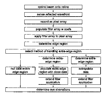
The creation and application of a spatial filter of the present invention is
shown in flowchart form in FIG. 13. The spatial filter permits a recomputation
of the
brightness of each pixel in a bitmap using a weighted-averaging technique that
considers surrounding pixels. In a par6cular application herein described for
illustration and byway of example, the spatial filter is designed to: yield a
maximum
value when centered on valid data; reduce an effect of individual bright
pixels or
small groups thereof; nomlalize background levels; smooth valid data profiles;
and
simplify the task of extracting the valid data from background noise or haze.
The spatial filter is embodied in a software package 261 resident on a
processor 262, which in tum is part of the wavefront analyzer 26 (FIG. 1)..
Another
software package 263 resident on the processor 262 determines aberrations in
the
eye based upon the filtered image data, as discussed in application Serial No.
09/566,668.
. A fifter employed in one embodiment of the present invention is square (n x
n) and includes real values (positive and negative) assigned to each pixel.
The filter
is designed to be optimally matched to images obtained from eyes with high,
yet
measurable, levels of aberration. By way of example, a cross-section through
the
filter is illustrated with reference to FIG. 8. An effect of applying such a
fitter
improves an image 500 such as illustrated with reference to FIG. 9 to the
image
500' illustrated in FIG. 10, by way of example, a cleaner image and one that
is
easily processed for identification and computation of cluster centroids. By
applying
the filter, images that would otherwise be deemed too noisy or of insufficient
quality
to process, can now be processed and desired wavefront information computed.
To illustrate the effect of applying this filter, a two-dimensional vari:ant
is applied to the image of FIG. 11 to yield the output of FIG. 12. An analysis
of
exemplary FIG. 11 yields several insights:
1. Valid data exist around locations 26, 93, 161, and 175 and are of an
expected profile (i.e., the same profile as the filter).
2. Noise spikes of varying magnitude are present at 10, 17, 129, 134,
139, 144, and 181. (Note: those between 129 and 144 inclusive are wider
and are not truly spikes.)
CA 02377852 2006-05-23
24
3. The background level varies across the plot.
It may be seen, therefore, that it is not possible to define a single
threshold
that would be exceeded by the valid data intensities and yet not by the noise
and
other unwanted data such as the high background levels in the middle of the
plot.
The result of applying a two-dimensional spatial filter Ci.e., one similar to
the profile
shown in FIG. 8) to these data is shown in FIG. 12. Note thatwith a two-
dimensional
filterthe coefficients at the edges have less effect than with a three-
dimensional filter,
and so the magnitudes of the coefficients need to be increased accordingly. In
this
parficular case the negative values at the edges need to be more negative.
In FIG. 12 the highest values correspond.to the valid data locations. The
problem of varying background levels has been removed and the use of a simple
threshold can uniquely identify the iocations of valid data. '
The three-dimensional filter (with coefficients at n x n x y locations) works
in
a very similar manner to yield the intensities shown in FIG. 10 from those in
FIG. 9.
The procedure by which the spatial filter is applied will now be describ-~!d:
1. The filter is created by populating an n x n array with the required
coefFicients. Note that a square array is used, but the coefficients are r.uch
that it is approximately radially symmetric.
2. The new intensities are'computed at all locations from a combination
of the fifter coefficients and the original intensities using the following
exemplary iterative approach:
initialize new intensities to 0
for all x locations (!x) to process in image
... for all y locations (1y) to process in image
. . . . . . for filter x offset (Fx) from -n/2 to +n/2
. . . . . . . . . for filter y offset (Fy) from -n/2 to +n12
. . . . . . . . . . . . newXaddress = lx + Fx
. . . . . . . . . . . . newYaddress =1y+ Fy
. . . . . . . . . . . . delta Intensity = filter coefficient(Fx,Fy) * Original
Intensity
(newXaddress, newYaddress)
. . . . . . . . . . . . new intensity(lx,ly) = new intensity(lx,/y) + delta
intensity
... ... ...end
. ... end
...end
end
CA 02377852 2006-05-23
Note the terminology "for all x locations (lx) to process an image." It is not
possible to apply the spatial filter in the normal way when the target pixel
is closer
than n/2 pixels to an edge, since the algorithm would aftempt to address
nonexistent
data.
There are a number of ways to address this:
1. Zero or null out all data within this edge region.
2. Allow filter to run all the way out to the edge and for data beyond the
image assuming that it is of the same intensity as the data closest
to this location at the edge of the image.
3. Extrapolate beyond the image (linearly or otherwise) to compute data
so that the filter may be used out to the edge of the image.
The most robust of these is to null the edge data.
In summary, by applying filters of the kind described here, images that would
otherwise be deemed too noisy or of insufficient quality to process can be
processed and the required wavefront information computed.
In the foregoing description, certain terms have been used for brevity,
clarity,
and understanding, but no unnecessary limitations are to be implied therefrom
beyond the requirements of the prior art, because such words are used for
description purposes herein and are intended to be broadly construed.
Moreover,
the embodiments of the apparatus illustrated and described herein are by way
of
example, and the scope of the invention is not limited to the exact details of
construction. -
Having now described the invention, the construction, the operation and use
of preferred embodiment thereof, and the advantageous new and useful results
obtained thereby, the new and useful constructions, and reasonable mechanical
equivalents thereof obvious to those skilled in the art, are set forth in the
appended
claims.