Note: Descriptions are shown in the official language in which they were submitted.
CA 02379175 2002-O1-15
WO 01/05448 PCT/CA00/00822
CONDUIT FOR A MECHANICAL CIRCULATORY DEVICE
FIELD OF INVENTION
The present invention relates to mechanical circulatory devices, and in
particular to a
conduit for a mechanical circulatory device.
BACKGROUND OF THE INVENTION
Mechanical Circulatory Devices (MCDs) such as artificial hearts, Ventricular
Assist
Devices (VADs) and other blood circulating systems have become increasingly
recognized as
life saving devices for patients whose heart is diseased or has been injured
by trauma or heart
attack or other causes. VADs in particular, are recognized as a major life
saving modality for
assisting patients who suffer from congestive heart failure.
MCDs must be connected to the natural blood circulation system of the body
such as
the heart and aorta. When designing an artificial heart or VAD, the inflow and
outflow
conduits are one of the most critical components. The conduits generally need
to deal with a
pulsatile or with a non-pulsatile flow, as well as with the flow negative
pressures created by
the MCD. The artificial conduit must function within or outside the host
patient's body. It
must not introduce or allow the entry of bacterial or other contamination into
the host's body
or circulatory system. If the conduit does not fulfil these requirements, it
may cause
thrombosis pannus formation, blockage, twisting, knocking, and pulling or
compressing the
heart and adjacent organs.
Almost all blood conducting devices exhibit some degree of thrombus (blood
clot
formation). Thrombosis is a multifactorial phenomenon. Two major factors are
the blood
flow pattern and the properties of the material in contact with the blood.
Research shows that
the major causes of clotting in the current blood flow conduits are the use of
thrombogenic
materials and of designs which create undesired flow patterns such as
turbulence, separation,
recirculation, stasis (pooling), and high and very low shear stresses. Among
specific factors
CA 02379175 2002-O1-15
WO 01/05448 PCT/CA00/00822
related to the above, undesired flow patterns are often generated by the
existence of crevices
or the lack of smoothness on interior surfaces or at joints in the conduits.
The risk of
twisting and folding of the conduits can be extremely dangerous.
Another persistent problem with current conduits relates to their durability.
Compression, tensile and torque forces act on the conduits, and current
conduits have an
insufficient fatigue resistance to these forces and are prone, to varying
degrees, to be
distorted. Moreover, suction generated by the MCD exposes the conduit to a
negative
pressure, which causes it to collapse.
Accordingly, there is a need of use of a conduit which provides sufficient
strength and
durability to prevent crevices or deformation where stress is exerted. In
order to use such a
strong conduit, there is a need to provide a suitable coupling to connect such
a conduit to the
MCD or a natural blood circulatory system. Flexibility, angulation, size and
orientation of a
conduit are all important factors that have to be considered in designing a
conduit that is
optimal in terms of performance, compression exerted on adjacent organs such
as the lungs,
heart, great vessels and displacement of the heart. The human chest anatomy,
with various
sizes and types for different bodies, is also one of the factors, dictating
how these factors have
to be considered in achieving anatomical fitness. The coupling needs to
provide smooth
internal transition between coupled components to reduce turbulence in the
blood flow.
Therefore, there is a need of a conduit assembly which allows use of a conduit
which
provides sufficient strength and durability, and also provides flexibility in
positioning such a
conduit and smooth internal transition between coupled components.
SUMMARY OF THE INVENTION
According to one aspect of the invention, there is provided a conduit assembly
for
attachment to an MCD having an orifice surrounded by an orifice rim and a
blood bag for
forming a blood chamber in the MCD. The blood bag has an open end extends
through the
orifice. The conduit assembly comprises a tube, a coupling and a washer. The
tube is
2
CA 02379175 2002-O1-15
WO 01/05448 PCT/CA00/00822
provided for conducting blood between a patient and the orifice of the MCD.
The tube has an
orifice end. The coupling attaches the orifice end of the tube to the orifice
of the MCD with
the open end of the blood bag folded over the orifice rim of the MCD. The
coupling is
movable between a rotatable position wherein the tube is rotatable relative to
the MCD, and a
locked position wherein the tube is immobile relative to the MCD. The washer
is placed
between the orifice end of the tube and the blood bag folded over the orifice
rim of the MCD.
The washer has an inner section which has a shape corresponding to a space
between the
orifice end of the tube and the blood bag folded over the orifice rim of the
MCD so as to
smooth the transition between the tube and the blood bag at the orifice of the
MCD to reduce
turbulence in blood flowing between the tube and the blood bag.
According to another aspect of the invention, there is provided a method for
implanting a circulatory apparatus in a patient. The apparatus comprises an
MCD having an
orifice surrounded by an orifice rim and a blood bag forming a blood chamber
in the MCD
and having an open end extending through the orifice, and a conduit assembly
for attachment
to the MCD. The conduit assembly comprises a curved rigid tube for conducting
blood
between a patient, the tube having orifice end; a coupling for attaching the
orifice end of the
tube to the MCD, the coupling being movable between a rotatable position
wherein the tube
is rotatable relative to the MCD, and a locked position wherein the tube is
immobile relative
to the MCD. The method comprises folding the open end of the blood bag over
the orifice
rim of the MCD; providing a washer on the blood bag folded over the orifice
rim of the
MCD, the washer having an inner section which has a shape corresponding to a
space
between the orifice end of the tube and the blood bag folded over the orifice
rim of the MCD
so as to smooth the transition between the tube and the blood bag at the
orifice of the MCD to
reduce turbulence in blood flowing between the tube and the blood bag;
attaching the orifice
end of the tube to the orifice rim through the washer and the blood bag as
being folded over
the orifice rim of the MCD with the coupling in the rotatable position;
positioning the MCD
relative to the patient; rotating the tube until a desired position of the
tube relative to the
patient is achieved; and moving the coupling to the locked position.
3
CA 02379175 2002-O1-15
WO 01/05448 PCT/CA00/00822
BRIEF DESCRIPTION OF THE DRAWINGS
The present invention will be further understood from the following detailed
description, with reference to the drawings in which:
Figure 1 is a schematical side view of a VAD in use to which an embodiment of
the
present invention is applied;
Figure 2A illustrates a lateral, exploded view of an inflow conduit assembly
in
accordance with an embodiment of the invention;
Figure 2B illustrates a lateral, exploded view of an outflow conduit assembly
in
accordance with an embodiment of the invention;
Figure 3 is a lateral cross-section of the tip body of the inflow conduit
assembly in
Figure 2A;
Figure 4A is a lateral view of the valve assembly in accordance with an
embodiment
of the present invention;
Figure 4B is a lateral view of a modified tissue valve;
Figure 4C is a perspective view of the valve enclosure in accordance with the
embodiment in Figure 4A;
Figure 4D is a bottom cross-sectional view of the outflow suture assembly in
accordance with the embodiment in Figure 4A;
Figure 4E is a cross-sectional view of the outflow suture assembly in Figure
4D;
Figure 4F is a lateral cross-sectional view of the inflow suture assembly in
accordance with
the embodiment in Figure 4A;
Figure SA is a perspective view of the inflow end of the valve assembly,
depicting the
suturing technique, in accordance with an embodiment of the invention;
Figure SB is a perspective view of the outflow end of the valve assembly,
depicting
the suturing technique, in accordance with an embodiment of the invention;
Figure 6 is a partial cross-sectional view of the inflow elbow conduit; and
Figure 7 is a partial cross-sectional view of another inflow elbow conduit.
Similar references are used in different figures to denote similar components.
4
CA 02379175 2002-O1-15
WO 01/05448 PCT/CA00/00822
DETAILED DESCRIPTION OF THE INVENTION
Mechanical Circulatory Devices (MCDs) include artificial hearts and
Ventricular
Assist Devices (VADs). An artificial heart is used in place of a natural
heart. A VAD is used
where a patient's natural heart that is diseased or injured is still partially
functioning. A VAD
is connected to such a natural heart and assists its functioning. Hereinafter
the present
invention is mainly described referring to VADs. However, the invention may be
also
applied to artificial hearts, other than those aspects respecting to
connection with a natural
heart.
Figure 1 shows a VAD 2 in use to which an embodiment of the present invention
is
suitably applied. The VAD 2 has a blood chamber 5 having an inflow orifice 6
and an
outflow orifice 7. At the inflow orifice 6, an inflow conduit assembly 100 is
provided to
connect the blood chamber 5 to a natural heart 1 of a patient. At the outflow
orifice 7, an
outflow conduit assembly 200 is provided to connect the blood chamber 5 to the
thoracic
aorta 3. The outflow conduit assembly 200 may be connected to a different part
of the blood
circulation system.
In operation, blood is pumped from the heart 1 into the blood chamber 5 of VAD
2
through the inflow conduit assembly 100. The VAD 2 then pumps the blood out of
the blood
chamber 5 into the thoracic aorta 3 through the outflow conduit assembly 200.
The inflow conduit assembly 100 comprises one or more conduits or tubes. A
proximal tube 175 that is connected to the inflow orifice 6 is made to be
rigid to provide
strength and durability of the conduit assembly 100. The proximal tube 175 is
preferably
curved to minimize interference with adjacent organs. In order to connect such
a curved rigid
tube 175, the conduit assembly 100 uses a coupling 180 which is movable
between a
rotatable position and a locked position. In the rotatable position, the
proximal tube 175 can
be rotated about its axis relative to the VAD 2 so that it can be positioned
at a desired angle to
avoid interference with adjacent organs. The axis of the proximal tube 175
curves as the tube
175 covers. The tube 175 rotates about the axis at the section engaging with
the VAD 2. The
5
CA 02379175 2002-O1-15
WO 01/05448 PCT/CA00/00822
VAD 2 may have one or more extension tubes. In that case, the tube 175 rotates
about the
axis at the section engaging with the nearest extension tube. After the angle
of the proximal
tube 175 is decided, the coupling 180 is moved to the locked position so that
the proximal
tube 175 is immovably locked relative to the VAD 2. Similar couplings are used
for
connection of other components as described later.
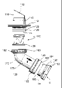
Figure 2A shows a lateral exploded view of the inflow conduit assembly 100 in
accordance with an embodiment of the invention. In this embodiment, the inflow
conduit
assembly 100 is connected to the VAD 2 by a coupling comprising a grand nut
180 and a
corresponding threaded connector 191.
The inflow conduit assembly 100 comprises two basic components, an apical tip
assembly 110 and an inflow elbow assembly 170. The apical tip assembly 110 and
the inflow
elbow assembly 170 are adapted to be connected together.
The inflow conduit elbow assembly 170 comprises an inflow elbow tube or
conduit
175 with a female threaded coupling or gland nut 160 at one end and a further
female
threaded gland nut 180 at the other end.
The inflow elbow conduit 175 is rigid and generally curved along its length.
Its shape
is dictated by the desire of minimizing interference with adjacent organs. The
inflow elbow
conduit 175 presented in Figure 2A has only one bend, however other shapes
such as an S-
shaped inflow elbow conduit may also be used.
On the VAD 2, an inflow plug 190 is mounted at the inflow orifice 6. The
inflow
plug 190 comprises an inflow port extension 6a having a rim 193 and a male
threaded
connector 191 for coupling to the grand nut 180. The inflow plug 190 also has
a flange 192
on its base surface near the inflow orifice 6. The flange 192 has a cross-
shaped outer surface.
It serves in gripping onto the inflow plug 190 when the grand nut 180 is being
tightened.
6
CA 02379175 2002-O1-15
WO 01/05448 PCT/CA00/00822
The gland nut 180 provides a rotatable union effect during the fitting
procedure. That
is, the gland nut 180 moves a rotatable position and a locked position. At the
rotatable
position, the grand nut 180 allows the inflow elbow conduit 175 to rotate
about its axis into
any rotated position relative to the VAD 2, when the VAD 2 is implanted. This
allows
flexibility in positioning of the conduit assembly 100. The positioning
flexibility is
advantageous, considering the difference in anatomies from patient to patient.
The
positioning flexibility is also useful during experiments performed on calves,
for example,
which present even a more dramatic difference in anatomy by comparison to the
human
anatomy. During the fitting of the VAD 2, an optimal position for the inflow
elbow conduit
175 is determined. Then, the inflow elbow conduit 175 is locked on in this
position by
tightening the gland nut 180 to the locked position. The optimal position of
the inflow elbow
conduit 175 defines a predetermined way into which the inflow conduit is to be
fitted within
its anatomical environment.
In order to achieve tight sealing, it is preferable that an end 183 of the
inflow elbow
conduit 175 closely mates the rim 193 of the inflow port extension 6a. One or
more sealing
rings may be used between the rim 193 of the inflow port extension 6a and the
end 183 of the
inflow elbow conduit 175 for tight sealing.
In order to facilitate the rotation of the inflow elbow conduit 175, it is
preferable that
the mating surfaces of rim 193 of the inflow port extension 6a and the end 183
of the inflow
elbow conduit 175 are smooth. It is also preferable that the rim 193 of the
inflow port
extension 6a and the end 183 of the inflow elbow conduit 175 are flat in a
plane
perpendicular to the axis of the inflow elbow conduit 175. It is also
preferable that they have
coincidental circular shapes so that the inflow elbow conduit 175 may be
rotated at any
desired angle. However, they may have unsmooth surfaces, such as a saw like
shape, or non-
circular shapes, such as octagonal shapes, as long as those surfaces can
achieve desired
sealing effects at different rotational angles, with or without the aid of
other sealing member.
The gland nut 180 may include bulges 182 on its external envelope surface. One
of
the purposes of the bulges 182 is to aid in gripping, by hand or by a wrench,
onto the nuts
7
CA 02379175 2002-O1-15
WO 01/05448 PCT/CA00/00822
180, during the fitting procedure. An alternative embodiment contemplates
small cavities or
holes instead of the bulges, to be gripped by a special instrument. Such an
embodiment may
be preferred because of the edges on the bulges 182.
The gland nut 180 is preferably manufactured to very high tolerances to ensure
an
extremely smooth seam between the component pieces. A biolization coating is
added, to
make the seam as non-thrombogenic as possible.
As shown in Figure 6, it is common that the VAD 2 uses an elastic bag 8 to
form the
blood chamber 5. It is preferable to extend the open end 9 of the elastic bag
8 over the rim
193 of the inflow port extension 6a to prevent leakage of blood from, the open
end 9. The
bended open end 9 is held by the engaging surface 183 of the inflow elbow
conduit 175. In
order to provide a constant blood flow, the inner diameter of the elbow
conduit 175 is
adjusted to be smaller than that of the inflow plug 190 for twice of the
thickness of the elastic
bag 8. By adjusting the inner diameter of the elbow conduit 175, the inner
wall of the elbow
conduit 175 may be aligned with the inner surface of the elastic bag 8.
However, in this
arrangement, due to the thickness of the elastic bag 8, the elastic bag 8
creates a ring space
185 having a semi triangle cross sectional shape is created at the bended
corner of the elastic
bag 8. This space 185 tends to cause turbulence in the blood flow. In order to
prevent such
turbulence, it is preferable to provide a washer 186 to mask the space 185.
As shown in Figure 7, the washer 186 preferably has an outer sleeve portion
186a to
form a port cap 187. The port cap 187 provides protection for the elastic bag
8.
The port cap 187 has a cap shoulder 186b extending from the port cap 187. The
end
183 of the inflow elbow conduit 175 has a corresponding surface 183b. Thus,
the port cap
187 provides alignment to the inflow elbow conduit 175 and the inflow port
extension 6a by
mating the cap shoulder 186b with the corresponding surface 183b of the inflow
elbow
conduit 175.
8
CA 02379175 2002-O1-15
WO 01/05448 PCT/CA00/00822
Referring back to Figure 2A, the apical tip assembly 110 comprises a tubular
tip body
112, a skirt 118 and an end section 120. The tubular tip body 112 is designed
for insertion
into the heart of the patient. The end section 120 is designed to stay outside
the heart. The
tip assembly 110 is sutured to the heart by the skirt 118.
Figure 3 is a lateral cross-sectional view of the tip body 112. The tip body
112 has a
tip section 11 S. The tip section 115 is preferably rigid for insertion into
the natural heart 1
through the heart muscle as shown in Figure 1. The length H of the tip body
112 is selected
to be long enough to protrude the cardiac tissue of the heart wall, but not
interfere with the
heart muscle pumping action.
For proper functioning, the length H of the tip section 115 is preferably
larger than the
thickness of the heart wall through which it penetrates. If the tip body 112
is too short and it
does not protrude the heart wall, the heart muscle tissue surrounding the open
end of the tip
body 112 grows and closes over the opening of the tip body 112, thereby
blocking the flow of
blood from the heart. Based on human and animal experiments and observations,
this length
H is preferably between 1.5 and 3.5 cm. The inside diameter of the tip body
112 must allow
sufficient blood flow to pass with acceptable velocities. If it is too slow,
the blood flow can
cause low washout. If it is too fast, the blood flow is disturbed causing
turbulence. In order
to let sufficient flow discharge for various body sizes and activities, a
diameter D, of 13 to 30
mm is recommended. The smaller end of the range may be suitable for small body
sizes, and
the larger one for larger body sizes.
The protruding tip section 115 has the above advantages. However, it tends to
cause
blood pooling around the protruding tip section 115. That is, around the tip
section 115 near
the hart wall, the blood flow becomes stagnant. In order to reduce such blood
pooling, it is
preferable to provide the tip section 115 with drainage holes 114 on its wall.
The size of the
holes 114 is such that the blood flows there through without clotting. It is
preferably
approximately 3 mm. The holes are preferably spaced approximately at a regular
distance
around the circumference of the tip section 115. They may be provided
approximately at
9
CA 02379175 2002-O1-15
WO 01/05448 PCT/CA00/00822
every centimetre around the circumference of the tip section 115. The
provision of the holes
114 prevents or reduces the risk of stasis and thrombosis.
It is preferable that the open end 113 of the tip section 11~ is angled
relative to the
axis of the tubular tip body 112. Thus, the tip section 115 has a longest side
1 I Sa having a
length ha and a shortest side 11 Sb having a length hb, as shown in Figure 3.
If the open end
113 of the tip section 115 is flat in a plane perpendicular to the axis of the
tip section 115, the
septum wall separating two blood chambers of the natural heart may interfere
the flow of
blood, and it could totally close the opening of the tip section 115. By
angling the open end
113, such interference by the septum wall can be avoided.
When implanting the VAD assembly, the tip section 115 is fitted to rest with
the
longest side 115a against the septum wall of the heart. Thus, the longest side
l I5a is defined
as the septum wall of the VAD assembly. The shortest side 115b of the tip
section 115 is
defined as the free wall.
In order to properly align the tip section 115, the tip assembly 110 further
includes a
male threaded connection 120 which form a union coupler with the female
threaded gland
nut 160 of the inflow elbow assembly 170 as shown in Figure 2A. The grand nut
160 is
similar to the gland nut 180 described above. By the gland nut 160 and the
male threaded
connection 120, the tubular tip body 1 I2 and the inflow elbow conduit 175 are
rigidly fixed
to one another in any relative angular position. The gland nut 160 may include
bulges 162 on
its external envelope surface, similar to the bulges 182 on the gland nut 180.
The skirt 118
also contributes to the proper alignment of the tip section 115 as described
below.
It is preferable that the tip body 112 accommodates a valve inset or assembly
140 to
regulate the blood flow as described below. In order to accommodate the valve
assembly 140
without generating disturbance in the blood flow, it is preferable that the
tubular apical tip
body 112 is cylindrical and presents a variable internal cross-section such
that an essentially
constant blood flow diameter is achieved when all parts of the conduit
assembly are fitted
together. The tip body 112 may comprise two sections of internal diameters D~
and D2,
CA 02379175 2002-O1-15
WO 01/05448 PCT/CA00/00822
respectively, as shown in Figure 3. An internal ridge 116 may be provided to
achieve the
change in the internal cross-section. Referring also to Figure 2A, the valve
assembly 140 has
an external diameter smaller than DZ but larger than D,. Thus, it slides into
the enlarged
elongated cylindrical opening within the apical tip body 112 up to the ridge
116. Preferably,
the internal diameter of the valve assembly 140 is approximately equal to D,,
for achieving an
essentially constant blood flow diameter. This is important for the prevention
of clot
formation in the conduit. From literature and experimental studies, it is seen
that an absolute
blood flow diameter close to 23 mm is well suited to optimize the prevention
of effects
leading to clotting.
The end section 120 of the tip body 112 may be provided with a hexagonal outer
cross
section 122. The hexed region 122 is intended to provide stability while
fitting the VAD 2
inside the patient. Stability may be provided through a wrench action for
example, so that the
torque applied to the natural heart 1 during fitting is minimized.
The apical tip assembly 110 is attached to the natural heart 1 by means of the
skirt
118. The skirt 118 is mounted on the tip body 112 between the tip section 115
and the end
section 120. The material from which the skirt 118 is manufactured is a
flexible material,
with tissue compatible characteristics. Many materials presenting such
properties are known
in the art. A commonly used material is a woven polyester velour. The shape of
the skirt 118
may be circular or any other shapes. In a preferred embodiment, the skirt 118
is made of a
flexible but strong material, it has approximately 1 to 12 cm in width. It is
glued to the tip
body 112 and sutured in place to the heart muscle inside the heart. The
procedure of suturing
the skirt 118 is similar to that known in the art as ventricular apical
cannulation. The skirt
118 is sufficiently flexible to conform to the curvature of the natural heart
1 and can be
pierced with relative ease by a surgical needle. Once sutured in place, the
heart tissue will
grow and surround the skirt 118, thus making an extremely strong bond.
In the embodiment of Figure 2A, the inflow conduit assembly 100 presents a
completely rigid structure to blood flowing through it. The use of a rigid
structure prevents
11
CA 02379175 2002-O1-15
WO 01/05448 PCT/CA00/00822
the conduit from collapsing, breaking or twisting under the various
compression, tensile,
torque forces and negative pressures exerted upon it. While the embodiment of
Figure 2A
has a completely rigid structure, it may also include an elastic tubular
member when less
pressures are exerted, e.g., between the tip assembly 110 and the elbow
assembly 170. A
diseased heart is generally swelled. Such a diseased heat tends to shrink as
the disease is
being cured. The elastic tubular member absorbs such changes in the size of
the heart, and
maintains the proper connection between the heart and the VAD 2.
As to the material of rigid components, it is preferable to use titanium for
several
reasons. There is evidence to support the idea that the use of titanium
provides the conduit
systems 100 and 200 with non-thrombogenic properties. Specifically, titanium,
when
exposed to oxygen, becomes titanium oxide, which is also believed to be non-
thrombogenic.
In order to improve the blood compatibility of titanium oxide, the interior
surface of the
conduit systems 100 and 200 may be coated with a gelatin coating. This
technique is known
as biolization.
Titanium is one of the strongest metals for its weight. It has proven to be
durable,
extremely strong and resistive to stress. Therefore, the use of titanium
allows for the
manufacturing of very thin conduits, of reduced size and small weight. The use
of titanium
for over 60 years in humans for such things as hip joints, finger joints,
orthopaedics and
prosthesis shows evidence of tissue compatibility and non-thrombogenic
properties.
Referring now to Figure 2B, the outflow conduit assembly 200 comprises two
basic
components, an outflow conduit 210 and an outflow elbow assembly 270, adapted
to be
connected together. Blood flows through the assembly 200 as shown by arrow B.
In the embodiment presented in Figure 2B, the outflow conduit 210 comprises a
tubular conduit section 21 S, having an outflow end 217 and an inflow end 218.
The outflow
end 217 is adapted to be sutured onto an artery or similar vessel. The inflow
end 218
comprises a female threaded coupling or gland nut 260. The gland nut 260 is
similar to the
gland nut 180 of the inflow conduit elbow assembly 100 in Figure 2A. The
conduit section
12
CA 02379175 2002-O1-15
WO 01/05448 PCT/CA00/00822
215 is made of a flexible, tissue compatible material, such as a woven
polyester velour. It is
manufactured sufficiently long so that it can be cut at a desired length
during the fitting
procedure, either in a patient, or in an animal during experimental studies.
Referring to
Figure 1, the outflow end 217 of the conduit section 215 is shown sutured to
the thoracic
S aorta 3.
The outflow conduit elbow assembly 270 is rigid and similar to the inflow
elbow
assembly 170 in Figure 2A. In the embodiment presented in Figure 2B, the
outflow conduit
elbow assembly 270 includes a gland nut 280 at its inflow end, and a male
threaded
connector 273 at the other end. The gland nut 280 is similar in function to
the gland nut 180
in the inflow conduit elbow assembly 170 in Figure 2A. The gland nut 280 is
adapted to be
coupled to a plug provided on the VAD 2 at the outflow orifice 7, similar to
plug 190. Thus,
the outflow elbow assembly 270 may be rotated around the axis of the elbow
conduit, and
then fixed at a desired angle relative to the VAD 2. The male threaded
connector 273 may
include a hexagonal region 274 for gripping while tightening the gland nut
260. In addition,
the elbow assembly 270 may further include an enlarged elongated cylindrical
opening within
it, to receive a valve assembly 240.
Preferably, the length and orientation of the components of the conduit
systems 100
and 200 are chosen so as to minimize compression on adjacent organs and great
vessels,
once the MCD is implanted within an anatomical environment. Optimal sizes,
geometries
and orientations of the various parts of the conduit systems 100 and 200 may
be determined
based on study of both the literature and the anatomy of the human chest, as
well as taking
actual measurements during both intra-operative procedures and from fresh
cadavers.
The outflow elbow assembly 270 may also include an enlarged elongated
cylindrical
opening similar to that in the apical tip assembly 110 shown in Figure 2A for
receiving a
valve assembly 240.
Referring back to Figure 2A, the valve assembly 140 having a one-way valve is
provided in the tip assembly 110 of the inflow conduit assembly 100. The one-
way valve
13
CA 02379175 2002-O1-15
WO 01/05448 PCT/CA00/00822
assembly 140 prevents back flow of the blood from the blood chamber 5 to the
heart.
Traditionally, such a one-way valve was provided at the inflow orifice 6.
However, it often
generated undesired flow patterns in the blood flow. Compared to the inflow
port location,
within the conduit assembly 100, the blood flow is more stable. Thus, by
providing the valve
in the conduit assembly 100, the disturbance in the blood flow by the
provision of the valve is
reduced.
Similarly, the outflow conduit assembly 200 is provided with the valve
assembly 240
having a one-way valve in the elbow conduit assembly 270, as shown in Figure
2B. The
valve assemblies 140 and 240 may be identical to each other. By using
identical valve
assemblies for the inflow and outflow conduit assemblies 100 and 200, these
assemblies may
be inserted in either conduit. This leads to an easier, more effective,
fitting procedure. The
valve assemblies will be described hereinafter refernng only to valve assembly
140, for
simplicity.
Referring to Figures 4A, SA and SB, the valve assembly 140 comprises a valve
enclosure assembly 141 and a one-way modified tissue valve 300. The valve
enclosure
assembly 141 comprises a valve enclosure 145, an outflow suture assembly 142
and an
inflow suture assembly 150. The tissue valve 300 is sutured to the inflow
suture assembly
150.
The modified tissue valve 300 is preferably a tricuspid or tri-foliate, having
three
leaflets 302. Each leaflet 302 has a semi-triangle shape having a semi-
circular base end 303.
The base end 303 is sutured on the inflow suture assembly 150. The other two
ends 304, 305
of the leaflet 302 are free ends. Three leaflets 302 are provided so that each
free ends 304,
305 is located closely to the free end 304, 305 of the neighboring leaflet
302. When the
blood flow comes in the direction shown with the arrow B in Figure 4A, the
leaflets 302 open
the spaces between the free ends 304, 305 by bending along the blood flow.
When the blood
flow comes in the other direction, the leaflets 302 close the spaces between
the free ends 304,
305 to block the blood flow. Each leaflet 302 is preferably made of natural or
artificial tissue.
14
CA 02379175 2002-O1-15
WO 01/05448 PCT/CA00/00822
While blood flows through the tissue valve 300, the leaflets 302 hits the
walls of the
conduit within which the valve 300 is mounted by their natural movements as
dictated by the
blood flow. The tip of the leaflets 302 is therefore impeded and repeatedly
contacted against
the wall. This may wear, deformation, and eventually tear in the leaflets 302.
In order to reduce the impacts on the leaflets 302, it is preferable to
provide a movable
wall 152. The movable wall 152 is attached onto the enclosure 145 by suturing
assemblies
142 and 150.
The movable wall 152 is preferably made of a natural or artificial tissue
material.
Such material is preferably grafted on a flexible, blood compatible fabric
156, such as woven
polyester velour, for further attachment within valve assembly 140 .
Figure 4B shows the movable wall 152. The movable wall 152 has a wall annulus
157 bordered by a sinusoidal wall inset 158. The wall inset 158 forms three
peaks 159,
corresponding to the three leaflets 302 of the tissue valve 300. The wall
inset 158 is made of
a natural or artificial tissue material. Thus, the wall inset 158 may expand
by the blood flow.
When blood flows through the tissue valve 300 as shown by arrow B in Figure
4A,
the movable wall 152 moves naturally as the wall inset 158 expands by the
blood flow. The
movement of the movable wall 152 occurs predominantly in a radial direction as
the wall
inset 158 is supported by the valve enclosure 145 and the wall annulus 157.
The maximum
radial deflection occurs at the points farthest from the center, which are at
the peaks 159 of
the wall inset 158. Thus, the tip of the leaflets 302 of the tissue valve 300
does not touch to
the movable wall 152.
Figure 4C shows the valve enclosure 145. The valve enclosure 145 comprises a
cylindrical body defined by an inflow base ring 146 in the plane of the
cylinder, an outflow
base ring 147 forming a flange at the base of the cylindrical body and three
legs 149 joining
the two rings to define three side windows 148. Preferably, the windows 148
are identical,
located approximately 120° apart.
CA 02379175 2002-O1-15
WO 01/05448 PCT/CA00/00822
When assembled, the windows 148 provides flexibility to the valve assembly
140.
They also allow use of a dead space between the outer diameter of the valve
enclosure 145
and the inner diameter of the tip body 112 (Figure 3) in which the valve
assembly 140 is
inserted, and prevent friction between the moving parts of the tissue valve
300 and the
movable wall 152 of the valve enclosure 145 as described above.
Referring to Figure 4A, in accordance with a preferred embodiment of the
invention,
in assembling the valve assembly 140 the movable wall 152 is sutured onto the
valve
enclosure assembly 141 by positioning the three peaks 159 of the sinusoidal
wall inset 158 in
the centers of the three windows 148 of the valve enclosure 145, respectively.
A preferred
embodiment also features a vertical distance gap 19 between the peaks 158 and
the outflow
end of the valve enclosure 145, for allowing the wall inset 158 freedom in
moving vertically,
unconstrained by the valve enclosure 145. Since the maximum deflection of the
wall inset
158 occurs at the peaks 159, this fashion of mounting the valve allows it to
function in its
normal free state, while also mounted in a rigid structure.
The inflow and outflow base rings 146 and 147, as well as the vertical legs
149, are
provided with holes 20, 25 and 30, respectively, for suturing the movable wall
152 onto the
valve enclosure assembly 141 in a manner which will be described below.
Figure 4A shows the inflow suture assembly 150, which comprises an inflow
suture
ring cover 151 that is attached to the inflow base ring 146 of the valve
enclosure 145.
The inflow base ring 146 is a rigid ring. It has holes 20 for stitching the
tissue valve
300 (Figures 4A, SA). The inflow suture ring cover 151 corresponds to the
inflow base ring
146 of the valve enclosure 145 (Figure 4C). The inflow base ring 146 is
wrapped by the
suture ring cover 151 around it. The inflow suture ring cover 151 is made from
a blood
compatible fabric material to be stitched around the inflow base ring 146 of
the valve
enclosure 145 which provides an inflow suturing support for the movable wall
152.
16
CA 02379175 2002-O1-15
WO 01/05448 PCT/CA00/00822
Turning back to Figure 4A and referring to Figures 4D and 4E, the outflow
suture
assembly 142 is sutured onto the outflow base ring 147 of the valve enclosure
145, forming a
flange 26. Flange 26 is designed to fit smoothly into the inflow elbow
assembly 170 of
Figure 2A. The two ends of the valve assembly 140 are distinguished by the
flange 26. The
flange 26 is matingly adapted only in one direction into the tip body 112 of
the apical tip
assembly 170. Thus, the valve assembly 140 can be installed within assembly
100 in only
one way. This feature eliminates the risks of wrongly inserting the valve
assembly 140 into
the conduits in a wrong direction. The provision of the flange 26 leads to an
easier, safer
fitting procedure.
The outflow suture ring 143 shown in Figure 4D is a rigid ring, of mating size
with
the outflow base ring 147 of the valve enclosure 145. The outflow suture ring
143 provides
an outflow suturing support for the movable wall 152 in assembling the valve
assembly 140.
The outflow suture ring 143 has holes 15 that register with holes 25 of the
outflow
base ring 147. The movable wall 152 is sutured to the outflow suture ring 143,
which is in
turn sutured to the outflow base ring 147. Thus, the movable wall 152 is
secured to the valve
enclosure 145 by the outflow suture ring 143.
The valve enclosure 145 also has holes 30 on the legs 149 near the outflow
suture
assembly 142, as shown in Figures 4A and 4C. These holes 30 allow suturing of
the
movable wall 152 to the valve enclosure 145.
Referring to Figures 4A-4E, the valve assembly 140 is assembled as follows:
First, the valve enclosure assembly 141 is assembled, by attaching the inflow
suture
assembly 150 and the outflow suture assembly 142 to the valve enclosure 145.
As described
above, the inflow suture assembly 150 comprises the inflow suture ring cover
151 wrapped
around the inflow base ring 146 of the valve enclosure 145. The outflow suture
assembly 142
comprises the outflow suture ring 143 with the outflow suture ring cover 144
wrapped around
it.
17
CA 02379175 2002-O1-15
WO 01/05448 PCT/CA00/00822
Second, the movable wall 152 is placed inside the assembled valve enclosure
assembly 141 with its inflow end facing the inflow side of the valve enclosure
assembly 141.
Third, the inflow end 153 of the grafted fabric 156 of the movable wall 152 is
stitched
S on the circumference of the inflow suture support provided by the inflow
suture ring cover
151 of the inflow suture assembly 150.
Finally, the outflow end 154 of the grafted fabric 156 of the movable wall 152
is
stitched on the circumference of the outflow suture support provided by the
outflow suture
ring cover 144 of the outflow suture assembly 142.
In a preferred embodiment, the suturing technique is such that the suturing
material,
which may be thrombogenic, does not come into contact with the blood flowing
through the
conduit assembly. Thus, stitching occurs only on surfaces that do not contact
the main blood
flow stream, when in operation. A method of assembling the valve assembly 140
and its
subassemblies, in accordance with such a preferred embodiment, is described in
detail next.
Assembling the Outflow Suture Assembly:
Referring now more specifically to Figures 4D and SB and as previously
described,
the outflow suture assembly 142 provides a suturing support on the outflow end
of the valve
enclosure assembly 141, for the movable wall 152. The support is provided
through the
outflow suture ring cover 144 which has to be wrapped and sutured around the
outflow suture
ring 143. The ring of sutures around the periphery of the outflow suture
assembly 142 thus
obtained is herein denoted by I.
According to a preferred embodiment of the invention, the assembling of the
outflow
suture assembly 142 comprises the following substeps:
1. A rectangular strip of a biocompatible fabric is cut at a suitable size,
for forming the
outflow suture ring cover 144.
18
CA 02379175 2002-O1-15
WO 01/05448 PCT/CA00/00822
2. The fabric forming the outflow suture ring cover 144 is wrapped around and
held
tight against the inside surface of the outflow suture ring 143 with the
excess material
lying outside the outflow suture ring 143.
3. Using a surgical suture, a first stitch is made at the edge of the fabric
forming outflow
suture ring cover 144. When passing the needle, one should preferably make
sure that
it is directly against the outer surface of the outflow suture ring 143
through both the
upper and lower layer of the outflow suture ring cover 144. The initial knot
is started
by passing the needle through the two layers twice in the same location.
During the
second pass, while the needle is still part way through the outflow suture
ring cover
144, the suture line is wrapped around the needle twice, the needle is pulled
through
the double loop, and then the knot is tightened.
4. A stitch is passed back through both layers of the outflow suture ring
cover 144 with a
stitch length of 5 +/- 1 mm, still being careful to have the needle directly
against the
outer surface of the outflow suture ring 143. Reversing the stitch direction,
a stitch is
made back through where the stitch initially started. When the stitch comes
through
the outflow suture cover ring 144, one makes sure the stitch passes between
the
previous stitch and the outflow suture ring 143 so that the suture line is not
cut by the
needle and the stitch remains tight.
5. A stitch is passed through both layers of the outflow suture ring cover 144
with a
stitch length of 7 +/- 1 mm.
6. With a stitch length of 5 +/- 1 mm, the direction is reversed and a stitch
is passed
through the outflow suture ring 143. A cover is made so that the suture
emerges near
the previous stitch. Again, for the reverse direction stitch, the needle is
passed
between the previous suture line and the outflow suture ring 143.
19
CA 02379175 2002-O1-15
WO 01/05448 PCT/CA00/00822
7. The sutures are continued with a stitch length of 5 +/- 1 mm, continuously
ensuring
that the outflow suture ring cover 144 stays tight against the inside surface
of the
outflow suture ring 143.
8. Once the outflow suture ring cover 144 has been sutured around the entire
circumference of the outflow suture ring 143, the outflow suture ring cover
144 is cut
to length such that the two edges of the outflow suture ring cover 144 abut
against
each other.
9. Two additional stitches are made across the gap in the outflow suture ring
cover 144,
then the suture is tied off with two double finishing knots, a final stitch is
passed
underneath the knot with each line and then cut the suture at the surface of
the outflow
suture ring cover 144 using the surgical scissors with the rounded cutting
edge.
10. The outflow suture ring cover 144 is cut 1.0 - 2.0 mm outside of the
periphery of the
completed ring of sutures.
11. Using a soldering iron set to 277 +/- 10°C, the seam is welded
along the two edges of
the outflow suture ring cover 144 starting at the interior surface and working
in the
radial direction, being careful not to touch the suture material which could
melt and
break upon contact.
12. In a similar manner, the soldering iron set to 277 +/- 10°C is used
to weld the seam
along the outer periphery of the outflow suture ring assembly 142.
Assembling the Inflow Suture Assembly:
Referring now more specifically to Figures 4A, 4C, 4F and SA and previously
described, the inflow suture assembly 150 provides a suturing support on the
inflow end of
the valve enclosure assembly 141. The support is provided by the inflow suture
ring cover
151 which has to be wrapped and sutured around the inflow base ring 146 of the
valve
CA 02379175 2002-O1-15
WO 01/05448 PCT/CA00/00822
enclosure 145. According to a preferred embodiment of the invention, the
assembling of the
inflow suture assembly 150 comprises the following substeps:
A) A first ring of sutures II is made in order to attach the inflow suture
ring cover 151 to
the inflow base ring 146 of the valve enclosure 145, by passing a surgical
suture
through the holes 20 in the inflow base ring 146. In detail, this may be done
as
follows:
1. A piece of uncrimped fabric shaped into a conduit having a diameter
approximately
equal to the diameter of the movable wall 152 and to the internal diameter of
the valve
enclosure 145, is fed through the valve enclosure 145 and wrapped around the
inflow
base ring 146 of the valve enclosure 145 such that 5.0 +/- 1.0 mm is hanging
over the
outside portion of the valve enclosure 145 and the remainder of the uncrimped
fabric
conduit is within the valve enclosure 145. In this embodiment, the uncrimped
fabric
conduit forms the inflow suture ring cover 151.
2. A cylindrical rubber stopper is placed into the valve enclosure's 145
inflow orifice to
hold the inflow suture ring cover 151 in place.
3. Using a surgical suture, the suturing of the inflow suture ring cover I 51
to the valve
enclosure 145 is started through one of the holes 20 in the inflow base ring
146.
Preferably, this stitch should start from the outside traveling towards the
inside.
4. The suture is then taken through the adjacent hole 20 from the inside out.
5. The two ends of the sutures are tied off with two double knots that are
made on the
inflow suture ring cover 151 in between two holes 20 on the inflow base ring
146 on
the external surface of the valve enclosure 145 .The sutures are tied off such
that there
are approximately equal lengths of suture on either side of the knot.
21
CA 02379175 2002-O1-15
WO 01/05448 PCT/CA00/00822
6. The knot is tightened so that it resides over one of the two holes 20 in
the inflow base
ring 146 that have just been tied off.
7. The next suture is started with the last hole 20 that was used coming from
the outside
in and then bringing the suture back out in the adjacent hole 20 in a similar
manner as
before.
8. Continue making sutures until all holes 20 are stitched.
9. Once the final knot has been tied around the periphery of the component,
the two
ends of the suture are left as they will be used later.
B) A second ring of sutures III is made in order to suture the inflow suture
ring cover 151
closed. where the two portions of the fabric conduit come together. In detail,
this may
be done as follows:
10. The fabric conduit making the inflow suture ring cover 151 is cut to the
same length
on the internal side of the valve enclosure 145 as the fabric is overhanging
on the
external side of the valve enclosure 145.
11. The suture line is passed underneath the external layer of the fabric
conduit so that it
emerges near the edge of the uncrimped fabric conduit using the longer portion
of the
suture left from point 9. Then, starting from the outside and working towards
the
center, a stitch is passed through both layers of the conduit uncrimped fabric
to start
the stitch.
12. As the stitch is being made, a knot is tied to complete each stitch. This
can be done
by wrapping the suture line around the needle before the needle is completely
pulled
through the material.
22
CA 02379175 2002-O1-15
WO 01/05448 PCT/CA00/00822
13. The suturing is continued around the periphery of the uncrimped fabric
conduit with
3-5 mm long stitches located on average ~ 2.0 - 2.5 mm apart.
14. When crossing between two of the windows 148 in the valve enclosure 145 ,
a single
stitch is made, that starts at one edge of the window that has just been
completed,
which passes under the fabric conduit on the external side of the valve
enclosure 145,
and emerges at the edge of the next window 148 on the valve enclosure 145.
15. The suture is finished off by making a single stitch back down near the
original knot
at the bottom of the fabric conduit and then make two double knots, and then
feeding
the two ends of the suture through the fabric under the knot, and then cutting
the ends
of the suture.
16. Once the suture is completed around the base of the uncrimped fabric
conduit making
the inflow suture ring cover 151, a soldering iron at 277 +/-10°C is
used to weld the
seam shut, being careful not to contact the suture which could melt.
Assembling the Valve Enclosure Assembly:
Referring now more specifically to Figures 4C, 4D and SB , as previously
described,
the assembling of the valve enclosure assembly 141 consists in attaching the
inflow suture
assembly 150 and the outflow suture assembly 142 together. In this embodiment,
this is
accomplished by the suture technique described in detail below, the result of
which is a new
ring of sutures IV, obtained by passing a suture through the holes 15 in the
outflow suture
assembly 142 and the holes 25 in the outflow base ring 147 of the valve
enclosure 145.
1. The assembled outflow suture ring assembly 142 is positioned over the
outflow base
ring 147 of the valve enclosure 145, so that their sets of holes, 1 S and 25,
respectively,
overlap.
2. Using a surgical suture, the suture is started by passing a stitch through
the holes 15
in the outflow suture ring assembly 142 and through the holes 25 in the
outflow base
23
CA 02379175 2002-O1-15
WO 01/05448 PCT/CA00/00822
ring 147 of the valve enclosure 145 and then passing the other needle through
the
adjacent pair of holes 15, 25 in the outflow suture ring assembly 142 and the
outflow
base ring 147, respectively.
3. The two free ends are tied with two double knots and the resulting knot is
located
directly over one of the hole-pairs 15, 25.
4. One end of the suture is passed through the hole-pair 15, 25 where the last
knot was
located and the suture is brought back through the adjacent hole-pair 15, 25.
5. The two loose ends of the suture are tied off with two double knots and the
knot is
located directly over the holes 15, 25.
6. The suturing of the outflow suture ring assembly 142 to the valve enclosure
assembly
141 is continued through the eighteen hole-pairs 15, 25.
7. Once the suturing is completed, the suture line is passed down through the
last hole
of the valve enclosure 145 but not through the corresponding hole 15 of the
outflow suture ring assembly 142.The suture line is passed between the valve
20 enclosure 145 and the outflow suture ring assembly 142 to the outside edge
of the
components. The suture is tied off on the outside edge of the outflow suture
ring
assembly 142 so that the knot is not located on the flat bottom of the
component.
8. After the knot is tied, a stitch is passed under the knot and then the
suture is cut close
25 to the surface using the surgical scissors with the rounded cutting edge.
Assembling the Valve Assembly:
Referring now to Figures 4A, 4B, SA and SB and as previously described, the
final
steps in assembling the valve assembly are the suturing of the movable wall
152 inside the
assembled valve enclosure assembly 141. This involves three main steps, as
described next:
24
CA 02379175 2002-O1-15
WO 01/05448 PCT/CA00/00822
A) The movable wall 152 is placed inside the assembled valve enclosure
assembly 141
with its inflow end facing the inflow side of the valve enclosure assembly
141. In
detail, this can be accomplished as follows:
1. A suitable movable wall 152 as previously described is removed from sterile
water
and its grafted fabric 156, shaped into a conduit, is cut approximately 5
corrugations
above and below the tissue valve, 153, 154.
2. The movable wall 152 is then inserted into the assembled valve enclosure
assembly
141 such that each of the peaks 159 of the wall inset 158 lies centered within
the
window 148 of the valve enclosure 145 and the peak 159 of the wall inset 158
lies 1 -
2 mm above the outflow side of the window 148 of the valve enclosure 149. The
movable wall 152 is in the proper orientation when the peaks 159 of the wall
inset
158 are pointing towards that outflow suture ring assembly 142.
B) The inflow end 153 of the grafted fabric 156 is stitched on the
circumference of the
inflow suture support provided by the inflow suture ring cover of the inflow
suture
assembly 150 by creating a new ring of sutures V. In detail, this can be
accomplished
as follows:
3. The movable wall 152 is trimmed so that it is flush to the base surface of
the inflow
orifice of the valve enclosure assembly 141. The inflow orifice should
preferably be
approximately 1.5 corrugations 153 above the inset 158 of the movable wall
152, as
shown in Figure 4B.
4. While ensuring to keeping the fabric taut, using a surgical suture, the
stitch starts by
passing a suture from the outer periphery of the valve enclosure assembly 141,
through the inflow suture ring cover 151 and back out through the movable wall
152,
as close to the valve enclosure 145 as possible, and then a double knot is
made.
Preferably the knot should be located on the outside edge of the valve
assembly 140.
CA 02379175 2002-O1-15
WO 01/05448 PCT/CA00/00822
5. A continuous stitch, forming a new ring of sutures V, with stitches
approximately 5
mm long and 2 - 3 mm apart is made around the inflow periphery of the valve
enclosure assembly 141, making sure that the stitch lies on the upper surface
of the
valve enclosure assembly 141, for a total of approximately 30 -50 stitches.
S
6. Once the continuous stitch has returned to the starting position, it shall
be tied off with
the starting loose end using two double knots.
7. After the knot has been tied, both loose ends of the suture are passed
through the
uncrimped conduit, under the knot and then cut with the surgical scissors with
the
rounded cutting edge.
C) The outflow end of the grafted fabric 156 is stitched on the circumference
of the
outflow suture support provided by the outflow suture ring cover of the
outflow suture
assembly 142, by creating a new ring of sutures VI. In detail, this can be
accomplished as follows:
9. The valve assembly 140 is turned over and the outflow orifice of the
modified tissue
valued 152 is trimmed down flush to the outflow suture ring 143 surface using
the
scalpel, leaving approximately 2.5 graft corrugations 154 between the peaks
159 of
the wall inset 158 and the end of the fabric 156 of the movable wall 152.
10. Using a surgical suture a stitch is started by passing the suture line
from the outside of
the valve assembly 140, in through the outflow suture ring assembly 142 and
out
through the fabric 156 of the movable wall 152. This stitch should be made in
between the ring of suture IV holding the outflow suture ring assembly 142 to
the
valve enclosure 145 and the ring of suture I holding the outflow suture ring
assembly
142 together.
11. A continuous stitch, forming a new ring of sutures VI, is made with
stitches
approximately 5 mm in length and 2-3 mm apart around the outflow orifice (for
a total
26
CA 02379175 2002-O1-15
WO 01/05448 PCT/CA00/00822
of 30 - 50 stitches) ensuring that the stitch is made on the flat surface of
the outflow
suture ring assembly 142. Care must be taken to ensure the grafted fabric 156
conduit
is stretched to fit the valve enclosure 145.
12. The suture is finished in a manner similar to that described above for the
assembling
of the inflow suture assembly 150 in steps 7 - 9.
D) In this embodiment, the movable wall 152 is also attached midways to the
valve
enclosure, by suturing its grafted fabric to the legs 149 of the valve
enclosure, through
the holes 30 provided in the legs. In more detail, this can be accomplished as
follows:
13. The final sutures will be done through the holes 30 in the legs 149 of the
valve
enclosure 145.
14. A stitch is passed from the outside of the valve enclosure 145 , through
one of the
holes 30 and the modified valued conduit and then back out the adjacent hole
30.
15. The two free ends of the suture are tied with three double knots and then
the free
ends of the suture will be cut off leaving approximately 3 mm of length at the
end of
the lines.
16. Steps 14 -15 are repeated for all three sets of holes 30 in the legs 149
of the valve
enclosure 145.
E) The valve enclosure 145 is visually inspected, and stored in a container.
F) A small portion of the graft conduit 156 that was trimmed off at substep 3
(approximately 1 x 2 cm) is cut and placed this into the container with the
valve
assembly 140 for future bacteria cultures.
27
CA 02379175 2002-O1-15
WO 01/05448 PCT/CA00/00822
Although a suturing technique has been described above in detail, it will to
be
appreciated by one skilled in the art that this description only pertains to a
specific
embodiment of the invention. Other suturing techniques may be employed for the
assembling of the various components and for attaching the movable wall 152 to
the valve
enclosure assembly 141. Moreover, other methods of attachment known in the
art, such as
glueing, can be used in assembling the various parts of the valve assembly 140
together.
Turning back to Figure 2A, the valve assembly 140 is mounted on the inflow
conduit
assembly 100 by sliding its inflow end into enlarged elongated cylindrical
opening of the
apical tip assembly 110, and its outflow end with flange 26 into the inflow
elbow assembly
170.
Referring now to Figure 2B, as indicated above, the outflow valve assembly 240
is
identical in structure to the inflow valve assembly 140. The outflow valve
assembly 240 is
mounted into the outflow conduit assembly 200 by sliding its outflow end with
flange 27 into
the outflow conduit 210 and its inflow end into enlarged elongated cylindrical
opening of
outflow elbow assembly 270.
Numerous modifications, variations, and adaptations must be made to the
particular
embodiments of the invention described above, without departing from the scope
of the
invention, which is defined in the claims.
28