Note: Descriptions are shown in the official language in which they were submitted.
CA 02382197 2002-02-19
BYO 01/13094 PCT/US00/22754
-I-
METHODS FOR DETERMINING THE
PHYSIOLOGICAL STATE OF A PLANT
Field of the Invention
The present invention relates generally to methods for measuring a
photosynthetic parameter, and to methods for determining the physiological
state of a
plant.
Back ,round of the Invention
Photosynthesis in green plants takes place in two stages, the light reactions,
which occur only when plants are illuminated, and the dark reactions, which
can
occur in the absence or presence of light. In the light reactions chlorophyll
and other
pigments of the photosynthetic cells absorb light energy and conserve it in
chemical
form as the two energy-rich products adenosine triphosphate (ATP) and
nicotinamide
adenine dinucleotide phosphate (NADPH); simultaneously, oxygen is evolved. In
the dark reactions, the ATP and NADPH generated in the light reactions are
used to
reduce carbon dioxide to form glucose and other organic products.
In eukaryotic, photosynthetic cells, both the light and dark reactions take
place in the chloroplast. Chloroplasts are surrounded by a continuous outer
membrane. An inner membrane system encloses the internal compartment. Inside
the latter, and often connected to the inner membrane, are many flattened,
membrane-
surrounded vesicles or sacs, called thylakoids, which are either single, or
arranged in
stacks called gram. The thylakoid membranes contain all the photosynthetic
pigments of the chloroplast and most of the enzymes required for the primary
light-
dependent reactions. The fluid in the compartment surrounding the thylakoid
CA 02382197 2002-02-19
w0 01/13094 PCT/US00/22754
-2-
vesicles, the stroma, contains most of the enzymes required for the dark
reactions (i. e.
COZ fixation).
Light energy is absorbed by photosynthetic pigments located within the
thylakoid membranes. The primary light-absorbing pigment is chlorophyll.
Photosynthetic cells of higher plants always contain two types of chlorophyll.
One is
always chlorophyll a, and the second in many species is chlorophyll b. In
addition to
chlorophylls, the thylakoid membranes contain secondary light-absorbing
pigments,
together called the accessory pigments, which include various carotenoids. The
carotenoid pigments absorb light at wavelengths other than those absorbed by
the
chlorophylls and thus are supplementary light receptors.
The light-absorbing pigments of thylakoid membranes are arranged in
functional sets or clusters called photosystems. The clusters can absorb light
over the
entire visible spectrum but especially well between 400 to 500 and 600 to 700
nanometers (nm). All the pigment molecules in a photosystem can absorb
photons,
but a special subset of the molecules, housed in complexes of proteins and
cofactors,
called the 'photochemical reaction centers' in each cluster ultimately convert
the light
energy into chemical energy. Other pigment molecules, that function to funnel
light
into the reaction centers, are housed in light-harvesting complexes. They
function to
absorb light energy, which they transmit at a very high rate to the reaction
center.
There are two different kinds of photosystems: photosystem I (PS I), which is
maximally excited by light at longer wavelengths, and has a high ratio of
chlorophyll
a to chlorophyll b; and photosystem II (PS II), which is maximally activated
by light
below 680 nm, and contains relatively more chlorophyll b and may also contain
chlorophyll c. Photosystem I and Photosystem II are functionally linked by a
chain
of electron carriers, as shown in FIGURE 1.
When light quanta are absorbed by photosystem I, energy-rich electrons are
expelled from the reaction center and flow down a chain of electron carriers
to
NADP+ to reduce it to NADPH. This process leaves a deficit of electrons (an
electron hole) in photosystem I. This hole is, in turn, filled by an electron
expelled
by illumination of photosystem II, which arrives via a connecting chain of
electron
carriers, including a pool of about 6 plastoquinone molecules per reaction
center, the
cytochrome b6f complex and plastocyanin. The resulting electron hole in
photosystem II is filled by electrons extracted from water. This pattern of
electron
flow is usually referred to as the "Z-scheme". Additionally, absorbed light
can be
reemitted in the form of fluorescence.
CA 02382197 2002-02-19
w0 01/13094 PCT/US00/22754
-3-
The thylakoid membrane has an asymmetric molecular organization. The
electron-transferring molecules in the connecting chain between photosystem II
and
photosystem I are oriented in the thylakoid membrane in such a way that
electron
flow results in the net movement of H+ ions across the membrane, from the
outside
of the thylakoid membrane to the inner compartment. Thus photoinduced electron
flow generates an electrochemical gradient of H+ ions across the thylakoid
membrane, so that: 1 ) the inside of the thylakoid vesicles becomes more acid
than the
outside, storing energy as a difference in pH (known as OpH); and 2) the
inside of the
thylakoid membrane becomes more positively charged than the outside, storing
energy as an electrical field (known as Ayr). The sum of energies stored as
4pH and
Dy drives the synthesis of ATP from ADP and inorganic phosphase, for later use
in
plant biochemical processes.
Lumen acidification also initiates processes that down-regulate the entire
photosynthetic apparatus. The down-regulatory processes reduce the amount of
light
transferred from the light harvesting pigments to the photosystem II reaction
centers,
thus protecting the reaction centers from over-exposure to light.
Another type of light-induced electron flow that can take place in
chloroplasts
is called cyclic electron flow, to differentiate it from the normally
unidirectional or
noncyclic electron flow of the "Z-scheme" that proceeds from H20 to NADP+. As
shown in FIGURE 2, cyclic electron flow involves only photosystem I. It is
called
cyclic because the electron boosted to the first electron acceptor in
photosystem I (an
iron-sulfur cluster) by illumination of photosystem I, instead of passing to
NADP+,
flows back into the electron hole of photosystem I by a shunt or bypass
pathway. As
shown in FIGURE 2, this shunt involves some of the electron carriers of the
chain
between photosystems I and II, including the pool of plastoquinone molecules,
the
cytochrome b~f complex and plastocyanin. Thus, illumination of photosystem I
can
cause electrons to cycle continuously out of the reaction center of
photosystem I and
back into it. During cyclic electron flow there is no net formation of NADPH,
nor is
there any oxygen evolution. However, cyclic electron flow is accompanied by
proton
pumping into the lumen (inside) of the thylakoid vesicle. Thus cyclic electron
flow
can generate ATP, and this process is referred to as cyclic
photophosphorylation.
Cyclic electron flow is thought to have two functions: to supply ATP when
amply
supplied with reducing power in the form of NADPH, and to initiate down-
regulation
by acification of the thylakoid lumen.
CA 02382197 2002-02-19
WO 01/13094 PCT/US00/22754
-4-
The methods of the invention allow one or more photosynthetic parameters of
a plant to be determined by measuring the steady-state turnover rates and
resistances
to turnover of several photosynthetic reactions and protein complexes just
after a
rapid light-to-dark transition. The relaxation processes that occur just after
switching
off the light reflect the processes that occurred in the light, and thus the
measurements provide information of the steady-state of photosynthesis. The
physiological state of a plant (such as whether the plant is subject to an
environmental stress) affects photosynthesis. Thus, the methods of the
invention can
be used to measure one or more photosynthetic parameters which, in turn, can
be
used to indicate the presence of one or more plant stresses before they become
apparent as lowered crop yields or other visible symptoms.
Summar~of the Invention
The present invention provides methods for measuring a photosynthetic
parameter. The methods of the invention include the steps of: (a) illuminating
a
plant leaf until steady-state photosynthesis is achieved; (b) subjecting the
illuminated
plant leaf to a period of darkness; (c) using a kinetic spectrophotometer or
kinetic
spectrophotometer/fluorimeter to collect spectral data from the plant leaf
treated in
accordance with steps (a) and (b); and (d) determining a photosynthetic
parameter
from the spectral data. In the practice of the present invention, the plant
leaf can be
attached or detached from its parent plant.
Typically, the illuminated plant is subjected to darkness for a period of from
2 milliseconds to 120 seconds, depending on the photosynthetic process that is
being
measured. It will be understood that the plant subjected to darkness is
nonetheless
illuminated (for at least a portion of the dark period) by one or more
measuring
beams of light generated by the kinetic spectrophotometer or kinetic
spectrophotometer/fluorimeter. Depending upon the wavelengths) of the
measuring
beam(s), many processes can be measured by their absorbance of light, which
can be
expressed as the differences in transmission normalized to a standard
transmission
(DI/Io). Wavelength of light is measured in units of nanometers (nm).
Representative examples of photosynthetic parameters that can be determined
using the methods of the invention are: one or more redox reactions of the
photosystem I primary electron donor (the required spectral data can be
obtained, for
example, by illuminating the plant leaf with a measuring beam of light having
a
wavelength of 703 nm, or a wavelength in the range of 800-850 nm); one or more
redox reactions of plastocyanin (the required spectral data can be obtained,
for
CA 02382197 2002-02-19
w0 01/13094 PCT/US00/22754
-5-
example, by illuminating the plant leaf with a measuring beam of light having
a
wavelength of 600 nm, or a wavelength in the range of 850-925 nm); one or more
redox reactions of cytochrome f (the required spectral data can be obtained,
for
example, by illuminating the plant leaf with a measuring beam of light having
a
wavelength selected from the group consisting of 435, 545, 554 and 560 nm);
one or
more redox reactions of cytochrome b (the required spectral data can be
obtained, for
example, by illuminating the plant leaf with a measuring beam of light having
a
wavelength selected from the group consisting of 420, 563 and 572 nm); one or
more
redox reactions of the primary quinone acceptor of photosystem II (the
required
spectral data can be obtained, for example, by illuminating the plant leaf
with a
measuring beam of light having a wavelength of 300 nm, or a wavelength
selected
from the group consisting of 545, 550 and 555 run (which measures the Stark-
shift of
the nearby pheophytin)); the conversion of violaxanthin to antheraxanthin and
zeaxanthin (in response to thylakoid lumen acidification) in the light
harvesting
complexes (the required spectral data can be obtained, for example, by
illuminating
the plant leaf with a measuring beam of light having a wavelength of 505 nm);
the
amount of energy stored across the thylakoid membrane (the required spectral
data
can be obtained, for example, by illuminating the plant leaf with a measuring
beam of
light having a wavelength selected from the group consisting of 470 and 520
nm);
and the fraction of open photosystem II reaction centers (the required
spectral data
can be obtained, for example, by illuminating the plant leaf with a measuring
beam of
light having a wavelength greater than 650 nm).
Additional examples of photosynthetic parameters that can be determined
from the spectral data obtained from plant leaves treated in accordance with
the
methods of the invention include: electron (e ) transfer through photosystem
I;
electron (e ) transfer through photosystem II; the quantum efficiency of the
photosystem I and II antennae complexes; proton transfer across the thylakoid
membrane; the percentage of electron transfer going through the cyclic
pathway; the
percentage of electron transfer going through the linear pathway (the so-
called Z-
scheme); the amplitude of the electrochromic shift (which is an indication of
the
amount of energy stored across the thylakoid membrane as proton motive force);
and
the chlorophyll content.
Spectral data is collected using a kinetic spectrophotometer or kinetic
spectrophotometer/fluorimeter. One example of a kinetic spectrophotometer is a
diffused-optics flash kinetic spectrophotometer (DOFS), which is specifically
CA 02382197 2002-02-19
w0 01/13094 PCT/US00/22754
-6-
designed to measure absorbance and fluorescence changes in leaves and to
decrease
interference from light scattering changes. A preferred kinetic
spectrophotometer
useful in the practice of the invention generates a measuring light beam
having a
direction that is randomized before and after passing through the plant leaf.
The determined photosynthetic parameters) can be used to provide
information about the type and amount of photosynthetic activity in a plant
leaf, or in
a whole plant, or population of plants. Additionally, the determined
photosynthetic
parameters) can be used to ascertain whether the subject plant is experiencing
one or
more of a variety of environmental and/or physiological stresses, such as
temperature
stress, drought stress and nutrient stress (including nitrogen stress). Thus,
in one
aspect, the present invention provides methods for determining the
physiological
state of a plant comprising: (a) illuminating a plant leaf until steady-state
photosynthesis is achieved; (b) subjecting the illuminated plant leaf to a
period of
darkness; (c) using a kinetic spectrophotometer or kinetic
spectrophotometer/fluorimeter to collect spectral data from the plant leaf
treated in
accordance with steps (a) and (b); (d) determining a value for a
photosynthetic
parameter from the spectral data; and (e) using the determined value for the
photosynthetic parameter to determine the physiological state of the plant.
Brief Description of the Drawings
The foregoing aspects and many of the attendant advantages of this invention
will become more readily appreciated as the same become better understood by
reference to the following detailed description, when taken in conjunction
with the
accompanying drawings, wherein:
FIGURE 1 shows the Z-scheme of electron transfer in the light reaction of
photosynthesis.
FIGURE 2 shows cyclic electron flow in the light reaction of photosynthesis.
FIGURE 3 shows an exploded schematic view of a diffused-optics flash
kinetic spectrophotometer.
FIGURE 4 shows an exploded perspective view, partially in cross-section, of
a primary scrambling chamber of the diffused-optics flash kinetic
spectrophotometer
of FIGURE 3.
FIGURE 5 shows a schematic diagram of the methods of the present
invention as applied to the measurement of the steady-state turnover of PS I
(P,oo).
FIGURE 6 shows data collected from an intact Poinsettia leaf using a
diffused-optics flash kinetic spectrophotometer to measure the relaxation
kinetics of
CA 02382197 2002-02-19
w0 01/13094 PCT/US00/22754
_7_
cytochrome,f and P,oo, and the electrochromic shift (Dyr) during a brief
shuttering of
continuous illumination (300 mole photons/m'/s).
FIGURE 7 shows the linear relationship between Oz evolution and dark-
interval relaxation kinetic analysis.
FIGURE 8 shows plots of electrochromic shift measured at a measuring beam
wavelength of 520 nm, and PS I reduction measured at a measuring beam
wavelength
of 820 rnn, versus delay time after shutter closure, as described in Example
2.
FIGURE 9 shows a schematic diagram of the methods of the present
invention as applied to the measurement of proton flux.
FIGURE 10 shows a plot of the ratio of half time for decay of the
electrochromic shift to half time for decay of P,oo reduction versus the half
time for
decay of the electrochromic shift (measured in milliseconds), as described in
Example 4 herein.
FIGURE 11 shows the ratio of initial rates of fluorescence and proton
pumping (520 nm absorbance changes) versus ~,I measured in the same leaf over
a
range of light intensities, as described in Example 5 herein.
FIGURE 12 shows measurements of the relaxation half times associated with
ATP synthesis (i. e. the signal at 520 nm) and P,oo reduction (i. e. at 820
nm). These
measurements were used to measure drought stress in a detached plant leaf as
described in Example 6 herein.
FIGURE 13 shows a plot of the ratio of the turnover times of P,oo to ATP
synthase. This plot was generated using the data set forth in FIGURE 7.
FIGURE 14 shows measurement of the ATP synthase reaction as a function
of time after detachment of the measured leaf from a plant.
FIGURE 15 shows the ratio of the change in the 820 nm signal versus the
change in the 520 nm signal in tobacco plants as a function of the
availability of
nitrogen as a nutrient. The data was obtained from plants exposed to
continuous 700
mole photons m Z s' light as described in Example 8 herein. The light was
shuttered
for a period of 200 ms, at which time the absorbance changes were measured.
FIGURE 16 A shows dark interval relaxation kinetics of P,oo in an intact
tobacco leaf illuminated adapted for at least 30 minutes at 145 (closed
squares), 310
(open squares), 470 (closed circles), 700 (open circles) and 1050 (closed
triangles)
qmol photons m Zs' red light, followed by a series of 750 ms dark intervals,
taken at
60 s intervals. Data was taken and deconvoluted as described in Example 9
herein.
CA 02382197 2002-02-19
CVO 01/13094 PCT/US00/22754
_g_
FIGURE 16 B shows dark interval relaxation kinetics of cyt f in an intact
tobacco leaf illuminated adapted for at least 30 minutes at 145 (closed
squares), 310
(open squares), 470 (closed circles), 700 (open circles) and 1050 (closed
triangles)
~mol photons m Zs-' red light, followed by a series of 750 ms dark intervals,
taken at
60 s intervals. Data was taken and deconvoluted as described in Example 9
herein.
FIGURE 17 shows short dark interval relaxation kinetics of P,oo (squares) and
cyt f (circles) in an intact tobacco leaf illuminated for at least 30 minutes
at 940 ~mol
photons m Zs' red light, followed by a series of 40 ms dark intervals, taken
at 10 s
intervals. Data was taken and deconvoluted as described in Example 9 herein.
FIGURE 18 shows the dependence of DIRK initial rates for cyt f and P,oo on
light intensity. The initial rates of DIRK for cyt f (open squares) and P~oo
(open
circles) were obtained from data under conditions in FIGURE 17, except that
the
light intensity was varied from 13 to 2100 pmol photons m Zs-'.
FIGURE 19 shows the DIRK initial rates for reduction of the hpc as a
function of A~. DIRK initial rates of reduction of the entire hpc were
calculated from
those for cyt f and P,oo as described in Example 9. Gross COZ assimilation
(AG) was
measured as described in Example 9. The error bars represent the standard
deviation
of measured values at each light intensity. The solid line represents the best
fit line,
with a slope of 4.8 a /C02 (r=.98). The dashed line represents the expected
relationship, i.e., four e- per COZ.
FIGURE 20 shows chlorophyll a fluorescence yield parameters during
steady-state photosynthesis in intact tobacco leaves. Fluorescence yields in
the
steady-state, FS (open circles), and during saturation pulses, FM' (closed
squares),
were obtained at varying light intensities using a diffused optics flash
spectrophotometer as described in Example 10. The quantum yield of photosystem
II
and associated light harvesting complexes (F~/F~,,' or ~I,, open diamonds) and
an
estimate of photosystem II electron flux (i* ~,I) were calculated as described
in
Example 10.
FIGURE 21A shows spectral and kinetic changes that occur upon rapid
shuttering of actinic light. Changes in absorbance, estimated by -DI/Io, were
obtained
in an intact tobacco plant leaf using a diffused optics flash
spectrophotometer at a
series of wavelengths as described in Example 10. The background actinic light
was
set at 900 pmoles photons m Zs' and shuttered for approximately 40 ms every 15
s.
Data was averaged over 8 traces at each wavelength. Measurements were made at
1.4, 3.4, 5.4, 7.4, 8.4, 10.4, 12.4, 15.4 and 25.4 ms after half shutter
closure for
CA 02382197 2002-02-19
WO 01/13094 PCT/US00/22754
_g_
curves represented by open squares, closed squares, open circles, closed
circles, open
triangles, closed triangles, open diamonds, closed diamonds and open hexagons,
respectively. FIGURE 21B shows dark interval relaxation kinetics at 520 nm
measured under the conditions described in the description of FIGURE 21A.
Shutter
was half closed at time zero.
FIGURE 22 shows a comparison of DIRKECS, estimating proton pumping, and
i*~", estimating electron flux through PS II. Values were calculated as
described in
Example 10. The open and closed symbols represent data taken from two separate
plants. The r-value of the best-fit line was 0.995.
FIGURE 23 shows a comparison of DIRKECS, estimating proton pumping, and
DIRKhP~., estimating electron flux through the cytochrome b~f complex and PS
I.
Values were calculated as described in Example 10. The different symbols
represent
data taken from three separate plants. The r-value of the best-fit line was
0.992.
Detailed Description of the Preferred Embodiment
As used herein, the term "steady-state photosynthesis" means that the
concentrations of photosynthetic intermediates in the light reactions of
photosynthesis are not changing significantly over the time scale of the
period during
which one or more photosynthetic parameters are being measured using the
methods
of the present invention. For example, if the concentrations of photosynthetic
intermediates in the light reactions of photosynthesis do not change signif
candy over
the time scale of one second, this state would be considered "steady-state
photosynthesis" in the context of using the methods of the invention to
measure
electron transfer during a time period of 10 milliseconds.
As used herein, the term "kinetic spectrophotometer" refers to an instrument
capable of measuring changes in the light absorbance of a material (such as a
plant
leaf) over time.
As used herein, the term "kinetic spectrophotometer/fluorimeter" refers to an
instrument capable of measuring changes in the light absorbance and/or changes
in
the fluorescent radiation emission of a material (such as a plant leaf) over
time.
As used herein the term "photosynthetic parameter" refers to any
photosynthetic reaction that can be quantitatively measured using a kinetic
spectrophotometer and/or kinetic spectrophotometer/fluorimeter. Representative
examples of photosynthetic parameters include: light-driven fluxes of protons
through photosystems I and II, the levels of light-driven ATP synthesis, the
control of
light capture by the antenna complexes, the storage of proton motive force
across the
CA 02382197 2002-02-19
WO 01/13094 PCT/US00/22754
-10-
thylakoid membrane (both as an electric field and as a difference in pH
values), the
redox states of the electron transfer components in the light and dark.
Many redox reactions in photosynthesis can be measured using the methods
of the invention, including the redox reactions of the photosystem I primary
electron
donor (P,oo, measured at 703 nm, and in the near infrared at about 800-850
nm),
plastocyanin (at about 600 nm, or in the near infrared at about 850-925 nm),
cytochrome f (at 435, 545, 554 and 560 rmu), cytochrome b (at 420, 563 and 572
nm),
the primary quinone acceptor of photosystem II (known as QA in the UV at about
300
nm or via the Stark-shift of the nearby pheophytin at 545, 550 and 555 nm).
The
down-regulatory process of conversion of violaxanthin to antheraxanthin and
zeaxanthin in the light harvesting complexes (in response to lumen
acidification) can
be measured at 505 nm. Two major signals indicate the energization of the
thylakoid
membrane. The energy stored as Dyr can be measured as a shifting of the
absorbance
spectrum of carotenoids in the light harvesting complexes, resulting in
signals at
about 520 and 470 nm. The OpH component of thylakoid energy induces changes in
the shape of the thylakoid vesicles, causing changes in the scattering of
light. In an
absorbance spectrophotometer, changes in light scattering appear as absorbance
changes, having a broad spectrum with peak at about 535 nm.
In addition to absorbance, changes in the chlorophyll fluorescence of plants,
measured at wavelengths greater than 650 nm, can yield important information
about
the state of the photosynthetic apparatus. Photons of light absorbed by
pigments in
the light harvesting complexes are called excitons. Excitons can decay by
several
pathways, the most prominent being photochemistry in the reaction centers,
fluorescence, non-radiative decay (to heat) and the formation of triplet
states
(intersystem crossing). The rates of exciton decay down these pathways are
modulated by the state of the chloroplast. When the photosystem II reaction
centers
are active (i.e. in 'open' states) most excitons are delivered to them, and
used for
performing photochemistry. When the photosystem II centers are closed,
excitons
decay by other routes, such as fluorescence. The increased flux of excitons
through
the fluorescence decay pathway is then an indicator that photosystem II
reaction
centers are in inactive states. During normal photosynthesis, photosystem II
reaction
centers are excited by light, and pass through several inactive, highly
fluorescent
states before returning to open states that can accept more light energy. When
the
input of light energy is high, the input of excitons into the reaction centers
competes
with the return to open states and the fraction of photosystem II centers in
closed
CA 02382197 2002-02-19
BYO 01/13094 PCT/US00/22754
-11-
states increases, increasing the fraction of excitons that decay through
fluorescence.
By analyzing fluorescence yield, the fraction of open photosystem II reaction
centers
can be estimated. In addition, the rate of photosystem II center reopening can
be
observed by measuring the kinetics of decay of highly fluorescent states after
light
exposure.
The major processes that downregulate photosynthesis decrease the fraction
of excitons that reach the reaction centers. This is accomplished by
"shunting"
excitons to heat, via non-radiative processes, and thus these processes are
collectively
termed non-photochemical quenching (NPQ) of excitation energy. The activation
of
NPQ affects fluorescence because the quenching process also competes with the
decay of excitons to fluorescence. Thus, the maximal fluorescence when all
reaction
centers are closed, decreases when downregulation is activated.
Other representative examples of photosynthetic parameters that can be
measured using the methods of the present invention include: cyclic electron
transfer
around photosystem I and photosystem II (see, e.g., Example 2 herein); light
driven
proton flux (see, e.g., Example 3 herein); resistance to turnover of
photosynthetic
complexes (see, e.g., Example 4 herein); measurement of the efficiency of
light
capture by photosystem II (see, e.g., Example 5 herein); and ATP synthase
activity
and P,oo reduction (see, e.g., Example 6 herein).
In the practice of the present invention, spectral data is collected using a
kinetic spectrophotometer or kinetic spectrophotometer/fluorimeter. One
example of
a kinetic spectrophotometer is a diffused-optics flash kinetic
spectrophotometer
(DOFS). The kinetic spectrophotometers and kinetic
spectrophotometer/fluorimeters
useful in the practice of the present invention preferably resolve spectral
changes that
occur in one millisecond or faster. In addition, the kinetic
spectrophotometers and
kinetic spectrophotometer/fluorimeters useful in the practice of the present
invention
preferably distinguish, either spectrally or kinetically, absorbance changes
from light-
scattering changes. The DOFS instrument does this by using diffused measuring
light.
FIGURE 3 shows a representative example of a diffused optics flash
spectrophotometer 10 useful in the practice of the present invention.
Spectrophotometer 10 includes primary scrambling chamber 12. As shown more
clearly in FIGURE 4, primary scrambling chamber 12 includes a generally
cylindrical body 14, defining a lumen 16, and a detachable cap 18. Primary
scrambling chamber body 14 can be made from any suitable, reflective and light-
CA 02382197 2002-02-19
w0 01/13094 PCT/US00/22754
-12-
scattering material, such as Spectralon plastic (manufactured by Labsphere,
North
Sutton, NH). Primary scrambling chamber body 14 also defines an actinic light
entrance port 20, a sample exit port 22, a reference exit port 24 and a probe
entrance
port 26 which receives a compound parabolic concentrator 28. Actinic light
entrance
port 20 is located directly opposite sample exit port 22. The entrance to
actinic light
entrance port 20 is covered by a dichroic blocking filter 30, such as a 6400
LP
Omega Optical filter (shown in FIGURE 3). Reference exit port 24 is covered by
a
color blocking filter 32 (also shown in FIGURE 3). In one embodiment, primary
scrambling chamber 12 has an outside diameter of approximately 9.0 cm, an
inside
diameter of approximately 4.0 cm, and an inside height of approximately 5.5
cm.
Referring again to FIGURE 3, primary scrambling chamber 12 is connected,
by a sample connecting portion 34, to a sample secondary scrambling chamber
36,
and to a reference secondary scrambling chamber 38 by a reference connecting
portion 40. Sample secondary scrambling chamber 36 is connected to a sample
detector 42, and reference secondary scrambling chamber 38 is connected to
reference detector 44. A sample blocking filter 46 is positioned between
sample
secondary scrambling chamber 36 and sample detector 42. Similarly, a reference
blocking filter 48 is positioned between reference secondary scrambling
chamber 38
and reference detector 44.
Diffused optics flash spectrophotometer 10 also includes an actinic light
source 50, a measuring light source 52, an actinic light lens system 54, and a
measuring light lens system 56. A sample 58 for analysis is placed between
sample
connecting portion 34 and sample secondary scrambling chamber 36. A reference
sample 60 is placed between reference connecting portion 40 and reference
secondary
scrambling chamber 38. Diffused optics flash spectrophotometer 10 is described
in
Kramer, D.M. and Sacksteder, C.A., Photosynthesis Research 56: 103-112 (1998),
which publication is incorporated herein by reference.
A feature of diffused optics flash spectrophotometer 10 shown in FIGURES 3
and 4 is that interference from light-scattering changes is minimized by
randomizing
the direction of the measuring beam both before and after passing through
sample 58
(such as a plant leaf). To this end, diffused optics flash spectrophotometer
10
includes primary scrambling chamber 12, sample secondary scrambling chamber
36,
and reference secondary scrambling chamber 38 that are each constructed from a
highly reflective and light-scattering plastic. Samples are placed between
primary
scrambling chamber 12 and sample secondary scrambling chamber 36, and between
CA 02382197 2002-02-19
WO 01/13094 PCT/US00/22754
-13-
primary scrambling chamber 12 and reference secondary scrambling chamber 38,
so
that light reaching sample detector 42 and reference detector 44 has been
diffused
both before and after passing through sample 58, and reference sample 60,
respectively.
Thus, in operation actinic light source 50 generates actinic light which
passes
through actinic light lens system 54 and enters primary scrambling chamber 12
through actinic light entrance port 20. Scattered actinic light is prevented
from
striking reference sample 60 by color blocking filter 32. Examples of actinic
light
sources 50 include a red light emitting diode (e.g., LED, HLMP-8103, Hewlett
Packard), or a heat-filtered, 100 W tungsten-halogen lamp, or a xenon lamp, or
a
laser.
Measuring light source 52 generates a measuring light flash which passes
through measuring light lens system 56 that includes a 25 mm focal length lens
and a
2-3 nm narrow bandpass interference filter. The measuring light flash is then
filtered
through compound parabolic concentrator 28 that both concentrates and diffuses
the
measuring light flash which then enters primary scrambling chamber 12. Primary
scrambling chamber 12 divides the measuring light flash equally between sample
exit
port 22 and reference exit port 24. Measuring light is prevented from escaping
from
actinic light port 20 by dichroic blocking filter 30 (e.g., 6400 LP Omega
Optical)
which reflects blue and green light back into primary scrambling chamber 12
while
allowing red or near IR actinic light to pass. Spectral data is collected by
shuttering
actinic light impinging on sample 58, thereby subjecting sample 58 to a period
of
darkness. During the dark period, measuring light source 52 generates a flash
of
measuring light of one or more desired wavelengths) which impinges on sample
58
and yields measurable spectral data.
Data collected using the methods of the present invention show the relaxation
of absorbance changes upon briefly shuttering actinic light impinging on a
sample
(such as a plant leaf). The initial changes reflect what occurred just prior
to shutter
closure. It is difficult to measure fluxes through a process in the steady-
state because
the concentrations of reaction intermediates (i.e., what is being measured) do
not
change. The steady-state must be disturbed to measure it. The inventive
techniques
do this in a non-invasive way, by inhibiting only the light-driven reactions,
and
following the progress (or relaxation) of the non-light driven reactions, in
plant
photosynthesis.
CA 02382197 2002-02-19
w0 01/13094 PCT/US00/22754
-14-
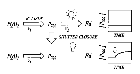
By way of example, FIGURE 5, shows how the methods of the invention can
be used to measure the steady-state turnover of PS I (P,oo). In the steady-
state, the
rate of light-driven oxidation of P,oo (v,) is precisely counterbalanced by
the rate of its
rereduction via turnover of the cytochrome bbf complex (v,), leading to a
stable P,oo
redox state. By briefly and rapidly shuttering the light, v' is temporarily
inhibited,
thus allowing the system to relax. The initial changes in the concentration of
reduced
P7oo (the dark relaxation) reflect v,, and are proportional to the flux
through the
system just prior to the shuttering. It should be noted that the methods of
the present
invention are not sensitive to changes in the PS I acceptor side redox state
(see,
Klughammer C and Schreiber U Planta 192: 261-268 (1994)) when used to measure
PS I flux, and so should be free from this potential artifact.
The measured photosynthetic parameters) can be used to determine whether
the subject plant is experiencing one or more of a variety of environmental
and/or
physiological stresses, such as temperature stress, drought stress and
nutrient stress
(including nitrogen stress). Thus, in one aspect, the present invention
provides
methods for determining the physiological state of a plant comprising: (a)
illuminating a plant leaf until steady-state photosynthesis is achieved; (b)
subjecting
the illuminated plant leaf to a period of darkness; (c) using a kinetic
spectrophotometer or kinetic spectrophotometer/fluorimeter to collect spectral
data
from the plant leaf treated in accordance with steps (a) and (b); (d)
determining a
value for a photosynthetic parameter from the spectral data; and (e) using the
determined value for the photosynthetic parameter to determine the
physiological
state of the plant. In one embodiment, the step of using the determined value
for the
photosynthetic parameter to determine the physiological state of a plant
comprises
the step of comparing the determined value for the photosynthetic parameter to
a
reference value for the same photosynthetic parameter determined from spectral
data
obtained from one or more reference plants. Typically a difference is observed
between the determined value for the photosynthetic parameter and the
reference
value for the photosynthetic parameter. The difference can typically be
correlated
with the presence of a physiological stress in the plant.
For example, utilizing the foregoing methods for determining the
physiological state of a plant, changes in the following, representative,
photosynthetic
parameters can be correlated with the presence of a physiological stress in a
plant: an
increase in ATP synthase activity (relative to ATP synthase activity in one or
more
reference plants) is correlated with the presence of drought stress in the
plant; an
CA 02382197 2002-02-19
w0 01/13094 PCT/US00/22754
-15-
increase in P,°° reduction (relative to P,°°
reduction in one or more reference plants) is
correlated with the presence of drought stress in the plant; an increase in
the
proton/electron resistance ratio (relative to the proton/electron resistance
ratio in one
or more reference plants) is correlated with the presence of drought stress in
the
plant; a decrease in ATP synthase activity (relative to ATP synthase activity
in one or
more reference plants) is correlated with the presence of nitrogen stress in
the plant; a
decrease in P,oo reduction (relative to P,°° reduction in one or
more reference plants)
is correlated with the presence of nitrogen stress in the plant; a decrease in
the
proton/electron resistance ratio (relative to the proton/electron resistance
ratio in one
or more reference plants) is correlated with the presence of nitrogen stress
in the
plant.
The following examples merely illustrate the best mode now contemplated for
practicing the invention, but should not be construed to limit the invention.
EXAMPLE 1
Relaxation Kinetics of Cytochrome f, P,°°. and the
Electrochromic Shift in an
Intact Poinsettia Leaf
FIGURE 6 shows data collected from an intact Poinsettia leaf using diffused
optics flash kinetic spectrophotometer 10 to measure the relaxation kinetics
of
cytochrome f and P,°°, and the electrochromic shift (D~r) during
a brief shuttering of
continuous illumination (300 pmole photons/mz/s).
Leaves on intact plants were placed in diffused-optics flash kinetic
spectrophotometer 10 and illuminated with continuous, actinic light. The light
was
shuttered either with a mechanical shutter or by switching off a bank of light-
emitting
diodes.
Cytochrome f redox changes were deconvoluted from the electrochromic shift
and other species by four different methods: 1 ) based on that reported by
Joliot and
Joliot (Biochim. Biophys Acta. 765: 219-226 (1984)), a straight baseline,
between
the 545 nm and 573 nm absorbance changes, was subtracted from the 544 nm
changes, i.e. DI/I~yt~= (DI/I554 - DI/I545) - 0.32(DI/I5~3 - DI/I545)~ 2) the
technique of
Nishio and Whitmarsh (Plant Physiol. 95: 522-528 (1990), i.e. DI/I~ytf -
(~I/I554 -
DI/I560); 3) the technique of Kramer and Crofts (Biochim Biophys Acta 976: 20-
41
(1989)) where a fraction (10-13%) of the 515 nm change is subtracted from the
difference between the absorbance at 545 nm and 554 run; and 4) assuming 545
nm
to be nearly isobestic for cytochrome f redox changes and all absorbance
changes at
this wavelength were to be caused by the electrochromic shift, the
electrochromic
CA 02382197 2002-02-19
WO 01/13094 PCT/US00/22754
-16-
shift spectrum published by Witt was scaled to the 545 nm absorbance changes
and
subtracted from the entire spectrum. Though the different techniques yielded
different signal extents, which most likely depended upon the differences in
the
extinction coefficients and the wavelengths employed in each, they all yielded
similar
kinetics when their amplitudes were normalized. Results using the technique of
Joliot and Joliot are reported herein, mainly because the data could be
directly
compared to a large body of previously accumulated data.
The results presented in FIGURE 6 show that rates of linear electron flow can
be readily measured with the methods of the present invention by monitoring
electron
transfer through PS I or cytochrome f. As expected, a linear relationship was
observed between estimates of electron transfer rates (shown as PS 1 flux)
obtained
by measurement of OZ evolution (in a leaf disk electrode) and analysis of the
820 nm
change (in this case measured by reflectance spectroscopy) in a tobacco leaf
disk (See
FIGURE 7). Oz evolution was measured using a Clark electrode using the methods
disclosed in Sacksteder and Kramer (1998), supra. An OZ concentration of 2%
was
used to minimize photorespiration.
EXAMPLE 2
Measurement of Cyclic Electron Transfer Around Photosystem I and
Photos~tem II
The methods of the present invention can also be used to measure cyclic
electron transfer around both photosystems as long as a measurement of the
fraction
of open photosystems can be made. For example, the number of open PS I and PS
II
centers was probed by exciting the leaf with saturating laser pulses and
monitoring
the extent of the rapid rise of the electrochromic shift (i. e., dA at about
520 nm). The
experiment was repeated with the laser flash given at a range of times after
shutter
closure. Plots of the extent of laser-induced electrochromic shift,
proportional to the
fraction of open centers against the delay time between shutter closure and
laser
flash, (FIGURE 8) represent the kinetics of center reopening (in a tobacco
leaf under
continuous 300 ~mol photons m 2 s' red light), the initial rate of which is
proportional to the steady-state turnover rate of both PS I and PS II centers.
The
absorbance changes that occurred at around 820 nm, upon shutter closure,
indicating
the reopening of photosystem I centers as P,oo was reduced, was also monitored
(FIGURE 8). As is clear from FIGURE 8, in healthy intact leaves, the initial
rates of
PS I and PS II reopening are nearly equal, as expected for linear electron
transfer.
CA 02382197 2002-02-19
WO 01/13094 PCT/US00/22754
-17-
EXAMPLE 3
Measurement of Light-Driven Fluxes of Protons
The methods of the present invention can also be used to measure light-driven
fluxes of protons (See FIGURE 9). During continuous illumination, protons are
pumped across the thylakoid membrane by the photosynthetic electron transport
chain and the resulting proton motive force (p.m.~) is dissipated by the
turnover of
the ATP synthase and proton leakage pathways (Mitchell P, Ann. Rev. Biochem.
46:
996-1005 (1977)). As with the electron transfer reactions discussed above, the
flux
of protons into the lumen (v,) will be precisely balanced by efflux (v,) in
the steady-
state. Upon a rapid light-dark transition, the light-driven proton pump (v1)
is halted,
leaving only proton efflux (v2).
Because protons are charged, their movement across the thylakoid membrane
is electrogenic and thus the magnitude of v~ can be measured by changes in the
transthylakoid electric field (which is probed by the electrochromic shift).
This
should hold true even if the steady-state transthylakoid electric field is
negated by
counter movements of ions (e.g., Vredenberg WJ Biochem. Biophys. Res. Comm.
42:111-118 (1971); Vredenberg WJ and Bulychev AA, Plant Science Letters
7:101-107 (1976) and Vredenberg WJ and Tonk WJM, Biochim. Biophys. Acta,
387:580-587 (1975)) as long as ion fluxes are significantly slower than proton
efflux.
This appears to be the case, as shown both from microelectrode measurements in
vitro (Vredenberg and Tonk (1975), supra) and from electrochromic shift
measurements in intact plants (see, e.g., FIGURE 6). Thus, the present
invention
provides for the first time, a non-invasive in vivo assay for light-driven
proton fluxes
and thus for the measurement of light-driven ATP synthesis.
EXAMPLE 4
Measurement of the "Resistance" to Turnover of Photosynthetic Complexes
The initial rate of the dark-interval relaxation of a number of photosynthetic
systems is a measure of the steady-state flux through those systems. The
relationship
between the amplitude of the relaxation signal with the initial relaxation
rate, and the
half time for the relaxation are measures of the "resistance to flux" that can
easily be
made using the methods of the present invention. FIGURE 10 shows data obtained
from two tobacco plants after irradiation with 100-1000 mmol photons m Zs'.
The
control plant was well-watered, while the test plant was deprived of water for
two
days.
CA 02382197 2002-02-19
w0 01/13094 PCT/US00/22754
-18-
The data in FIGURE 10 is plotted as the ratio of the half time for
electrochromic shift decay to the half time for the electron transfer to P,oo
versus the
half time for the decay of the electrochromic shift. This way of presenting
the data
more clearly shows that the resistances to proton pumping (measured by the
electrochromic shift) and electron transfer (measured by the 820 nm change)
are both
increased by drought stress, but that the resistance to proton pumping is
affected
more strongly than the resistance to electron transfer.
EXAMPLE 5
Measurement of the Efficiency of Light Capture by Photosystem II
The methods of the present invention can also be used to measure the
efficiency of light capture by photosystem II. Light capture efficiency is
related to
antenna regulation, and thus to the physiological status of the plant.
Prior to the present invention this efficiency parameter (termed ~I,) was
determined using super-saturating pulses of light as the ratio of the change
in
fluorescence induced by the saturating pulse (F~) over the maximal chlorophyll
a
fluorescence obtained during the saturating pulse (F,~,,). This requires bulky
and
expensive light sources and can potentially harm or disturb the plant being
measured.
The methods of the present invention make this measurement using an
alternative
approach: by comparing the initial slopes of fluorescence yield, reflecting PS
II
photochemistry and photochemical efficiency (or antenna downregulation), with
absorbance changes at 820 nm, reflecting PS I photochemistry, or 520 nm,
reflecting
proton pumping.
FIGURE 11 shows data from a greenhouse grown tobacco plant, comparing
the ratio of initial rates of fluorescence and proton pumping (520 nm
absorbance
changes) measured using the methods of the present invention with ~" measured
in
the same leaf over a range of light intensities. The ~I, data was obtained by
periodically applying saturating light pulses (1 s long, >10,000 mole photons
rriz s'
white light from a xenon arc lamp, filtered to eliminate infrared and
ultraviolet
radiation). A good correlation was observed indicating that the methods of the
present invention can be used to obtain qualitative, and relative, estimates
of antenna
downregulation.
CA 02382197 2002-02-19
w0 01/13094 PCT/US00/22754
-19-
EXAMPLE 6
Determining the Physiological State of a Plant in Response to Drought Stress
The data described in this example show how the methods of the present
invention can be used to correlate changes in either of two photosynthetic
parameters
(ATP synthase activity and P,oo reduction) to drought stress in plants.
FIGURE 12 shows measurements of the relaxation half times for signals
associated with ATP synthesis (i.e. the signal at 520 nm) and P,oo reduction
(i.e. at
820 nm) in a drought-stressed plant and in a control plant that was not
subjected to
drought stress. The data is expressed as the inverse of the relaxation time
(or the time
it takes to decay to 1 /2.718 of the original value). The tobacco plants were
grown in
a green house in pots. One plant was watered (control), the other was
subjected to
water stress (drought-stressed) by not watering it for two days.
There was a marked increase in the steady state turnover times, indicating a
slowing of electron transfer and ATP synthase activity in response to drought
stress.
The reaction of the plants to drought conditions and its effects on
photosynthesis is more clearly observed by plotting the ratio of the turnover
times of
P7oo to ATP synthase (See FIGURE 13), i.e., this ratio, referred to as the
proton/electron "resistance" ratio, is higher in drought stressed plants than
in
unstressed, control plants. The ratio of the two measurements in a control and
drought stressed plant shows the ability of the methods of the present
invention to
distinguish a normal, non-stressed plant from one undergoing drought stress.
The
advantage of making a ratio is that plant-plant fluctuations can be accounted
for, thus
allowing an immediate recognition of the stress status of a plant.
EXAMPLE 7
Correlation of the Rate of Change of the ATP Synthase Reaction with
Drought Stress
FIGURE 14 shows measurements of the ATP synthase reaction as a function
of time after detachment of the measured leaf from a plant. The half time for
relaxation of the electrochromic shift was measured using diffused-optics
flash
kinetic spectrophotometer instrument 10 to generate a measuring beam of light
at
520 nm. A leaf from an intact tobacco plant was measured, then detached from
the
plant. Additional measurements were made on the detached leaf over a time
course.
Detachment of the leaf resulted in the induction of a condition of plant
stress (i.e.,
drought stress) in the leaf. As shown in FIGURE 12, the onset of leaf plant
stress
was reflected in an increase in the relaxation time for the turnover of the
ATP
CA 02382197 2002-02-19
w0 01/13094 PCT/US00/22754
-20-
synthase. These results showed that the ATP synthase reaction in leaves
responded
sensitively and rapidly to drought stress.
EXAMPLE 8
Response of ATP S~nthase/Electron Transfer (P,o~~Resistances to Nitro~n
Stress
The data set forth in this example and in FIGURE 15 show that the ratio of
ATP synthase resistance/electron transfer (P,oo) resistance decreases in
response to
nitrogen stress.
Plants subjected to nitrogen fertilizer deficits exhibited a lowered ratio of
ATP synthase resistance/electron transfer (P,oo) resistance, as indicated by a
low ratio
of the change in the 820 nm signal versus the change in the 520 nm signal (see
FIGURE 15). This observation is in contrast to the increased ratio observed
with
drought stressed plants. This observation shows that the methods of the
present
invention can be used to not only measure plant stress, but distinguish
between
various kinds of plant stress based upon the ratio of ATP synthase/electron
transfer
resistances observed in control versus test plants.
EXAMPLE 9
Dark Interval Relaxation Kinetics of Absorbance Changes as a Quantitative
Probe of Steady-State Electron Transfer
Abbreviations used in this Example: A~ - gross carbon assimilation; cyt -
cytochrome; DIRK - dark-interval relaxation kinetics; DIRKhP~ - the initial
rate of
change, upon shutter closure, of the redox states of all high potential chain
components; DOFS - diffused optics flash spectrophotometer; EPR - electron
paramagnetic resonance; E,,P~ - the redox poise of the high potential chain;
Fe,S, - the
'Rieske' iron sulfur cluster; hpc - the high potential chain of electron
carriers,
consisting of cyt f, plastocyanin and P,oo; IR - infrared; IRGA - infrared gas
analyzer;
KhP~ - the equilibrium constant for sharing of electrons between (cyt f +
plastocyanin)
and P,oo; PC - plastocyanin; PS - photosystem; PQ - plastoquinone; PQHz
plastoquinol; QA - the primary quinone acceptor of PS II; Qo - the
plastoquinol
oxidase site of the cytochrome b~f'complex;
Plant material. Nicotiana tobacum (tobacco) plants were grown under
greenhouse conditions 0900 qmol photons m' s' maximum light intensity). The
plants were watered daily and fertilized once a week. Young, fully-expanded,
attached leaves were used for all experiments.
CA 02382197 2002-02-19
WO 01/13094 PCT/US00/22754
-21-
Dark interval ~°elaxation kinetics. Steady-state rates of
photosynthetic
electron transfer were estimated by following the absorbance changes upon
rapid
light-dark transitions using diffused optics flash spectrophotometer 10.
Actinic light
intensities ranging from 200 to 1650 ~mol photons m' s' of red light were
achieved
by filtering a high-intensity xenon arc lamp through a series of two dichroic
filters
(Optical Coatings Inc, CA), one cutting off below 600 nm, the other above 750
nm.
Dark intervals were achieved by shuttering the light with an electromechanical
shutter (UniblitzT"~ VS25) with 3.25ms shutter closure time, synchronized to
measuring pulses by the DOFS control computer. Kinetics at each light
intensity
were averaged over 3 traces with the initial slope measurement beginning less
than 2
ms after final shutter closure encompassing 5 data points over 6 ms.
Cytochrome f
redox changes were measured and deconvoluted as described in Kramer DM,
Sacksteder CA (1998) Photosynth Res 56: 103-112. The re-reduction kinetics of
P,oo+ were detected at 820 nm, where the P,oo+ canon absorbs (Katoh S,
Shiratori I,
Takamiya A (1962) Biochemistry 51: 32-40). We considered possible interference
from other species at 820 nm (Klughammer C, Schreiber U ( 1991 ) Z Naturforsch
46c: 233-244; Kramer DM, Crofts AR. (1996) Control of photosynthesis and
measurement of photosynthetic reactions in intact plants. In: N Baker, (ed).
Photosynthesis and the Environment. Advances in Photosynthesis pp. 25-66.
Dordrecht, The Netherlands: Kluwer Academic Press; Klughammer C, Schreiber U.
(1998) Measuring P700 absorbance changes in the near infrared spectral region
with
a dual wavelength pule modulation system. In: G Garab, (ed). Photosynthesis:
Mechanisms and Effects pp. 4357-4360. Dordrect: Kluwer Academic Publishers),
but
concluded that over the time range of the initial rate measurements, these
species did
not significantly compromise the data.
C02 fixation rates. Carbon exchange and absorbance kinetics were measured
simultaneously using an open flow system containing two infrared gas analyzers
(IRGAs) in series, an Anarad AR-500-R, set up in differential mode, and a
Qubit
Systems S 151 set up in absolute mode. Similar assimilation measurements were
obtained with both detectors while the Qubit IRGA was used to measure absolute
COz levels. The leaf chamber/gas exchange cuvette was constructed in-house,
based
on the Qubit Systems design, but with a significantly smaller leaf aperture to
accommodate the light path of DOFS instrument 10. Two Plexiglas windows fitted
with ports for gas flow were fastened together with a neoprene washer to seal
the
chamber and protect the leaf. As in the Qubit chamber, the leaf boundary
resistance
CA 02382197 2002-02-19
w0 01/13094 PCT/US00/22754
-22-
layer was minimized with an array of evenly spaced gas inlet and outlet ports
on both
sides of the chamber. A premixed, compressed gas mixture containing 350 ppm
CO,,
1 % Oz and a balance of N~, was flowed through the sample chamber at a
constant
flow rate of 75 ml~miri'. A low flow rate was used because the sample leaf
area,
l.7cm2, was correspondingly small, allowing for better resolution of the COZ
signal
changes. The depression in [COz] over the leaf was less than 50 ppm under all
conditions. A thermocouple wire was used to obtain leaf temperature readings
while
the Qubit system sensors were used to monitor air temperature and humidity.
The
COZ, temperature and humidity data were collected using the Qubit systems
Universal Lab interface and Logger Pro Software (Vernier). The air in the leaf
chamber was maintained at average temperature and relative humidity of
23°C and
62% respectively. Gross CO, assimilation (A~) was calculated as the difference
of
light and dark CO, uptake measurements. Respiration during photosynthesis was
estimated using the earliest stable dark respiration rate directly following
each
illumination interval.
Analysis of dark-interval relaxation kinetics in intact leaves under steady-
state illumination FIGURE 16 A and FIGURE 16 B show DIRK traces, with a dark
interval of 750 ms, for P~oo (820 nm) and cyt f respectively in an attached
tobacco leaf
under steady-state light conditions. During the first 100 ms, the time course
over
which this type of analysis is typically taken, the re-reduction of both P~oo
and cyt f
appeared essentially monophasic. The half times changed little with increasing
light
intensity, varying from llms to 6ms for P,oo and l8ms to lams for cyt f as
light
intensity was changed from 145 to 1050 ~mol~m'~s'. These data are consistent
with
that observed previously by several groups (Harbinson J, Hedley CL ( 1989)
Plant
Cell Environ 12: 357-369; Laisk A, Oja V (1994) Photosynth Res 39: 39-50;
Laisk
A, Oja V (1995) Photosynth Res 45: 11-19; Kramer DM, Sacksteder CA, Cruz JA
(1999) Photosynth Res 60: 151-163; Ott T, Clarke J, Birks K, Johnson G (1999)
Planta 209: 250-258). A significantly slower phase in the reduction kinetics
for both
P7oo and cyt f appears when the decay over the longer, 750 ms, dark interval
was
considered. It is possible that a fraction of the long phase of the 820 nm
absorbance
kinetics is due to re-reduction of PC or to light scattering changes
(Klughammer C,
Schreiber U (1991) Z Naturforsch 46c: 233-244). However, this is not likely to
be
the case with cyt f because extensive spectral analysis (as described in
Kramer DM,
Sacksteder CA (1998) Photosynth Res 56: 103-112) showed that the cyt f signal
is
essentially uncontaminated by other contributions (not shown). The slow phases
of
CA 02382197 2002-02-19
w0 01/13094 PCT/US00/22754
-23-
P7oo and cyt f reduction may be due to a relative imbalance in the excitation
of PS I
over PS II, resulting in a relatively small extent of PQ pool reduction. The
apparent
pseudo-first order behavior of the initial kinetics of the rapid ( 10-20 ms)
re-reduction
phase can be explained by a preferential binding of PQH, over PQ to the Qo
site (as
proposed in Kramer DM, Joliot, A., Joliot, P., Crofts, A. R. (1994) Biochim
Biophys
Acta: 251-262), ensuring that the cyt b~f complex will be at least partially
charged
with quinol substrate even with a relatively oxidized quinone pool. Bound PQHZ
will
be rapidly oxidized upon a light-dark transition, giving rise to the rapid
phase. If the
PQ pool is predominantly oxidized, the binding of fresh PQHz to the Qo site
will be
slow or inhibited, resulting in the slower reduction phase.
DIRK estimates of flux using the summation method. The summation method
was used to account for differential partitioning of electrons. The total
number of
electrons entering the hpc over a particular time period, D[hpc(red)], is
defined as:
O[hpc(red)] _ ~ ~''~
where n is the number of species in the hpc, DA;,, is the absorbance change at
a particular wavelength due to the reduction of species i at wavelength or set
of
wavelengths ~,, and DE;,, is the effective reduced-minus-oxidized difference
extinction coefficient at ~., which includes scatter-induced enhancement and
flattening effects. As discussed above, we assume that PC and cyt f are nearly
isopotential and rapidly equilibrating, allowing us to treat them as a single
redox
pool. Previous measurements (Graan T, Ort DR (1984) J Biol Chem 259: 14003-
14010; Hope AB (1993) Biochim Biophys Acta 1143: 1-22) indicate that the ratio
of
PC:cyt f: P,oo is approximately 2:1:1, and thus, the total hpc redox changes
(i.e.,
0[hpc(red)]), as measured by the spectroscopically accessible redox changes of
cyt f
and P,oo, can be described by the following:
0[hpc(red)] =3 ~'''J + ~8zo
~'yl
where DA'y~ f represents the deconvoluted absorbance change for cyt f using
wavelengths around its alpha band. If the effective extinction coefficients
are
accurate, the initial rate of change of 0[hpc(red)] upon shutter closure,
termed here
CA 02382197 2002-02-19
WO 01/13094 PCT/US00/22754
-24-
DIRKhP~, should be equal to the rate of electron transfer through the
photosynthetic
electron transfer chain.
There are a number of factors that alter the effective extinction coefficient,
particularly flattening and scattering-induced enhancement (see Latimer P,
Eubanks
CAH (1962) Arch Biochem Biophys 98: 274-285; Klughammer C, Schreiber U
(1991) Z Naturforsch 46c: 233-244; Kramer DM, Crofts AR. (1996) Control of
photosynthesis and measurement of photosynthetic reactions in intact plants.
In: N
Baker, (ed). Photosynthesis and the Environment. Advances in Photosynthesis
pp.
25-66. Dordrecht, The Netherlands: Kluwer Academic Press). It is therefore
necessary to estimate their values in situ. To accomplish this, absorbance
changes
were measured that are associated with both cyt f and P,°° (at
820 nm) redox changes
that occurred upon full oxidation of hpc components by exposure to 60 seconds
of far
red (>730 nm) light. This was judged to give essentially full oxidation of hpc
components since increased intensities resulted in no increase in the signal
extents.
Reported values for the extinction coefficient for the alpha band of cyt f at
554 nm (s~ytf) range between 19.5 mM-'cm' in parsley and lettuce (Ford ML,
Bertole
G, Zanetti G (1965) Biochim Biophys Acta 109: 33-40; Nelson N, Neumann J
(1972)
J Biol Chem 247: 1817-1824) and 25 mM-'cm' in spinach by Metzger SU, Cramer
WA, Whitmarsh J (1997) Biochim Biophys Acta 1319: 233-241. Nishio and
Whitmarsh have found that the effective extinction coefficient for the alpha
band of
cyt f in leaves is nearly identical to that found in isolated membrane, i.e.,
it appears
relatively uncorrupted by flattening and scatter-induced enhancement. We thus
converted our absorbance changes to concentrations using two values, the newer
value (Metzger SU, Cramer WA, Whitmarsh J (1997) Biochim Biophys Acta 1319:
233-241) and the average of the older and the newer. When adjusted to reflect
our
method of deconvolution, these resulted in effective extinction coefficients
for cyt f
of 21.9 and 19.5 mM-'cm' respectively. From this, and a ca. 1:1 stoichiometry
for
cyt f and P,°° (e.g., Graan T, Ort DR (1984) J Biol Chem 259:
14003-14010; Kramer
DM (1990). Ph.D. Thesis thesis. University of Illinois, Urbana, Illinois),
effective
extinction coefficients of 121 and 108 mM-'crri' for P,°° were
estimated. These
estimates do not include possible contributions of PC oxidation to the 820 nm
absorbance change of up to 30% (Klughammer C, Schreiber U (1991) Z Naturforsch
46c: 233-244). However, altering the relative extinction coefficient of
P~°° by 30%
had only small effects on the linearity DIRK initial rates vs. A~, although it
did affect
the slope of this relationship. In addition, key experiments were repeated
using an
CA 02382197 2002-02-19
WO 01/13094 PCT/US00/22754
-25-
alternate method for the calculation of the hpc in which contributions from PC
reduction were independently estimated from near IR absorbance changes as
reported
by Klughammer and Schreiber (Klughammer C, Schreiber U (1991) Z Naturforsch
46c: 233-244; Klughammer C, Schreiber U. (1998) Measuring P700 absorbance
changes in the near infrared spectral region with a dual wavelength pule
modulation
system. In: G Garab, (ed). Photosynthesis: Mechanisms and Effects pp. 4357-
4360.
Dordrect: Kluwer Academic Publishers). This approach also yielded linear data,
very
similar to that where PC redox changes were estimated from cyt f absorbance
changes. In this way it is also possible to make DIRK measurements exclusively
in
the near infrared. The IR approach has significant advantages from an
instrumentation standpoint, because these wavelengths are non-actinic and
intense
light-emitting diodes in this spectral range are readily available. For the
purposes of
the experiments reported herein, cyt f was used because the effective
extinction
coefficient in leaves is most likely close to published values from isolated
material,
allowing quantitative estimates of electron flux to be made.
DIRK analysis of linear electron transfer. For comparison with results from
DIRK analysis COZ assimilation (A~) under non-photorespiratory conditions
(i.e.,
low oxygen) was measured. To avoid perturbation of steady-state electron
transfer
rates, the dark intervals were kept short (40 ms). Representative kinetic
traces, taken
at 940 ~mol photons m Zs' are shown in FIGURE 17. The sigmoidal reduction
kinetics of cyt f (upon shutter closure) and oxidation kinetics of P,oo (upon
shutter
opening) indicate that Khp~>1 and confirm the necessity of accounting for
differential
electron partitioning.
FIGURE 18 shows the light-intensity dependence of DIRK initial rates of cyt f
and P~°o reduction. Both cyt f and P,oo DIRK rates were nearly linear
with light
intensities between 0 and 250 pmol photons m's'. At about 500 p,mol photons m
Zs
', cyt f DIRK reached a maximum, above which it fell, reflecting the
appearance of
the distinct lag phase in cyt f reduction (as seen in FIGURE 17). On the other
hand,
DIRK of P,°o continued to rise, nearing saturation at the highest light
intensity. This
behavior could be explained by differential partitioning effects.
FIGURE 19 shows that DIRK,,P~, i.e., the total hpc reduction rates of all hpc
components, is linearly dependent on A~ over light intensities from ~10% to
essentially fully saturating. The symbols represent DIRKhP~ values derived
using the
extinction coefficient of Metzger SU, Cramer WA, Whitmarsh J (1997) Biochim
Biophys Acta 1319: 233-241. Within the noise level, the dependence of DIRKhP~
CA 02382197 2002-02-19
WO 01/13094 PCT/US00/22754
-26-
initial rate on A~ was linear and a best-fit line (FIGURE 19, solid line) gave
an
r-value of 0.98 with a y-intercept near zero. The slope of this line gave an
estimated
4.8 electrons per COZ fixed. Not surprisingly, the slope was sensitive to
chosen cyt f
extinction coefficients. When the average of literature values was used, an
estimate
was obtained of 5.4 electrons passed through the hpc per COZ fixed (FIGURE 19,
dotted line). The dashed line in FIGURE 19 indicates the expected value of
4 electrons passed through the hpc per CO, fixed. Given the good
correspondence
between estimated and predicted e-:COZ ratio, it was concluded that light-
dependent
partitioning of electrons can be well accounted for by the summation
technique.
The ca. 15-20% deviation in electron and COz measurements can be ascribed to
errors in the estimates of effective extinction coefficients, the
stoichiometry of PC to
cyt f, contributions of PC to the 820 nm signal, or in the measurement of A~.
For
example, an effective extinction coefficient for cyt f alpha band of 30 mM-
'crri'
resulted in a ratio of 4 for electrons per CO, fixed. On the other hand, an
excess of
hpc electron flux over AG could be an indicator of significant flux to
alternative
electron acceptors, including residual photorespiration and nitrite reduction,
or of the
participation of PS I cyclic electron transfer or the Mehler-peroxidase/water-
water
cycle (see reviews in Baker NR, Oxborough K, Andrews JR. (1995) Operation of
an
alternate electron transfer acceptor to COz in maize crops during periods of
low
temperatures. In: P Mathis, (ed). Photosynthesis: From Light to Biosphere pp.
771-
776. The Netherlands: Kluwer Academic Publishers; Heber U, Gerst U, Krieger A,
Neimanis S, Kobayashi Y (1995) Photosynth Res 46: 269-275; Asada K. (1996)
Radical production and scavenging in the chloroplasts. In: NR Baker, (ed).
Photosynthesis and the Environment pp. 123-150. The Netherlands: Kluwer
Academic Publishers; Kramer DM, Crofts AR. (1996) Control of photosynthesis
and
measurement of photosynthetic reactions in intact plants. In: N Baker, (ed).
Photosynthesis and the Environment. Advances in Photosynthesis pp. 25-66.
Dordrecht, The Netherlands: Kluwer Academic Press; Ivanov B, Kobayashi Y,
Bukhov NG, Heber U (1998) Photosynth Res 57: 61-70; Cornic G, Bukhov NG,
Wiese C, Bligny R, Heber U (2000) Planta 210: 468-477; Eichelmann H, Laisk A
(2000) Plant Cell Physiol 41: 138-147; Miyake C, Yokota A (2000) Plant Cell
Physiol 41: 335-343). The fact that a linear relationship was observed between
DIRK,,P~ and A~ would indicate that the magnitude of such fluxes were
proportional
to linear electron transfer (reviewed in Kramer DM, Crofts AR. (1996) Control
of
photosynthesis and measurement of photosynthetic reactions in intact plants.
In: N
CA 02382197 2002-02-19
w0 01/13094 PCT/US00/22754
-27-
Baker, (ed). Photosynthesis and the Environment. Advances in Photosynthesis
pp.
25-66. Dordrecht, The Netherlands: Kluwer Academic Press).
The DIRK approach is particularly useful under conditions where COZ and O,
gas exchange estimates are not readily achieved or are significantly affected
by
alternate electron sinks (e.g., cyclic electron transfer or photorespiration).
Moreover,
neither the use of saturation pulses nor knowledge of the amount of light
absorbed by
the plant--both of which are required for the chlorophyll fluorescence
techniques--are
required for DIRK flux estimates.
EXAMPLE 10
Measurement of Proton Transfer Using the Methods of the Invention
Abbreviations: A~, the rate of gross photosynthetic COz assimilation;
ATPase, (C)Fo-(C)F, ATP synthase; cyt, cytochrome; DIRK, dark interval
relaxation
kinetics; DIRKhp~, DIRK analysis of absorbance changes associated with
reduction of
cyt f and P~oo yielding an estimate of electron flux through the high
potential chain of
photosynthesis; DIRKECS, DIRK analysis of absorbance changes associated with
the
ECS yielding an estimate of light-induced proton fluxes through the ATPase;
DOFS,
diffused optics flash spectrophotometer; ECS, the electrochromic shifting of
light-
harvesting pigments in response to delocalized transthylakoid electric field;
FS, the
steady-state chlorophyll a fluorescence yield; FM, the maximal chlorophyll a
fluorescence yield obtained under conditions in which non-photochemical
fluorescence quenching is minimal; FM', the yield of chlorophyll a
fluorescence
obtained during steady-state illumination, during application of light pulses
that
saturate PS II photochemistry; H+/e', the stoichiometry of protons to
electrons passed
through the photosynthetic apparatus; pmf, proton motive force; PS,
photosystem; ~I,,
the quantum efficiency of PS II and associated antenna; qNp, non-photochemical
quenching of chlorophyll a fluorescence.
Plants and growth conditions. Nicotiana tabacum (tobacco) was grown in a
greenhouse with midday light intensity of about 900 moles photons m Zs-', as
described in Example 9.
Measurements of absorbance changes in the steady-state. Steady-state rates
of photosynthetic electron transfer and proton flux through the ATPase were
estimated by following the absorbance changes upon rapid light-to-dark
transitions
using the methods of the invention. Use of DOFS instrument 10 significantly
attenuates the interfering light scattering changes in the 500-560 nm range,
allowing
observation of changes in the redox states of cyt f and P,oo as well as
changes in the
CA 02382197 2002-02-19
WO 01/13094 PCT/US00/22754
-28-
extent of the ECS. Wavelength selection was provided by a wheel of 2 or 3 nm
bandpass interference filters (Omega Optical, Brattleboro, Vt.) under computer
control. Actinic light was provided by a 500 W Xenon arc lamp, collimated with
quartz lenses. A series of three aluminum discs for holding optical filters
were
mounted on computer-controlled servo motors and placed in the light path of
the high
xenon actinic light. One of these filter discs held a series of two dichroic
filters
(passing light from 640 to 730 nm, Omega Optical, Brattleboro, Vt.), while the
other
two held a series of neutral density filters (New Focus, Santa Clara, Ca). A
heat
reflecting filter (57401, Oriel, Corp., Stratford, Ct) was kept in place at
all times to
remove excess heat from the actinic beam and to prevent saturation of the
detectors.
The control computer automatically selected actinic light intensities from
about 15 to
>2010 moles photons m 2 s~' by switching the neutral density filter
combinations.
Young, fully expanded, intact leaves were gently clamped into the leaf
chamber of DOFS instrument 10, which was perforated with two 5 mm diameter air
holes to allow free exchange of gasses. At least 3 minutes was allowed to
establish
steady-state conditions after each change in illumination intensity. After
steady-state
conditions were established, the actinic beam was shuttered for approximately
40 ms
periods at 15 s intervals to allow decay of photo-activated processes, and the
associated absorbance changes were measured at a range of wavelengths. The
actinic
light was shuttered (half time for closure of approximately 3.4 ms) using an
electromechanical shutter (Uniblitz, Vincent and Associates). Selected
measurements were also performed with a smaller shutter that closed with a
half time
of ca. 0.5 ms, but allowing for less light throughput, with nearly identical
results (not
shown). During measurements in the 500-575 nm region, the detectors were
protected from actinic light and fluorescence by Schott BG-18 filters, whereas
in the
case of infrared-measurements, protection was provided by Schott RG730
filters.
The blocking filters were mounted on metal discs and positioned by computer
control
via servo motors. The detector circuit was AC-filtered and thus sensitive to
the
pulsed measuring beam but not to offsets due to changes in chlorophyll
fluorescence
or to leakage of actinic light through the filters. The temperature of the
leaves,
measured by a thermocouple, deviated from room temperature by less than 1
°C
during the experiments.
Deconvolution of redox and electrochromic signals. In order to measure
relative changes in the transthylakoid electric field, Ayr, generated by
movement of
protons through the ATPase, changes in the ECS were measured that followed
rapid
CA 02382197 2002-02-19
w0 01 /13094 PCT/US00/22754
-29-
shuttering of the actinic light. To ensure that interfering signals did not
affect the
results, several different estimates of the electrochromic shift were
compared,
including the straight -DI/Ip changes at 520 or 515 nm, and -~I/Io differences
between
the following wavelength pairs: 515-545 nm, 520-530 nm, 520-510 nm, and 510-
500
nm. For the relatively rapid changes reported in this example, all estimates
were
found to be proportional within the noise levels, and changes at 520 nm were
used
throughout. Cytochrome f redox changes were deconvoluted from the
electrochromic
shift and other background signals by the method described in Kramer, D. M. &
Sacksteder, C. A. (1998) Photosynth. Res. 56, 103-112, and in Joliot, P. &
Joliot, A.
(1984) Biochim. Biophys. Acta 765, 219-226. The re-reduction kinetics of P,oo+
were
followed by observing the absorbance changes at 820 nm (Katoh, S., Shiratori,
I. &
Takamiya, A. (1962) Biochem. 51, 32-40) as described in Example 9.
Saturation pulse fluorescence changes. DOFS instrument 10 was modified to
measure saturation pulse fluorescence changes. Saturation pulses of either
9000 or
5500 pmoles photons m' s' white light lasting ca. 2 s were achieved by
removing, by
computer control, both the red and neutral density filters from the actinic
light path.
Full saturation was assumed because both pulse intensities gave essentially
identical
results. The time required for switching all filters was between 100 and 200
ms,
depending upon starting and ending servo positions. Chlorophyll a fluorescence
yield changes were measured essentially as in Kramer, D. M., Robinson, H. R. &
Crofts, A. R. (1990) Photosynth. Res. 26, 181-193, but using the DOFS optics
and
detector 10. The pulsed measuring beam, which struck the adaxial side of the
leaf
where the actinic beam struck, consisted of the DOFS xenon measuring flash
blocked
with a 425 nm (5 nm bandpass) interference filter (Omega Optical, Brattleboro,
Vt)
and an infrared rejecting filter (51962, Oriel Corp., Stratford Ct.).
Fluorescence was
measured on the abaxial side of the leaf with the DOFS sample detector. To
minimize
the effects of fluorescence re-absorption, the detector was blocked with a
color glass
filter that transmitted above 750 nm, where chlorophyll absorption is minimal
(Vogelmann, T. C., Bornman, J. F. & Josserand, S. (1989) Phil. Traps. R. Soc.
Lond.
323, 411-421). Steady-state fluorescence yields, FS were taken just prior to
application of saturation pulses, while fluorescence yields with all PS II
centers
closed, FM', were taken during the saturation pulse. The parameter ~I, was
calculated
as (FM'-FS)/FM (Genty, B., Harbinson, J., Briantais, J.-M. & Baker, N. R.
(1990)
Photosynth. Res. 25, 249-257).
CA 02382197 2002-02-19
WO 01/13094 PCT/US00/22754
-3 0-
Saturation pulse fluorescence assays of PS II electron transfer rates.
Electron transfer flux through PS II was estimated by the saturation-pulse
fluorescence rise technique introduced by Genty et al. (Genty, B., Harbinson,
J.,
Briantais, J.-M. & Baker, N. R. (1990) Photosynth. Res. 25, 249-257). The
application of supersaturating pulses of light saturates all photochemical
reaction
centers, and changes in chlorophyll a fluorescence yield reflect the
photochemical
quantum efficiency of PS II associated antenna (~") (e.g. Genty, B. &
Harbinson, J.
(1996) in Photosynthesis and the Environment, ed. Baker, N. R. (Kluwer
Academic
Publishers, The Netherlands), pp. 67-99). Multiplying ~" by the absorbed
actinic
light intensity has been shown to yield a good estimate of photosynthetic
electron
transfer rates (see e.g. Genty, B. & Harbinson, J. (1996) in Photosynthesis
and the
Environment, ed. Baker, N. R. (Kluwer Academic Publishers, The Netherlands),
pp.
67-99; Genty, B., Harbinson, J., Briantais, J.-M. & Baker, N. R. (1990)
Photosynth.
Res. 25, 249-257; Falkowski, P. G., Kolber, Z. & Mauzerall, D. (1994) Biophys.
J.
66, 923-928; Havaux, M., Strasser, R. J. & Greppin, H. (1991) Photosynth. Res.
27,
41-55; Krause, G. H. & Weis, E. (1991) Annu. Rev. Plant Physiol. Plant Mol.
Biol.
42, 313-349; Genty, B., Harbinson, J. & Baker, N. R. (1990) Plant Physiol.
Biochem.
28, 1-10; Govindjee (1995) Aust. J. Plant Physiol. 22, 20-29; Joshi, M. K. &
Mohanty, P. (1995) J. Scient. Ind. Res. 54, 155-174; Edwards, G. E. & Baker,
N. R.
(1993) Photosynth. Res. 37, 89-102; Edwards, G. E., Johnson, E., Lal, A. &
Krall, J.
P. (1993) Plant Cell Physiol. 34, 1205-1212; Baker, N. R. (1996) in
Photosynthesis
and the Environment, ed. Baker, N. R. (Kluwer Academic Press, The
Netherlands),
pp. 469-476). In the experiments reported herein, we multiplied ~" by the
incident
light intensity which provides, in arbitrary units, a measure of PSII electron
flux.
It is noteworthy that, under extreme conditions, estimates of electron flux
derived from ~" have sometimes deviated from those derived from alternative
techniques, e.g. COZ measurements (reviewed in Baker, N. R. (1996) in
Photosynthesis and the Environment, ed. Baker, N. R. (Kluwer Academic Press,
The
Netherlands), pp. 469-476; Baker, N. R., Oxborough, K. & Andrews, J. R. (1995)
in
Photosynthesis: From Light to Biosphere, ed. Mathis, P. (Kluwer Academic
Publishers, The Netherlands), Vol. IV, pp. 771-776; Kramer, D. M. & Crofts, A.
R.
(1996) in Photosynthesis and the Environment. Advances in Photosynthesis, ed.
Baker, N. (Kluwer Academic Press, Dordrecht, The Netherlands), pp. 25-66). It
has
been argued that, in some cases, these deviations may be due to secondary
effects of
high-intensity light pulses on other properties of the system (van Gorkom, H.
J.,
CA 02382197 2002-02-19
BYO 01/13094 PCT/US00/22754
-31-
Tamminga, J. J. & Havema, J. (1974) Biochim. Biophys. Acta 347, 417-438;
Vernotte, C., Etierme, A. L. & Briantais, J. M. (1979) Biochim. Biophys. Acta
545,
519-527; Kramer, D. M., DiMarco, G. & Loreto, F. (1995) in Photosynthesis:
From
Light to Biosphere, ed. Mathis, P. (Kluwer Academic Publishers, The
Netherlands),
Vol. I, pp. 147-150; Samson, G. & Bruce, D. (1996) Biochim. Biophys. Acta
1276,
147-153; Haveman, J. & Mathis, P. (1976) Biochim. Biophys. Acta 440, 346-355;
Best, J. A. V. & Mathis, P. (1978) Biochim. Biophys. Acta 503, 178-188) or
from the
operation of alternative electron acceptors or cycles around one or the other
photosystems (e.g. Ivanov, B., Kobayashi, Y., Bukhov, N. G. & Heber , U.
(1998)
Photosynth. Res. 57, 61-70; Cornic, G., Bukhov, N. G., Wiese, C., Bligny, R. &
Heber, U. (2000) Planta 210, 468-477; Heber, U., Gerst, U., Krieger, A.,
Neimanis,
S. & Kobayashi, Y. (1995) Photosynth. Res. 46, 269-275; Baker, N. R.,
Oxborough,
K. & Andrews, J. R. (1995) in Photosynthesis: From Light to Biosphere, ed.
Mathis,
P. (Kluwer Academic Publishers, The Netherlands), Vol. IV, pp. 771-776;
Peterson,
R. B. (1991) Plant Physiol. 97, 1388-1394; Foyer, C., Furbank, R., Harbinson,
J. &
Norton, P. (1990) Photosynth. Res. 25, 83-100; Foyer, C. H., Lelandais, M. &
Harbinson, J. (1992) Plant Physiol. 99, 979-986; Harbinson, J. & Foyer, C. H.
(1991)
Plant Physiol. 97, 41-49; Klughammer, C. & Schreiber, U. (1994) Planta 192,
261-
268; Asada, K. (1996) in Photosynthesis and the Environment, ed. Baker, N. R.
(Kluwer Academic Publishers, The Netherlands), pp. 123-150; Eichelmann, H. &
Laisk , A. (2000) Plant Cell Physiol 41, 138-147; Miyake, C. & Yokota, A.
(2000)
Plant Cell Physiol 41, 335-343). All experiments in this example were
performed
under permissive, non-stressed conditions, and so interference from such
phenomena
should not occur.
FIGURE 20 shows typical light-dependencies of fluorescence parameters
investigated in the experiments reported in this example. The amplitude of
F",,'
decreased steadily as light intensity increased, reflecting the progressive
engagement
of non-photochemical quenching mechanisms (e.g. Owens, T. G. (1996) in
Photosynthesis and the Environment, ed. Baker, N. (Kluwer Academic Publishers,
Dordrecht, The Netherlands), pp. 1-23). The steady-state fluorescence, FS,
rose from
light intensities between 17 and 800 ~mol photons m 2 s', reflecting a net
steady-state
reduction of QA. It then fell between 800 and 2000 mole photons m' s',
reflecting
the onset of strong non-photochemical quenching (reviewed in Genty, B. &
Harbinson, J. (1996) in Photosynthesis and the Environment, ed. Baker, N. R.
(Kluwer Academic Publishers, The Netherlands), pp. 67-99; Govindjee (1995)
Aust.
CA 02382197 2002-02-19
WO 01/13094 PCT/US00/22754
-32-
J. Plant Physiol. 22, 20-29; Norton, P., Ruban, A. & Walters, R. ( 1996) Annu.
Rev.
Plant Physiol. Plant. Mol. Biol. 47, 655-684). Under dark-adapted conditions,
the
photochemical quantum efficiency of PS II and associated antenna, estimated by
~",
was between 0.75 and 0.81 for all leaves measured. The value of ~" decreased
as
light intensity was increased, reaching between 0.15 to 0.22 at about 2000
~mol
photons m z s-'. The flux of electrons through PS II, as estimated by i*~",
showed a
typical light saturation curve, reaching half maximal value at about 450 pmol
photons m 2 s ' .
Dark interval relaxation kinetics. Typical -DI/Io changes that occurred during
DIRK experiments are shown in FIGURE 21 and were essentially as described
previously. The pronounced signature of the ECS was observed between 500 and
545 run (Witt, H. T. (1979) Biochim. Biophys. Acta 505, 355-427), whereas
contributions from redox changes in the cyt b6f complex occurred in the 545-
570 nm
region, most notably an absorbance increase due to reduction of cyt f (oc-band
peak at
554 nm). Moreover, the spectral changes were consistent with major
contributions
from ECS and cyt f over the entire time course of the decay, from 1 to 40 ms
after
shutter closure, indicating that the deconvolution procedures utilized herein
yielded
good representations of cyt f and ECS signals. Absorbance changes at 830 nm,
associated with P,oo reduction, were similar to those reported earlier (e.g.
Klughammer, C. & Schreiber, U. (1994) Planta 192, 261-268; Laisk, A. & Oja, V.
(1994) Photosynthesis Research 39, 39-50; Klughammer, C. & Schreiber, U.
(1991)
Z. Naturforsch. 46c, 233-244; Klughammer, C. & Schreiber, U. (1998) in
Photosynthesis: Mechanisms and Effects, ed. Garab, G. (Kluwer Academic
Publishers, Dordrect), Vol. V, pp. 4357-4360). The half times for the
relaxation
ranged from approximately from 10 to 6 ms for P,oo and 17 to 12 ms for cyt f
as light
intensity was changed from 10 to 1600 ~moles~rri Z-s-', in line with data
presented by
several groups (Laisk, A. & Oja, V. (1994) Photosynthesis Research 39, 39-50;
Harbinson, J., Genty, B. & Baker, N. R. (1989) Plant Physiol. 90, 1029-1034;
Laisk,
A. & Oja, V. (1995) Photosynth. Res. 45, 11-19; Ott, T., Clarke, J., Birks, K.
&
Johnson, G. (1999) Planta 209, 250-258; Kramer, D. M., Sacksteder, C. A. &
Cruz,
J. A. (1999) Photosynth. Res. 60, 151-163). The halftimes for the decay of the
ECS
remained in a narrow range from 18 to 20 ms over the entire range of light
intensities.
This implies that neither electron transfer nor ATP synthesis were hindered by
product inhibition, substrate depletion, or feedback processes. The initial
rates of
CA 02382197 2002-02-19
WO 01/13094 PCT/US00/22754
-33-
relaxation for the electrochromic shift, i.e. DIRKECS, were estimated by
fitting a line
through the 520 nm data 2-8 ms after closure of the shutter.
Within the noise level, the magnitudes of DIRKECS and DIRK,,P~ initial rates
were proportional to PS II electron flux as measured by i*~", (FIGURES 22 and
23).
Moreover, the relationship remained linear when the initial rate for DIRKESC
was
taken at a wide range of time intervals, or when the full extent of ECS signal
was
used. This indicates that residual cyt b6f complex turnover did not impact the
DIRKECS signal. These relationships are further substantiated in a preliminary
report
(Sacksteder, C. A. & Kramer, D. M. (1998) in Photosynthesis: Mechanisms and
Effects, ed. Garab (Kluwer Academic Publishers, Dordrect), Vol. 3, pp. 1621-
1624),
in which a linear relationship was found between O~ evolution and DIRKECS
under
conditions similar to those reported here. Experiments performed with longer
pre-
illumination times, as well as in the reverse light-intensity order, gave
nearly
identical results, indicating that induction phenomena did not significantly
impact the
results.
The relationships between relative fluxes of electrons transferred through PS
II and protons passed through the ATPase, were found to be linear within the
noise
level (FIGURES 22 and 23). This indicates that H+/e remained constant over the
entire light saturation curve. Most recent measurements agree that the H+/e-
ratio for
linear electron flow is 3 at low light intensities (Berry, S. & Rumberg, B.
(1999)
Biochim. Biophys. Acta 1410, 248-261; Kramer, D. M., Sacksteder, C. A. & Cruz,
J.
A. (1999) Photosynth. Res. 60, 151-163; Kobayashi, Y., Neimanis, S. & Heber,
U.
(1995) Plant Cell Physiol. 36, 1613-1620). From the data presented herein, it
is
inferred that the H+/e- ratio remains at 3 in healthy, unstressed plants.
Finally, it is worthwhile noting that the linear relationships between the
DIRK
techniques and the Iraqi parameter (Genty, B., Harbinson, J. & Baker, N. R.
(1990)
Plant Physiol. Biochem. 28, 1-10) as well as gas exchange (see Example 9
herein,
and; Sacksteder, C. A. & Kramer, D. M. ( 1998) in Photosynthesis: Mechanisms
and
Effects, ed. Garab (Kluwer Academic Publishers, Dordrect), Vol. 3, pp. 1621-
1624)
tend to validate the use of these measurements as linear indicators of flux.
The DIRK
approach should be particularly useful as an independent test of fluorescence
estimates under extreme conditions where the validity of the more commonly
used
technique has not been fully established.
CA 02382197 2002-02-19
w0 01 /13094 PCT/US00/22754
-34-
While the preferred embodiment of the invention has been illustrated and
described, it will be appreciated that various changes can be made therein
without
departing from the spirit and scope of the invention.