Note: Descriptions are shown in the official language in which they were submitted.
CA 02383328 2002-03-22
WO 01/21112 PCT/US00/40949
-1-
HEART VALVE PROSTHESIS WITH ROTATABLE CUFF
FIELD OF THE INVENTION
The present invention relates generally to
heart valve prostheses. More specifically, the present
invention relates to heart valve prostheses which have
a sewing cuff and which have a valve body that is
rotatable relative to the sewing cuff.
BACKGROUND OF THE INVENTION
Prosthetic valves are used to replace
defective natural valves in human hearts. The
prosthetic heart valves permit blood flow in one
direction through the valve, and block blood flow in the
other direction. In general, prosthetic heart valves
include an orifice ring which forms the valve housing
and which provides a central orifice or lumen for
passage of blood. A valve mechanism, such as one or
more occluders or leaflets, is mounted in the orifice
and opens and closes to regulate the passage of blood.
The housing and occluders collectively form the valve
body. One such valve is disclosed in U.S. Patent No.
4,276,658.
To attach the valve body to the tissue of the
heart, typically a sewing cuff (also called a suture
cuff) is provided. The sewing cuff for heart valve
prostheses is generally a soft, flexible torus-like
element through which sutures may pass to secure the
sewing cuff, and consequently the heart valve, to the
heart tissue.
One technique for attaching a sewing cuff to
a valve body is illustrated in U.S. Patent No.
4,276,658. In that embodiment, the valve body includes
a groove that is used in coupling the sewing cuff to the
valve body.
CA 02383328 2002-03-22
WO 01/21112 PCT/US00/40949
-2-
Another method of coupling the sewing cuff to
the valve body is shown in U.S. Patent No. 5,071,431 to
Sauter et al. Sauter et al. discloses a heart valve
where a sewing cuff is attached to a stiffening ring,
with the stiffening ring being coupled to the valve body
by a lock ring which rides in grooves in the outer
periphery of the valve body and the inner periphery of
the stiffening ring.
After a damaged or diseased natural valve
structure is removed from the patient, the prosthesis is
typically seated in the proper orientation and the
sewing cuff is sewn to the peripheral heart tissue.
Depending on the particular valve structure, care must
be taken to ultimately orient the valve to ensure that
the valving mechanism is in the most favorable
anatomical position to provide proper blood flow and to
ensure that the valve operates without interference from
surrounding heart tissue. This must either be done
before the sewing cuff of the valve is sutured into
place, or if the sewing cuff is rotatable relative to
the valve body (rotatable sewing cuff valve), this can
be done after the sewing cuff is secured to the heart
tissue. While this latter arrangement is convenient and
can obviate the need to remove and resuture a valve to
effect a rotation, a rotatable sewing cuff valve must
meet several criteria.
For example, the torsional force required to
rotate the valve body relative to the sutured cuff must
be low enough so that the surgeon is able to
rotationally position the valve with ease and without
damage to the surrounding tissue. Once implanted,
however, the valve body must maintain the desired
position during the remainder of the surgery, and
thereafter. Consequently, the torque required to
CA 02383328 2002-03-22
WO 01/21112 PCT/US00/40949
-3-
initiate rotation must be great enough to prevent
spontaneous rotation in vivo. Thus, the torsional force
required to rotate the valve body within the sewing cuf f
should be predictable and fall within a narrow
predetermined range such that the valve body may be
easily rotated by the surgeon, yet is resistant to
undesirable in vivo rotation once implanted.
The torsional force required to rotate the
valve body relative to the sewing cuff will be
determined by the manner in which the sewing cuff is
retained on the valve body. Various methods have been
proposed to rotatably secure the sewing cuffs of heart
valve prostheses to the valve bodies . For example, U. S .
Patent No.4,197,593 to Kaster et al. discloses a heart
valve where a sewing cuff is sutured to a polymeric slip
ring that slides along the surface of the valve body.
U.S. Patent No. 4,535,483 to Klawitter et al. discloses
a heart valve where the sewing cuff is carried by
deformable metal retainer rings that engage a stiffening
ring disposed in and secured to a peripheral groove in
the valve body. U.S. Patent No. 5,104,406 to Curicio et
al. discloses a heart valve where the fabric of the
sewing cuff is stitched to a core, which directly abuts
and rides the groove in the valve body. The core and
the valve additionally sandwich the fabric along the
annular space where the fabric is stitched to the core.
U.S. Patent No. 5,178,633 to Peters discloses a heart
valve where the sewing cuff is coupled to the valve body
by continuous fastener bands. The frictional engagement
between the fabric tube and the valve body or "orifice
ring" is controlled by the internal diameter of the
fastener bands, which may be manufactured with
precision. U.S. Patent No. 5,876,463 to Vanney et al.
discloses a rotatable heart valve which employs a spring
CA 02383328 2002-03-22
WO 01/21112 PCT/US00/40949
-4-
for actively and independently exerting a controlled
force directed substantially radially inward onto the
outer circumference of the heart valve prosthesis.
Although several rotatable sewing cuff valves
are available, these prior art devices typically suffer
from one or more shortcomings. These shortcomings may
include but are not limited to complexity of
manufacture, undesirable variation in torque needed for
rotation, excessive bulk, or insufficient radiopacity.
Therefore, a need exists for an improved rotatable heart
valve prosthesis. Moreover, the desired torque
characteristics should be repeatable from valve to valve
without surgically significant variation. Limitations
of space within the implant site require that the cuff
retention mechanism preferably be compact. Preferably,
the retention mechanism should provide enhanced
radiopacity to the valve.
SUMMARY OF THE INVENTION
A prosthetic heart valve is provided that
includes an orifice ring adapted to carry blood
therethrough. An annular recess is formed in an outer
surface of the orifice ring and has first and second
axially spaced walls. A sewing cuff is adapted to be
coupled to a native tissue annulus of a heart and
includes an inner annular cuff portion adapted to
conform to the first and second recess walls in the
orifice ring. A cuff retaining ring extends around the
inner annular fabric portion such that the inner annular
cuff portion is positioned between the cuff retaining
ring and the annular recess. The cuff retaining ring is
further adapted to exert a substantially axially
directed force directed against the annular cuff portion
and first and second axially spaced walls of the annular
recess whereby a controllable torque to rotate the cuff
CA 02383328 2002-03-22
WO 01/21112 PCT/US00/40949
-5-
relative to the orifice ring is developed substantially
due to friction between the first and second recess
walls cuff portion and the annular recess.
BRIEF DESCRIPTION OF THE DRAWINGS
Figure 1 is a perspective view of a prosthetic
heart valve including a rotatable cuff in accordance
with the present invention.
Figure 2 is a sectional perspective view of a
portion of the prosthetic heart valve of Figure 1.
Figure 3 is a cross-sectional view of a
portion of the prosthetic heart valve of Figure 1.
Figure 4 is a perspective view of a cuff
retaining ring that is a component of the prosthetic
heart valve of Figure 1.
Figure 5 is an exploded perspective view
showing steps of assembling an orifice ring of a valve
body with a fabric tube and a cuff retention ring in
accordance with the present invention.
Figure 6 is a perspective view showing another
step in the assembly in which the cuff retention ring
and the fabric tube have been placed adjacent to and
over the orifice ring, respectively.
Figure 7 is a perspective cut away view of
another step in the assembly in which the fabric has
been folded over the retention ring.
Figure 8 is a perspective cut away view of the
assembly process in which the fabric and the cuff
retention ring have been placed in an intermediate
position.
Figure 9 is a cut away perspective view
showing a step in the assembly process in which the
retention ring has been moved into a final position.
Figure 10 is a plan view of an apparatus used
in the assembly of the sewing cuff to the valve body to
CA 02383328 2002-03-22
WO 01/21112 PCT/US00/40949
-6-
form prosthetic heart valve of Figure 1 and used in the
steps illustrated in Figures 5-9.
DETAILED DESCRIPTION OF THE PREFERRED EMBODIMENTS
Figure 1 is a perspective view of an assembled
heart valve prosthesis 10 in accordance with the present
invention. Prosthesis 10 includes orifice ring (or
housing) 12 which carries pivot guards 14. Leaflets
(occluder or occluders) 16 extend between pivot guards
14 and are pivotably carried in depressions 18 formed in
pivot guards 14. The orifice housing 12 retains the
leaflets 16 thus forming the valuing mechanism also
known as the valve body 28. In Figure 1, leaflets 16
are shown in their open position.
However, leaflets can pivot to a closed
position such that blood passage through orifice ring 12
is substantially blocked. In Figure 1, valve 10 is
positioned to allow blood flow in the direction
indicated by arrow 29 and to block blood flow in the
reverse direction.
A sewing cuff 20 extends around the outer
diameter of orifice ring 12 and is secured in a recess
44 (not shown in Figure 1) formed between distal rim 22
and proximal rim 42 (not shown in Figure 1).
Prosthetic valve 10 is attached to the tissue
annulus of a patient's heart after the natural tissue
valve has been excised by a surgeon. Attachment of the
valve 10 is through the use of sewing cuff 20. Sutures
are run through sewing cuff 20 and the natural tissue
annulus to secure valve 10 to the heart tissue. As will
be described below in greater detail, after the sewing
cuff 20 has been attached to the heart tissue, the
orifice ring 12 can be rotated relative to cuff 20 to
achieve a desired angular position of the valve body 28,
CA 02383328 2002-03-22
WO 01/21112 PCT/US00/40949
including orifice ring 12 and leaflets 16 relative to
the heart.
Figure 2 is a sectional perspective view of
heart valve prosthesis 10 which illustrates the
attachment of cuff 20 to orifice ring 12 in greater
detail. Cuff 20 includes a cuff retaining ring 40 and
is positioned between distal rim 22 and proximal rim 42
in the recess 44 formed therebetween. Recess 44 is
spaced between a first wall 46 and a second wall 48
which are axially spaced apart and formed by distal rim
22 and proximal rim 42, respectively. Recess 44 is
bounded by annulus 60 formed by an outer circumference
of orifice ring 12. As set forth in the description of
Figure 3, cuff retaining ring 40 has a width and witdh
which place a substantially axially directed force
against walls 46 and 48 to achieve a desired rotation
torque . In the embodiment illustrated in Figure 2 , cuf f
retaining ring 40 is carried within sewing cuff 20 and
any pressure or force from cuff retaining ring 40
against orifice ring 12 is transmitted through an inner
annular portion 51 of the sewing cuff 20. Figure 4 is
a perspective view of cuff retaining ring 40 showing an
inner annulus 62, an outer annulus 64, a distal edge 66
and a proximal edge 68.
Figure 3 is a cross-sectional view of a
portion of orifice ring 12 showing the attachment of
cuff 20 to ring 12 in greater detail. As illustrated in
Figure 3, cuff retaining ring 40 is carried within folds
of fabric tube 50 which form suture sewing cuff 20. In
the particular embodiment shown, a single layer of
fabric separates cuff retaining ring 40 from orifice
ring 12. The sewing cuff is folded upon itself and
secured or self -adhered with suture 21 to form exterior
fold 23. A preferred material for cuff retention ring
CA 02383328 2002-03-22
WO 01/21112 PCT/US00/40949
_g_
40 is a cobalt-nickel-chromium-molybdenum alloy known as
MP-35N, but other high strength biocompatible metals
including alloys of cobalt or of titanium may be used.
Fabric tube 50 is preferably made from a biocompatible
material such as polyester fabric. However, any
biocompatible material that can be readily compressed
and that provides a moderate back force when somewhat
compressed, such as elastomers including silicones and
polyurethanes and fabrics including those made with
polyterefluoroethylene (PTFE) may be used to form fabric
tube 50. The sewing cuff preferably is composed of at
least an outer layer of fabric since fabric typically
provides good tissue ingrowth. The material of fabric
tube 50 is preferably biocompatible and should also be
somewhat pliable . Preferably, the fabric of fabric tube
50 is organized into a structure which is most
preferably knitted, but in other embodiments, the fabric
tube 50 may be a woven or non-woven structure.
Figure 3 also shows radial width W and axial
length L of cuff retaining ring 40, the axial spacing S
between walls 46 and 48 and the radial depth or height
H of recess 44 relative to the circumference of distal
rim 22 and proximal rim 42. To achieve a desired torque
and retention of cuff 20 in recess 44, the length L of
cuff retaining ring 40 is selected to provide a
relatively small clearance relative to S, the spacing
between walls 46 and 48. This significantly compresses
an inner portion 51 of fabric tube 50 against walls 46
and 48. This compression causes cuff retaining ring 40
to apply a substantially axially directed force against
walls 46 and 48 which largely determines the torque
required to rotate orifice ring 12 relative to cuff 20.
Further, cuff retaining ring 40 is configured to provide
a relatively large clearance with respect to the inner
CA 02383328 2002-03-22
WO 01/21112 PCT/US00/40949
_g_
annulus ring 62 and recess 44. Specifically, the
difference between the height H and the width W is such
that there is little or no compression of fabric tube 50
in the radial direction against annulus 60. This
combination of clearances, with S minus L smaller than
H minus W, provides a very consistent torque required to
rotate sewing cuff 20 relative to orifice ring 12 while
also securely attaching cuff 20 to ring 12. The portion
of fabric tube 50 which fits within recess 44 provides
an inner annular cuff portion 51.
In a preferred embodiment, the outer diameter
DR of the cuff retaining ring is less than or equal to
the maximum outer diameter D~ of each of the proximal
rim 42 and distal rim 22, to minimize bulk which may
interfere with patient' s tissue or with surgeon' s suture
needle.
Figures 5-9 are perspective views showing the
steps of assembling sewing cuff 20 onto orifice ring 12
of prosthetic valve 10. Figure 5 shows an exploded view
of valve body 28, which includes orifice ring 12, fabric
tube 50, and cuff retaining ring 40. In Figure 6, valve
body 28 including orifice ring 12 is supported in an
assembly apparatus 100 (not shown in Figure 6, see
Figure 10) and has been placed inside of fabric tube 50.
Cuff retaining ring 40 is placed around fabric tube 50.
Preferably, leaflets 16 are inserted into orifice ring
12 forming valve body 28 prior to attachment of cuff 20
because the presence of the cuff retaining ring 40 may
interfere with the process of inserting the leaflets 16.
Preferably, valve body 28 is placed into the valve
assembly apparatus 100 with pivot guards 14 facing down
to allow assembly of the fabric tube 50 and cuff
retaining ring 40 over the proximal rim 42.
CA 02383328 2002-03-22
WO 01/21112 PCT/US00/40949
-10-
In Figure 7, fabric tube 50 is shown with .fold
70 which extends around cuff retaining ring 40. Ring 40
is positioned adjacent proximal rim 42. In Figure 8,
cuff retaining ring 40 and fold 70 are shown in an
intermediate position achieved by operation of assembly
apparatus 100 (Figure 10). In the intermediate
position, one side 72 of cuff retaining ring 40 is
positioned in recess 44. The other side of cuff
retaining ring 40 which is 180° from side 72 has not yet
been placed into recess 44 and is pressed against
proximal rim 42. The movement of cuff retaining ring 40
is made possible due to the compressibility of the
fabric of fabric tube 50 and the elasticity of the ring
40 and orifice ring 12. To prevent undesirable
distortion or fracture of cuff retaining ring 40 during
the assembly process, several factors must be
considered. The ring 40 must be of a biocompatible
material which is relatively strong and the inside
diameter and thickness of ring 40 must be carefully
matched to the diameter of proximal rim 42.
In Figure 9, cuff retaining ring 40 is shown
completely seated within recess 44 achieved by operation
of assembly apparatus 100 shown in Figure 10. Next,
fabric tube 50 is folded exterior to the valve body 28
and retention ring 40 and adhered to itself, preferably
with sutures 21 (not shown in Figure 9) to form a sewing
cuff 20 as illustrated in Figure 3. While the preferred
self-attachment method uses sutures, other self-
attachment methods include use of chemical adhesive and
metal staples. In a preferred embodiment, the cuff 20
includes only the fabric of fabric tube 50. In one
embodiment, a biocompatible filler material, such as a
silicone ring, is included in the exterior fold 23 of
sewing cuff 20.
CA 02383328 2002-03-22
WO 01/21112 PCT/US00/40949
-11-
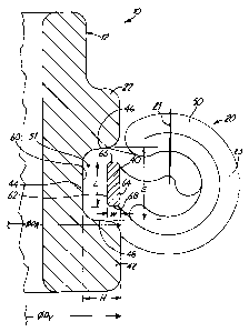
Figure 10 is a side plan view of a cuff
assembly apparatus 100 for use in assembling a
prosthetic heart valve body and sewing cuff in
accordance with the present invention. Assembly
apparatus 100 includes base 102 and orifice ring support
104 which carries orifice ring 12 (not shown in Figure
10) by means of mating feature 105. Mating feature 105
captures pivot guards 14 and prevents rotation of the
valve body relative to the orifice ring support 104. A
cuff insertion finger 107 is fixed to and projects
downward from a cuff insertion head 106 and is adapted
to press upon the fabric tube 50 and cuff retaining ring
40 during the assembly process with respect to Figures
5-9. Head 106 is coupled to wheel 110 and handle 111
through shaft 108, which is rotatably carried in support
112. During assembly, fabric tube 50 is placed over
orifice ring 12. Then cuff retaining ring 40 is placed
over fabric tube 50 and above proximal rim 42. A fold
70 of fabric tube 50 is then brought over retaining ring
40. Wheel 110 is then rotated while head 106 moves
downward, limited by stop 109, to engage finger 107 with
the cuff retaining ring 40 which is covered by fold 70.
The force conveyed through finger 107 thereby
momentarily stretches a subjacent portion of cuff
retaining ring 40 while momentarily compressing a
corresponding volume of fabric tube 50. Thus, the
subjacent portion of ring 40 is moved over proximal rim
42 and into recess 44. As the head 106 is rotated, the
finger 107 pushes an increasing portion of retaining
ring 40 into recess 44, until the entire cuff retaining
ring 40 resides in recess 44 over the inner annular cuff
portion of fabric tube 50 when finger 107 has
sufficiently traversed around orifice ring 12. Other
CA 02383328 2002-03-22
WO 01/21112 PCT/LTS00/40949
-12-
devices can be used to assemble the valve and device 100
is provided as one example.
In an embodiment, the annular cuff portion of
fabric tube 50 is heated to above a glass transition
temperature of its fabric under a compressive load prior
to assembly. This results in a reduction in thickness
in the annular cuff portion to a generally uniform
value, which is beneficial in maintaining a consistent
range of torque. In an embodiment, the material of
fabric tube 50 includes a coating or impregnation such
as an ion-beam implantation of a substance, for example,
silver, toxic to bacteria or other microbes.
A heart valve prosthesis in accordance with
the present invention provides a substantially
controllable and predictable level of rotation
resistance torque over a desired range. Further, such
prosthesis requires only a single cuff retention ring
which can be manufactured to be relatively thin to
thereby reduce the bulk of stiff cuff retention
mechanism. Indeed, for a preferred embodiment, the cuff
retention ring does not extend past the outermost
surface of the valve housing. This prevents
interference with patient's tissues and provides a
maximum volume of sewing cuff available for suturing.
Thus, minimizes the chance of the ring impeding suture
needle penetration. Furthermore, the thin cross-
sections of the valve housing and cuff retention ring
over-all allows an increase in the lumen area of the
prosthesis, thereby improving blood flow. This
improvement in blood flow is highly beneficial to the
patient.
The rotation mechanism of the invention
provides a relatively low profile (i.e., thin in an
axial direction) design in comparison to prior art
CA 02383328 2002-03-22
WO 01/21112 PCT/US00/40949
-13-
configurations. The present rotation mechanism requires
only a relatively small area. Further, the mechanism is
relatively thin in a radial direction. Preferably, the
ring 40 does not extend in a radial direction beyond the
outer radius of rings 22 and 42 such that a large amount
of cuff 20 is available for suturing.
Other benefits of the present invention
include the following. The metal cuff retaining ring
provides radiopacity. The components can be
manufactured using standard prosthetic valve
manufacturing techniques. The assembly steps do not
require any critical adjustments by an operator to
achieve a desired resistance to rotation.
Although the present invention has been
described with reference to preferred embodiments,
workers skilled in the art will recognize that changes
may be made.in form and detail without departing from
the spirit and scope of the invention.