Note: Descriptions are shown in the official language in which they were submitted.
CA 02385420 2007-08-27
1 SUTURE MATERIAL HAVING ANTIMICROBIAL CHARACTERISTICS
2
3 The present invention relates to a suture material
4 having antimicrobial characteristics.
6 Sutures are the threads or wires used to stitch two
7 bodily surfaces together. Typically, sutures are
8 required to close surgical incisions and to treat deep
9 lacerations inflicted on a patient.
11 Suture types fall into two main categories; absorbable
12 and non-absorbable. Additionally, the sutures can be
13 of monofilament or multifilament structure, with the
1-1 multifilament sutures beirig braided or twisted. A
variety of sizes of sutures are available. Typical
16 commercially available suture types are listed below:
17
18 Non-absorbable:
19
Silk twisted, braided & multifilament
21 Nylon polyamide monofilament
-1-
CA 02385420 2007-08-27
7
1 Polypropylene monofilament
2 Polyester braided multifilament
3 PTFE monofilament
4 PVDF monofil-ament
Stainless steel monofilament
6 Linen multifilament
7
8 Absorbable:
9
PGA monofilament & multifilaments
11 PLA monofilament & multifilaments
12 Lactide/Glycolide
13 Copolymers monofilaments & multifilaments
14 Catgut monofilament
Collagen monofilament
16
17 In general braided multifilaments have a smoother
18 surface than the alternative twisted multifilaments and
19 so remain more cohesive when stitched. Monofilaments,
being formed from a single fibre, cannot unravel and
21 thus lose cohesiveness.
22
23 To improve the lubrication along the surface of the
24 suture and to provide friction to improve knot
strength, the sutures may be coated. Conventionally
26 however monofilament sutures are not coated. Coatings
27 which may be applied include 1001; beeswax BP,
28 Silicone, PTFE (e.g. TeflonTM), PVP, polylactic acid
29 (PLA), polyglycolactide (PLG), polycaprolactones and
copolymers thereof. Often the coatings will
CA 02385420 2008-07-23
-3-
1 incorporate detergents or other lubricating substances,
2 e.g. calcium stearate.
3
4 However, sutures used for surgical wound closure are
associated with increased bacterial infectivity. Sutures
6 draw contaminants into the wound closure and provide a
7 surface along which micro-organisms can track as a
8 biofilm. Contamination of the wound via the suture can
9 arise from the local environment (particularly in gut
surgery), the closure area around the wound, inappropriate
11 handling of the suture or from contaminated suture stock.
12
13 It is an object of the present invention to reduce the
14 risk of infection due to suturing a wound, by providing
sutures having antimicrobial characteristics.
16
17 Thus, in one aspect, the present invention provides a
18 surgical suture material having either: (a) an external
19 surface at least partially coated with an anti-microbial
composition comprising a water-soluble, metal ion-
21 releasing glass containing from about 30 to about 60%
22 phosphorous pentoxide; or (b) a water-soluble, metal ion-
23 releasing glass containing from about 30 to about 60%
24 phosphorous pentoxide as an anti-microbial agent
incorporated therein.
26
27 The surgical suture material may be formed from any
28 suitable substance and may be absorbable or nonabsorbable.
29 Mention may be made of silk, polyester, nylon,
polypropylene, polyvinylidenefluoride, linen, steel wire,
31 catgut (beef serosa or ovine submucosa),
CA 02385420 2002-03-15
WO 01/28601 PCT/GBOO/04049
4
1 polyglycolactide, polyamide (e.g. polyamide nylon),
2 fibroin, polyglycolic acid and copolymers thereof. The
3 sutures may be monofilament or may be braided or
4 twisted multifilament yarns.
6 The anti-microbial composition if to be applied as a
7 coating may be applied to the suture surface in the
8 same way as a conventional coating. Indeed, a
9 conventional coating material admixed with or including
an anti-microbial agent is suitable for use in the
11 present invention.
12
13 Preferably the anti-microbial agent is biodegradable
14 over a period of time compatible with the timescale of
wound healing. A slow-release of the anti-microbial
16 active ingredient of the agent over a period of weeks
17 or months is thus desirable.
18
19 A preferred anti-microbial agent is a water-soluble
metal ion-releasing glass, especially in particle (e.g.
21 fine powder) form that may be simply admixed with a
22 conventional coating and applied to the suture
23 material. Advantageously the metal released by the
24 glass is silver.
26 Thus we have found that by incorporating a comminuted
27 anti-microbial water soluble glass either into the
28 suture material itself or coated onto the external
29 surface thereof, the infectivity of a wound site is
reduced, whilst the handling characteristics
31 (knotability and insertion lubricity) are maintained.
CA 02385420 2007-08-27
1 Phosphorous pentoxide (P205) is used as the
2 glass former of the biodegradable glass used in the
3 coating.
4
5 The mole percentage of phosphorous pentoxide
6 in the glass composition is between 30-600.
7
8
9 Alkali metals, alkaline earth metals and lanthanoid
oxides or carbonates are preferably used as glass
11 modifiers. Generally, the mole percentage of alkali
12 metals, alkaline earth metals and lanthanoid oxides or
13 carbonates is less than 60%, preferably between 40-60 s.
14
Boron containing compounds (eg B203) are preferably used
16 as glass additives. Generally, the mole percentage of
17 boron containing compounds is less than 15% or less,
18 preferably less than 5%.
19
Other compounds may also be added to the glass to
21 modify its properties, for example SiO2, A1203, SO3,
22 sulphate ions (S042-), transition metal compounds (eg.
23 first row transition metal compounds) or mixtures
24 thereof.
26 Typically the soluble glasses used in this invention
27 comprise phosphorus pentoxide (P205) as the principal
28 glass-former, together with any one or more
29 glass-modifying non-toxic materials such as sodium
oxide (Na20), potassium oxide (K20) , magnesium oxide
31 (MgO), zinc oxide (ZnO) and calcium oxide (CaO) or
CA 02385420 2002-03-15
WO 01/28601 PCT/GBOO/04049
6
1 mixtures thereof. The rate at which the glass
2 dissolves in fluids is determined by the glass
3 composition, generally by the ratio of glass-modifier
4 to glass-former and by the relative proportions of the
glass-modifiers in the glass. By suitable adjustment
6 of the glass composition, the dissolution rates in
7 water at 38 C ranging from substantially zero to
8 25mg/cm2/hour or more can be designed. However, the
9 most desirable dissolution rate R of the glass is
between 0.01 and 2.Omg/cm2/hour.
11
12 The water-soluble glass is preferably a phosphate
13 glass, and preferably comprises a source of silver ions
14 which may advantageously be introduced during
manufacture as silver orthophosphate (Ag3PO4) . The
16 glass preferably enables controlled release of silver
17 or other metal ions, for example Zn, Cu, Mg, Ce, Mn,
18 Bi, Se, Cs and mixtures thereof (preferably Ag, Cu, Zn
19 and Mg and mixtures thereof) and other constituents in
the glass and the content of these additives can vary
21 in accordance with conditions of use and desired rates
22 of release, the content of silver generally being up to
23 5 mole o. While we are following convention in
24 describing the composition of the glass in terms of the
mole o of oxides, of halides and of sulphate ions, this
26 is not intended to imply that such chemical species are
27 present in the glass nor that they are used for the
28 batch for the preparation of the glass.
29
The optimum rate of release of the metal ions (eg Ag,
31 Cu, Zn or Mg, or any of the other metal ions mentioned
CA 02385420 2002-03-15
WO 01/28601 PCT/GBOO/04049
7
1 above) into an aqueous environment may be selected by
2 circumstances and particularly by the specific function
3 of the released metal ion. The invention provides a
4 means of delivering metal ions to an aqueous medium at
a rate which will maintain a concentration of metal
6 ions in said aqueous medium of not less than 0.01 parts
7 per million and not greater than 10 parts per million.
8 In some cases, the required rate of release may be such
9 that all of the metal added to the system is released
in a short period of hours or days and in other
11 applications it may be that the total metal be released
12 slowly at a substantially uniform rate over a period
13 extending to months or even years. In particular cases
14 there may be additional requirements, for example it
may be desirable that no residue remains after the
16 source of the metal ions is exhausted or, in other
17 cases, where the metal is made available it will be
18 desirable that any materials, other than the metal
19 itself, which are simultaneously released should be
physiologically harmless. In yet other cases, it may
21 be necessary to ensure that the pH of the resulting
22 solution does not fall outside defined limits.
23
24 Generally, the mole percentage of these additives in
the glass is less than 250, preferably less than 10%.
26
27 In a preferred embodiment the biodegradable glass
28 comprises 20-35 moleo Na20; 18-30 mole% CaO and 45-60
29 mole o P2O5 .
CA 02385420 2002-03-15
WO 01/28601 PCT/GBOO/04049
8
1 It is a further object of the invention to provide a
2 method of reducing the risk of infection and provide
3 faster and more efficient healing of the wound by using
4 the suture material of the invention to close the
wound.
6
7 The present invention will now be further described by
8 reference to the following, non-limiting, examples and
9 to figures, in which:
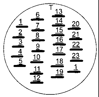
11 Fig. 1 shows the template used in the example
12 to facilitate regular application of the
13 suture lengths on the plates.
14
Figs. 2-6 : show digitally generated photographic
16 images showing the results of Example 2.
17
18 EXAMPLE 1: Suture Coating Preparation
19
Glasses were prepared according to Table 1.
21
22
23
24
26
27
28
29
31
CA 02385420 2002-03-15
WO 01/28601 PCT/GB00/04049
9
1 Table 1
2
Annealed Mode m Composition Code
Solution Rate
Mg. cm. -2 hr"1
Na20 CaO P205 AgZ0
0.14 23.71 22 26.5 47.0 4.5 01
1.42 19.44 33. 16.5 47.0 3.0 02
0.27 19.96 27.5 22 47.0 3.5 03
1.42 6.50 33 16.5 47.0 3.0 04
16.05 14.02 30 10 47.5 6.5 05
6.02 12.64 36 13 47.5 3.5 06
3.48 25.44 34.5 14.5 47.5 3.5 07
11.28 12.20 36 11.5 47.5 5.0 08
3
4 These glasses were prepared as powders (mode size given
in m in Table 1 above) for incorporation into a suture
6 coating.
7
8 Testing
9
Physical/Mechanical
11
12 It is important that addition of silver ion releasing
13 glass into the coating does not compromise the physical
14 or mechanical properties of the suture. The smoothness
of the coating is essential in ensuring smooth
16 insertion of the suture. The coating should not slough
17 off on insertion and the knot properties should not be
CA 02385420 2007-08-27
Lo
1 reduced. Test samples show that up to 2.501 wt/wt
2 (final dry weight of coating) of glass powder could be
3 added to the coating without affectirig these properties
4 and up to 55% wt/wt may be possible with some samples.
6 Samples
7
8 Glass samples 01 and 04 were applied to
9 glycolide/lactide copolymer braided multifilament
sutures in a glycolide/caprolactone coating at various
11 weights. The coat weight applied was 2% wt/wt dry
12 weight coating onto the suture. Samples G1 to Gl0
13 contain glass 01 from 0.25-2.5 s wt/wt dry weight in the
14 coating. Gil to G20 contain glass 04 at 0.25 to 2.5's
wt/wt dry weight in the coating. G21 is a nylon
16 monofilament with 2% wt/wt coating containing 2.5%
17 wt/wt of 04. This coating did not bond well with the
18 suture G22 and G23 and control copolymer and control
19 nylon sutures respectively.
21 EXAMPLE 2: Anti-microbial Activity
22
23 Gl to G23 were screened against 17 test organisms.
24
Suture Material
26
27 Gl to G20-Violet Polysorb size 0 sutures
28 G21-DacronTM suture size 2/0
29 G22-Violet PolysorbTMcontrol
G23-DacronTMcontrol
31
CA 02385420 2002-03-15
WO 01/28601 PCT/GBOO/04049
11
1 Test Organisms
2
3 A panel of "wild-type" clinical isolates was used
4 except for organism 5, Staph epidermidis NCTC 11047.
This organism is a reference organism noted to be
6 sensitive to test sutures utilised in a previous
7 experiment.
8
9 Gram-positive Isolates
11 1. Enterococcus faecalis
12 2. Staphylococcus aureus
13 3. Enterococcus faecalis - vancomycin resistant (VRE
14 - VanA genotype)
4. Methicillin-resistant Staphylococcus aureus (MRSA
16 - epidemic type 15)
17 S. Staphylococcus epiderrnidis NCTC 11047.
18 6. Streptococcus agalactiae (Group B streptococcus)
19
Gram-negative Isolates
21
22 7. Stenotrophomonas maltophilia (formerly Xanthomonas
23 maltophilia)
24 8. Pseudomonas aeruginosa - strain 1
9. Pseudomonas aeruginosa - strain 2
26 10. Serratia marcescens
27 11. Enterobacter cloacae
28 12. Morganella morganii
29 13. Escherichia coli
14. Klebsiella pneumoniae
31 15. Acinetobacter sp.
CA 02385420 2002-03-15
WO 01/28601 PCT/GBOO/04049
12
1 Yeasts
2
3 16. Candida albicans
4 17. Candida gl abra ta
6 Method
7
8 Media - 9 cm plates of Oxoid Iso-sensitest agar were
9 used for all organisms except the candida isolates
which were plated on Yeast Morphology Agar.
11
12 Inoculum - Overnight plate cultures of the test
13 organisms were emulsified in physiological saline to
14 achieve a semi-confluent growth on the agar plates.
16 Inoculum procedure - The plates were pre-dried at 37 C
17 for 2 hours. The inoculum was applied using a sterile
18 swab using a cross-streaking technique.
19
Suture application - The suture was cut into
21 approximate 1 cm lengths using sterile instruments.
22 Where possible, straight sections of suture were used.
23 A template was constructed to facilitate regular
24 application of the suture lengths. Each plate of test
organism had the series of 21 test and 2 control
26 sutures applied, with a replicate of suture Gl as an
27 internal control on the far side of the plate (see
28 Figure 1 for template). Each suture was pressed down
29 with sterile forceps to optimise contact with the agar
surface.
31
CA 02385420 2002-03-15
WO 01/28601 PCT/GBOO/04049
13
1 Incubation - 37 C for 18 hours. The plates were
2 reassessed after a further 24 hours.
3
4 Recording of results - The maximum width of the zone of
inhibition at right angles to the suture length was
6 recorded to nearest 0.5 mm (the maximum width was
7 recorded to avoid skewing of results due to incomplete
8 contact of parts of the suture with the agar surface,
9 resulting in irregular zones - see photographic
results).
11
12 Results
13
14 See digitally generated photographic images provided as
Figs. 2 to 6 and Table 2.
16
17 Conclusions
18
19 G21-Dacron suture:
Zones of inhibition were seen with all test organisms
21 except Candida albicans (organism 16).
22
23 G23-Dacron control suture:
24 No demonstrable activity.
26 G1-G20-violet Polysorb suture:
27 There was a general trend towards increasing activity
28 with the higher Polysorb suture numbers, with zone
29 sizes plateauing with G14, 15 and 16 followed by a
slight decline.
31
CA 02385420 2002-03-15
WO 01/28601 PCT/GBOO/04049
14
1 Activity was seen against most organisms in the panel.
2 No zones were seen with two candida isolates (organisms
3 16 and 17) and the zones for Stenotrophomonas
4 maltophilia (organism 7) and Enterobacter cloacae
(organism 11) tended to be smaller, or absent compared
6 to the other Gram-negative isolates.
7
8 Activity against the staphylococcal isolates (organisms
9 2, 4 and 5) was seen with virtually all sutures. This
is of note given the particular importance of
11 staphylococci in the aetiology of stitch abscesses.
12
13 The enterococci and streptococci (organisms 1, 3 and 6)
14 demonstrated the largest zones of inhibition.
Interestingly, the control suture (G22) also yielded
16 significant zones for all three organisms, indicating
17 that one of the constituents of the suture has
18 antimicrobial activity in its own right. This
19 constituent must be released from the suture and be
able to diffuse through the agar. There is apparent
21 interaction with the components of the test sutures -
22 G2 consistently gave zones smaller than the control.
23
24 As will be seen from the digital images (Figs. 2 to 6)
the inoculum ranged from semi-confluent to near
26 confluent growth. The Gram-negative organisms tended
27 to a heavier inoculum. Despite the significant
28 challenge, zones of inhibition were seen. At this
29 stage the duration of activity of the test sutures
cannot be stated - however, transient contact with the
CA 02385420 2002-03-15
WO 01/28601 PCT/GBOO/04049
1 surface of the agar (duration less than 5 seconds)
2 resulted in a small zone of inhibition.
3
4 EXAMPLE 3: Anti-microbial Activity
5
6 Protocol
7
8 As for Example 2.
9
10 The experiment was performed to confirm the results
11 from the previous experiment, in particular the
12 activity of the G22 control suture against the
13 enterococci and streptococci, and the effect of a lower
14 inoculum on the results from the Gram-negative
15 organisms.
16
17 Results
18
19 See Table 3.
21
22
23
24
26
27
28
29
31
CA 02385420 2002-03-15
WO 01/28601 PCT/GBOO/04049
16
1 Table 3 : Maximum width of zone of inhibition measured
2 at right angles to the suture (millimetres)
3
ORGANISM
1 5 Staph 11047 6 Gp B strept 13 E Coli
Enterococcus
Suture
G4 9 m 2 m 8 m 0 m
G9 9 m 2.5 m 9 m 1 m
G11 8 m 2.5 m 10 m 1.5m
G14 7.5 m 3.5 m 9 m 2 m
G17 8 m 2.5 m 8 m 2 m
G22 9 m 0 m 12 m 0 m
4 Key: m microcolonies present within zone of inhibition
6 Conclusions
7
8 Zone sizes were similar to the results from Example 2.
9 Control suture G22 again demonstrated activity against
both enterococci and Gp B streptococci. The zone sizes
11 for the E coli using a lighter inoculum were similar to
12 previous results.
13
14 EXAMPLE 4: Controlled Release
16 Suture Material
17
18 Gil - previously noted to yield a small zone of
19 inhibition with NCTC 11047.
CA 02385420 2002-03-15
WO 01/28601 PCT/GBOO/04049
17
1 G16 - previously noted to yield a large zone of
2 inhibition with NCTC 11047.
3
4 Test organism
6 Staphylococcus epidermidis NCTC 11047.
7
8 Method
9
A single plate of Oxoid Iso-sensitest agar (Plate 1)
11 was seeded with the test organism to achieve a semi-
12 confluent growth. Four Gll sutures were applied to one
13 side of the plate, with four G16 sutures on the
14 opposite side. Each suture had been bent to yield a 90
kink in the middle. After 24 hours incubation at 37 C
16 the zones of inhibition at right angles to the sutures
17 were recorded and the sutures were transferred to a
18 freshly seeded Iso-sensitest plate (Plate.2). The kink
19 in the suture ensured that the same aspect of the
suture was in contact with the agar surface on each
21 occasion. The new plate was incubated for a further 24
22 hours and the sutures were removed prior to assessment
23 of zones of inhibition.
24
Results
26
27 Plate 1 - Each of the Gll sutures yielded a zone of
28 inhibition 1.5 mm in (maximum) width. The G16 sutures
29 yielded zones 2.0 mm in width.
CA 02385420 2002-03-15
WO 01/28601 PCT/GBOO/04049
18
1 Plate 2 - After transfer to Plate 2, zones of
2 inhibition were not seen for either suture. On removal
3 of the sutures it was observed that there was confluent
4 growth of the test organism under the Gil sutures, but
there was inhibition of growth under G16.
6
7 Conclusions
8
9 After 24 hours in contact with the agar surface of
Plate 1, suture G1l had no demonstrable activity
11 against the test organism on Plate 2. Suture G16
12 demonstrated marginal activity on Plate 2, with
13 inhibition of growth directly underneath the suture
14 material.
16 EXAMPLE 5: Controlled Release
17
18 Suture material
19
G15 - previously noted to yield a large zone of
21 inhibition with NCTC 11047.
22
23 Test oraanism
24
Staphylococcus epidermidis NCTC 11047.
26 Method
27
28 A single plate of Oxoid Iso-sensitest agar was seeded
29 to yield a semi-confluent growth of NCTC 11047.
Sixteen sutures were applied with sterile forceps and
31 the plate was incubated at 37 C. At various time
CA 02385420 2002-03-15
WO 01/28601 PCT/GB00/04049
19
1 intervals sutures were removed. Two sutures were
2 assessed for each time except for "24 hours" where 4
3 sutures were used. At the end of the 24-hour period
4 the zones of inhibition were assessed and the plate was
photographed.
6
7 Results and Conclusions
8
9 Suture G15 exhibited activity against the test organism
when in contact with the agar surface for only 5
11 minutes. Activity increases up to the 3 hour point,
12 after which no increased activity is seen.
13
14 EXAMPLE 6: Duration of Anti-microbial effect
16 Suture
17
18 G16.
19
Test organism
21
22 Staphylococcus epidermidis NCTC 11047.
23
24 Method
26 An Oxoid Iso-sensitest agar plate was seeded with the
27 test organism to achieve a semi-confluent growth. Six
28 G16 sutures were applied with sterile forceps and the
29 plate was incubated for 24 hours at 37 C. An
uninoculated Iso-sensitest plate was also incubated as
31 the Control.
CA 02385420 2002-03-15
WO 01/28601 PCT/GBOO/04049
1 After the initial incubation period each suture was
2 surrounded by a zone of inhibition. Three of the six
3 sutures were then removed. Each zone of inhibition was
4 challenged using a calibrated loop to apply a drop of
5 standardised suspension of test organism. Each drop
6 contained approximately 104 colony forming units. An
7 identical drop was applied to the Control plate. Both
8 plates were then incubated for a further 24 hours and
9 the zones were challenged again, and a further drop was
10 added to the Control plate. The procedure was repeated
11 on a daily basis. The end point of the experiment was
12 when growth appeared in the original zones of
13 inhibition following challenge, or when the Control
14 plate lost the ability to support organism growth due
15 to progressive dehydration. (This was minimised by
16 incubating the plates in an atmosphere with high
17 humidity.)
18
19 Results
21 Over the thirteen days of the experiment, no growth was
22 seen in any of the zones of inhibition. There was no
23 difference between the zones where the suture remained
24 in place and the zones where the suture had been
removed. The experiment was terminated at the 13 day
26 point even though the Control plate continued to
27 support growth of the challenge organism. This was
28 because the test plate appeared to be dehydrating more
29 rapidly, presumably because of the influence of the
lawn of growth of NCTC 11047 on its surface.
31
CA 02385420 2002-03-15
WO 01/28601 PCT/GBOO/04049
21
1 Conclusions
2
3 Experiment 1 demonstrated that much of the activity of
4 the suture is released in the first 24 hours.
Experiment 2 showed that activity is present within 5
6 minutes of contact with the agar. Experiment 3
7 illustrates that even though the suture may be
8 depleted, the surrounding area returns antimicrobial
9 activity over a period in excess of one week.
11 EXAMPLE 7: Cytotoxity
12
13 1. Objective
14
To determine the cytotoxity of a series of suture
16 samples using a standard extraction/elution test, after
17 ISO 10993 part 5.
18
19 2. Scope
21 The test procedure applies to all suture samples which
22 were received sterile.
23
24 3. Equipment and Materials
3.1 Equipment
26 3.1.1 Laminar air flow hood.
27 3.1.2 Incubator maintained at 37 C/5o carbon
28 dioxide.
29 3.1.3 Refrigerator at 4 C.
3.1.4 Freezer at -18 C.
31 3.1.5 Vacuum source.
CA 02385420 2002-03-15
WO 01/28601 PCT/GB00/04049
22
1 3.1.6 Phase contrast microscope.
2
3 3.2 Materials
4 3.2.1 Sterile plastic-ware pipettes.
3.2.2 Sterile glass pipettes.
6 3.2.3 24 well sterile dishes.
7 3.2.4 Surgical grade forceps.
8 3.2.5 Surgical grade scissors.
9 3.2.6 Sterile Universal containers.
3.2.7 L929 cell culture line (ATCC NCTC Clone
11 929).
12 3.2.8 TCPS negative control.
13 3.2.9 Natural rubber latex control.
14 3.2.10 Other control samples were supplied in
suture form.
16
17 4. Procedure
18
19 4.1 Test sample preparation
4.1.1 Test samples and controls were cut to
21 the appropriate size (see Section
22 4.2.1).
23 4.1.2 Tissue culture polystyrene was employed
24 as a negative control. Natural rubber
latex was employed as a positive
26 control. The controls were not in the
27 same physical form as the test material.
28
29 4.2 Extraction/elution method
CA 02385420 2002-03-15
WO 01/28601 PCT/GBOO/04049
23
1 All procedures carried out within laminar air
2 flow.
3 4.2.1 Sutures were prepared to provide a
4 surface area equivalent to 120 cm sq.
for each 20 mL of extracting medium.
6 4.2.2 Suture samples (typically 6 cm in
7 length) were transferred to Sterile
8 Universal containers.
9 4.2.3 Each container was labelled with the
test material code number.
11 4.2.4 20 mL of mammalian cell culture medium
12 (199) was added to each container.
13 4.2.5 The containers were placed in the
14 incubator 37 C/5o carbon dioxide for 24
hours.
16
17 4.3 Cell preparation
18 4.3.1 A cell subculture was prepared on the
19 same day the extracts were initiated.
4.3.2 Cells were plated into 24 well dishes at
21 a cell concentration of approximately 1
22 x 105 cells mL. Enough wells were
23 prepared to allow four wells per test
24 sample. 2 mL of serum supplemented
medium was added to each well.
26 4.3.3 The 24-well plates were incubated for 24
27 hours at 37 C/5o carbon dioxide.
28
29 4.4 Test procedure
4.4.1 After 24 hours all 24 well plates were
31 examined by phase-contrast microscope
CA 02385420 2002-03-15
WO 01/28601 PCT/GBOO/04049
24
1 (x20 objective lens) to ensure healthy
2 monolayer of >80% confluence.
3 4.4.2 The culture medium is aspirated.
4 4.4.3 The Universal containers are removed
from the extraction conditions, the pH
6 monitored using phenol red indicator.
7 4.4.4 2 mL of extracted medium is placed in
8 each well and the plates re-incubated
9 for a 48-hour period.
11 4.5 Interpretation of results
12 4.5.1 At the conclusion of the incubation
13 period the plates are removed from the
14 incubator and examined under phase
contrast microscope using xlO and x20
16 objective lenses.
17 4.5.2 Each test and control material was
18 evaluated using the scoring system
19 detailed below.
Reactivity Response Table
Grade Reactivity Conditions of all cultures
0 None Discrete intracytoplasmic granules; no
cell lysis
1 Slight No more than 20% of the cells are round,
loosely attached and without
intracytoplasmic granules; occasional
lysed cells are present
2 Mild No more than 50% of the cells are round
and devoid of intracytoplasmic granules;
extensive cell lysis and empty areas
between cells
3 Moderate No more than 70% of the cell layers
contain rounded cells and/or are lysed
4 Severe Nearly complete destruction of the cell
layers
CA 02385420 2002-03-15
WO 01/28601 PCT/GB00/04049
1 4.6 Results
2
3 The following table (Table 4) highlights the
4 results obtained following two separate tests:
5 Two readings were taken at each test. In all
6 cases negative control (TCPS) provided a 0 grade
7 and positive control provided a 2 grade.
8
9 Table 4
Material Grade Material Grade Material Grade
Code Test 1 Test 2 Code Test 1 Test 2 Code Test 1 Test 2
Gl 0 0 0 0 G9 0 0 0 0 G17 1 0 0 0
G2 0 0 0 0 G10 0 0 0 0 G18 1 0 0 0
G3 0 0 0 0 Gil 0 0 0 0 G19 1 1 0 0
G4 0 0 0 0 G12 0 1 0 0 G20 1 1 0 0
G5 0 0 0 0 G13 1 1 0 0 G21 1 1 0 0
G6 0 1 0 0 G14 1 1 0 0 G22 2 1 0 1
G7 0 0 0 0 G15 1 1 0 0 G23 1 1 0 0
G8 0 0 0 0 G16 1 1 0 0
11 Comments
12
13 The results as detailed provide a very subjective
14 assessment of material cytotoxity. Where a grade 0 is
shown, there was no evidence of toxicity and a
16 confluent healthy monolayer of cells was present.
17 Where there was any evidence of floating cells or
18 morphological abnormality or sub-confluent growth a
19 grade 1 was allocated. It should be noted that
floating cells do not necessarily indicate toxicity.
21 It should also be noted that the test 2 indicated less
22 evidence of toxicity than test 1. The extracts (with
CA 02385420 2002-03-15
WO 01/28601 PCT/GB00/04049
26
1 suture material removed) had been maintained in a
2 frozen state for 72 hours before re-testing.
CA 02385420 2002-03-15
WO 01/28601 PCT/GBOO/04049
27
TABLE 2: Maximum width of zone of inhibition measured
at right angles to the suture (millimetres)
ORGANISM
1 2 3 4 5 6
Enterococcus Staphylococcus VRE MRSA Staph Gp B
11047 Strep
Suture
Gi 6m (7m) 0 (0) 8 (9) 0 (1) 1 (0) 9 (8)
G2 3.5m 0 3 1 1 1.5
G3 5m 1 7 1.5 1 7
G4 7m 1 8.5 1.5 1.5 7
G5 3.5m 1 7.5 1.5 1.5 9
G6 5m 2 8 1.5 1.5 3
G7 6.5m 2 8 1.5 2 7
G8 6.5m 2 8 2 2.5 8
G9 7m 2 9 1.5 1.5 8
G10 7m 2 8 1 1.5 7
G11 6m 2m 8 2m 1.5 7
G12 2m 2m 7 2m 2 8
G13 5m 2.5m 7.5 2m 1.5m 8
G14 6m 2.5m 7 1.5m 2.5m 3.5
G15 6m 2.5m 8 2m 2.5 8.5
G16 7m 2.5m 8 2.5m 2.5 8.5
G17 5.5m 2 9 1.5 2 7.5
G18 7m 2 9 1.5 1.5 7
G19 8m 1.5 12 1.5 1.5 8
G20 8m 1.5 13 1 1.5 8
G21 1.5m 2m 2 2m 1.5 2.5
G22 8m 0 9 0 0 12
Control
G23 0 0 0 0 0 0
Control
CA 02385420 2002-03-15
WO 01/28601 PCT/GBOO/04049
28
TABLE 2 (CONT'D): Maximum width of zone of inhibition
measured at right angles to the suture (millimetres)
ORGANISM
7 8 9 10 11 12
Steno Malto Pyo 1 Pyo 2 Serr Enter Morg
marcescens cloacae morganii
Suture
G1 0 (0) 0 (0) 0 (0) 0 (0) 0 (0) 0 (0)
G2 0 0 1 0 0 0
G3 0 1 1 0 0 1
G4 0 1 1 1 0 0
G5 0 1 1.5 1 0 1
G6 0 1.5 1.5 1.5 0 1.5
G7 0 1.5 2 1.5 1 2
G8 0 1.5 2 1.5 0 2
G9 0 1 1 1.5 0 1.5
G10 0 1 1 1.5 0 1.5
Gil 0 0 1 1.5 0 1.5
G12 0 0 1.5 1.5 0 0
G13 1 2 1.5 1 0 2
G14 1 0 2 2 1 1
G15 1 2 2 2 1 2.5
G16 1 1.5 2.5 2 1 2.5
G17 1 1 1.5 1.5 1 2
G18 0 0 1.5 1 0 1.5
G19 0 0 1.5 0 0 1.5
G20 0 1 1 1 0 1
G21 1 1.5 1 1.5 1 2
G22 0 0 0 0 0 0
Control
G23 0 0 0 0 0 0
Control
CA 02385420 2002-03-15
WO 01/28601 PCT/GBOO/04049
29
TABLE 2 (CONT'D): Maximum width of zone of inhibition
measured at right angles to the suture (millimetres)
ORGANISM
13 14 15 16 17
E Coli K1 pneumoniae Acinetobacter sp C albicans C glabrata
Suture
G1 1 (0) 0 (0) 0 (0) 0 (0) 0 (0)
G2 0 0 0 0 0
G3 0 1 0 0 0
G4 1 1 0 0 0
G5 1 1 1 0 0
G6 1.5 1.5 1.5 0 0
G7 1.5 1.5 0 0 0
G8 1.5 1.5 1.5 0 0
G9 1 1 1 0 0
G10 1.5 1 1 0 0
Gil 1.5 1.5 1 0 0
G12 1 1 1 0 0
G13 1.5 2m 1.5m 0 0
G14 2m 2m 2m 0 0
G15 2m 2m 2m 0 0
G16 2m 2m 2m 0 0
G17 1.5 1.5 1.5 0 0
G18 1 1 1.5 0 0
G19 0 1 1.5 0 0
G20 1.5 1 1 0 0
G21 1.5 1.5 1.5m 0 1
G22 0 0 0 0 0
Control
G23 0 0 0 0 0
Control
Key
() - Gl Replicate result
m - microcolonies present within zone of inhibition