Note: Descriptions are shown in the official language in which they were submitted.
CA 02386504 2007-06-06
52723-1
-1-
Facet Arthroplasty Devices And Methods
FIELD OF THE INVENTION
The present invention generally relates to
devices and surgical methods for the treatment of various
types of spinal pathologies. More specifically, the
present invention is directed to several different types
of spinal joint replacement prostheses, surgical
procedures for performing spinal joint replacements, and
surgical instruments which may be used to perform the
surgical procedures.
BACKGROUND OF THE INVENTION
Back pain is a common human ailment. In fact,
approximately 50% of persons who are over 60 years old
suffer from lower back pain. Although many incidences of
back pain are due to sprains or muscle strains which tend
to be self-limited, some back pain is the result of more
ckironic fibromuscular, osteoarthritic, or ankylosing
spondolytic processes of the lumbosacral area.
Particularly in the population of over 50 year olds, and
most commonly in women, degenerative spine diseases such
CA 02386504 2007-06-06
52723-1
- 2 -
as degenerative spondylolisthesis and spinal stenosis
occurs in a high percentage of the population. Iida, et
al, 1989.
Degenerative changes of the adult spine' have
traditionally been determined to be the result of the
interrelationship of the three joint complex; the disk and
the two facet joints. Degenerative changes in the disc
lead to arthritic changes in the facet joint and vice
versa.
One cadaver study of 19 cadavers with
degenerative spondylolisthesis showed that facet
degeneration was more advanced than disc degeneration in
all but two cases. Farfan. In mild spondylolisthetic
cases, the slip appeared to be primarily the result of
predominantly unilateral facet subluxation. Other studies
into degenerative changes of the spine have revealed
extensive contribution of facet joint degeneration to
degenerative spinal pathologies such as degenerative
spondylolisthesis, central and lateral stenosis,
degenerative scoliosis, and kypho-scoliosis, at all levels
of the lumbar spine.
it has been determined that facet joint
degeneration particularly contributes to degenerative
spinal pathologies in levels of the lumbar spine with
sagittally oriented facet joints, i.e. the L4-L5 level.
When intractable pain or other neurologic
involvement results from adult degenerative spine
diseases, such as the ones described above, surgical
procedures may become necessary. Traditionally, the
surgical management of disease such as spinal stenosis
CA 02386504 2007-06-06
52723-1
- 3 -
consisted of decompressive laminectomy alone.
Wide decompressive laminectomies
remove the entire lamina, and the marginal osteophytes
around the facet joint. Because a lot of degenerative
spine disease has been demonstrated to be caused by facet
joint degeneration or disease, this procedure removes
unnecessary bone from the lamina and insufficient bone
from the facet joint.
Furthermore, although patients with one or two
levels of spinal stenosis tend to do reasonably well with
just a one to two level wide decompressive laminectomy,
patients whose spinal stenosis is associated with
degenerative spondylolisthesis have not seen good results.
Some studies reported a 65% increase in
degree of spondylolisthesis in patients treated with wide
decompressive laminectomy.
The increase in spinal slippage
especially increased in patients treated with three or
more levels of decompression, particularly in patients
with radical laminectomies where all of the facet joints
were removed.
To reduce the occurrence of increased
spondylolisthesis resulting from decompressive
laminectomy, surgeons have been combining laminectomies,
particularly in patients with three or more levels of
decompression, w1t h muiti-ievei arthrodesls. Altholzgh
patients who undergo concomitant arthrodesis do
demonstrate a significantly better outcome with less
chance of further vertebral slippage after laminectomy,
arthrodesis poses problems of its own. Aside from the
occurrence of further spondylolisthesis in some patients,
CA 02386504 2007-06-06
52723-1
- 4 -
additional effects include non-unions, slow rate of fusion
even with autografts, and significant morbidity at the
graft donor site. Furthermore, even if the fusion is
successful, joint motion is totally eliminated at the
fusion site, creating additional stress on healthy
segments of the spine which can lead to disc degeneration,
herniation, instability spondylolysis, and facet joint
arthritis in the healthy segments.
An alternative to spinal fusion has been the use
of an invertebral disc prosthesis. There are at least 56
artificial disc designs which have been patented or
identified as being investigated.
Although different designs
achieve different levels of success with patients, disc
replacement mainly helps patients with injured or diseased
discs; disc replacement does not address spine pathologies
such as spondylolisthesis and spinal stenosis caused by
facet joint degeneration or disease.
SUMMARY OF THE INVENTION
There is a need in the field for prostheses and
prosthetic systems to replace injured and/or diseased
facet joints, which cause, or are a result of, vari-ous
spinal diseases. There is also a need for surgical
methods to install such prostheses. There is also a need
for prostheses and prosthetic systems to replace spinal
fushion procedures.
The present invention overcomes the problems and
disadvantages associated with current strategies and
designs in various treatments for adult spine diseases.
The present inventive spinal arthroplastic systems avoid
the problems of spine stiffness, increased loads on
CA 02386504 2007-06-06
52723-1
-5-
unfused levels, and predictable failure rates associated
with spinal arthrodesis.
The present invention pertains to spinal
prostheses designed to replace facet joints and/or part of
the lamina at virtually all spinal levels including Ll-L2,
L2-L3, L3-L4, L4-L5, L5-S-l, T11-T12, and T-12-Ll. Various
types of joint replacement prostheses are described for
treating different types of spinal problems.
According to a first aspect of the present
invention, there is provided a prosthesis for replacing a
caudal portion of a natural facet joint on a vertebral body
comprising: a stem including a fixation member adapted to
be fixed to the vertebral body at a pedicle, and an
artificial facet joint structure adapted and configured to
replace the caudal portion of the natural facet joint
wherein the artificial facet joint structure is carried by
the stem.
According to a second aspect of the present
invention, there is provided a prosthesis system for
replacing a natural facet joint between adjoining inferior
and superior vertebral bodies comprising: a first
prosthesis according to the first aspect of the present
invention, wherein the fixation member is adapted to fix the
stem to the interior vertebral body, and a second prosthesis
adapted to fix to the superior vertebral body at a pedicle,
including a second artificial facet joint structure adapted
and configured to replace a cephalad portion of the natural
facet joint and to articulate with the artificial facet
joint structure, thereby forming an artificial facet joint
between the adjoining vertebral bodies.
CA 02386504 2007-06-06
52723-1
-5a-
According to a third aspect of the present
invention, there is provided a prosthesis assembly for
replacing a caudal portion of a left natural facet joint and
a caudal portion of a right natural facet joint on a
vertebral body, the prosthesis comprising: a right caudal
prosthesis comprising a prosthesis according to the first
aspect of the present invention, wherein the fixation member
is adapted to fix the stem to the vertebral body at a left
pedicle and the artificial facet joint structure is adapted
and configured to replace a caudal portion of the left
natural facet joint and a left caudal prosthesis comprising
a prosthesis according to the first aspect of the present
invention, wherein the fixation member is adapted to fix the
stem to the vertebral body at a right pedicle and the
artificial facet joint structure carried by the left caudal
prosthesis is adapted and configured to replace a caudal
portion of the left natural facet joint.
According to a fourth aspect of the present
invention, there is provided a prosthesis to replace all or
a portion of a natural facet joint on a vertebra,
comprising: a fastening element adapted to be removably
attached to the vertebra; a structure removably attached to
the fastening element; and a prosthetic facet joint
articulating surface removably attached to the structure.
Another aspect of the invention provides a facet
prosthesis, which suitable for use in virtually all levels
of the spine, including all lumbar levels, lower thoracic
levels, and the first sacral level. The facet prosthesis
may comprise, e.g., a body which attaches to a pedicle and
includes a surface defining a facet.
Another aspect of the invention provides a
bilateral facet arthroplasty system. The bilateral facet
CA 02386504 2007-06-06
52723-1
-5b-
arthroplasty system may comprise, e.g., an inferior
lamina/facet prosthesis that spans the distance from one
inferior facet joint to another and replaces both inferior
facet segments and any inferior section of a lamina which
has been cut. The bilateral facet arthroplasty system may
also comprises, e.g., facet prostheses which have replaced
the superior facets to form a complete prosthetic facet
joint with the inferior facet prosthesis.
Another aspect of the invention provides a hemi-
lamina/facet prosthesis, which may replace parts of a lamina
and inferior facet which have been removed in a
hemiarthroplasty with or without wide decompressive
laminectomy.
Another aspect of the invention provides surgical
procedures for performing replacements of various facets and
lamina in the spine, as well as surgical instruments
CA 02386504 2002-04-19
WO 01/30248 PCT/US00/29347
- 6 -
for facilitating performance of the disclosed surgical
procedures, including spinal fushion.
Another aspect of the invention allows sequential
replacements of all facet joints from Sl to Tll, allowing
for motion on all levels.
Features and advantages of the inventions are set
forth in the following Description and Drawings, as well
as in the appended Claims.
BRIEF DESCRIPTION OF THE DRAWINGS
FIG. 1 is a lateral view of a spine with
degenerative spondylolisthesis at L4-L5;
FIG. 2 is a front view of a universal facet
replacement prosthesis;
FIGS. 2A, 2B, and 2C are view of an alternative
embodiment of a universal facet replacement prosthesis;
FIG. 3 is a lateral view of a spine with a
superior universal facet prosthesis installed in a L5
vertebra;
FIG. 4 is a superior view of a L5 vertebra with
an installed superior universal facet prosthesis;
FIG. 5 is a superior view of a L5 vertebra
depicting removal of the prominent bone of the superior
articular process;
FIG. 6 is a diagram illustrating the trimming of
the superior facet to decompress a nerve root prior to
reaming;
FIG. 7 is a superior view of a L5 vertebra
depicting the reaming of the facet into the pedicle;
FIG. 8 is a front view of a facet reamer;
FIG. 9 is a superior view of a vertebral body
depicting broaching an opening into a vertebral body;
CA 02386504 2002-04-19
WO 01/30248 PCT/US00/29347
- 7 -
FIG. 10 is a superior view of a vertebral body
depicting two universal facet prostheses which have been
installed in a vertebral body to form two superior facets;
FIG. 11 is a posterior view of a spine depicting
an installed inferior lamina/facet prosthesis;
FIG. 12 is a superior view of a vertebral body
depicting complete prosthetic facet joints comprising an
inferior lamina/facet prosthesis and two superior
universal facet prostheses;
FIG. 13 is a lateral view of an installed
complete prosthetic facet joint;
FIG. 14 is a superior view of a vertebral body
depicting sagittally oriented arthritic facets with
lateral stenosis;
FIG. 15 is a superior view of a vertebral body
depicting removal of the inferior one eighth of the
spinous process;
FIG. 16 is a superior view of a vertebral body
after an inferior lamina/facet resection;
FIG. 17 is a posterior view of a spine at an
L4-L5 showing a spinous process resection line and
inferior facet resection line;
FIG. 18 is a posterior view of an L4-L5 after
part of the lamina and inferior facets have been removed,
showing an installed universal facet prosthesis;
FIG. 19 is a posterior view of an L4-L5 after
part of the lamina and inferior facets have been removed
with an alternative V-type laminal cut, showing an
installed universal facet prosthesis;
FIG. 20 is a posterior view of a L4 vertebra with
an alternative shaped inferior lamina/facet prosthesis
installed over a V-type laminal cut;
CA 02386504 2002-04-19
WO 01/30248 PCT/US00/29347
- 8 -
FIG. 21 is a posterior view of one embodiment of
an installed hemi-lamina/facet prosthesis of the present
invention;
FIG. 22 is a front view of one embodiment of a
hemi-lamina/facet prosthesis of the present invention;
FIG. 23 is a posterior view of a spine, at an
L4-L5 joint which has undergone hemiarthroplasty with wide
decompressive laminectomy, with two base members of a
hemi-lamina/facet prosthesis in the process of being
installed onto the L4-L5;
FIG. 24 is a posterior view of one embodiment of
an installed hemi-lamina/facet prosthesis of the present
invention;
FIG. 25 is a posterior view of one embodiment of
an installed hemi-lamina/facet prosthesis of the present
invention;
FIG. 26 is a posterior view of the L4-L5
depicting various cuts which may be made into the lamina
a facets for a hemiarthroplasty with or without wide
decompressive laminectomy;
FIG. 27 is a lateral view of the L4 and L5
vertebrae;
FIG. 28 is a superior view of the L4 and L5
vertebrae in a separated condition;
FIG. 29 is a front elevation view of a single-
side prosthesis that embodies the feature of the
invention;
FIG. 30 is a side elevation view of the
prosthesis shown in FIG. 29;
FIG. 31 is a lateral view of the L3, L4, and L5
vertebrae, with the prosthesis shown in FIG. 29 secured to
the L4 vertebral body;
CA 02386504 2002-04-19
WO 01/30248 PCT/US00/29347
- 9 -
FIG. 32 is a lateral view of the L3 and L4
vertebrae, with a link secured to the L4 vertebral body;
FIG. 33 is a lateral view of the L3 and L4
vertebrae, with a link secured to the L4 vertebral body;
FIG. 34 is a front elevation view of another
single-side facet prosthesis that embodies the feature of
the invention;
FIG. 35 is a lateral view of the L3 and L4
vertebrae, with the prosthesis shown in FIG. 34 secured to
the L4 vertebral body;
FIG. 36 is a front elevation view of a double-
side facet joint link assembly that embodies the feature
of the invention, being formed of two criss-crossing,
mating link bodies;
FIGS. 37 and 38 are front elevation views of the
link bodies forming the joint link assembly shown in Fig.
36, being shown in a mutually separated condition;
FIG. 39 is a front elevation view of an
alternative embodiment of a link body that, when assembled
with a mating link body, forms a joint link assembly like
that shown in Fig. 36;
FIG. 40 is a front elevation view of the double-
side facet joint link assembly shown in FIG. 36 in
relation to its location on a vertebral body;
FIG. 41 is a side view of a prosthesis, like that
shown in FIGS. 29, 34, or 36, secured for use on the
pedicle of a vertebral body (shown in lateral view); and
FIG. 42 is a side view of the lower end of the
prosthesis shown in FIG. 41, forming the inferior half of
a facet joint, the superior half of the facet joint being
formed by a superior universal facet prosthesis shown in
Fig. 2.
CA 02386504 2002-04-19
WO 01/30248 PCT/US00/29347
- 10 -
The invention may be embodied in several forms
without departing from its spirit or essential
characteristics. The scope of the invention is defined in
the appended claims, rather than in the specific
description preceding them. All embodiments that fall
within the meaning and range of equivalency of the claims
are therefore intended to be embraced by the claims.
DETAILED DESCRIPTION OF THE PREFERRED EMBODIMENTS
I. Anatomy of Lumbar Vertebrae
FIGS. 27 and 28 show the fourth and fifth lumbar
vertebrae L4 and L5, respectively, in a lateral view
(while in anatomic association) and in a superior view
(separately). The lumbar vertebrae (of which there are a
total of five) are in the lower back, also called the
"small of the back."
As is typical with vertebrae, the vertebrae L4
and L5 are separated by an intervertebral disk 25. The
configuration of the vertebrae L4 and L5 differ somewhat,
but each (like vertebrae in general) includes a vertebral
body 10, which is the anterior, massive part of bone that
gives strength to the vertebral column and supports body
weight. The vertebral arch 12 is posterior to the
vertebral body 10 and is formed by the right and left
pedicles 14 and lamina 16. The pedicles 14 are short,
stout processes that join the vertebral arch 12 to the
vertebral body 10. The pedicles 14 project posteriorly to
meet two broad flat plates of bone, called the lamina 16.
Seven other processes arise from the vertebral
arch. Three processes -- the spinous process 18 and two
transverse 20 processes -- project from the vertebral arch
12 and afford attachments for back muscles, forming levers
that help the muscles move the vertebrae. The remaining
CA 02386504 2002-04-19
WO 01/30248 PCT/US00/29347
- 11 -
four processes, called articular processes, project
superiorly from the vertebral arch (and are thus called
the superior articular processes 22) and inferiorly from
the vertebral arch (and are thus called the inferior
articular processes 24). The superior and inferior
articular processes 22 and 24 are in opposition with
corresponding opposite processes of vertebrae superior and
inferior adjacent to them, forming joints, called
zygapophysial joints or, in short hand, the facet joints
or facets. The facet joints permit gliding movement
between the vertebrae L4 and L5. Facet joints are found
between adjacent superior and inferior articular processes
along the spinal column.
The facet joints can deteriorate or otherwise
become injured or diseased, causing lack of support for
the spinal column, pain, and/or difficulty in movement.
As described in this Specification, a facet joint
has a superior half and an inferior half. The superior
half of the joint is formed by the vertebral level below
the joint, and the inferior half of the joint is formed by
the vertebral level above the joint. For example, in the
L4-L5 facet joint, the superior half of the joint is
formed by structure on the L-5 vertebra, and the inferior
half of the joint is formed by structure on the L-4
vertebra.
II. Superior Universal Facet Prosthesis
A. Structure
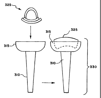
A superior universal facet prosthesis 330 is
shown in Fig. 1 that embodies features of the invention.
The prosthesis 330 is designated "superior" because it
creates an artificial facet surface for the superior half
of the facet joint. The artificial surface articulates
CA 02386504 2002-04-19
WO 01/30248 PCT/US00/29347
- 12 -
with the inferior half of the facet joint. The prosthesis
330 allows for the replacement of injured, diseased and/or
deteriorating components along the superior half of facet
joints, to provide improved support for the spinal column.
The universal facet prosthesis 330 may be
constructed and configured in various ways. The universal
facet prosthesis 330 may, e.g., comprise a cup member 315.
The cup member 315 itself may be made of various materials
commonly used in the prosthetic arts including, but not
limited to, polyethylene, rubber, titanium, titanium
alloys, chrome cobalt, surgical steel, or any other total
joint replacement metal and/or ceramic, bony in-growth
surface, sintered glass, artificial bone, any uncemented
metal or ceramic surface, or a combination thereof. The
cup member 315 may also be any appropriate shape
including, but not limited to, rectangular, disc shaped,
trough shaped, or cup shaped. The cup member may be fixed
or anchored directly to a vertebra with
poly(methylmethacrylate) bone cement, hydroxyapatite,
screws, nails, bolts, anchors, break-away anchors and/or
wires to facilitate any future removal of the prosthesis,
or a combination thereof, or any other means known in the
art.
As shown in FIG. 2, the cup member 315 is made of
any joint materials commonly used in the prosthetic arts,
including, but not limited to, metals, ceramics, titanium,
titanium alloys, tantalum, chrome cobalt, surgical steel,
bony in-growth surfaces, artificial bone, uncemented
surface metals or ceramics, or any combination thereof,
preferably covered with a bony in-growth surface.
In the illustrated embodiment, the cup member 315
is fixed to a stem 310, e.g., pre-welded, or glued with a
CA 02386504 2002-04-19
WO 01/30248 PCT/US00/29347
- 13 -
biocompatible adhesive, or removably secured using a
frictional Morse taper. If desired, the stem 310 can
incorporate one or more fins or ribs (not shown),
extending outward from the stem 310, which desirably
reduce and/or eliminate rotation of the stem 310 once
positioned within the targeted bone. In addition, the stem
310 can be cannulated, if desired, to allow the use of
guide pins during insertion of the stem, as is well known
in the art.
The stem 310 may itself be made of any joint
materials commonly used in the prosthetic arts, including,
but not limited to, metals, ceramics, titanium, titanium
alloys, tantalum, chrome cobalt, surgical steel, bony
in-growth surfaces, artificial bone, uncemented surface
metals or ceramics, or a combination thereof. In a
preferred embodiment, the stem 310 is covered with a bony
in-growth surface.
In the illustrated embodiment, the cup member 315
carries a surface member, which is made of a material,
e.g. polyethylene, ceramic, or metal, which provides glide
and cushioning ability for any potential contacting
components, such as the articular head members described
below. In one embodiment (see Fig. 2b), the surface member
325 can be formed in a gently upwardly curving shape,
similar in shape to a catcher's mitt. In another
embodiment (see Fig. 2c), the surface member 325 is
rectangular in shape with rounded corners. The cup member
315 is sized to be larger than the articulating superior
half of the facet joint, to allow for motion of the joint.
The surface member 325 may be a separate
component that is fixed to the cup member 315, e.g., with
a biocompatible adhesive, screws, nails, or comprise a
CA 02386504 2002-04-19
WO 01/30248 PCT/US00/29347
- 14 -
formed part of the cup member 315. The surface member 325
may also be held into the cup member 315 with compressive
forces or friction (e.g., using a Morse taper).
As shown in FIGS. 2a and 2b, the stem 310a could
alternately comprise a threaded portion, such as in a
pedicle screw, with the head or pedestal 315a
incorporating a depression 316a sized to accommodate a
hexagonal driver or other surgical driving tool well know
in the art. In addition, the prosthesis 320a could
incorporate a lower insert 321a sized to fit into the
depression 316a in the head 315a. If desired, the insert
321a could comprise a Morse taper. In this embodiment, the
stem 310a can be screwed into the bone, with the insert
321a positioned or otherwise secure within the depression
316a. The stem 310a could be placed by tapping without
screwing. If revision surgery is required, or some other
condition required removal of the prosthesis, the insert
321a can be removed from the stem 310a, and the stem 310a
can subsequently be removed from the bone.
As FIG. 2a shows, the stem 310a can also include
an enlarged projection or collar 311a abutting the cup
member 315a. The collar 311a serves to prevent unintended
ingress of the stem 310a further into the pedicle, beyond
a desired distance.
FIG. 1 depicts a spondylolisthetic spine with
slippage at the L4-L5 joint between the L4 and L5
vertebrae. FIG. 3 and FIG. 4 depict a universal facet
prosthesis 330 which has been installed into an L5
vertebra 105 to replace the inferior half 305 of a facet
joint. In one embodiment, the stem 310 of universal facet
prosthesis 330 is fixed into the L5 vertebra 105 with poly
(methylmethacrylate) bone cement, hydroxyapatite, a ground
CA 02386504 2002-04-19
WO 01/30248 PCT/US00/29347
- 15 -
bone composition, or a combination thereof. In another
embodiment, both the stem 310 and the cup member 315 are
fixed to a vertebra with stainless steel wire to provide
addition stability.
The new support provided by a universal facet
prosthesis 330 helps correct degenerative spine diseases
such as spondylolisthesis, spinal stenosis, or any spine
disease. As demonstrated by comparing FIG. 1 showing a
spondylolisthetic spine with slippage between the L4
vertebra 100 and the L5 vertebra 105 with FIG. 3 where the
diseased superior half 305 of the facet joint has been
replaced with a superior universal facet prosthesis 330 of
the present invention, correcting spondylolisthesis at the
L4-L5 joint and preventing further spondylolisthesis.
Similarly, where correction of scoliosis and/or kypho-
scoliosis is desired, the size and/or shape of the
prosthesis may be chosen to re-orient the affected
level(s) of the spine.
The superior universal facet prosthesis 330
described above may be used as a replacement for the
superior half of one or more of facet joints at any facet
joint at any level of the spine. In the preferred
embodiment, the universal facet prosthesis 330 is used to
replace the superior half of one or more facet joints in
one or more facet joints. The superior facet prosthesis
330 is designed such that it has the appropriate cephalad
and caudad directions as well as the appropriate
medial/lateral angulation for the given level of the spine
where the implant occurs.
In further embodiments, one or more surfaces of
a universal facet prosthesis 330 may be covered with
various coatings such as antimicrobial, antithrombotic,
CA 02386504 2007-06-06
52723-1
- 16 -
and osteoinductive agents, or a combination thereof. See,
e.g., U.S. Pat. No. 5,866,113.
These agents may further be carried
in a biodegradable carrier material with which the pores
of the stem and/or cup member of certain embodiments may
be impregnated. See, e.g., U.S. Pat. No. 5,947,89,,
in still further embodiments of the present
invention, a universal facet prosthesis may be attached to
strengthened or fortified bone. Vertebrae may be
strengthened prior to or during fixation of the prostheses
using the methods, e.g., described in U.S. Pat. No.
5,827,289.
This type of bone strengthening is particularly suggested
for osteoporotic patients who wish to have facet
replacement.
B. Surgical Method for Facet Replacement Using
the Superior Universal Facet Prosthesis
A surgical procedure that embodies features of
the invention replaces the superior half of a facet joint
with the superior universal facet prosthesis 330 described
above. The surgical procedure comprises exposing the
spinous process, lamina, and facet joints at a desired
level of the spine using any method common to those of
skill in the medical arts. The prominent bone 306b (see
FIG. 5) may then be rongeured using any means common in
the field. The superior facet 305 may also be trimmed, as
depicted in FIG. 6, to decompress the nerve root 203. A
reamer 400, or any other instrument that is useful for
grinding or scraping bone, may be used to ream the facet
305b into the pedicle 304b as depicted in FIG. 7 and FIG.
8.
CA 02386504 2007-06-06
52723-1
- 17 -
in a preferred embodiment (see FIG. 9), an
opening 407 is made into the vertebral body 107 with a
broach 405. The universal facet prosthesis 330b is
.installed into the opening 407 made by the broach 405, as
shown in FIG. 10. The opening 407 may be partly filled
with bone cement, hydroxyapatite, or any bone adhesive
before installation of the universal facet prosthesis
330b.
In an alternative embodiment, the stem 310 of the
superior universal facet prosthesis 330 may be constructed
in such a way that the superior universal facet prosthesis
330 can be directly screwed or tapped into the vertebral
body 107.
In another arrangement, the cup member 315 of the
universal facet member 330 may additionally be fixed to
the vertebral body 107 with bone cement, hydroxyapatite,..
or any other biocompatible adhesive. In yet, another
arrangement, a universal facet prosthesis without a stem
310 may be attached to the vertebral body with
poly(methylmethacrylate) bone cement, hydroxyapatite,
screws, nails, bolts, anchors, break-away anchors to
facilitate later removal of the prosthesis, or a
combination thereof, or any other means known in the art.
in a further embodiment of the present invention,
the universal facet prostYiesis 330 may be fixed into
strengthened or fortified bone. Vertebrae may be
strengthened prior to or during fixation of the prosthesis
using the methods described in U.S. Pat. No. 5,827,289.
This type of
bone strengthening procedure is particularly suggested for
osteoporotic patients who wish to have facet replacement
surgery.
CA 02386504 2002-04-19
WO 01/30248 PCT/US00/29347
- 18 -
III. Inferior Lamina/Facet Prosthesis
A. Structure
An inferior lamina/facet prosthesis 500 that
embodies features of the invention is shown in FIG. 11.
The prosthesis 500 is designated "inferior" because it
creates an artificial facet surface for the inferior half
of a facet joint. The artificial surface articulates with
the superior half of the facet joint. The prosthesis 330
allows for the replacement of injured, diseased and/or
deteriorating components along the inferior halves of
facet joints to provide improved support for the spinal
column.
The prosthesis 330 may span the distance from a
region on one side of a vertebra to a region of the other
side of the vertebra. It can thus replace both inferior
halves of a facet joint.
FIG. 14 depicts a superior view of a vertebral
body depicting sagitally oriented arthritic facets with
lateral stenosis, showing how the spinal process 631
presses forward onto the nerve roots 205 and 200. The
prosthesis 500 allows for replacement of diseased and
deteriorating inferior regions of the vertebra and partial
.replacement of lamina (see FIG. 12), which may be pressing
on the spinal nerves, to relieve pain. The prosthesis 500
creates artificial facet surfaces for the inferior half of
facet joints in the spine, which provide improved support
for the spinal column.
As FIG. 12 shows, a superior universal facet
prosthesis 330, as described above, may also be installed
to replace the superior halves of the facet joints and,
with the inferior lamina/facet prosthesis 500 replacing
the inferior halves of the facet joints, forming a total
CA 02386504 2002-04-19
WO 01/30248 PCT/US00/29347
- 19 -
facet replacement system that can result in entire
artificial facet joints along a length of the spinal
column. Alternatively, just the inferior half one or more
facet joints, or just the superior half of one or more
facet joints, may be replaced. The inferior and/or
superior halves of facet joints may be replaced on one
side of a given vertebra (unilateral), on the both sides
of a given vertebra (bilateral), or a combination of each
along a length of the spinal column.
The inferior lamina/facet prosthesis 500 may be
constructed in various ways. As shown in FIG. 11, the
prosthesis 500 can comprise a base member 505. The base
member 505 may be made of any joint materials commonly
used in the prosthetic arts, including, but not limited
to, metals, ceramics, titanium, titanium alloys, tantalum,
chrome cobalt, surgical steel, bony in-growth surfaces,
artificial bone, uncemented surface metals or ceramics, or
a combination thereof. The base member 505 may also be any
appropriate shape to give appropriate support to the spine
and to appropriately and sturdily attach to the inferior
portions of a vertebral body. The base member 505 may be
fixed or anchored directly to the inferior portion of a
vertebral body with poly(methylmethacrylate) bone cement,
hydroxyapatite, screws, nails, bolts, anchors, break-away
screws to facilitate any future removal of the prosthesis,
or a combination thereof, or any other means known in the
art.
In a preferred arrangement, as depicted in FIG.
11, FIG. 12, and FIG. 13, the base member 505 of the
inferior lamina/facet prosthesis 500 is attached to each
pedicle 102a and 102b with bilateral pedicle screws 520a
and 520b. The base member 505 of the inferior lamina/facet
CA 02386504 2002-04-19
WO 01/30248 PCT/US00/29347
- 20 -
prosthesis 500 may further be attached to the spinous
process 630 with a trans-spinous-process screw 515 to
provide additional stability.
In another embodiment, the inferior lamina/facet
prosthesis 500 may have a head member 510 for articulation
with the cup member 315 of a superior universal facet
prosthesis 330 or with a superior articular process of the
adjoining vertebral body. The head member 510 may be made
of various materials commonly used in the prosthetic arts
including, but not limited to, polyethylene, rubber,
tantalum, titanium, chrome cobalt, surgical steel, bony
in-growth surfaces, ceramics, artificial bone, or a
combination thereof. The head member 510 may further be
any shape which facilitates attachment to the rest of the
inferior lamina/facet prosthesis 500 and to smooth
connection to, and movement in orientation to, a universal
facet prosthesis 330 or a superior articular process of an
adjoining vertebral body. In one embodiment, a head member
510 is attached to the base member 505 of the inferior
facet/lamina prosthesis 500 with poly(methylmethacrylate)
bone cement, hydroxyapatite, screws, nails, bolts,
anchors, or any other means known in the art. The head
member 510 may also be removably attached by frictional
engagement (e.g., using a Morse taper)
In a preferred embodiment (see FIGS. 11 and 12),
the inferior facet/lamina prosthesis 500 comprises two
head members 510a and 510b formed in the shape of an
articular head. The head members 510a and 510b preferably
each have a Morse taper 512 at their upper surface to
allow them to lock into the base member 505 of the
inferior facet/lamina prosthesis 500. Of course, either
or both head members 510a and 510b could be formed
CA 02386504 2007-06-06
52723-1
- 21 -
integrally with the prosthesis 500. In the preferred
arrangement, a complete prosthetic facet joint 560 is
provided (see FIG. 11), in which the head members 510a and
510b articulate with the cup member 315 of the superior
universal facet prosthesis 330.
in further embodiments, one or more surfaces of
the inferior lamina/facet prosthesis 500 may be covered
with various coatings such as antimicrobial,
antithrombotic, and osteoinductive agents, or a
combination thereof. See, e.g., U.S. Pat. No. 5,866,113.
These agents
may further be carried in a biodegradable carrier material
with which the pores of the base member and/or any screws,
bolts, or nails of certain embodiments may be impregnated.
See, e.g., U.S. Pat. No. 5,947,89-3.
In other arrangements; an.inferior lamina/facet
prosthesis 500 may be attached to strengthened or
fortified bone. Vertebrae may be strengthened prior to or
during fixation of the prosthesis using the methods
described, e.g., in U.S. Pat. No. 5,827,289.
This type of bone
strengthening is particularly suggested for osteoporotic
patients who wish to have facet replacement.
B. Surgical Method for Partial Inferior
Lamina/Facet Replacement Using the Inferior
Lar-ina/Facet Prosthesis
A surgical procedure that embodies features of
the invention replaces inferior lamina and articulated
processes with the inferior lamina/facet prosthesis 500 as
described above. The surgical procedure exposes the
spinous process, lamina, and facet joints at a desired
CA 02386504 2002-04-19
WO 01/30248 PCT/US00/29347
- 22 -
level of the spine using any method common to those of
skill in the medical arts. As FIG. 15 shows, an inferior
one eighth to one half of the spinous process 302 may be
cut along the spinous process resection line 610 and
removed, if the spinous process appears diseased or
damaged. The cutting and removal of the spinous process
may be performed using any means common in the field.
As shown in FIGS. 16 and 17, the inferior half of
the facet joint may also be cut at or near the inferior
facet 'resection line 600. In a preferred embodiment (see
FIGS. 16 and 17), most of the lamina 615 is preserved, as
is the facet joint capsule 625, which may be opened and
folded back. In a preferred embodiment, the facet joint
capsule 625 may be cut perpendicular to its direction.
The inferior half 621 of the facet joint 620 may then be
retracted from the superior half 622. Once the facet
joint 620 is separated, the cut inferior bone 615 of the
upper joint (i.e. the cut inferior portion of the L4
vertebra in the L4-L5 joint) may be removed.
Alternatively, it may be possible to remove the cut
inferior bone 615 while simultaneously separating the
facet joint 620.
In a preferred embodiment (see FIGS. 18 and 19),
a superior universal facet prosthesis 330 is then
installed as previously described. Alternatively, the
superior universal facet prosthesis 330 may be installed
before the inferior bone is removed or even cut.
An inferior lamina/facet prosthesis 500 as
described above may be placed onto the facet joints and
over the spinous process. The inferior lamina/facet
prosthesis 500 may be fixed or anchored to the vertebral
body with poly(methylmethacrylate) bone cement,
CA 02386504 2007-06-06
52723-1
- 23 -
hydroxyapatite, screws, nails, bolts, anchors,.break-away
screws, or a combination thereof to facilitate any future
removal of the prosthesis, or any other means known in the
art. In the preferred embodiment (see FIG. 11, FIG. 12,
and FIG. 13), the inferior lamina/f acet prosthesis 500 is
attached.to each pedicle 102a and 102b of the inferior
facets with bilateral pedicle screws 520a and 520b and is
further attached to the spinous process 630 with a
trans-spinous-process screw 515 to provide additional
stability.
A head member 510 of an inferior lamina/facet
prosthesis 500 may articulated into the cup member 315 of
the superior universal facet prosthesis 330, or into a
inferior half of a facet joint if the inferior half has
not been replaced, to create a complete prosthetic facet
joint.
In an alternative embodiment, as depicted by FIG.
19, the inferior facet resection line 610 may be a V-type
cut. if a V-type cut is used, an appropriately shaped
inferior lamina/facet prosthesis 550 should be used, such
as depicted in FIG. 20. The inferior facet resection line
may alternatively be cut in other ways, which are apparent
to one of skill in the art of orthopedic surgery and will
require inferior lamina/facet prostheses of varying shapes
to appropriately fit the cut vertebra.
In a further embodiment of the present invention,
a universal facet prosthesis and/or an inferior
lamina/facet prosthesis may be fixed into strengthened or
fortified bone. Vertebrae may be strengthened prior to or
during 'fixation of the prosthesis using the methods
described, e.g., in U.S. Pat. No. 5,827,289.
This type of bone
CA 02386504 2002-04-19
WO 01/30248 PCT/US00/29347
- 24 -
strengthening procedure is particularly suggested for
osteoporotic patients who wish to have facet replacement
surgery.
IV. Hemi-Lamina/Facet Prosthesis
A. Structure
A hemi-lamina/facet prosthesis 700 that embodies
features of the invention (see FIG. 21) may be used to
replace parts of a lamina and inferior processes, some or
all which may have been removed in a primary procedural
bone resection, (i.e. with or without wide decompressive
laminectomy). The hemi-lamina/facet prosthesis 700 may be
designed similarly, or even identically, to the inferior
lamina/facet prosthesis 500 described above, depending on
how much of the bone is removed.
The hemi-lamina/facet prosthesis 700 may be
constructed in various ways. In one embodiment,
hemi-lamina/facet prosthesis 700 may, e.g., comprise a
base member 705. The base member 705 may be made of any
joint materials commonly used in the prosthetic arts,
including, but not limited to, metals, ceramics, titanium,
titanium alloys, tantalum, chrome cobalt, surgical steel,
bony in-growth surfaces, artificial bone, uncemented
surface metals or ceramics, or a combination thereof. The
base member 705 may be any shape which gives appropriate
support to the spine and can be appropriately attached to
the bone of the remaining lamina. The base member 705 may
be fixed or anchored directly to the inferior portion of
a vertebral body with poly(methylmethacrylate) bone
cement, hydroxyapatite, screws, nails, bolts, anchors,
break-away screws to facilitate any future removal of the
prosthesis, a combination thereof, or any other means
known in the art.
CA 02386504 2002-04-19
WO 01/30248 PCT/US00/29347
- 25 -
In a preferred embodiment (see FIG. 21) of a
prosthesis for hemiarthroplasty (depicted as cut line 800
and further described below) without decompressive
laminectomy, the base member 705 of the hemi-lamina/facet
prosthesis 700 is attached to superior pedicle 102b with
a pedicle screw 720. In another preferred embodiment, the
base member 705 of the hemi-lamina/facet prosthesis 700
may further be attached to the spinous process 630 with a
trans-spinous-process screw 715 to provide additional
stability.
In a preferred embodiment (see FIGS. 24 and 25)
of a prosthesis for hemiarthroplasty with wide
decompressive laminectomy, the hemi-lamina/facet
prosthesis 700 comprises at least one base member 705.
The base member 705 may further comprise a pedicle
attachment hole 725 through which a pedicle screw 720, or
a nail, anchor, break-away anchor, bolt, or any other
fastening means, may be installed to help secure the
hemi-lamina/facet prosthesis 700 to the inferior pedicle.
In the preferred embodiment, the base member 705 may also
have at least one lamina attachment hole, with two lamina
attachment holes 741 and 742 pictured in FIG. 22, to
further secure the hemi-lamina/facet prosthesis 700 to the
remaining laminal bone with screws, nails, anchors,
break-away anchors, bolts, or any other fastening means.
Parts of the hemi-lamina/facet prosthesis 700 which
overlap bone may be additionally fixed with bone cement,
or any biocompatible adhesive.
A hemi-lamina/facet prosthesis 700 may further
comprise a connection plate, similar to the connection
plate 750 depicted in FIG. 24, to connect two base
members, i.e. 705a and 705b, together. The connection
CA 02386504 2002-04-19
WO 01/30248 PCT/US00/29347
- 26 -
plate 750 may be fixed to each base member 705a and 705b
with a biocompatible adhesive, screws, nails, bolts,
compressive force, a combination thereof, or any other
means common to those of skill in the art. Alternatively,
a hemi-lamina/facet prosthesis 700 may further comprise at
least one stabilization bar, similar to the stabilization
bars 761 and 762 depicted in FIG. 25. A stabilization bar
or bars may be fixed to each base member 705a and 705b
with a biocompatible adhesive, screws, nails, bolts,
compressive force, a combination thereof, or any other
means common to those of skill in the art. A
hemi-lamina/facet prosthesis 700 may have any type of
bridging or stabilizing members, or no bridging members at.
all, and may be comprised of any number of base members to
provide appropriate stability to the spine. The bridging
members may be made of any joint materials commonly used
in the prosthetic arts, including, but not limited to,
metals, ceramics, titanium, titanium alloys, tantalum,
chrome cobalt, surgical steel, bony in-growth surfaces,
artificial bone, uncemented surface metals or ceramics, or
a combination thereof.
In another embodiment, a hemi-lamina/facet
. prosthesis 700 may have a head member 710 for articulation
with the cup member 315 of a superior universal facet
prosthesis 330 or with the superior articular process of
an adjoining superior pedicle. The head member 710 may be
made of various materials commonly used in the prosthetic
arts including, but not limited to, polyethylene, rubber,
titanium, chrome cobalt, surgical steel, bony in-growth
sintering, sintered glass, artificial bone, or a
combination thereof. The head member 710 may further be
any shape which allows it to attach to the rest of the
CA 02386504 2007-06-06
52723-1
- 27 -
hemi-lamina/facet prosthesis 700 and to smoothly connect
to, and move in orientation to, the universal facet
prosthesis 330 or superior articular facet of the
adjoining superior pedicle. In one embodiment, the head
member 710 is attached to the rest of the
hemi-lamina/facet prosthesis with poly(methylmethacrylate)
bone cement, hydroxyapatite, screws, nails, bolts,
anchors, a combination thereof, or any other means known
in the art. The head member 710 may be removably
attached, using, e.g., a Morse taper.
In a preferred embodiment, hemi-lamina/facet
prosthesis 700 comprises a head member 710 made in the
shape of an articular head. The head member 710 preferably
has a Morse Taper at its upper surface to allow it to lock
into hemi-lamina/facet prosthesis 700.
In further:embodiments, one or more surfaces of
a hemi-lamina/facet prosthesis 700 may be covered with
various coatings such as antimicrobial, antithrombotic,
and osteoinductive agents, or a combination thereof. See,
?_c?., U.S. Pa.t. No. 5,866,11?.
These agents may further be carried
in a biodegradable carrier material with which the pores
of the base member and/or any screws, bolts, or nails of
certain embodiments may be impregnated. See, e.g., U.S.
Pat. No. 5,947,893.
In still further embodiments of the present
invention, a hemi-lamina/facet prosthesis 700 may be
attached to strengthened or fortified bone. Vertebrae may
be strengthened prior to or during fixation of the
prosthesis using the methods described, e.g., in U.S. Pat.
No. 5,827,289.
CA 02386504 2002-04-19
WO 01/30248 PCT/US00/29347
- 28 -
This type of bone strengthening is particularly suggested
for osteoporotic patients who wish to have facet
replacement.
B. Hemiarthroplasty With or Without Wide
Decompressive Laminectomy Using the Hemi-
Lamina/Facet Prosthesis
A surgical procedure that embodies features of
the invention removes at least part of a lamina and at
least one superior portion of a facet joint and replacing
it with a hemi-lamina/facet prosthesis 700 as described
above. The general surgical procedure is generally-similar
to the inferior lamina/facet replacement previously
described, with the main difference being the types of
cuts made into the laminal bone, and that two separate
prostheses are used to replace the superior portions of
two facet joints (left and right) of a given vertebra.
One embodiment of the surgical procedure
comprises exposing the spinous process, lamina, and facet
joints at a desired level of the spine using any method
common to those of skill in the medical arts. The
inferior facet joint and part of the lamina may be cut
with a hemiarthroplasty resection line 800 as depicted in
FIG. 26 for a hemiarthroplasty. The lamina may
additionally be cut for a wide decompressive laminectomy
along the decompression resection line 810 as depicted in
FIG. 26. The inferior facet joint may be cut on one side
or both sides of the lamina. Likewise, the lamina may be
cut along a decompression resection line on one side or
both sides.
In a preferred embodiment of a hemiarthroplasty
without a wide decompressive laminectomy, leaving the cut
inferior facet bone 300 in place, the facet joint capsule
CA 02386504 2002-04-19
WO 01/30248 PCT/US00/29347
- 29 -
625 may be opened and folded back. In the preferred
embodiment, the facet joint capsule 625 may be cut
perpendicular to its direction. The inferior half 621 of
the facet joint 620 may then be retracted from the
superior half 622. Once the facet joint 620 is separated,
the cut inferior facet bone 825 may be removed.
Alternatively, it may be possible to remove the cut
inferior facet bone 825 while simultaneously separating
the facet joint 620.
In a preferred embodiment of a hemiarthroplasty
with a wide decompressive laminectomy, a superior
universal facet prosthesis 330 is then installed as
previously described, and depicted in FIG. 18.
A base member 705 of hemi-lamina/facet prosthesis
700 as described in any of the embodiments above may be
placed onto at least one facet joint and at least one
pedicle as depicted in FIG. 23, and over the spinous
process if it has not been removed for hemiarthroplasty
without decompressive laminectomy as depicted in FIG. 21.
The hemi- lamina/facet prosthesis 700 may be fixed or
anchored to the vertebral body with
poly(methylmethacrylate) bone cement, hydroxyapatite,
screws, nails, bolts, anchors, break-away screws to
facilitate any possible future removal of the prosthesis,
a combination thereof, or any other means known in the
art. In the preferred embodiment, as depicted in FIG. 21,
FIG. 24, and FIG. 25, the hemi-lamina/facet prosthesis 500
is attached to each pedicle with bilateral pedicle screws
720.
A hemi-lamina/facet prosthesis 700 that may be
used in hemiathroplasty without wide decompressive
laminectomy, depicted in FIG. 21, may further be attached
CA 02386504 2002-04-19
WO 01/30248 PCT/US00/29347
- 30 -
to the spinous process 630 with a trans-spinous-process
screw 715 to provide additional stability. A hemi-lamina
prosthesis 700 that may be used in hemiathroplasty with
wide decompressive laminectomy, as depicted in FIGS.. 23,
24, and 25, may further be attached to remaining laminal
bone with screws, bolts, nails, anchors, or breakaway
anchors through at least one lamina attachment hole 741 to
provide additional stability.
In embodiments where a hemi-lamina/facet
prosthesis 700 with more than one base member 705 is
installed, a connection plate, depicted as connection
plate 750 in FIG. 24, at least one stabilization bar,
depicted as stabilization bars 761 and 762 in FIG. 25, or
any other connecting or stabilizing means known in the
art, may be installed with the base members to provide
additional stability to the spine.
At least one head member, depicted as head member
710 in FIGS. 21, 23, 24, and 25, of a hemi-lamina/facet
prosthesis 700 may be articulated into a cup member of a
superior universal facet prosthesis 330 to create a
prosthetic facet joint capsule.
The embodiments may be used to replace one or
more facet joints for the entire length of the spine from
Si to Tll, on one side of a given vertebra, or both sides
of a given vertebra, or a combination thereof along a
length of the spine. If only one facet joint at a given
level is to be replaced, the unilateral arthroplasty
prosthesis for the inferior half of the joint may be fixed
to the superior ipso-lateral pedicle and include a box
fitted over the spinous process, combined with screw
fixation. The spinous process box is similar to the
CA 02386504 2007-06-06
52723-1
- 31 -
spirnous process box, in the bilateral total facet
arthroplasty embodiment previously discussed.
In a further embodiment of the present invention,
a universal facet prosthesis 330 and/or a hemi-
lamina/facet prosthesis 700 may be fixed into strengthened
or fortified bone. The vertebrae may be strengthened prior
to or during fixation of the prosthesis using the methods
described, e.g., in U.S. Pat. No. 5,827,289.
This type of bone
strengthening procedure is particularly suggested for
osteoporotic patients who wish to have facet replacement
surgery.
V. Other Facet Prostheses
A. Single Side
FIGS. 29 and 30 show an inferior prosthesis 26
that embodies features of' the invention. The prosthesis 26
is designated "inferior" because it creates an artificial
facet surface in the inferior half of a facet joint. _The
artificial surface articulates with the superior half of
the facet joint. The prosthesis 26 is particularly well
suited to single-sided procedures and/or for procedures
involving vertebral bodies which are not symmetrical.
When the processes on one side of a vertebral
body are differently spaced from those on the other side
of the' same body, the prostheses- on each side would
desirably be of differing sizes as well. Moreover,'it is
often difficult and/or impossible for a surgeon to
determine the precise size and/or shape necessary for a
prosthesis until the surgical site has actually been
prepared for receiving the prosthesis. In such a case, the
surgeon typically needs a family of prostheses possessing
differing sizes and/or shapes immediately available during
CA 02386504 2002-04-19
WO 01/30248 PCT/US00/29347
- 32 -
the surgery. The surgeon cannot wait for a custom-fitted
device to be created during the surgery, so a number of
prostheses of varying sizes and/or shapes must be
available for each procedure.
The prosthesis 26 can be conveniently formed in
different sizes and shapes, to offer an array of
prostheses 26 from which the surgeon can pick and choose
as surgery proceeds. This allows a surgeon to create a
"custom" implant during.the surgical procedure.
In the illustrated embodiment (see FIGS. 29 and
30), the prosthesis 26 comprises a body 28 sized and
shaped to span the distance between a pedicle 14 and an
inferior articular process 24 on the same side of a
vertebral body (see FIG. 31). The body 28 may be formed
of a material commonly used in the prosthetic arts
including, but not limited to, polyethylene, rubber,
titanium, chrome cobalt, surgical steel, bony in-growth
sintering, sintered glass, artificial bone, or a
combination thereof.
The upper section of the body 28 desirably
includes an opening 32. The opening 32 accommodates a
pedicle screw 34 (see FIG. 41), which secures the upper
end of the body 28 into the pedicle 14 of the vertebral
body. The opening 32 could be elongated, to allow for
varying orientations and/or sizes of the pedicle screw 34.
The remainder of the link body 28 can be secured to the
exterior of the vertebra using, e.g., biocompatible
adhesive.
The lower section of the body 28 is oriented to
serve as the superior half of a facet joint. The lower
section of the body 28 desirably incorporates a head 30.
The head 30 can be permanently affixed to the body 28,
CA 02386504 2002-04-19
WO 01/30248 PCT/US00/29347
- 33 -
using, e.g., adhesive. Alternatively, the head can be
frictionally secured, e.g., using a Morse taper, for
removal and replacement (as FIG. 41 shows). Like the body
28, the head 30 can be formed of a material commonly used
in the prosthetic arts including, but not limited to,
polyethylene, rubber, titanium, chrome cobalt, surgical
steel, bony in-growth sintering, sintered glass,
artificial bone, or a combination thereof. The head 30
possesses a curvilinear shape that desirably curves along
a gradual arc (as FIG. 42 shows), or can present a
"button" shape.
If desired, the lower section of the joint link
body 28 could be angled, to more naturally mimic the
orientation of a non-diseased facet joint. In one
alternative embodiment, the lower section of the joint
link body 28 could rotate relative to the upper section,
and could be rotationally secured in a desired position by
a surgeon using a locking screw or other locking means
known in the art. Such an embodiment would allow the
surgeon to alter the orientation of the lower section to
fit the particular needs of a patient during the actual
surgical procedure.
In use (see FIG. 31), the head 30 articulates
with the superior half of the facet joint. The superior
facet 22 can comprise the natural superior articular
process itself (as FIG. 31 shows), or it can comprise a
superior prosthetic facet created, e.g., by the previously
described universal facet prosthesis 330 (as FIG. 42
shows). The surface member 320 of the universal facet
prosthesis 330 can comprise a metal material made of,
e.g., titanium, cobalt, chrome, etc., or a plastic
material such as, e.g., polyethylene, or a ceramic
CA 02386504 2002-04-19
WO 01/30248 PCT/US00/29347
- 34 -
material. Thus the surgeon can select the same or
different materials to form the joint interface between
the head 30 and facet prosthesis 330.
FIGS. 34 and 35 show another embodiment of an
inferior universal prosthesis 36 that embodies features of
the invention. The prosthesis 36, like the prosthesis 26,
is designated "inferior" because it creates an artificial
facet surface in the inferior half of the facet joint. The
artificial surface articulates with the superior half of
the facet joint. Like the prosthesis 26, the prosthesis 36
is particularly well suited to single-sided procedures
and/or for procedures involving vertebral bodies which are
not symmetrical.
The prosthesis 36 comprises a body 38 sized and
15, shaped to span the distance between a pedicle 14 and an
inferior articular process 24 (see FIG. 35). The body 38
may be formed of the same types of material as the link
body 28. Like the link body 28, the upper section of the
joint link body 38 desirably includes an opening 42, to
accommodate a pedicle screw 34 (see FIG. 35), which
secures the upper end of the body 38 into the pedicle 14
of the vertebral body, in similar fashion as generally
shown in FIG. 41. As before described with reference to
the link 26, the opening 42 in the link body 38 could be
elongated, to allow for varying orientations and/or sizes
of the pedicle screw 34. The remainder of the link body 28
can be secured to the exterior of the vertebra using,
e.g., biocompatible adhesive.
Unlike the link body 28, the link body 38
includes an intermediate opening 44. In use (see FIG. 35),
the spinous process 18 (if present) can extend through the
opening 44, to stabilize the link body 38 on the vertebral
CA 02386504 2002-04-19
WO 01/30248 PCT/US00/29347
- 35 -
body. Desirably, a trans-spinous-process screw 45 can be
used to provide additional stability
The lower section of the joint link body 38 is
oriented to serve as the inferior half of a facet joint.
The lower section of the joint link body 38 desirably
incorporates a head 40, which can be constructed in the
same fashion as the head 30 of the link 26. Like the head
30, the facet head 40 can be permanently affixed to the
body 38 or can be secured in with a frictional fit (e.g.,
using a Morse taper) for removal and replacement. Like the
head 30, the head 40 can be formed of a material commonly
used in the prosthetic arts.
In use (see FIG. 35), the head 40 articulates
with the superior half of the facet joint with the next
adjacent vertebra level. As before explained for the link
26, the superior facet 22 can comprise the natural
superior articular facet 22 itself, or it can comprise a
prosthetic facet created, e.g., by the previously
described universal facet prosthesis 330.
FIG. 32 shows a superior prosthetic link 26' that
also embodies features of the invention. The prosthetic
link 26' is designated "superior" because it creates an
artificial facet surface in the superior half of a facet
joint. The artificial surface articulates with the
inferior half of the facet joint. The superior prosthesis
link 26', like the prosthesis 26, is particularly well
suited to single-sided procedures and/or for procedures
involving vertebral bodies which are not symmetrical.
A stem 37 extends out from the upper end of the
link 26'. The stem 37 is inserted (by screwing or
tapping) into the pedicle, to thereby secure the link 26'
to the vertebral body.
CA 02386504 2002-04-19
WO 01/30248 PCT/US00/29347
- 36 -
As FIG. 32 shows, the upper end of the link 26'
is shaped to form a cup 36, which articulates with the
inferior half of the facet joint.
The inferior half of the facet joint can comprise
the natural inferior articular process 24 itself (as FIG.
32 shows), or it can comprise the head 30 of an inferior
prosthesis 26 or link 26' attached to the next adjacent
upper vertebra level (as FIG. 33 shows).
The lower end of the link 26' can also carry a
head 30 for articulation with the superior half of a facet
joint with the next adjacent lower vertebra. The superior
half of the facet joint can comprise the natural superior
articular process 22 itself, or it can comprise the cup of
a link 26' attached to the next adjacent lower vertebra
level.
It can thus be appreciated that the link 26' is
well suited for use in procedures requiring replacement of
multiple levels of facet joints, and can be interlinked in
superior and inferior pairs, like a structure formed out
of interlinking tinker-toy pieces. The link 26' also allow
subsequent surgeries to build upon already replaced
levels, rather than requiring the removal and replacement
of an existing implant to accommodate replacement of
failing facet joints in an adjacent level. It should be
appreciated that the upper end of the prosthesis 36 can
also be shaped to form a cup to articulate with the
superior half of the facet joint with the next adjacent
upper vertebra level.
The prosthesis 26, 36, or link 26' are well
suited for use in a single side of the vertebral body,
such as where the facet joints need only be replaced on a
single side of the vertebral body. The prosthesis 26, 36,
CA 02386504 2002-04-19
WO 01/30248 PCT/US00/29347
- 37 -
or link 26' are also well suited for use in a dual-sided
procedure where the vertebral body is either symmetrical
or non-symmetrical. In this arrangement, other prostheses
26, 36, or links 26' can be secured on the opposite side
of the vertebral body, allowing both sides of the
vertebral body to be treated. Because the surgeon can pick
prostheses 26, 36, and links 26' of varying sizes,
depending upon the size of the vertebral site, and can
individually position each prosthesis 26 or link 26'
relative to the vertebral body, the surgeon can tailor the
linked implant system to the individual's needs.
B. Multiple Level, Sequential Link Assemblies
FIG. 36 shows a universal prosthetic joint link
assembly 56 that embodies features of the invention. The
joint link assembly 56 is particularly well suited to
double-sided procedures and for sequential, multiple level
procedures.
In the illustrated embodiment (see FIG. 36), the
joint link assembly 56 comprises two criss-crossing link
bodies 58 and 60. Each body 58 and 60 (shown mutually
separated in FIGS. 37 and 38, respectively) may be formed
of a material commonly used in the prosthetic arts
including, but not limited to, polyethylene, rubber,
titanium, chrome cobalt, surgical steel, bony in-growth
sintering, sintered glass, artificial bone, or a
combination thereof.
As FIG. 36 shows, the link bodies 58 and 60 are
desirably locked together for use at an intermediate key-
way 62, to form the x-shaped, criss-crossing assembly 56.
The key-way 62 is formed by a shaped opening 68 in one
body 60 (see FIG. 37) and a mating shaped key 70 in the
other body 58 (see FIG. 38). The key 70 nests within the
CA 02386504 2002-04-19
WO 01/30248 PCT/US00/29347
- 38 -
opening 60 (as FIG. 36 shows), to frictionally hold the
bodies 58 and 60 together and resist relative rotation
between the bodies 58 and 60.
Of course, the shape of the opening 68 and key 70
can vary. In FIGS. 36, 37, and 38, the opening 68 and key
70 are generally square or rectilinear in shape. In FIG.
39, an alternative link body 58 is shown, which possesses
a key 70' that is generally octagonal in shape, sized to
nest within a corresponding octagonal opening in the other
link (not shown) . In this arrangement, the two link bodies
58 and 60 can be mutually assembled in different arcuately
spaced orieritations, allowing for variations in facet
joint size and positioning. If desired, the key-way 62
could alternately be formed in a tooth and gear
arrangement, which would desirably allow a multiplicity of
potential arcuately spaced orientations for the two link
bodies 58 and 60 forming the assembly 56.
The key 70 desirable peripherally defines an
opening 72 (see FIG. 38), through which the spinous
process 18 can (if present) project during use. This is
generally shown in phantom lines by FIG. 41.
Alternatively, the link bodies 58 and 60 could be
formed in a criss-crossing shape as a single, unitary
body.
The upper section of each link body 58 and 60
desirably includes a cup 64. The cups 64 form the left and
right superior halves of a facet joint and, in use,
articulate with the left and right inferior halves of the
facet joint.
A stem 65 extends out from the upper end of each
link body 58 and 60. The stem 67 is inserted (by screwing
or tapping) into the pedicle, to thereby secure the link
CA 02386504 2002-04-19
WO 01/30248 PCT/US00/29347
- 39 -
bodies 58 and 60 to the vertebral body. In use, the stems
67 secure the upper end of the bodies 58 and 60 into an
opposite pedicle 14 of a vertebral body.
As FIG. 40 best shows, the bodies 58 and 60 are
each sized, shaped and mutually oriented to span the
distance between a pedicle 14 on one side of the vertebral
body and the region of the inferior articular process on
the opposite side of the vertebral body. The remainder of
the link bodies 58 and 60 can be secured to the exterior
of the vertebra using, e.g., biocompatible adhesive. A
trans-spinous-process screw 63 can also be used to provide
additional stability
The lower section of each link body 58 and 60 is
oriented to serve as the inferior half of a facet joint.
As FIG. 40 shows, the link body 58, secured to the right
pedicle, is positioned to serve as the inferior half of
the facet joint on the left side of the vertebra. The link
body 60, secured to the left pedicle, is positioned to
serve as the inferior half of the facet joint on the right
side of the vertebra. For this purpose, the lower section
of each link body 58 and 60 desirably incorporates a head
66. As before explained, the head 66 can be permanently
affixed to each body 58 and 60 or it can be secured in a
frictional way using, e.g., a Morse taper for removal and
replacement. Like the bodies 58 and 60, the head 66 can
be formed of a material commonly used in the prosthetic
arts including, but not limited to, polyethylene, rubber,
titanium, chrome cobalt, surgical steel, bony in-growth
sintering, sintered glass, artificial bone, or a
combination thereof.
In use, the heads 66 articulate with the superior
halves of the left and right facet joints with the next
CA 02386504 2002-04-19
WO 01/30248 PCT/US00/29347
- 40 -
adjacent vertebra level. As earlier described with
reference to the single link structures, the superior
halves of the facet joints can comprise the natural
superior articular process 22 itself, or it can comprise
a prosthetic facet created, e.g., by the cups 64 of
another link assembly 56 secured to the next adjacent
lower vertebra.
The interlocking of the criss-crossing link
bodies 58 and 56 increases the strength of the overall
link assembly 56. The link assembly 56 distributes forces
to both of the pedicles (and the spinous process, if
desired), rather than relying upon fixation to a single
pedicle.
Like the link 26', the link assembly 56 is well
suited for implantation in procedures requiring
replacement of multiple levels of facet joints, and can be
interlinked in superior, and inferior pairs, like a
structure formed out of interlinking tinker-toy pieces.
Like the link 26', the link assembly 56 also allows
subsequent surgeries to build upon already replaced
levels, rather than requiring the removal and replacement
of an existing implant to accommodate replacement of
failing facet joints in an adjacent level.
The size and shape of any prosthesis disclosed
herein are desirably selected by the physician, taking
into account the morphology and geometry of the site to be
treated. The shape of the joint, the bones and soft
tissues involved, and the local structures that could be
harmed if move inappropriately, are generally understood
by medical professionals using textbooks of human anatomy
along with their knowledge of the site and its disease
and/or injury. The physician is also desirably able to
CA 02386504 2007-06-06
52723-1
- 41 -
select the desired shape and size of the prosthesis and
its placement in and/or around the joint based upon prior
analysis of the morphology of the targeted joint using,
for example, plain film x-ray, fluoroscopic x-ray, or MRI
or CT scanning. The shape, size and placement are
desirably selected to optimize the strength and ultimate
bonding of the prosthesis to the surrounding bone and/or
tissue of the joint.
Other embodiments and uses of the invention will
be apparent to those skilled in the art from consideration
of the specification and practice of the invention
disclosed herein. The
specification and examples should be considered exemplary
only with the true scope and spirit of the invention
indicated by the following claims. As will be easily
understood by those of ordinary skill in the art,
variations and modifications of each of the disclosed
embodiments can be easily made within the scope of this
invention as defined by the following claims.