Note: Descriptions are shown in the official language in which they were submitted.
4.
CA 02386829 2002-05-17
-1-
COMPUTER-BASED VIDEO RECORDING AND MANAGEMENT
SYSTEM FOR MEDICAL DIAGNOSTIC EQUIPMENT
BACKGROUND OF THE INVENTION
1. Field of the Invention
The present invention is related to a computer-based
video recording and management system, which is used in
conjunction with medical diagnostic equipment. In
particular, the system allows a physician or medical
personnel to record and time-mark significant events during
a medical procedure, to index patient data with the video
footage, and then to later edit and/or access the video
footage with patient data from a database in an efficient
and accurate manner.
2. Discussion of Background Information
An endoscope is an instrument which is formed from a
flexible tube which can be inserted inside the human body
through natural openings such as the mouth, the nose, etc.
The endoscope allows the physician performing a medical
examination to visually observe the state of natural body
cavities such as the throat, esophagus, stomach, pancreatic
and biliary ducts, colon, etc.
Typically, during a medical procedure in which a
physician uses an endoscope, he/she can press buttons on the
endoscope to capture still images of the most relevant
portions of the procedure. These images can then be stored
into a computerized system. The computerized system will
associate each picture taken during the procedure with the
patient's records. In particular, during the procedure, the
endoscope user simply presses a button on the scope and an
associated computer system captures the images. The system
a
CA 02386829 2002-05-17
-2-
saves the images into a database containing the patient's
medical records. As a result, the system allows one, after
the endoscopic procedure is complete for instance, to
assimilate and analyze the data so that a medical report can
be generated.
Following the endoscopic examination, the physician
reviews all the medical data generated throughout the
procedure and issues a report. The medical report normally
includes text and images. For example, a textual
description and diagnosis of what the physician found during
the procedure (i.e. malignancies, polyps, etc.) is always
included in the medical report. Furthermore, images can be
included to pictorially illustrate the medical condition of
which the physician is actually referring to in the textual
description.
Still imaging systems for endoscopes have been
available for several years. For example, U.S. Patent No.
5,111,306 to Kanno et al. teaches an endoscope image filing
apparatus wherein endoscope image information and search
information are recorded in the same medium by a recording
apparatus.
U.S. Patent No. 5,124,789 to Hiyama et al,, discloses a
system including multiple image signal generating
apparatuses which each generate an image signal using an
electronic endoscope, an ultrasonic scope or the like. The
scopes are connected to a common large capacity filing
apparatus through an interface so that image data can be
recorded together with image information with respect to the
data in the unit of any number of images for a single
examination. The image data recorded can then be searched
for in the unit of a single examination.
CA 02386829 2002-05-17
-3-
As endoscope technology progresses, video endoscopes
have been developed which generate live images on a video
monitors by capturing the visual information using an
electronic sensor placed at the distal end of the endoscope.
Video taping an entire endoscopic procedure is also well-
known. For instance, the entire endoscopic procedure, from
beginning to end, can be recorded on a VCR in the procedure
room. Such video recordings have been traditionally used
only for specific reasons, such as presentational,
educational or training purposes, etc.
An example is provided in U.S. Patent No. 6,184,922 to
Saito et al., in which a camera control unit for processing
a signal output from an imaging device incorporated in an
endoscope is taught. The system includes an analog video
signal output terminal through which a video signal is
output to a monitor, and a digital video signal output
terminal to which a still image-specific or motion picture-
specific expansion unit is coupled in a freely detachable
manner. By handling a release switch, a still image or
motion picture can be recorded digitally.
However, none of the above-noted references provide
features which allow for the video footage to be
incorporated or combined with important patient data such as
name, medical finding, finding location, and free text to be
associated with each video clip. Furthermore, none of the
above-noted references provide features which allow for
storage of the video with patient data, so that it can be
easily edited and/or stored in a database.
One of the main reasons video has not been incorporated
into the patient's records is because of system constraints
(e.g., computer memory). Another reason is because of the
extensive amount of time required to edit the video and to
I.
CA 02386829 2002-05-17
-4-
incorporate the video into the patient's records. Whether
the editing is performed on a VCR editing machine or on a
computer system with video editing capabilities, it requires
an extensive amount of time, which in turn, increases
medical costs.
Typically, the medical staff has higher priorities
during the medical procedure than editing the video of the
procedure in real-time. Furthermore, such procedures are
often unpredictable. Because one does not know what may
occur or be discovered during the procedure, it is somewhat
imprudent to selectively record only phenomena of which is
expected to be of significant interest during the procedure.
For instance, during the procedure of unexpected problem may
occur, and later full documentation will suddenly become
very important and relevant. Therefore, traditionally, if a
video clip of the procedure is desired, one must initially
record the entire procedure.
Depending upon the medical procedure, the video
recording time can be anywhere between ten minutes to two
and one-half hours for each procedure. Once the procedure
is finished and the medical staff has a better perspective
of the results of the operation, it is then normally a more
appropriate time to edit the video.
It is becoming increasingly more important to provide
systems in which critical data can be extracted from medical
devices and then organized in an efficient and effective
manner. Currently in medical database research, if one
tries to carry out a visual search of medical
investigations, often the databases do not have enough
materials to accurately implement a complete search. Many
medical databases are incomplete and only have random bits
and pieces of medical data. When research is being
1 1;
CA 02386829 2002-05-17
-5-
conducted, often the researcher does not know why certain
procedures have been recorded.
Ultimately, when conclusions are drawn from statistical
research, the conclusions drawn are only as good as the data
in the database. Therefore, in order to perform any kind of
statistical research based on a database, a need exists for
ensuring that the database is significantly populated in a
systematic way.
SUMMARY OF THE INVENTION
An aspect of the present invention is to provide a
computer-based video picture recording and management system
for medical diagnostic equipment (hereinafter referred to as
"motion picture studio" or "MPS system"). In particular,
the disclosed MPS system features techniques, which allow
the physician and medical staff to record video clips during
an endoscopic procedure while simultaneously marking
significant events, to include important patient data with
each video clip, and then to later edit the video clips with
great efficiency and accuracy.
The present invention provides many distinct advantages
over the traditional manner of recording video during a
medical procedure. First, the present invention offers the
advantage of significantly reducing editing time for
developing a patient's medical report. It eliminates the
need for handling video cassettes, DVD's or memory cards
because all the video data is electronically and directly
stored into the memory of the MPS system.
A second major advantage of the present invention is a
feature that immediately associates the video clips with the
patient's current medical procedure record. Traditionally,
this would require labeling and proper archival of a
CA 02386829 2002-05-17
-6-
videocassette. Not only is such a procedure time consuming,
but also the storage of a videocassette for each individual
patient becomes a problem. More storage space required for
each patient's records equates to higher medical costs.
Furthermore, as a result of the underlying digital
video recording technology, the present invention offers a
greater quality video playback on a computer screen compared
to the quality offered by a VCR. This is especially true
when the user desires to pause on a specific view, in which
case the present invention continues to display a clear high
quality view based upon digital technology, whereas the VCR
offers at best a blurry picture with multiple horizontal
blanking lines.
Another aspect of the present invention is that the MPS
system has been designed to be integrated with medical
instruments which provide a video source from a variety of
devices. For example, the MPS system is also capable of
being integrated with medical instruments used in
ultrasound, fluoroscopy, x-ray, and/or surgical cameras.
The present invention also offers additional benefits
due to its ability to be integrated with currently available
video endoscopic instrumentation, such as the line of
products manufactured by Pentax Corp. For instance, the MPS
system is designed to be tightly integrated with a variety
of Pentax video processors and a variety of Pentax video
endoscopes.
The present invention also provides indexing capability
for time-marks and their associated useful segments. In one
embodiment, the index includes a title, a finding, a
location and free text of unlimited length. The index
information comes into view during playback of the video
footage while the focus is within the time span of the
CA 02386829 2002-05-17
_7_
useful segment associated with the indexed time-mark.
Through existing "ENDOPRO" software developed and marketed
by Pentax Corp. and advancements to the ENDOPRO software
package disclosed herewith, it is now possible to search and
locate specific video clips that relate to a certain
findings, locations, and free-text criteria.
Another aspect of the present invention relates to a
feature referred to as "Auto-Editing" or "Auto-Editor".
This feature allows one to expediently produce a summary
output of the entire video. It allows one to reduce a two
to three hour video down to a two to three minute "Executive
Summary Video" which focuses only on the most significant
events during the procedure in a matter of seconds. The
"Auto-Editing" is a software function which can be performed
after the procedure is complete. Therefore, editing time or
production time to produce a medical report with still
pictures or video is decreased dramatically.
Another aspect of the present invention provides a very
systematic tool in order to compile, edit or correct video
information so that the medical information having
significance can be input into a database. Such feature
allows an increased efficiency in medical research due to
the fact that information about medical conditions can be
assimilated in a much more effective and efficient manner.
Thus, medical research should be improved because the
compilation of medical data will be immediately organized so
that it is easily accessible.
Furthermore, once the information is assimilated, the
MPS system allows one to easily transfer the data into
databases so that expedient searching may be performed.
Therefore, not only is the editing time or production time
required to produce a medical report with still pictures or
a.
CA 02386829 2002-05-17
-8-
video decreased dramatically, but the invention. allows for
efficient assimilation and storing of data so it can be
searched more effectively.
According to an aspect of the present invention, a
computer readable medium storing a computer program that
provides a computer-based video recording and management
system for medical procedures is provided. The medium
includes a source code segment that inserts at least one
time-mark into video footage upon receiving input from a
user, the at least one time-mark capable of being inserted
into the video footage real-time while the video footage is
being recorded and post procedure during review; and a
source code segment that associates an index with the at
least one time-mark, data capable of being input into the
index real-time during a medical procedure and post-
procedure during review.
According to another aspect of the present invention
the index includes data for at least one of a patient's
name, medical finding, finding location, and free text. In
yet another aspect of the invention, the data is transmitted
from at least one of a medical instrument, microphone,
footpedal/switch, mouse and computer keyboard operated by a
user of the system.
In another aspect of the present invention, a source
code segment is provided that extracts at least one portion
of the video footage starting at a predetermined period of
time before the at least one time-mark and ending at a
predetermined period of time after the at least one time-
mark. According to a further aspect of the present
invention, the at least one portion of video footage is
concatenated with at least another portion of video footage
into a shortened summary video clip.
CA 02386829 2002-05-17
-9-
According to a further aspect of the present invention,
a computer readable medium storing a computer program is
provided that enables recording and time-marking of
significant events during a medical procedure in video
footage, indexing patient data with the video footage, and
then editing and accessing the video footage with patient
data and diagnostic information from a database in an
efficient and expedient manner. The medium includes a
source code segment that inserts at least one time-mark into
the video footage; a source code segment that associates an
index with the at least one time-mark; a source code segment
that extracts at least one portion of the video footage at
the at least one time-mark, wherein the at least one portion
begins before the at least one time-mark and ends after the
at least one time-mark; a source code segment that
concatenates the extracted at least one portion of video
footage together with at least another portion of video
footage into a shortened summary video clip; and a source
code segment that stores, both the video footage and
shortened summary video clip with associated indices, into a
searchable database.
In another aspect of the present invention, a source
code segment is provided that maintains and updates at least
one patient's medical record with at least one of data from
the index, video footage, and still pictures from the
medical procedure. According to a still further aspect of
the present invention, the index includes data fields for at
least one of a name, medical finding, finding location, and
free text. Other aspects of the present invention include
wherein data for the index is capable of being input real-
time during a medical procedure and post-procedure during
review.
CA 02386829 2002-05-17
-10-
Further aspects of the present invention include
wherein the time-mark is inserted according to a user input
device. According to other aspects of the present invention
include wherein the time-mark is capable of being real-time
during a medical procedure and post-procedure during review.
According to another aspect of the invention, the user is
notified whether the insertion of the at least one time-mark
was successful or failed, by displaying a message on a
monitor.
According to still a further aspect of the present
invention, a source code segment is provided which includes
a specialty video player. According to another aspect of
the present invention, the specialty video player includes a
playback speed control which provides for playback speeds
ranging from a reduced speed to an accelerated speed as
compared to a normal speed. In yet another aspect of the
present invention, a source code segment is provided that
enables jumping backward to a previous time-mark or jumping
forward to a next time-mark.
According to a further aspect of the present invention,
a source code segment is included that provides a capture
still image feature which stores a still picture within at
least one patient's medical record. In another aspect of
the present invention, a source code segment is included
that provides a create marker and delete marker feature
which allows for the creation and deletion of the at least
one time-marker within the video footage. According to a
still further aspect of the present invention, a source code
segment is included which provides a voice activated data
entry system allowing data to be entered via voice.
Furthermore, according to another aspect of the present
invention, a computer-based video recording and management
CA 02386829 2002-05-17
-11-
system is provided. It is used in conjunction with medical
diagnostic equipment, which allows recording and time-
marking of significant events during a medical procedure on
video footage, indexing patient data with the video footage,
and then editing or access the video footage with patient
data from a database in an efficient manner. The system
includes at least one input device that inserts at least one
time-mark into the video footage; and at least one
workstation that associates an index with each time-mark,
extracts at least a portion of the video footage at the at
least one time-mark beginning before and ending after the at
least one time-mark, concatenates the at least one portion
of the video footage with at least another portion of video
footage into a shortened summary video clip, and stores both
the video footage and summary video clip into a searchable
database.
In another aspect of the present invention, the at
least one input device includes a medical instrument having
a video source, the video source being connected to the at
least one workstation. According to a still further aspect
of the present invention, the at least one workstation
maintains at least one patient's medical record. According
to another aspect of the present invention, the index
includes data fields for at least one of a name, medical
finding, finding location, and free text.
According to a still further aspect of the present
invention, data for the index is capable of being input
real-time during a medical procedure and post-procedure
during a review period. Other aspects of the invention
include wherein the at least one workstation is connected to
a network. Furthermore, other aspects of the present
invention include wherein the at least one workstation is
^
CA 02386829 2002-05-17
-12-
connected to the network via an Internet connection.
According to other aspects of the present invention, at
least one file server having a video storage array is
connected to the network which stores at least one patient's
medical record.
According to other aspects of the present invention,
the medical instrument includes an endoscope. According to
another aspect of the present invention, the medical
instrument includes one of an ultrasound device, flouroscopy
device, x-ray device and surgical camera. According to a
further aspect of the present invention, the input device
includes a foot pedal/switch, microphone, mouse, and
computer keyboard.
Additionally, other aspects of the present invention
include wherein when the input device is activated, the
system encapsulates data with the video footage for indexing
purposes. In yet another aspect of the present invention,
the network includes a peer-to-peer network. And in another
aspect of the present invention, the database is located in
one of the at least one workstation and the at least one
file server.
Other exemplary embodiments and advantages of the
present invention may be ascertained by reviewing the
present disclosure and the accompanying drawing.
BRIEF DESCRIPTION OF THE DRAWINGS
The present invention is further described in the
detailed description which follows, in reference to the
noted drawings by way of non-limiting examples of exemplary
embodiments of the present invention, in which like
reference numerals represent similar parts throughout the
several views of the drawings, and wherein:
CA 02386829 2002-05-17
-13-
Figure 1 is a system diagram of an embodiment of the
present invention in which several MPS workstations are
networked together with an MPS file server and video storage
array;
Figure 2 is a flow diagram of a prior art ENDOPRO
Workstation Main Module;
Figure 3 is a flow diagram of an exemplary MPS Main
Module according to an aspect of the present invention;
Figure 4 is a flow diagram of an exemplary sequence for
initializing the MPS Main Screen window according to an
aspect of the present invention;
Figure 5 is a flow diagram of an exemplary MPS
Recording Mode Initialization sequence according to an
aspect of the present invention;
Figure 6 is a flow diagram of an exemplary User
Interaction Processing (MPS Recording Mode) sequence
according to an aspect of the present invention;
Figure 7 is a flow diagram of an exemplary Single Frame
Capture Process sequence according to an aspect of the
present invention;
Figure 8 is a flow diagram of an exemplary Video
Recording ON Process sequence according to an aspect of the
present invention;
Figure 9 is a flow diagram of an exemplary Video
Recording OFF Process sequence according to an aspect of the
present invention;
Figure 10 is a flow diagram of an exemplary Live Video
ON Process sequence according to an aspect of the present
invention;
Figure 11 is a flow diagram of an exemplary Live Video
OFF Process sequence according to an aspect of the present
invention;
I 9
CA 02386829 2002-05-17
-14-
Figure 12 is a flow diagram of an exemplary User
Interaction Processing (MPS Playback Mode) sequence
according to an aspect of the present invention;
Figure 13 is a flow diagram of an exemplary MPS Multi-
Media Player Module according to an aspect of the present
invention;
Figure 14 is a flow diagram of an exemplary MPS Multi-
Media Player User Controls according to an aspect of the
present invention;
Figure 15 is a screen shot of the prior art ENDOPRO
Main Menu according to an aspect of the present invention;
Figure 16 is an exemplary screen shot of the Today's
Room Schedule screen with the Adding Patient/Procedure
window open according to an aspect of the present invention;
Figure 17 is an exemplary screen shot of the MPS Main
Screen according to an aspect of the present invention;
Figure 18 is an exemplary screen shot the MPS Main
Screen having the thumbnail bar displayed with captured
still and video images according to an aspect of the present
invention;
Figure 19 is an exemplary screen shot the Video Clip
Viewer according to an aspect of the present invention;
Figure 20 is an exemplary screen shot of the Annotation
Text window according to an aspect of the present invention;
Figure 21 is an exemplary screen shot of the MPS
Specialty Video Player (SVP) displaying the Playback Window
according to an aspect of the present invention;
Figure 22 depicts an exemplary time line, index zones,
index selector, and index data box according to an aspect of
the present invention;
Figure 23 depicts an exemplary use of the index
selector according to an aspect of the present invention.
11 CA 02386829 2002-05-17
-15-
Figure 24 is an exemplary screen shot of the Index
Table Management window according to an aspect of the
present invention;
Figure 25 is an exemplary instrumentation diagram of
the endoscope buttons, foot pedal and voice actuated
microphone according to an aspect of the present invention;
and
Figures 26(a) and (b) illustrate packet transmission
from the video processor to the imaging platform according
to an aspect of the present invention.
DETAILED DESCRIPTION OF THE PRESENT INVENTION
The particulars shown herein are by way of example and
for purposes of illustrative discussion of the embodiments
of the present invention only and are presented in the cause
of providing what is believed to be the most useful and
readily understood description of the principles and
conceptual aspects of the present invention. In this
regard, no attempt is made to show structural details of the
present invention in more detail than is necessary for the
fundamental understanding of the present invention, the
description taken with the drawings making apparent to those
skilled in the art how the several forms of the present
invention may be embodied in practice.
A. MPS System Overview:
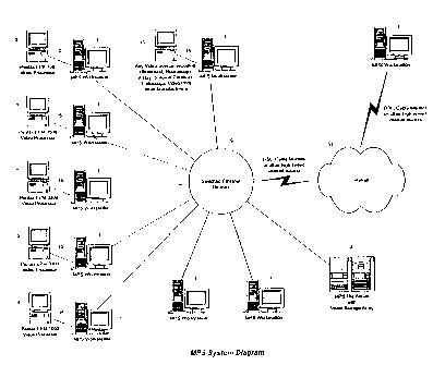
Figure 1 is a system diagram which depicts an
embodiment of the MPS system. This embodiment includes a
variety of video processors 5 and video sources 13 each
connected to a MPS workstation 1. The MPS workstations are
connected to a switched Ethernet network 9. Also connected
to the Ethernet network 9 is a MPS file server 3 which has a
I
CA 02386829 2002-05-17
-16-
video storage array. As shown in Figure 1, a MPS
workstation 1 can also be connected to the Ethernet network
9 via DSL, cable modem or any other high-speed Internet
access 11.
The data line 15 connecting the video processing or
source equipment to the MPS workstation is a logical
connection commonly referred to as an "ENDONET Connection"
which is a product manufactured by Pentax Corp. The ENDONET
Connection includes a video connection per RGBS, S-VIDEO,
NTSC, or DV standards, which transmits the visual
information generated by the video source to the MPS
workstation 1. The ENDONET Connection may also include a bi-
directional or mono-directional data communication line per
the RS232C or other standards such as USB or TCP/IP, which
essentially carries patient or procedure related information
from the MPS workstation 1 to video source 13 and/or
processor 5, and receives image capture, or video recording
requests from video source 13 and/or processor 5 to the MPS
workstation 1.
All data transactions between the video source 13
and/or processor 5 and MPS workstation 1 are performed over
data line 15 (from Figure 1) using a data exchange protocol
known as the ENDONET Communication Protocol Version 2.21 and
also referred to as Dynamic Device Recognition (DDR)
Protocol. The DDR Protocol, which is a Pentax product, will
be explained in greater detail later in the specification.
The MPS system has been designed to be capable of being
tightly integrated with readily available video medical
instruments 2 (from Figure 25), such as a line of products
manufactured by Pentax Corp. The MPS system is also
compatible with a variety of other medical devices such as
video gastroscopes, duodenoscopes, colonoscopes,
CA 02386829 2002-05-17
-17-
sigmoidoscopes, bronchoscopes, rhino-laryngoscopes,
cystoscopes, and choledochoscopes. The medical instruments
are, such as a video endoscope, are not illustrated on
Figure 1, but would be connected to the video processors, as
shown in Figure 25.
A exemplary list of endoscopes (and the respective part
numbers) compatible with the MPS system and of which Pentax
manufactures is as follows:
Video gastroscopes and duodenoscopes include
EG-2901/62019, EG-3400/62020, EG-3410/62014, EG-2530/62090,
EG-2731/62075, EG-2931/62146, EG-3431/62125, EG-3830T/62079,
ED-2330/62118, ED-3230/62093, ED-3430/62130, ED-3430T/62061,
VSB-3430/62105, EG-1840/62092, EG-2540/62082, EG-2940/62068,
EG-3440/62083, EG-3840T/62073, ED-3240/62132, ED-3440/62091,
ED-3440T/62062, VSB-3440/62106, EG-2930K/62095,
EG-3430K/62145, ED-3430K/62110, ED-3430TK/62147,
EG-3631U/62153, EG-2930Q/62099, and EG-3430Z/62124.
Video colonoscopes and sigmoidoscopes include
EC-3400L/62003, EC-3801L/62009, EC-3800TL/62006,
ES-3801/62025, EC-3430F/62081, EC-3430L/62080,
EC-3830F/62089, EC-3831L/62135, EC-3830TL/62065,
ES-3831/62157, EC-3440F/62084, EC-3440L/62085,
EC-3840F/62070, EC-384OL/62069, EC-3840TL/62067,
ES-3840/62071, EC-3430LK/62149, EC-3430FK/62143,
EC-3830LK/62096, EC-3830TLK/62151, EC-3830FK/62101,
ES-3830K/62097, EC-3430LZ/62159, and EC-3830LZ/62098.
Video bronchoscopes include EB-1530T3/62136 and
EB-1830T3/62141. Video rhino-laryngoscopes include
VNL-1330/62109 and VNL-1530T/62111. Video cystoscopes
include ECY-1530/62116. Video choledochoscopes include
ECN-1530/62115.
1 i
CA 02386829 2002-05-17
-18-
If the medical instrument 2 to be used with the MPS
system is a Pentax product, it will require a video
processor 5 which, may also be manufactured by Pentax. For
instance, the MPS system can be integrated with the Pentax
EPK-700, EPM-3500, EPM-3300, EPM-3000, and EPM-1000 video
processors, as illustrated in Figure 1. The above-noted
video processors are connected directly to the MPS
workstations 1.
It should be noted that when using the above-mentioned
Pentax products, the Pentax endoscope and Pentax video
processor 5 are coupled together. As a result, the video
source is a function of the video processor S. Therefore,
the Pentax endoscope and Pentax video processor 5 may be
considered together as one unit.
The MPS system is also designed with flexibility in
mind with respect to compatibility with non-Peritax products.
Any other device which provides a standard video output can
also be used in conjunction with the MPS system. For
instance, the present invention is capable of being
integrated with any other video source 13 including
ultrasound, fluoroscopy, x-ray, surgical cameras, or
endoscopic video from other manufacturers. In this case, a
foot pedal/switch 18 could be used to capture the still
image or to toggle the video recording on or off. An
exemplary MPS foot switch 18, which is also a Pentax
product, is part number MPS-FS/FS200.
Figure 25 depicts a medical instrument 2, for instance
a Pentax video endoscope, which may be used in conjunction
with a Pentax video processor 5 and MPS workstation 1 to
capture video. The button 8 labeled "V" (for video) allows
the physician to toggle the video recording process with a
single key (video turn ON/OFF). The button 10 labeled "C"
CA 02386829 2002-05-17
-19-
(for capture) permits the capture of a single still picture
by the computer-based video imaging system, but
simultaneously tags the video footage with time-marks in
association with the snap-shot captured.
As this operation (i.e., pressing the "C" button to
capture a still image) is repeated several times throughout
the medical examination, the invention encapsulates within
the video footage all necessary information for indexing.
This allows the viewer to access during playback the most
useful views directly, without having to watch the entire
footage.
The time-marks are further used to identify segments of
the video footage that are the most useful and worth being
kept permanently in the medical report. The MPS software
applies a set of predefined rules to construct segments of
video which were marked during the procedure. In default
mode, the MPS system takes the ten seconds immediately prior
to the time-mark and the twenty seconds following the time-
mark to identify a "useful segment" within the video
footage.
It is important to note that all of the controls of the
MPS system are primarily based from software functions.
Thus, every peripheral device used in the MPS system is
eventually mapped to software buttons, including the buttons
8 and 10, foot switch button 18, mouse input, and keyboard
input. In other words, the MPS system architecture is
designed in a manner such that all control functions operate
from software command sequences and, therefore, are the
primary basis for user input with respect to channeling such
data signals into the MPS system. On the other hand,
buttons 8 and 10 and the foot switch 18 provide signals
which mimic the signals of recording controls 76, 78 and 80
J!
CA 02386829 2002-05-17
-20-
(from Figure 17) and are, therefore, mapped to the same data
entry point of which the software based control buttons
originate from.
The Playback Window (see Figure 21) provided in the MPS
system offers an additional way to modify the length of a
segment, and functions to extract and save one or several
segments created in the fashion described above. This
technique provides for the reduction of the original video
volume to a fraction of the original length while preserving
the most relevant and useful material contained within.
Thus, for illustrative purposes only, it can be
imagined that the user of the present invention can mark a
video recording 4.5 minutes into the procedure, then
initiate a second mark at 15 minutes into the procedure, and
a third mark 17.3 minutes into the procedure. Once the body
of the video material is marked, the user can then make the
assumption that the important moments of the procedure are
known to be around those shots. Then, the MPS system can
be commanded, via the "Auto-Edit" command, to automatically
extract a sufficient amount of video to create a summary
output video (e.g., ten seconds before and twenty seconds
after). This summary video is referred to as the "Executive
Summary Video".
In this manner, a two hour video clip can be reduced to
a much shorter time, e.g., two to three minutes. The amount
of time before and after the snapshots is also adjustable.
For instance, the physician can adjust the amount of video
that is played depending on his/her preference. Or the
physician can choose an auto setting to receive the
automatic results that the MPS system produces. In this
case, Auto-Editing is performed by the MPS system with
absolutely no intervention on the part of the physician. As
CA 02386829 2002-05-17
-21-
a result, a two hour video can be reduced to a much shorter
amount of video virtually automatically without. the
intervention of anyone.
Thus, the MPS system has the capability to combine the
marked video clips together to create a summary output video
and then store it in the patient's records. The user still
has the option to keep the original footage, which can be
several hours of video, for archival purposes if desired.
Depending on the parameters that are set up in the MPS
system, the entire footage can be saved just to be on the
safe side for reference in the future or it can be deleted
to save storage space in the system memory.
Noteworthy features of the MPS system include: (1) an
indexing feature, (2) a video database, (3) Auto-Editing
capabilities, (4) a Specialty Video Player, (5) a real-time
diagnostic information capture feature, (6) a specialty data
format feature, and (7) a distributed video storage and
management feature. Each of the aforementioned features
will be further discussed in greater detail below.
1. Indexing Feature
As described above, the indexing feature provides the
MPS system the ability to create markers (or "time-marks")
within the body of video footage and to associate each time-
mark with a name, medical finding, finding location, and
free text.
During the medical examination, the physician may
request the MPS system to capture still images by pressing
button 10 (often labeled as "C" for capture) on the handle
of the medical instrument, as illustrated in Figure 25. The
indexing can be created in real-time during the performance
of the medical examination or post procedure during the
CA 02386829 2002-05-17
-22-
review time. When indexing is performed during the
procedure, the scope button "C", or a foot pedal/switch 16
is used to create the index. When indexing is performed post
procedure, the user can click on a software button 26 (the
"Create" marker) displayed on the MPS Playback Window (see
Figure 21).
Each time the "C" button 10 or foot switch 18 is
pressed, the request is transmitted to the MPS workstation 1
across the data link 15 described above, and the capture
result is sent back to the medical instrument 2 via the same
channel. The physician is made of about the outcome of the
request by a flashing "CAPTURE OK" or "CAPTURE FAILED"
message on the video processor monitor 5.
The physician can also toggle another button 8 on the
handle of the medical instrument 2 (often labeled by the
letter "V" for video), in order to start and stop the
recording of video footage directly into the MPS system. The
data communications pertaining to these actions is also
transmitted over data line 15 described above, and the
physician is provided with feedback indicating that video
recording is ON or OFF.
When snap shots are taken while the video recording is
ON, the video footage is marked with the exact time when the
capture requests were performed. Each time- mark is
associated also with an index, which includes informative
components such as the medical finding, the location of the
finding, and comments in a free text format. These data
elements can then be filled in post-procedure, when the
physician performs his procedure record review.
2. Video Database
The indexing data generated by the system is stored in
CA 02386829 2002-05-17
-23-
a specialty database located in the MPS file server 3 (see
Figure 1), which offers a variety of tools for retrieval of
the indexed material and provides instantaneous access to
it.
Normally, it can take a long time to accomplish the
required editing with a video cassette. For instance,
assuming that the video material was originally recorded on
a video cassette, that the cassette was properly labeled,
and that the physician has an idea with regard to where the
relevant view is stored with respect to the time line of the
video clip; the physician would have to seek the cassette
from storage, insert it in the VCR, start playback, fast
forward at high speed, watch the monitor, identify the right
sequence, and switch from fast forwarding to normal
playback.
On the otherhand, the MPS system automatically
accomplishes the same result in a matter of a few seconds.
Furthermore, the MPS system keeps a consistent link between
the video footage and other computerized data elements
pertaining to the medical procedure record of the patient.
The MPS database maybe developed from structured query
language (SQL). Exemplary database software includes Sybase
SQL Anyware Version 5.5Ø4 If the MPS system is a
standalone configuration, the SQL database is located on the
local workstation. If the MPS system is in a network
configuration, the database is located in the MPS file
server 3 (see Figure 1).
With an embodiment as provided in Figure 1, initially
all information recorded during the procedure is stored in
the database located in the MPS file server 3, except for
still image files and video recordings. The still image
files are stored in an "Active Image" subdirectory which is
1 81
CA 02386829 2002-05-17
-24-
stored on the local MPS workstation 1. With regard to the
video footage, only after the video footage has been edited,
it is then transferred to MPS file server 3. Otherwise, MPS
file server 3 would have to store a tremendous amount of
data which is truly not relevant or needed. If not in a
network configuration, all information is saved to the local
hard disk drive in the individual MPS workstation 1 utilized
during the medical procedure.
3. Auto-Editing Capabilities
The Auto-Editing function results from the utilization
of several novel features disclosed herein. Initially, the
Auto-Editor associates a pre-determined video clip size with
each video index. As previously discussed, a video clip
starts, e.g., sometimes a few seconds prior to the index
mark and ends sometimes, e.g., several seconds past the
index mark. The timing in question is typically set to ten
seconds before and twenty seconds after the time mark by
default, for a resulting clip of thirty seconds per index.
However, the default values can be overwritten during review
time on a case per case basis, or replaced with new default
values according to the user's needs.
The Auto-Editing function utilizes the Specialty Video
Player (referred to as "SVP"; discussed in next section; see
Figure 21) to perform its function. The SVP offers the
ability to extract the video clips as individual files one
at a time or all at once. This feature provides for the
ability to extract the most useful portions of the video
material.
An additional function offered on the Specialty Video
Player to support Auto-Editing is a software button 34 which
permits the extraction of all video clips created through
CA 02386829 2002-05-17
-25-
automatic or manual indexing and their concatenation into a
single output file called the "Executive Summary Video".
Referring to Figure 21, a time line 40 is provided with
a cursor 48. Underneath the time line 40 are three index
zones 42, 44, 46 which are representative of the video
footage recorded. The Auto-Editor allows one to extract the
index zones to create a video output.
Auto-Editing occurs in a series of steps. The first
step involves the function of extracting the video clips.
In this sense, the video footage is truly only being copied,
thus, the original full length video footage is always left
intact. Next, the clips are concatenated together in a
concise video clip. As seen in Figure 21, an Auto-Edit
button 34 is provided in the upper lefthand corner of the
SVP screen.
The transitions from sequence to sequence (clip to
clip) within the Executive Summary Video are implemented
with fade-in, fade-out type effects so that when viewing the
summary clip the user is made aware of the fact that there
may be a significant drift in the time line. Furthermore,
all indexing information (title of the index, finding,
location, and free text) for all markers are included with
the Executive Summary Video. The action of creating the
summary video is typically left to the decision of the user,
but it can also be automated in such a way that without any
intervention on the part of the user, the computer can
generate it based on default video footage extraction rules
provided in the MPS system software.
Auto-Editing also encompasses the choice of disposing
of the original full-length video footage. The user is
offered several choices, such as discarding it completely
right away, or keeping it for a certain amount of time prior
CA 02386829 2002-05-17
r t
-26-
to deleting it, or reducing it significantly by applying a
compression scheme and transferring to a near on-line long
term storage medium such as a DVD disk or like. This last
feature is provided to help keep the amount of data stored
in the MPS computer memory minimized.
4. Special Video Player
The Specialty Video Player (referred to as SVP; and
illustrated in Figure 26) offers VCR or DVD like
functionality, which has been specifically enhanced for use
with the indexing and editing techniques described above.
It offers all standard features such as Play 52, Pause 54,
and Stop 56. It also offers frame-by-frame forwarding 64
and back stepping 62, second-per-second forwarding 68 and
back stepping 58.
The SVP indicates all the markers along the video time
line 40. Furthermore, it shows all the indexing information
(marker, video clip, index title, finding, location, free
text) using a "smart display" technique. This technique
includes bringing up only the relevant information when
needed during playback.
The Specialty Video Player also provides software
buttons to perform the following: single frame capture 22,
video title frame replacement 24 with currently paused view,
saving highlighted clip as a separate video clip 30, saving
all clips in individual separate video files 32, and
producing a summary video footage 34. The SVP and the
operation thereof, will be discussed in much greater detail
in a following section of the specification.
5. Real-time Diagnostic Information Capture Feature
The present invention offers two different modalities
CA 02386829 2002-05-17
-27-
for entering the diagnostic information into the video index
selected based upon on whether the user wants to provide the
data during the procedure or post procedure. After the
procedure is finished, the physician may elect to use more
traditional data entry techniques using the computer
keyboard and the attached pointer device (such as a mouse).
The diagnostic information may also be entered via a
voice activated recognition data entry system which can be
used for the purpose of inputting data immediately during
the execution of the medical procedure by issuing verbal
commands to the MPS system. As the procedure is performed
and the physician decides to take a picture, he can speak
into a microphone 16 (i.e., to state a finding or
observation, etc.) as shown in Figure 25, and the MPS system
will immediately tag the index with the stated finding and
location.
If any voice commands are issued within a short period
of time either before or after the marker is created, then
the command is related to that marker. For instance, the
physician may press "C" button 10 and within seconds speak
"finding is polyp in sigmoid colon". MPS creates the new
marker and the associated index and parses the voice
command, recognizing the finding "polyp" and location
"sigmoid colon". It then fills in the data fields. If voice
information comes too late or not at all, the fields are
left empty so that the physician can fill the fields in
later during the review process.
One embodiment of the voice recognition system uses a
lapel microphone 16 in order to capture and interpret vocal
commands issued by the physician during the procedure. As
long as the voice instructions are issued within a time
frame sufficiently close to the instant the "C" button 10 or
CA 02386829 2002-05-17
-28-
foot switch 18 is pressed, the MPS system interprets the
content of the oral information, matches the voice data to
existing expressions, which are previously provided
standardized nomenclatures for medical findings and
physiologic locations, and associates the outcome of the
voice processing with the index created by the button
action. After the procedure is finished, a similar voice
controlled data entry job can still be performed.
Exemplary voice recognition software for the
aforementioned embodiment is provided by Microsoft Speech
API software. This software is found within the Microsoft
Tools packages/library for audio recognition. In this
embodiment, a sound card should be provided in each MPS
workstation if the voice recognition feature is to be used.
6. Specialty Data Format Feature
Since the association between the video indexing
information and the video footage is unique to the present
invention, there is a need to enclose both entities together
prior to transferring the overall data from one point to
another. The Specialty Data Format defines how those
different components are encapsulated together, and provides
for the encapsulation of the Specialty Video Player so that
when data is transferred, the receiving party has the
ability to observe all the relevant material in a coherent
way.
This format feature is designed so that video clips
which are recorded on the MPS system, can be viewed with
other video viewing systems. The video used in the MPS
system is based on standard video formats such as AVI,
MPEG1, MPEG2, or MJPEG. However, the MPS Specialty Video
Player is capable of displaying more than just video
I
CA 02386829 2002-05-17
-29-
playback. It also shows indexing information, such as
finding, location, and comments from the physician, etc.
Yet, this information will not be shown if the video
footage, which was originally recorded on the MPS system, is
viewed on a foreign system.
Thus, the Special Data Format feature is designed to
encapsulate the standard video footage with the index
information, finding, location. etc., which is referred to
as the "MPS Video Format". The MPS Format is compatible
with non-MPS Format video players; however, the indexed data
will not be shown.
Two formats are provided. The first is MPS video
stream only. The second is MPS video stream plus the MPS
Specialty Video Player encapsulated into the MPS video
stream. Thus, the second would appear as an executable
file, which when opened, would cause the MPS SVP to appear
and then upon activation, would be capable of showing the
MPS video stream.
7. Distributed Video Storage and Management Feature
As previously discussed, Figure 1 is an illustration of
an embodiment of the present invention which depicts a
configuration in which several MPS workstations 1 are
networked together. The system initially records the video
material to its local hard disk drive (referred to as "Local
Storage"). However, the MPS file server 3 with video
storage array provides a massive amount of centralized data
storage optimized for fast access.
Since the MPS workstations 1 are interconnected in a
peer-to-peer fashion, in addition to being connected to a
centralized file server 3, the user is not confined to the
MPS workstation 1 where the procedure record was originally
CA 02386829 2002-05-17
-30-
created. Since the MPS workstations 1 are connected in a
peer-to-peer network design, each MPS workstation 1 acts as
a server on the Ethernet network 9. Thus, each MPS
workstation 1 is capable of initiating and terminating
sessions, exchanging data and files, and processing
information.
After finishing the medical procedure, the physician
may walk to any other MPS workstation 1 and start working on
the procedure data. High speed network lines 9 (e.g., 100
to 1000 Megabit per second switched Ethernet topology)
supply the required networking performance to make the
system application provide the user with the appropriate
responsiveness regardless of whether the data is being
accessed from a local hard disk drive or from the MPS system
network (see Figure 1).
After the initial review, the MPS system software
automatically compresses the video material and uploads it
to the MPS file server 3 so that the finite amount of
storage space available on the MPS workstation is freed up
for future use. The MPS system software also manages the
storage volume on the MPS File Server. The oldest video
material can be further compressed and then moved to
removable high-density data storage media such as optical
disks, DVD, Jukebox, etc.
B. MPS Procedure Overview:
An MPS endoscopic procedure is performed according to a
standardized action plan. The patient calls in to schedule
an appointment. A patient record, albeit incomplete, is
created if it does not exist yet in the MPS file server 3
database. An appointment is scheduled for a certain date.
On the scheduled date, the patient shows up at the medical
CA 02386829 2002-05-17
-31-
facility. Additional demographic information is put in the
patient record. A pre-procedure interview may be conducted
by a nurse or physician. The patient and procedure records
in the MPS file server 3 database get fed with additional
data. The patient is taken to the procedure room and the
procedure is performed. Photos, video, and additional
procedure data are fed to the database. When the procedure
is over, the physician reviews the procedure record to
complete the record and issues the medical report. He also
sends feedback to the referring physician.
All the above-noted steps are tracked by the MPS
system. Each step is associated with a status flag. These
flags are constantly monitored by the software to determine
which actions are allowed. For instance, if the procedure is
not over yet, a medical report cannot be issued. If the
procedure is over then one cannot include additional video
material into the record, etc. As will be explained in the
next section, the MPS Main Module performs the
aforementioned status check in the database at s26 (see
Figure 3).
C. MPS System Software:
1. Overview
Portions of the software provided for the present
invention are based on an existing software package, known
as "ENDOPRO", which is currently produced and marketed by
Pentax Corp. The program is mostly a mix of C++ code and
some Power Builder applications.
A series of flow diagrams of the MPS system software
for implementing the MPS system is provided in Figures 2-
14. Figure 2 is representative of the portion of existing
CA 02386829 2002-05-17
-32-
ENDOPRO software which is currently available on the market.
Figures 3-14, illustrate the noteworthy and inventive
portions of the software, which are considered an
advancement over the current existing ENDOPRO software
package provide by Pentax Corp.
Figure 2 is representative of the ENDOPRO Workstation
Main Module Ml which performs the function of initializing
the ENDOPRO Main Menu (see Figure 15) and accepting a user
input response for a desired task or operation to be
performed on the MPS system. At the start of the procedure,
the MPS system database, which is located in the MPS file
server 3 with the video storage array (see Figure 1) is
accessed and initialized at s2. Next, system parameters and
preferences are retrieved at s4. At s6 the MPS Main Menu
(Figure 15) is displayed on the MPS workstation 1 CRT. At
s8 the MPS software waits for user input which designates
the task that the user desires to accomplish with the MPS
system.
The MPS Main Menu provides the user with six basic
selections or tasks: (1) "Patient Data Management", (2)
"Today's Room Schedule", (3) "Advance Scheduling", (4)
"Image Review/Reporting", (5) "Database Analysis/Reports",
and (6) "Administration". It should be noted that the
features of the present invention are accessed by either
invoking the Today's Room Schedule Module M3 or the Image
Review/Reporting Module M5.
Now referring to Figure 2, steps s10 through s20
reflect the determination of which of the above-noted tasks
the user selects and the appropriate initiation of each
respective module M2 through M7. After the functions of the
modules M2 through M7 are complete, the program returns to
s8 to either accept another input from the user indicating a
CA 02386829 2002-05-17
-33-
desired task M2 through M7, or the operator can initiate a
sequence to power down the entire MPS system at s9.
Figure 3 depicts an exemplary flow diagram for the MPS
Main Module M8. The Main Module M8 will be entered or
initiated by either accessing the Invoke Today"s Schedule
Room Module M3 or the Invoke Procedure Record Review Module
M5, as illustrated in Figure 2. The software link to the
MPS Main Module M8 is preferably designed so that it can
process one specific patient and one specific procedure at a
time.
The patient information is extracted from data that has
been input into the MPS system prior to the procedure. Such
general information about the patient, can either be entered
into the MPS system by invoking the Patient Data Management
Module M2 or the Today's Room Schedule Module M3 illustrated
in Figure 16. Once the procedure has been initiated and the
MPS Main Module M8 has been invoked, the data assimilated in
the Patient Data Management Module M2 or the Today's Room
Schedule Module M3, will be accessible to the MPS Main
Module M8 so that the same data can eventually be associated
with the patient's video clip.
The purpose of the MPS Main Module M8 is to initialize
the MPS Main Screen and to then determine which mode of the
MPS system is desired, i.e., Recording Mode or Playback
Mode. At s22, the Main Module first initializes the MPS
Main Screen (or window) illustrated in Figure 17. Then the
thumbnail bar 74 is populated at s24 as illustrated in
Figure 18. The thumbnail bar 74 (also referred to as "Image
bar") provides reduced-sized images for user reference. It
is located on the lefthand side of the MPS Main Screen. In
regards to the embodiment depicted in Figure 18, the
thumbnails are vertically stacked. However, thumbnail bar
CA 02386829 2002-05-17
-34-
74 can also be displayed in a horizontal configuration (not
shown).
Next at s26, the software performs a procedure status
check in the database 3. Note, this procedure has been
already discussed above in a previous section. At s27, the
software then determines which mode has been selected based
upon the user's input. If the user selects Today's Room
Schedule 88 from the MPS Main Menu (see Figure 15), the MPS
system is initialized in the "Recording Mode" at s28. If
the user selects the Image Review/Reporting button 89 from
the MPS Main Menu, the MPS system is initialized in
"Playback Mode" (or review mode) at s38.
Details of the Initialization of Recording Mode are
described below with respect to Figure 5. After the MPS
system is initialized in Recording Mode at s28, the
recording controls (or buttons) are shown on the MPS Main
Screen at s30 (as shown in Figure 17). It is noted that
when the MPS system is initialized in the Recording Mode,
the recording controls which are initialized are the second
row of buttons comprising the following three buttons:
Toggle Preview Window ON/OFF button 76, Toggle Video Capture
ON/OFF 78, and the Single Frame Capture button 80.
Next, the Live Video Preview Screen 82 (see Figure 17)
is activated at s32. It is noted that the connection of the
video source to the screen (GUI) is actually performed in a
two-step process. First, the video source is routed to the
screen via a physical connection or "connector"without the
video actually being shown on the GUI. This step is
accomplished during the initialization step at s28. The
second step at s32, actually turns on the video so that it
can be viewed by the user.
ti
CA 02386829 2002-05-17
-35-
Then at s34 the MPS system is ready to accept and
process user interactions. Details of processing User
Interactions (MPS recording Mode) are described below with
reference to Figure 6. Once recording is complete, the
software deactivates the video circuit at s36. This is done
because the video flow/source to the screen (GUI) is
provided in the physical connection or "connector" (as
compared to frame-to-frame) and the deactivation of the
video circuit reduces the burden on the MPS operating
system. Essentially, the deactivation step at s36 is the
opposite of the activation occurring during the
initialization of MPS Recording Mode in step s28. Finally,
the MPS Main Processing Module ends.
If Review Mode is not selected, the MPS system is
initialized in Playback Mode at s38 to inhibit the MPS
recording capabilities. Upon Playback Mode initialization,
the software turns OFF the Live Preview feature at s40 for
safety measures (i.e. initialization/deinitialization
housekeeping), deactivates the video circuit at s42 to
minimize the use of system resources, and then enters the
User Interaction Processing (MPS Playback Mode) sequence
(see Figure 12) which processes user interactions/inputs at
s44. It is important to note, that even if the Live Preview
was ON in this circumstance, the video would not appear on
the screen because the video source is not connected as it
is in the MPS Recording Mode. In other words, in the
Playback mode, live video is not connected. Finally, the
MPS Main Module processing ends.
Figure 4 illustrates the MPS Window Initialization
sequence which is depicted as s22 in the Main Module M8 (see
Figure 3). At s46, the work space and thumbnail bar space
are laid out. At s48, the common controls and menu items
CA 02386829 2002-05-17
-36-
are shown. With respect to the disclosed invention, when
the term "common controls" is noted, it refers to the
standard functions of Microsoft Windows (e.g., functions to
allow the user to move or maximize a window). Next, the
thumbnail bar object is initialized at s50. At this point,
the MPS Window initialization sequence is complete and the
logic returns to Figure 3.
Figure 5 depicts the MPS Recording Mode Initialization
sequence which is depicted as s28 in the Main Module M8 (see
Figure 3). This sequence sends the patient information to
the monitor associated with the medical instrument 2 so that
the physician can verify that the information being provided
by the MPS system is accurate (i.e., to make sure the
patient information actually matches with the person being
operated on). This feature is provided so that information
is not mistakenly recorded to the wrong record.
First, the patient and procedure parameters are
retrieved from the database 3 at s52. At s54, it is
determined whether a medical instrument link is required.
This step is provided to determine whether the instrument is
a compatible product which utilizes the Dynamic Device
Recognition (DDR) Protocol. If a link is required, a
communications channel with the video processor 5 is
initialized at s56. At s58, DDR Protocol will be activated.
The DDR Protocol will be explained in greater detail later
in the specification.
Once a physical channel is established and operational,
the patient and procedure information is sent to the video
processor 5 and medical instrument at s60 which becomes an
overlay on the video screen. Thus, when the physician is
watching the main monitor while the procedure is being
performed, not only will the video be displayed on the
I
CA 02386829 2002-05-17
-37-
screen, but also the patient information will be displayed.
Once again, this information is displayed as a measure to
verify that the information being recorded and indexed
during the procedure accurately describes the subject being
operated on. Finally, DDR originated control requests are
then mapped at s62 and the control then returns to
processing described with respect to Figure 3.
In some instances, non-compatible medical instruments
are employed. If this is the case, because the non-
compatible devices do not use DDR Protocol, and thus have no
data exchange capability, a foot switch 16 is required to
activate still image capture or toggle the video recording
ON or OFF. Therefore, if a medical instrument link is not
included, the MPS system software determines whether a foot
switch 18 is being utilized at s64. If a foot switch 18
will be utilized, the software will map the foot switch
requests to the corresponding software functionality at s66.
Upon completion of the MPS Recording Mode Initialization
sequence, the program will continue to the MPS Main Module
M8 at s30 by showing the recording controls.
Another option that the MPS system provides, if a foot
switch is not available, is a basic mouse or keyboard input
device. In this case, the user would simply input data into
the MPS system via the MPS workstation 1, either by mouse or
keyboard input. In this case, s66 is skipped.
Figure 6 depicts the User Integration Processing (i.e.,
MPS Recording Mode) sequence which is depicted as s34 in the
Main Module M8 (see Figure 3). Typically there are two
major functions which are performed in this sequence. One
is to toggle the video recording ON or OFF. The other is to
perform the single frame capture.
CA 02386829 2002-05-17
-38-
At s68, the program is ready to receive a user input
command. At s70 if Toggle Video Preview is actuated ON, the
software further determines whether the Preview feature has
been previously turned ON at s72. If not, a live video
preview is activated at s74; if yes, the live video preview
is turned OFF at s76. In both cases, the logic returns to
s68 to await the next user input command.
If at s70 the toggle video preview is not selected, the
software then determines whether the toggle video recording
has been selected at s78. If yes, it is next determined
whether the recording feature has been previously turned ON
at s80. If not, the video recording is turned ON at s82; if
yes, the video recording is turned OFF at s84. In both
cases, the logic returns to s68 to await the next user input
command.
If at s78 the toggle video recording is not activated,
the software then determines whether a still frame is to be
captured at s86. If yes, the MPS system will perform a
single frame capture at s88. If not, the program determines
whether any other common control is activated at s90. At
this step, for instance, the software would process mouse
controls or keyboard inputs to minimize or maximize the
window, or menu items, etc. If a common control command has
been received, the program then determines whether the
command indicates that the user wants to exit the program at
s92. If an exit is not desired, the common control is
processed at s94 in a known manner. After this step, the
next command is accepted at s68. If an exit has been
requested, the sequence will terminate. If no other common
control is selected at s90, then the logic returns to get
the next command at s68.
I
CA 02386829 2002-05-17
-39-
Figure 7 illustrates the Single Frame Capture Process
sequence which is depicted as s88 of Figure 6 User
Interaction Processing (MPS Recording Mode). At s96, the
software grabs a frame. Eventually the frame will be saved
later in the sequence, but first the sequence determines
whether the Video Recording is ON at s98. If a recording is
being performed, the video recording will be ON. If yes,
the video footage is marked with a time offset at s100. For
example, assuming the recording has been ON for five
minutes, the system knows that the footage that is being
grabbed relates to the instance five minutes into the
footage.
At s102 an index is created, meaning that in the
database, the instance is marked. It is noted that whether
the index is marked during the original procedure or post-
procedure (i.e., when the video is being played back on the
screen), the same sequence or algorithm for creating the
index is used.
An additional index record is created which identifies
the time-offset of the footage being referred to and within
the footage the offset that is being referred to. Within
the index, three data fields are created which remain empty
but will later be filled in. The three fields include: (1)
the finding, (2) the location of the finding, and (3) free
text comments with regard to the finding and the location.
The information to be provided for indexing can be
provided in at least two ways. For example, the data can be
provided during the procedure, with a voice recognition
system. Alternatively, the information can be provided
after the procedure when the review is being performed. At
that time, the physician or medical staff will incorporate
the findings.
CA 02386829 2002-05-17
-40-
At s98, if the video recording is not ON, and also
after the video index is created in the database at s102,
the software next retrieves useful video area coordinates
from the database at s104. With respect to the grabbed
frame from s96, it is important to identify what area of
the image is actually useful because medical instruments
often do not provide an edge to edge clear picture.
Instead, many times the medical instrument provides a useful
picture and next to it is a black margin area with a variety
of encoded information, such a the patient's name, color
bars on the bottom of the screen, etc. Thus, to eliminate
the undesired margin area, the captured image is cropped to
the proper area at s106. Next, the photo is saved to the
MPS local hard drive at s108, the procedure record is
updated in the database at s110, and finally a new picture
is shown on the thumbnail bar 74 at s112 and the logic
returns.
Further detail will now be provided about how the
useful area video area coordinates from the database at s104
are determined, and furthermore, how the captured image is
cropped at s106. As discussed previously, the ENDOPRO
software is provided with a command protocol known as
ENDONET Serial Data Communication Protocol Version 2.2.1
(hereinafter DDR). This software is basically the
conversation between the medical instruments and the MPS
system so that when there is a request coming from the
medical instruments for image captures, recording, etc., it
identifies itself by sending two identifiers. One is a
video processor identifier and the other is a video
endoscope identifier. Typically, the two combined together
define the camera eye which is being used. Further
CA 02386829 2002-05-17
-41-
discussion on how DDR functions will be provided in a
following section.
At any given time, when the MPS system is interacting
with the medical instruments, the MPS system is provided
identifying data so that it can identify which specific
instrument is being used. Based on this definition, tables
are provided which identify the useful area of the captured
image. The endoscope identifier gives the physical
dimensions of the images (i.e., horizontally and
vertically). The video processor identifier gives the
bottom left corner of the useful image against the overall
screen area. Therefore, by using both identifiers, there is
sufficient information to identify the useful portion of the
image or video footage.
This information is also saved when a capture is
performed for either an image or video footage. This is
done because in the middle of the medical procedure, it may
be necessary to change the instrument due to failure or
contamination. If such an instance occurred, it would be a
much longer process to change the entire video processor.
It is possible the replacement instrument may have a
different sized CCD.
If a playback is being performed, because the
information was recorded during the procedure, the
information is saved with the video recording. Thus, it is
known what type of video camera or video source (i.e.
medical instrument 2) and what type of video processor, was
being used, and this information is provided with the video
footage. Thus, base on the two parameters (i.e., endoscope
identifier and video processor identifier) it is possible
to check look-up tables to determine the useful coordinates
to properly crop the image or video footage.
CA 02386829 2002-05-17
-42-
If the video is being recorded, the original video is
kept intact until after the procedure is complete. Thus,
the cropping information during a live recording is provided
by a handshake between the medical instrument 2 and video
processor S.
Figure 8 illustrates the Video Recording On Process
sequence which is depicted as s82 from Figure 6 User
Interaction Processing (MPS Recording Mode). At the
beginning of the sequence, the software determines whether
the video preview is ON at s114. If not, the live video
preview is activated at s116. If the video preview is ON at
s114, then the video stream is connected to the file stream
at s118. Also, after the live video preview is activated at
s116, the video stream is connected to the file stream at
s118. With respect to the present invention, the lower
layers of the Microsoft Windows operating system are
notified that a specific video stream is going to be
connected to a file stream at s118. Once the connection is
created, the connection has to be activated, which is done
using a standard Microsoft Windows operating system
protocol. Thus, the connection must be established before
it can be activated.
Next, at s120 the file transfer filter is activated and
then the logic returns to Figure 6. The transfer filter
from s120 relates to how video is processed within the
Microsoft Windows operating system; in particular, by the
"Active-X"/"Direct Show" layer of Windows. The Active-
X/Direct Show feature basically provides First.-In First-Out
buffers as filters. A filter has an entry point and exit
point. The entire incoming video from an outside source
(i.e., video processor 5) goes through a number of filters
which are connected to each other. A filter's output is
U.
CA 02386829 2002-05-17
-43-
connected to another filter's input. Each filter performs a
variety of functions which are standard Microsoft Windows
operating system functions. The data which goes through a
filter is referred to as a stream. In simplified terms, the
incoming video stream is connected to a filter and the
output of the filter is connected to another device, such as
the hard disk drive.
Therefore, the purpose of steps s118 and s120 is to
ensure that the incoming video stream from s118 is mapped to
the file on the hard disk drive so that all video data
coming through the video channel is properly stored to the
hard disk drive and at the same time the video stream is
displayed on the screen.
Figure 9 illustrates the Video Recording OFF Process
sequence which is depicted as s84 from Figure 6 User
Interaction Processing (MPS Recording Mode). At s122 the
sequence initially deactivates the video stream to the file
stream filter. Next, the video file on the storage unit is
closed at s124, the last frame of the video footage is
picked as a title frame at s126, and useful video area
coordinates are retrieved from the database at s128. At
s130, the title frame is cropped to the proper area, then
saved to memory at s132, the procedure record is updated in
the database at s134, and finally a new picture is shown in
the thumbnail bar is shown at s136 and the logic returns to
Figure 6.
It is important to note that the live video footage
which is being recorded to the hard disk drive keeps it full
size and is not cropped. But when a title frame is being
created (e.g., the last frame of the video clip), the frame
is cropped at s130 to the proper useful dimensions. This
function is considered to be a "dynamic" process. With
CA 02386829 2002-05-17
-44-
compatible instrumentation, the process is accomplished on
an image per image basis. Every time a video toggling or
video capture request is created, it is reanalyzed to verify
what specific video device is connected to the system or
what is the source of video to be provided (i.e., device
recognition logic).
If the medical instrument device is not a compatible
product, through the administrative functions of the ENDOPRO
software, a device can be simulated or arbitrarily it can be
associated with useful image coordinates. For example,
there are x-ray machines about which it is known the most
useful areas of the screen. The ENDOPRO software provides a
table to identify third party devices which provides useful
image coordinates for each respective device. In this case,
the function is considered a "static" process; i.e., the
user has to input into the MPS system what non-compatible
device is being used.
Figure 10 illustrates the Live Video Preview On
sequence which is depicted as s74 User Interaction
Processing (MPS Recording Mode) from Figure 6. It basically
details how the Video Preview is toggled on. This sequence
comprises setting up a video preview area in a child window
at s138 and connecting the live video feed to the video
preview window at s140. Subsequently, the logic returns to
Figure 6.
Also at s140, the color adjustments are being activated
so that if the user wants to adjust the color, he/she can
call up a menu item and change parameters such as
brightness, contrast, red, green, blue, etc. This is
accomplished by building blocks provided by the Microsoft
Windows operating system Active-X/Direct Show component
library.
CA 02386829 2002-05-17
-45-
Figure 11 illustrates the Live Video Preview OFF
sequence which is depicted as s76 from Figure 6, User
Interaction Processing (MPS Recording Mode). It basically
details how the Video Preview is toggled on. This sequence
comprises disconnecting the live video feed from the video
preview window at s142 and then destroying the video review
window at s144. Subsequently, the logic returns to Figure
6.
Figure 12 depicts the User Interaction Processing
(i.e., MPS Playback Mode) sequence which is depicted as s44
in the Main Module M8 (see Figure 3). The purpose of this
sequence is to scan for incoming commands, such as mouse
interactions on software buttons and direct appropriate
responses. At s146, the program is ready to receive a user
input command. At s148 if video stream playback is invoked,
the video file to be played back is identified at s150. At
s148 there are several subfunctions which can invoke the
playback of a video clip. For example, a double click on a
thumbnail image 74, or a right click on the thumbnail 74 in
conjunction with the display of a drop down menu in which
the user chooses "Playback Video".
S150 entails passing a fully qualified name and path
for the video clip, whether on the local disk drive or file
server, to the MPS Multi-Media Player. The player then
recognizes that it has received video footage which will be
played back. Next, the MPS Multi-Media Player Module M9
(see Figure 13) is invoked at s152. This step can be
accomplished by double clicking on any video thumbnail 74,
for instance, "Video #1" from Figure 18. Then, the logic
returns to s146 to await the next command.
If at s148 the video stream playback is not invoked,
the software then determines whether any other common
CA 02386829 2002-05-17
-46-
control is activated at s154. If at s154 a common control
command is input by the user, the program then determines
whether the user wants to exit at s156. If an exit is
desired, the logic returns to Figure 3. If an exit is not
desired, the common control is processed at s158 and the
logic returns to s146 to await the next command.
Furthermore, if at s154 no common controls are activated,
the logic also returns to s146 to await the next command.
Figure 13 diagrams the MPS Multi-Media Player Module M9
which is depicted at s152 in User Integration Processing
(MPS Playback Mode) from figure 12. Initially, a new window
(i.e., child window) is created with common controls at
s160. Next, the playback area is laid out at s162.
Exemplary standard playback controls, including Play 52,
Pause 54, and Stop 56, are shown at s164 and are illustrated
in Figure 21.
Next, the Indexing and Extraction Controls are shown at
s166, including: Capture Still Frame 22, Replace Title Frame
24, Add Index 26 (or Create Marker), Delete Index 28 (or
Delete Marker), Save Current Clips 30 and Save All Video
Clips 32 as illustrated in Figure 21. Then the Specialty
Stream Controls are shown at s168, including First frame 64,
Last Frame 62, One Second Rewind 58, One Second Forward 68,
One Frame Rewind 60, One Frame Forward 66, Previous Index
70, and Next Index 72, as illustrated on Figure 26.
Finally, the MPS Multi-Media Player user controls are
process at s170 and the logic returns to Figure 12.
The Capture Still Frame feature 22, which has been
previously discussed with respect to Figure 7, is now
further detailed. When the video is being played back, the
user can pause and capture a frame and save it. The single
CA 02386829 2002-05-17
-47-
frame image may be saved as a JPEG file and a thumbnail is
provided on thumbnail bar 74 as illustrated in Figure 18.
Another feature is the Replace Title Frame 24. The
image which is shown as a thumbnail can be used to
characterize or symbolize the video footage. This frame is
referred to as the "Title Frame". In default mode, the MPS
system will take the last frame from the video footage and
use it as the title frame. But most of the time, the last
frame of the footage is irrelevant and does not represent
anything which summarizes the gist of the video footage.
Thus, the Replace Title Frame feature 24 allows the user to
choose any image throughout the video footage to become the
title frame.
Another feature is the Add Index 26 (Create Marker) and
Delete Index 28 (Delete Market) features. These features
will be further discussed in the following MPS Specialty
Video Player section. The features basically allow the user
to create or delete an index during playback. Thus, the MPS
system not only allows for the creation of indexes while
recording the procedure, but it also allows for the creation
or deletion of indexes in the review mode. For instance, a
physician may during the procedure create more indexes than
are required. Thus, this gives the user the ability to
delete unnecessary duplicated indexes.
Figure 14 illustrates the MPS Multi-Media User Controls
as depicted as s170 of the MPS Multi-Media Player Module
(see Figure 13). Initially at s172, the software receives a
command from the user. At s174, the software determines
whether the video playback area has been selected. If yes,
the play/pause playback is toggled at s176. If not, the
software determines whether the "play" button has been
clicked on at s178. If yes, play/resume is executed at
CA 02386829 2002-05-17
-48-
-180. If not, the software determines whether the "First
Frame " button has been selected at s182. If yes, the
playback focus is set on the first frame at s184. If not,
the software determines whether the "One Second Rewind"
button has been selected at s186.
If the "One Second Rewind" button has been selected,
the software sets the playback focus on one second earlier
at s188. If the "One Second Rewind" button has not been
selected, the software will check to see if the "One Frame
Rewind" button has been selected at s190. If yes, the
software will set the playback focus on one frame earlier at
s192. If the "One Frame Rewind" button has not been
selected, the software will check to see if the "Previous
Index" button has been selected at s194. If yes, the
playback focus is selected and set to the previous index
video footage at s196. If not, the program determines
whether the "Capture Still Frame" button has been selected
at s198. If so, a single frame capture is performed at
s200. If not, the program determines whether the "Add
Index" button was selected at s202. If yes, a new index is
created at the current focus at s204. If not, it is next
determined whether the "Stop" button has been selected at
s206. If yes, the video playback is stopped at s208.
If the "Stop" button is not selected at s206, the
software then determines whether the "Pause" button has been
selected at s210, and if so, the video playback will be
paused at s212. If the "Pause" button is not selected at
s210, the software determines whether the "Last frame"
button has been selected at s214. If so, the playback
focus on the last frame is set at s216. If the "Last Frame"
button has not been selected, it is next determined whether
the "One Second Forward" button has been selected at s218.
CA 02386829 2002-05-17
-49-
If the "One Second Forward" button has been selected, the
playback focus is set on one second later at s220, if not,
the software then determines whether the "One Frame Forward"
button at s222 has been selected. If it has been selected,
the playback focus is set on one frame later at s224.
If the "One Frame Forward" button at s222 has not been
selected, it is next determined whether the "Next Index"
button has been selected at s226. If so, playback focus on
the next video clip will be selected and set at s228. If
not, the software then determines whether the "Replace Title
Frame" button has been selected at s230, and if so, it
replaces the titles frame with the current frame at s232.
If the "Rep Title Frame" button has not been selected, it
is next determined whether the "Delete index" button has
been selected at s234, and if so, the current index is
selected at s236.
If the "Delete Index" has not been selected, it is next
determined whether the "Save Current" button has been
selected at s238. If it has been selected, the video clip
associated with the current index is saved at s240. If not,
it is next determined at s242 whether "Auto Edit" has been
selected, and if so, an "Executive Summary Video" footage is
created at s244. If "Auto-Edit" has not been selected at
s242, the program determines whether the "Save All" button
has been selected at s246, and if it has, all video clips
will be saved at s248.
Finally, if the "Save All" button has not been selected
at s250, the software determines whether the user wants to
set the playback rate at s250, and if so, the video playback
rate is set to a new value at s252. If the user does not
want to set the playback rate at s250, the program returns
to s172 and is ready to receive the next command.
I.
CA 02386829 2002-05-17
-50-
Furthermore, after each occurrence of all of the above-noted
control functions, the software is directed back to s172 to
receive the next command.
2. Dynamic Device Recognition (DDR) Protocol
It should noted that the present application
incorporates by reference, the attachment entitled "ENDONET
Serial Data Communication Protocol Version 2.2.1" which was
disclosed in Provisional Application No. 60/291,638.
The ENDONET Serial Data Communication protocol version
2.2.1, otherwise referred to as Dynamic Device Recognition
(DDR) Protocol is the command protocol that allows for bi-
directional communications between the video processor 5 and
imaging platforms provided in the MPS system software
installed on the MPS workstation 1.
In order to send the patient information to video
processor 5, and receive video capture requests from it, the
MPS workstation 1 communicates, via a serial link, with a
universal data interface (UDI). In one embodiment, the UDI
is located within the video processor S. The UDI provides
serial input/output port capability. In another embodiment,
the UDI may be a separate component interfaced with the
video procesor 5, such as the EPM-3000 and EPM-3310 models
manufactured and offered by Pentax.
The communication takes place on an asynchronous basis
in compliance with the RS232C specifications. The lines TX,
RX, RTS (request to send), CTS (clear to send), DTR (data
terminal ready), DSR (date set ready), and GND are
effectively used, and CD and RI are both left out
Data transfer between both sides is bi-directional and
full duplex, even though the packet based protocol is built
upon a half-duplex paradigm. Both sides of the link perform
CA 02386829 2011-05-02
-51-
interrupt driven interactions with their respective host
circuitry for a reliable communication. Additionally, the
protocol includes error detection capabilities.
At the transport layer, information to be exchanged
with the other party is encapsulated into well defined data
packets. The packets are clearly identified with beginning
and ending flags in such a way that the network layer can
identify them without having to analyze their content. Each
packet also includes a one-byte packet identification key
that indicates to the system layer which method to use in
order to process the packet. A check-digit included in the
packet allows the network layer to check for data integrity
and to disregard the data if problems during data transfer
are detected.
Data packets are transmitted from the UDI to a video
capture utility (VCU). The VCU provides a communications
link having a serial data line and a video line. The
function of the VCU is to provide image capture capability
on computer based hardware, such as the MPS workstation 1.
In one exemplary embodiment, the VCU function may be
provided within the MPS workstation 1 via software. In
another exemplary embodiment, a separate VCU machine with
eight channel capability may be connected to the entire MPS
network 9 to provide video capture for multiple MPS
workstations 1. Hence, one VCU machine, can be alloted to
an entire MPS network. The packet transmission from the
UDI to VCUis illustrated in Figure 26(a). Here, the UDI
checks its CTS input for high condition. When it has data
to transfer and CTS is high, it starts sending data over the
TX line. If CTS goes low during its transmission, it
indicates that VCU is asking for a halt, therefore it stops
CA 02386829 2002-05-17
-52-
transmitting right away. When CTS high condition is
reestablished, it resumes sending data again.
The packet transmission from VCU to UDI is illustrated
in Figure 26(b). Here, the UDI keeps its RTS high and
receives the incoming data on RX. Any time it needs to stop
VCU, it pulls down its RTS line at which time VCU sees its
CTS to go low and stops data transmission toward UDI.
Six different handshakes are supported by the protocol
and together suffice to conduct the entire information
exchange between the two ends of the link. In the first
handshake, the UDI inquires for patient data. The VCU
answers by sending patient records. With the second
handshake, the UDI inquires for image capture after
identifying the CCD and video source type. The VCU performs
the capture and answers by responding with a positive or
negative acknowledgment record. In the third handshake, the
UDI inquiries for VCU presence and the VCU answers that it
is available. With the fourth handshake, the VCU inquiries
for CCD and video source type. The UBI answers by
identifying the CCD and video source type. In the fifth
handshake, the UDI sends press-button status information to
the VCU as a result of user action on the "V" or "C"
buttons. In the sixth handshake, the VCU (or the computer)
sends any of the command packets on its own initiative.
The liaison between VCU and UDI is performed over a
serial line in compliance with RS232C specifications. The
baud rate may be, for example, 1200. Characters are sent
asynchronously with 1 start, 8 data, no parity, and 1 stop
bits. Both the computer and the UDI act as DTE (data
terminal equipment) type devices. Therefore, a "crossover"
cable is used in order to establish the physical link
between the devices. Thus, TX/RX and RTS/CTS are cross
CA 02386829 2002-05-17
-53-
wired, DTR and DSR are also cross linked. CD and RI are not
used.
Both devices raise the DTR signal when they are powered
up and they check the presence of DTR in order to verify the
on-line presence of the other device. Data transmission is
conditioned by the CTS signal's presence at the transmitting
end. The receiving party can lower its outgoing RTS line
during reception in order to indicate to the other device
that data transmission should stop as there is an overload
condition at the receiving end. Once the receiving device
is ready to resume reception, it reasserts the RTS line to
let the transmitting device know that it can continue
sending data.
D. MPS System Operation:
1. Basic System Operation
The following passage will explain exemplary procedures
for entering data, capturing still images and video clips,
and producing prints using the MPS system. Initially, the
user should ensure that the MPS File Server 3, MPS
workstation 1, video processor 5 and corresponding procedure
monitor are all powered up. After the MPS system is powered
up, a MPS Main Menu 84 will appear as illustrated in Figure
15.
Prior to the execution of the medical procedure, the
nurse or administrative clerk identifies to the MPS
workstation 1 which will be assigned to the patient who is
to be examined. Therefore, the MPS system knows the patient
and procedure record with which all the data (visual and
textual) collected during the examination should be
associated. This procedure is performed from the Patient
CA 02386829 2002-05-17
-54-
Data Management Module M2 (see Figure 2) which is initiated
from the "Patient Data Management" button 86 on the MPS Main
Menu 84 (see Figure 13).
Next, the user should view the Today's Rooms Schedule.
This procedure can be initiated by selecting or clicking on
the "'Today's Room Schedule" button 88 provided on Main Menu
84 (see Figure 15).
The "Today's Room Schedule" window 90 appears having a
grid-like matrix 98 with rows representing time slots/blocks
92 and columns representing types of procedures/equipment
available during the time slots. To start the scheduler,
the user then clicks on the right hand button of the mouse
on the required time slot for the room where the MPS
workstation 1 is located, as illustrated in Figure 16.
Next, an "Adding Patient/Procedure" window 94 appears
when a time slot is selected from the schedule.. The user is
requested to enter the following procedure data: (1) the
endoscopist name is selected from the drop down list of
names; (2) the patient's last name and first name should be
entered; (3) the patient identification number (this is
optional, if not entered, the computer will generate a
unique ID). Next, the user should select a procedure type
from a drop down list of procedures. Furthermore, the user
can enter the sex of the patient. When all entered
information is correct, the user should click on the "Add"
button 96. As a result, the patient's name and procedure
details will appear on the schedule grid 98. To edit any
details, the user can right click using the mouse on the
procedure on the schedule grid 98.
At this point, the user is ready to start the
procedure. The procedure can be initiated by right clicking
the mouse button from the drop down list of procedures from
CA 02386829 2002-05-17
-55-
the Today's Rooms Schedule screen 90. Next, the user should
select "In Room" from the Today's Room Schedule Screen. The
procedure data will appear on the procedure monitor in the
operation room. Also, the MPS Main Screen will appear on
the MPS workstation monitor as illustrated in Figure 17.
The monitor area on the MPS main screen will display a
window 82 showing the same image as the procedure monitor.
During the procedure, motion pictures or video clips
can be captured by pressing the "V" button 8 on the scope
head to start the clip (see Figure 25). Pressing the "V"
button 8 a second time will stop the video clip. Statistics
about the captured clip can be viewed on the status bar
after video recording is completed. The last frame of the
captured video clip will appear on the thumbnail bar 74, as
shown in Figure 18.
Still images can be captured by pressing the "C"
button 10 on the scope head. When the image has been
successfully stored, the message "CAPTURE OK" will appear on
the procedure monitor. The captured images will also appear
on the thumbnail bar 74 as illustrated in Figure 18. When
the procedure is complete, the user clicks on the Exit
toolbar button. When the button is pressed, the "Today's
Room Schedule" will re-appear. To signify a completed
procedure, the color of the procedure on the schedule will
change.
After the procedure is complete, images from any
completed procedure can be reviewed. To do this, the user
right clicks on the required procedure and selects Image
Review to view images and video clips and also to print
images. At this instance, all the captured images and video
clips will appear on the thumbnail bar 74, as illustrated
in Figure 19. If there are more images than can be seen on
CA 02386829 2002-05-17
-56-
the Thumbnail Image Bar 74 at one time, the remaining images
can be seen by using the scroll controls on the image bar.
Video clips will be marked with a video number and still
images will be marked with an image number.
To view video clips, one right clicks the mouse over
the required video clip on the thumbnail bar 74 and selects
"`Play". At that point, the Video Clip Viewer Screen will
appear and the video will start playing as illustrated in
Figure 19. To pause the clip, the user should click on the
"Pause" button. To restart the clip, the user click on the
Play button. To capture a still image from a video clip,
the user can clicks on the "Capture Still Frame" button 98.
Captured still frames can be edited in the same ways as
still images captured from the endoscope. When the user is
finished with the video clip, the user can click on the
"Close" button 100 to return to the MPS Main Screen (see
Figure 17).
Images can be selected for printing by right clicking
the mouse over the required image on the thumbnail bar and
choosing "Select". An image will appear in the print area
which is in the center of the screen. In the embodiment
being discussed, up to twelve images may be printed per
command. As images are added to the print area the size of
the images is adjusted to make the best use of the available
space. If an image is selected by mistake, it can be
deselected from the print area by right clicking the mouse
over the image and selecting deselect.
To see how the print will appear with the selected
images, the user can press a Print Preview button. When
the Print Preview button is selected, a "Print Preview"
window appears. To create prints, the user can press the
Print button, select the required number of prints, and
CA 02386829 2002-05-17
-57-
click on OK to start the printing process. To return to the
previous screen, the user should press the cancel button.
2. Advanced Editing Techniques
The MPS system also provides alternate methods of
selecting images. Still images can be selected for printing
in three ways. The first method involves clicking the mouse
on the image and then choosing "Select". The second method
includes a drag and drop method. The user places the cursor
over the image, holds down the left mouse button and moves
the cursor to the location where the user wants to place the
image. The cursor will change to point to the place where
the image will be inserted if there are two or more images
already selected in the image area. The third method is
executed by a double click. With this method, the user
holds the mouse still over the image and quickly clicks the
left mouse button two times. Of course other methods are
possible.
Adding text to still images can be accomplished by
right clicking on the image and selecting "Annotation"
whereupon the Annotation window 110 will appear. Also, as
illustrated in Figure 21, another pop-up window entitled
"Annotation Text", appears which will show a list of
findings and a list of locations. Any text can be typed in
the text box. If a finding or location is required, then
the user should click on the finding or location to select
it. If both finding and location are chosen, the two will
be shown separated by a comma, e.g. Blood, Cecum. The user
then clicks on "OK" to accept the text or "Cancel" to go to
the main Annotation window 108. If text was selected, the
text can be dropped at the location of the pen cursor by
clicking on the image. The cursor points to the top left
CA 02386829 2002-05-17
-58-
corner of the text. To add more text, the user should then
click on the "Text" tool button from the button on the left
of the image.
Lines, ellipses, and rectangles, can be added to the
image by clicking on one of the drawing tools from the
buttons on the left of the image. The user should hold down
the left mouse key to anchor on an end of the object, then
move the mouse to the position for the other end of the
object and release the mouse button.
Annotation objects must be selected before they can be
edited. To do so, the user should click on the "Selection"
tool from the annotation toolbox. The user then clicks on
the object to be selected. As a result, the object will be
highlighted. Lines, ellipses, and rectangles will have
control handles. To select multiple objects, the user holds
down the Ctrl key when clicking on each new object.
Any annotation object (i.e., text, line, ellipse,
rectangle) can be dragged and dropped to another location on
the image. The size of the drawing objects (lines, ellipses
and rectangles) can be adjusted by selecting the object and
then dragging the drawing object handles to the required
size. Any annotation added to the image will be in the
color currently selected in the object properties bar. The
color of an object can be changed by selecting the object
and changing the color in a Color Property box. Also, the
width of the drawing objects can be changed by selecting the
object and selecting a new width from a width property box.
For text objects, the font, point size and color can be
changed in the same way.
Images can be enhanced with the Image Enhancement and
Optimization tools. Some tools use a slider under the image
to change the properties of the image and others are applied
a.
CA 02386829 2002-05-17
-59-
automatically when the tool button is clicked. To optimize,
the user automatically adjusts the contrast and brightness
to produce a good balance of light and dark areas by
clicking on a button that automatically applies the image
optimization. A brightness control lightens or darkens the
image. A contrast control changes the difference between
the light and dark parts of an image. A sharpness control
can be used to bring out detail in the image. To remove
noise, a noise tool smooths the image where there is a small
sharp change in brightness caused e.g., by noise or pinpoint
reflections from air bubbles. Clicking on this button
automatically applies the noise reduction. The color tool
can individually adjust each of the three color channels
red, green, and blue. To remove the enhancements, the user
should right click the mouse over the image and select
"Filters", then "Remove All Filters".
3. Operating the Specialty Video Player (SVP)
The following passage describes exemplary operation of
the MPS Specialty Video Player (SVP). As illustrated in
Figure 21, the SVP looks like a traditional computer
software based multi-media player. On the bottom left
corner of the SVP window, the traditional playback controls
are displayed in addition to SVP's special controls.
The first three buttons correspond to traditional-type
Play 52, Pause 54, and Stop 56 functions of the player.
These buttons are also referred to as Standard Playback
Functions. The SVP also provides for reduced speed and
accelerated speed playback of the video, otherwise referred
to as Playback Speed Control. The playback rate is
indicated in percentage from in a small box 36 at the top
right hand side of the SVP window. It should be noted that
CA 02386829 2002-05-17
-60-
Figure 21 shows an embodiment which ranges from 25% to 200%,
however, the range may vary. For instance, the range may
vary from 25% to 1000 %. The range can be adjusted as
appropriate. For example, when a video playback rate is
adjusted to 25%, the video is played back at one quarter of
the real-time rate. As the real-time rate is 30 frames per
second (fps), the minimum speed (in that embodiment) is 7.5
fps. At full acceleration, the playback is performed at ten
times speed (i.e. 300 fps).
The 1000% ability is ideal for a scenario when the
physician does not time-mark or index the entire video
footage (e.g., a crisis situation during the procedure when
time-marking loses all priority). Now, the situation
requires the entire footage has to be manually reviewed.
The 1000% rate allows one to review a 120 minute video in
twelve minutes with a stable video. Then the video footage
can be marked and indexed when a significant portion of the
video is located.
To change the playback rate, three methods are
available. The rate can be entered in the text window
directly by typing on the keyboard. The indicator showing
the playback rate can be dragged left for slower, and right
for faster speed. The standard rates 25, 50, 100 and 200
(as illustrated in Figure 21), can be achieved by clicking
with the mouse on the numbers shown in the playback rate
box.
While the playback is paused, the buttons designated
with the symbols " < " 62 or " > " 64, otherwise referred to
as "Frame per Frame Movement", allow for one frame at a
time reverse or forward shift within the video clip. The
purpose of this function is to pick a better image with less
CA 02386829 2002-05-17
-61-
motion blur in the event a still image capture or
replacement of the title frame is desired.
The buttons designated with the symbols " I1< " 58 or
>II " 68, otherwise referred to as Second by Second
Movement, provide one second at a time reverse or forward
motion within the video clip. The purpose of these buttons
58, 68 is to manually "scan" the video to get to the exact
location of the desired view once that view is within a few
seconds of motion. The buttons designated with the symbols
" << " 60 or " >> " 66, otherwise referred to as "Jump to
First and Last Frame Functions", permit rapid movement to
the beginning and ending of the video clip. Buttons 70 and
72 allow the user to jump to the previous index marker or
jump forward to the next index marker.
The SVP also provides a Slider 48, Index Zones (for
example 42, 44, 46), Index Selector Window 38, and Index
Data Box 39, as illustrated in Figure 22. Dragging the
Slider 48 left or right also performs movements within the
body of the video clip. During playback, Slider 48 moves
along a Time Line 40 at the playback speed selected. Slider
48 is paused while the video is paused. Slider 48 also
qualitatively indicates the relative instance of the video
clip which is currently being reviewed.
On the top left corner of the SVP window, there are
eight buttons side by side as illustrated in Figure 21.
From left to right, the first button shows an exit door 20,
and when selected, exits or closes the SVP window function.
The following two buttons, referred to as "Capture Still
Image" 22 and "Replace Title Frame" 24, respectively, are
controls for still image capture during playback. Once the
video playback is paused on the view which is desired to be
captured, one of the two buttons could be pressed. The
CA 02386829 2002-05-17
-62-
Capture Still Image button 22 will generate a still picture
within the current medical procedure record. The Replace
Title Frame button 24 will replace the title frame of the
video clip being played back with the current view.
The next couple of software buttons, otherwise referred
to as "Create" Marker 26 and "Delete" Marker 28,
respectively, provide for the creation and deletion of a
marker at the currently paused offset within the video clip.
A marker is usually created to indicate that there is
something interesting to view at that moment within the
video clip. Each marker is automatically associated with an
index entry in the database, and also provided with an Index
Zone, which is a certain amount of time which starts prior
to the marker and ends past the marker. The duration of the
Index Zone is set by default but can be overwritten by the
user. A marker can also be created by pressing the "C"
button 10 of the video endoscope during the medical
examination. By doing so, a still frame is also captured
immediately, and a label is affixed to the time line. If
the marker is created during playback using the software
button, no label is set on the Time Line 40 other than the
standard Index Zone.
The finding and location fields are fed with data
coming off standard lookup tables (as can be provided by
Pentax ENDOPRO software). The comment field 140 is for free
text entry. It is equally searchable using the database
management tools. Also, two buttons 30, 32 (from Figure 21)
are provided for the copy of one or all-video clips
respectively into individual video files. Button 30 copies
only the video clip associated with the Index Zone currently
highlighted. Button 32 does the same for all video clips
associated with all Index Zones. Another button 34 is also
I ~.
CA 02386829 2002-05-17
v 1
-63-
provided that allows for the automatic concatenation of all
video clips into the "Executive Summary Video Procedure", as
discussed earlier in the specification.
Figure 22 also illustrates the Time Line of the SVP.
Underneath, three Index Zones are identified in a
highlighted color. The first Index Zone indicates that the
view currently displayed corresponds to that time interval.
Note that when the slider is within the first Index Zone 42,
the Index Selector indicates "Index 1-Bleeding", and the
Index Data Box 39 on the right hand side shows the finding,
location and comment files of the index record.
The Index Zone (for example 42, 44, 46) corresponding
to a marker can be expanded or reduced by dragging the
starting and ending extremities of the Index Zone Line.
While the playback is paused within an Index Zone, an Index
Entry 134 from the Index Selector drop down box 132 can be
chosen for association with regard to the current Index
Zone, as illustrated in Figure 23.
If the Index Selector does not have the appropriate
Index Entry 134 already created, a new Index Entry 134 can
be created by clicking on the button 136 immediately to the
right of the selector. By doing so, the Index Table
Management Window (or Edit Index List), as illustrated in
Figure 24, is brought up. In its top portion 138, all the
existing index entries are shown. One of them is
highlighted, e.g., in blue. The Index Entry highlighted is
further detailed in the lower section of the window.
Namely, its title, the finding, the location of the finding,
and the comments of the physician regarding this entry are
displayed and can be edited. This window also permits the
creation of new Index entries. It is important to
understand that an Index Entry can be associated with
s
CA 02386829 2002-05-17
-64-
several Index Zones. Each Index Record is made of four
components or data fields including Title text, Finding,
Location, and free text. The title text can be created
automatically if the "Use finding and Location as Title" box
is checked.
One feature of the indexing system is that it can
actually be representative of multiple still images or video
footage for one area of concern. For instance, it is not
uncommon for a physician, while approaching a significant
discovery, to move the probe back and forth around the
area/point of interest to verify that the best perspective
of the medical condition has been observed. Thus, the same
area can be re-examined a series of times throughout the
entire procedure. Also, the same malignancy could be
examined as the probe is being inserted and while the probe
is being removed from the patient. In other words, while
entering the patient's body a malignancy could be discovered
four minutes into the procedure and at the end of the
procedure, perhaps twenty three minutes later, as the
endoscope is being removed, the physician will capture more
footage of the same malignancy. Yet since it is the same
malignancy, it truly is not appropriate to label both sets
of images or video clips as two different findings.
Whenever there are multiple images or video footage for
the same finding, the MPS system allows for one index to be
created for that same finding. Thus, it is appropriate to
only associate one index for one finding no matter how many
different shots have been taken or no matter when the series
of vantage points were taken during the procedure. This
approach then actually could provide multiple images or
video clips for one significant finding. Therefore, the MPS
system allows multiple images and or video clips to share
CA 02386829 2002-05-17
-65-
the same index, which in turn reduces the possibility of
confusion during the review of the entire procedure.
For example, if three malignancies are discovered
during a procedure, and the physician records footage while
entering the body canal and while egressing the same, it is
possible that confusion could occur when the clip is
reviewed. It is possible that the footage could be
interpreted as having six different malignancies, when it
truly had only three. Thus, the above-noted feature allows
the same indexes to be used twice for the first, second, and
third malignancy. With this feature, it is much easier to
keep track of the entire procedure and it also reduces the
possibility of errors or misinterpretation of the video clip
when reviewed later on.
It is noted that the foregoing examples have been
provided merely for the purpose of explanation and are in no
way to be construed as limiting of the present invention.
While the present invention has been described with
reference to exemplary embodiments, it is understood that
the words which have been used herein are words of
description and illustration, rather than words of
limitation. Changes may be made, within the purview of the
appended claims, as presently stated and as amended, without
departing from the scope and spirit of the present invention
in its aspects. Although the present invention has been
described herein with reference to particular means,
materials and embodiments, the present invention is not
intended to be limited to the particulars disclosed herein;
rather, the present invention extends to all functionally
equivalent structures, methods and uses, such as are within
the scope of the appended claims.
c
CA 02386829 2002-05-17
-66-
In accordance with various embodiments of the present
invention, the methods described herein are intended for
operation as software programs running on a computer
processor. Dedicated hardware implementations including,
but not limited to, application specific integrated
circuits, programmable logic arrays and other hardware
devices can likewise be constructed to implement the methods
described herein. Furthermore, alternative software
implementations including, but not limited to, distributed
processing or component/object distributed processing,
parallel processing, or virtual machine processing can also
be constructed to implement the methods described herein.
It should also be noted that the software
implementations of the present invention as described herein
are optionally stored on a tangible storage medium, such as:
a magnetic medium such as a disk or tape; a magneto-optical
or optical medium such as a disk; or a solid state medium
such as a memory card or other package that houses one or
more read-only (non-volatile) memories, random access
memories, or other re-writable (volatile) memories. A
digital file attachment to E-mail or other self-contained
information archive or set of archives is considered a
distribution medium equivalent to a tangible storage medium.
Accordingly, the invention is considered to include a
tangible storage medium or distribution medium, as listed
herein and including art-recognized equivalents and
successor media, in which the software implementations
herein are stored.
Although the present specification describes components
and functions implemented in the embodiments with reference
to particular standards and protocols, the invention is not
limited to such standards and protocols. Each of the
CA 02386829 2002-05-17
67
standards for Internet and other packet switched network
transmission (e.g., TCP/IP, UDP/IP, PPP, FTP), and
peripheral control (IrDA; RS232C; USB; ISA; ExCA) represent
examples of the state of the art. Such standards are
periodically superseded by faster or more efficient
equivalents having essentially the same functions.
Accordingly, replacement standards and protocols having the
same functions are considered equivalents.