Note: Descriptions are shown in the official language in which they were submitted.
CA 02389821 2002-07-10
TIP FORMATION FOR INSERTING A FLEXIBLE
MEMBRANE INTO AN EYE
FiF.LD OF THE INVENTION
The present imrention pertains to a tip formation for an instrument for
inserting a
flexible intraocular lens or other flexible membrane into an eye.
BACKGROUND OF THE INVENTION
The natural crystalline lens of the eye plays a primary role in focusing light
onto
the retina for proper vision. However, vision through the natural lens may
become
impaired because of injury, or due to the formation of a cataract caused by
aging or
disease. To restore vision, the natiual lens is typically replaced with an
artificial lens. An
artificial lens may also be implanted as a replacement or a supplement to the
natural lens
in order to make a refractive or other vision correction.
A natural lens is generally removed through the use of a slender implement
which
is inserted through a small incision in the eye. The implement includes a
cutting tool that
is ultrasonically vibrated to emulsify the lens. The emulsified fragments of
the lens are
aspirated out of the eye through a passage provided in the cutting tool. The
slender
nature of the implement enables extraction of the lens through a small
incision in the eye.
The use of a small incision over other procedures requiring a large incision
can lessen the
trauma and complications experienced during surgery and postoperatively.
The artificial lens is composed of a flexible material so that the lens can be
folded
and/or compressed to a smaller cross-sectional size, and thus avoid
enlargement of the
CA 02389821 2002-07-10
incision during implantation of the lens. To this end, inserters ordinarily
include a lens
reducing structure which functions to reduce the cross-sectional size of the
lens, and a
cannula with a lumen to direct the lens into the eye. The lens reducing
structure has taken
many different forms including, for example, hinged sections which close about
a lens and
tapering lumens which compress the lens as it is advanced toward the eye. The
cannula
is a slender, thin-walled tube at its distal end that guides the lens through
the incision and
into the eye. The lumen along the distal portion of the cannula generally has
a
substantially uniform configuration and size (i.e., with only a slight taper
for molding
purposes) to avoid additional high forces needed to further compress the lens.
By
maintaining a substantially uniform lumen, the risk of rupturing the thin
walls is alleviated.
While there is great interest in making the distal end of the inserter as
narrow as
possible, there are practical considerations which have limited the extent to
which the size
of the cannula can be reduced. For instance, as mentioned above, large
inwardly directed
forces are needed to further reduce a lens which is already tightly
compressed. As a
result, merely reducing the diameter of the lumen at its distal end to achieve
a smaller
cannula will at some point increase the inwardly directed forces so as to
impede the
advance of the lens or cause rupture of the waUs. Also, further thinning of
the walls to
reduce the cannula without narrowing the lumen will also at some point lead to
rupture
of the cannula walls during use.
SUMMARY OF THE INVENTION
The present invention pertains to a tip formation for an instrument used to
insert
a flexible intraocular lens or other flexible membrane into an eye. The
present tip
formation is formed at the distal end of a cannula which directs a lens into
the eye. The
-2-
; I.C I il i Gi CA 02389821 2002-07-10
distal end is beveled so as to provide ease of entry into the incision and to
orient the
discharge opening for the lens at an inclination to the longitudinal axis of
the lumen. The
cannula walls about the beveled end are tapered to form a smaller sized end
without
impeding the advance of the lens. The tip formation reduces the circumference
and
diameter of the cannula distal end without a concomitant reduction of the
cross-sectional
area of the path through which the lens is passed or a lessening of the
thickness of the
cannula's sidewalls. As a result, use of an inserter with the present tip
formation enables
the size of the incision to be minimized and the ease of inserting the
instrument into the
eye enhanced.
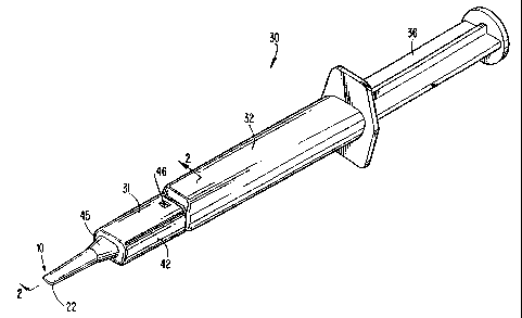
BIZn+'F DEsCIZ~iP'-'ION oF THE DRAWINGS
Figure 1 is a perspective view of an inserter with a tip formation in
accordance
with the present invention.
Figure 2 is an exploded cross-sectional view taken along line 2-2 in Figure 1.
Figure 3 is a partial, side elevational view of the tip member of the inserter
showing the present tip formation.
Figure 4 is a partial, top plan view of the tip member.
Figure 5 is a pardal, bottom view of the tip member taken along line 5-5 in
Figure
2.
Figure 6 is a cross-sectional view taken along line 6-6 in Figure 4.
Figure 7 is a cross-sectional view taken along line 7-7 in Figure 3.
Figure 8 is an exploded, side elevational view of a second embodiment of an
inserter with a tip formation in accordance with the present invention.
-3-
J I p
CA 02389821 2002-07-10
Figure 9 is a side elevational view of the cartridge of the inserter of the
second
embodiment.
Figure 10 is a perspective view of the cartridge.
DETAIL D DESCRIPTION OF'PHE PREFERRED EMBODIMENTs
The present invention pertains to a tip formation for an instrument used to
insert
a flexible intraocular lens or other flexible membrane into an eye. The tip
formation is
formed on the distal end of a cannula and could be used with virtually any
lens insertion
device which uses a tubular member to direct the lens into an eye.
In a prefetred embodiment, tip formation 10 of the present invention is formed
on
the distal or free end 22 of a cannula 16 (Figs. 2-7). The cannula includes a
lumen 18
which defines a generally linear path along a longitudinal axis 19 for
directing a lens of
reduced size through a small incision in an eye (Figs. 2 and 3). The distal
end of lumen
18 is open to form a discharge opening 23 for implanting the lens into an eye.
The distal
end 22 of cannula 16 is beveled to orient the discharge opening 23 at an
inclination to
longitudinal axis 19. The term beveled as used in this application is intended
to indicate
a surface which is oriented at least in part at an inclination to the
longitudinal axis of the
lumen, irrespective of the angle of the inclination, whether the cut is linear
or curved, or
whether the surface is regular or irregular.
The beveled free end 22 defines a front edge 26 and a rear edge 27 of
discharge
opening 23 (Figs. 2, 3, 5 and 7). The wall portion 24 of cannula 16 that
extends between
rear edge 27 and front edge 26 converges in a forward direction. In the
preferred
construction, the tapering of wall portion 24 begins at an imaginary
perpendicular plane
extending through rear edge 27 and continues to front edge 26. Further, wall
portion 24
-4-
, ~ k J1 IINi 1 CA 02389821 2002-07-10
preferably conforms substantially to the shape of a cone segment, such that
the entire
periphery converges toward longitudinal axis 19. Nonetheless, the convergence
of wall
portion 24 could have a different shape, be discontinuous, or extend along
only a part of
the distance between rear edge 27 and front edge 26. The convergence of waU
portion
24 toward axis 19 functions to reduce the size of the cannula's distal end to
ease insertion
of the inserter instrument into the incision, and to minimize the size of the
incision.
Moreover, the reduction of the distal end 22 is achieved without thinning of
the cannula
walls or impeding the advance of the lens into the eye.
The provision of a beveled free end across a tubular member creates a
discharge
opening which is larger than the discharge opening would be if formed to be
perpendicular
to the passage of the tubular member. In accordance with the present
invention, the
additional space gained by providing an inclined discharge opening is
advantageously used
to reduce the size of the free end of the cannula. In other words, wall
portion 24 adjacent
the inclined dischakge opening 23 converges to narrow the external surface of
cannula 16
forward of rear edge 27 without causing the discharge opening to have a
smaller area than
the perpendicular cross-sectional area of lumen 18 at rear edge 27.
In the preferred construction, the distal waUs of the cannula are formed as a
thin
tube to minimize the size of the tip to be passed into the eye. The portion
18a of lumen
18 which is rearward or upstream of rear edge 27 has a substantially uniform
inner
configuration and size (i.e., with the conventional slight taper for molding
purposes) in
order to direct the lens into the eye without the application of additional
high forces
associated with further compression of a lens. The discharge opening 23 is
inclined at a
45 angle to axis 19 to define front edge 26 and a rear edge 27 (Figs. 2 and
3). Wall
portion 24 converges forwardly at an included angle of about 20 (i.e., 10
relative to axis
-5-
CA 02389821 2002-07-10
19) from an orthogonal plane aligned with rearward edge 27. Despite the
reduced exterior
of the cannula, the perpendicular cross-sectional area of lumen 18 at rearward
edge 27 is
substantially equal to the area of discharge opening 23 to avoid impeding the
advance of the
lens into the eye (Figs. 5 and 6). In a preferred example, the area of the
discharge opening
23 and the perpendicular cross-section of the lumen at rearward edge 27 are
equal to about
0.004 square inches.
Many variations can be made to the preferred tip formation without departing
from the
spirit of the invention. For example, the converging wall portion 24 can
converge at a
changing rate or begin converging at locations forward or downstream of rear
edge 27.
Also, the beveled surface can be set at different inclinations or provided
with a non-linear
shape. The area of the discharge opening may also, of course, continue to be
somewhat
larger than the perpendicular cross-sectional area at the rear edge 27, if the
convergence
begins forward of the rear edge or a smaller level of convergence is used.
In the preferred embodiment, tip formation 10 is an integral part of a
discrete, one-
piece tip member 31 for an inserter 30 (Figs. I and 2). Inserter 30 has a
construction as
disclosed in U.S. Patent No. 5,873,879. In general, inserter 30 includes a
tubular member
32, a cover 34, and a plunger 36 along with tip member 31. Tubular member 32
has a
rearwardly opening cavity 34 for receiving plunger 36, and a forwardly
projecting shelf
38 for receiving a lens 37. Cover 34 overlies the shelf to enclose the lens
and define station
40 for holding and folding the lens.
In use, the lens is placed onto shelf 38 and enclosed with cover 34. Base
portion 42 of
tip member 31 is pushed over the shelf and cover, and locked in place with a
latch
46 to form an integral unit with tubular member 32. Plunger 36 is pushed
forward to
-6-
I'tl, ~ p
CA 02389821 2002-07-10
advance the lens through station 40, which folds the lens, and into tapering
segment 45 of
lumen 18. The combined folding and compression effects of station 40 and
tapering
segment 45 define a lens reducing structure which reduces the size of lens to
a cross-
sectional size small enough to fit through the narrow incision in the eye.
Portion 18a of
lumen 18 has a substantially uniform configuration and size to guide the lens
up to the rear
edge 27 of discharge opening 23 without any further significant compression of
the lens.
The lens is then fed through tip formation 10, out discharge opening 23, and
into the eye.
Yet, despite the convergence of wall portion 24, the area of the discharge
opening is still
the same or larger than the perpendicular cross-sectional area of the lumen at
the rear
edge 27.
Alternatively, tip formation 10 could be fonmed on the end of other inserters
which use
a cannula to direct the lens into an eye, irrespective of the type of lens
reducing structure
which is used, whether the inserter is composed of one piece or multiple
pieces, or whether
the tip formation is a part of a cartridge or inserter tip. For example, tip
formation 10 could
be provided on the distal end a cartridge 50 (Figs. 8-10). Cartridge 50 has a
tapering lumen
53 within the cartridge as a lens reducing structure. After the lens is loaded
into the lumen,
the cartridge is placed into insertion device 54 having a plunger 56 for
moving the lens into
an eye.
Similarly, tip formation 10 can also be fonmed on the end of a cartridge as 20
disclosed
in U.S. Patent No. 5,494,484 to Feingold. In this case, the cartridge includes
hinged
sections as the lens reducing structure which close about the lens to reduce
the cross-
sectional size of the lens. The cartridge is then placed within an insertion
device which
advances the lens into an eye.
-7-
CA 02389821 2002-07-10
As another example, tip formation 10 could be used with an inserter which has
a one-piece tubular member as disclosed in U.S. Patent No. 5,944,725. In this
device, the
lens reducing structure includes a laterally movable compressor for providing
an initial
reduction in the lens' cross-sectional size, and a tapering lumen which
further reduces the
size of the lens as it is moved toward the eye. Tip formation 10 is formed at
the distal
end of the cannula in the same way as described above.
The above discussion concerns the preferred embodiments of the present
invention. Various other embodiments as well as many changes and alterations
may be
made without departing from the spirit and broader aspects of the invention as
defined in
the claims.
-8-