Note: Descriptions are shown in the official language in which they were submitted.
CA 02391277 2002-05-10
WO 01/34145 PCT/US00/31067
TREATING CANCER BY INCREASING INTRACELLULAR MALONYL COA LEVELS
Review of Related Art
A number of studies have demonstrated surprisingly high levels of
fatty acid synthase expression (FAS, E.C. 2.3.1.85) in virulent human breast
cancer
(Alo, P. L., Visca, P., Marci, A., Mangoni, A., Botti, C., and Di Tondo, U.
Expression of fatty acid synthase (FASO as a predictor of recurrence in stage
I breast
carcinoma patients., Cancer. 77: 474-482, 1996; Jensen, V., Ladekarl, M., Holm-
Nielsen, P., Melsen, F., and Soerensen, F. B. The prognostic value of
oncogenic
antigen 519 (OA-519) expression and proliferative activity detected by
antibody
MIB-1 in node-negative breast cancer., Journal of Pathology. 176: 343-352,
1995),
as well as other cancers (Rashid, A., Pizer, E. S., Moga, M., Milgraum, L. Z.,
Zahurak, M., Pasternack, G. R., Kuhajda, F. P., and Hamilton, S. R. Elevated
1 S expression of fatty acid synthase and fatty acid synthetic activity in
colorectal
neoplasia., American Journal of Pathology. ISO: 201-208, 1997; Pizer, E., Lax,
S.,
Kuhajda, F., Pasternack, G., and Kurman, R. Fatty acid synthase expression in
endometrial carcinoma: correlation with cell proliferation and hormone
receptors.,
Cancer. 83: 528-537, 1998). FAS expression has also been identified in
intraductal
and lobular in situ breast carcinoma; lesions associated with increased risk
for the
development of infiltrating breast cancer (Milgraum, L. Z., Witters, L. A.,
Pasternack, G. R., and Kuhajda, F. P. Enzymes of the fatty acid synthesis
pathway
are highly expressed in in situ breast carcinoma., Clinical Cancer Research.
3: 211 5-
2120, 1997). FAS is the principal synthetic enzyme of fatty acid synthesis (FA
synthesis) which catalyzes the NADPH dependent condensation of malonyl-CoA
and acetyl-CoA to produce predominantly the 16-carbon saturated free fatty
acid,
palmitate (Wakil, S. Fatty acid synthase, a proficient multifunctional
enzyme.,
Biochemistry. 28: 4523-4530, 1989). Ex vivo measurements in tumor tissue have
revealed high levels of both FAS and FA synthesis indicating that the entire
genetic
program is highly active consisting of some 25 enzymes from hexokinase to FAS.
CA 02391277 2002-05-10
WO 01/34145 PCT/US00/31067
2
Cultured human cancer cells treated with inhibitors of FAS, including
the fungal product, cerulenin, and the novel compound, C75, demonstrated a
rapid
decline in FA synthesis, with subsequent reduction of DNA synthesis and cell
cycle
arrest, culminating in apoptosis (Pfizer, E. S., Jackisch, C., Wood, F. D.,
Pasternack,
G. R., Davidson, N. E., and Kuhajda, F. Inhibition of fatty acid synthesis
induces
programmed cell death in human breast cancer cells., Cancer Research. 56: 2745-
2747, 1996, Pfizer, E. S., Chrest, F. J., DiGiuseppe, J. A., and Han, W. F.
Pharmacological inhibitors of mammalian fatty acid synthase suppress DNA
replication and induce apoptosis in tumor cell lines., Cancer Research. 58:
4611-
4615, 1998). Pharmacological inhibition of mammalian fatty acid synthase
activity
lead to inhibition of DNA replication within about 90 minutes of drug
application.
These findings suggested a vital biochemical link between FA synthesis and
cancer
cell growth. While generating a great deal of interest, the question of how
inhibition
of fatty acid synthase triggered this phenomenon remained unknown.
Importantly,
these effects occurred despite the presence of exogenous fatty acids in the
culture
medium derived from fetal bovine serum. While it has been possible to rescue
the
cytotoxic effect of cerulenin on certain cells in fatty acid-free culture
conditions by
the addition of exogenous palmitate, most cancer cells were not rescued from
FA
synthesis inhibition by the pathway endproduct (data not shown) (Pfizer, E.
S.,
Wood, F. D., Pasternack, G. R., and Kuhajda, F. P. Fatty acid synthase (FAS):
A
target for cytotoxic antimetabolities in HL60 promyelocytic leukemia cells.,
Cancer
Research. 1996: 745-751, 1996). Thus, it has been unresolved whether the
cytotoxic
effect of FA synthesis inhibition on most cancer cells resulted from end
product
starvation, or from some other biochemical mechanism.
Summary of the Invention
This invention describes a method to kill cancer cells by acute
elevation of cellular malonyl Coenzyme A (Malonyl CoA) which leads to
apoptosis.
Elevation of malonyl CoA induced by inhibition of fatty acid synthase (FAS),
is
correlated with both inhibition of fatty acid synthesis and also with
inhibition of
carnitine palmitoyltransferase-1 (CPT-1). Any combination of drugs which
CA 02391277 2002-05-10
WO 01/34145 PCT/US00/31067
3
produces an analogous physiologic effect may be expected to lead to the same
effect
on susceptible tumor cells. For example, combination therapy with drugs) that
inhibit the fatty acid synthesis by inhibiting acetyl CoA carboxylase (the
first
enzyme in the fatty acid synthesis pathway) and drugs) that inhibit CPT-1 may
be
expected to induce apoptosis in tumor cells. Therefore, this invention
encompasses
any method to systemically inhibit the activity of CPT-1 in cancer cells
including
but not limited to direct inhibition of CPT-1 through small molecule
inhibitors such
as etomoxir, as well as inhibition of CPT-1 incidental to increasing the level
of
malonyl CoA in cancer cells.
This therapeutic strategy will lead to novel chemotherapeutic agents
for a wide variety of human cancers. In addition, as this is a novel pathway
leading
to apoptosis which is not shared by other cancer drugs, it may be anticipated
that
induction of high levels of malonyl CoA and/or CPT-1 inhibition may potentiate
other commonly utilized cancer therapeutic agents.
In one embodiment, this invention provides a method for inhibiting
growth of tumor cells in an organism by administering to the organism a
composition which causes a rise in intracellular malonyl CoA in tumor cells of
the
organism. Preferably, the intracellular malonyl CoA in at least the tumor
cells of the
organism rises abruptly (i.e., acutely or sharply), and more preferably, the
intracellular malonyl CoA rises prior to any significant rise in consumption
rate of
malonyl CoA. Typically, the intracellular malonyl CoA in cells of the organism
rises within 3 hours of administration, and intracellular malonyl CoA may be
expected to rise prior to growth inhibition of the cells.
In a preferred mode of the method of this invention, the rise in
intracellular malonyl CoA is correlated with reduced consumption of malonyl
CoA.
For example, the rise in intracellular malonyl CoA may be correlated with
reduced
intracellular activity of malonyl CoA decarboxylase (MCD) or reduced
intracellular
activity of fatty acid synthase; and optionally, the composition may comprise
an
inhibitor of MCD. In another preferred mode, the rise in intracellular malonyl
CoA
CA 02391277 2002-05-10
WO 01/34145 PCT/US00/31067
4
is correlated with increase synthesis of malonyl CoA and/or the rise in
intracellular
malonyl CoA is correlated with increased intracellular activity of acetyl-CoA
carboxylase (ACC).
In one embodiment of the method of this invention, the composition
comprises an agent selected from the group consisting of an activator of ACC,
an
activator of citrate synthase, an inhibitor of 5'-AMP-activated protein kinase
(AMPK), and/or an inhibitor of acyl CoA synthase. In another mode of this
invention, the composition comprises an inhibitor of carnitine
palmitoyltransferase-1
(CPT-1), which may be etomoxir, preferably administered in combination with an
agent from the proceeding group. In yet another embodiment of the method of
this
invention, a second chemotherapeutic agent is administered to the organism,
said
second chemotherapeutic agent being non-inhibitory to fatty acid synthesis.
Preferably, the method of this invention is used to treat organisms
having, prior to administration of the composition, intracellular malonyl CoA
level
in tumor cells of at least 2-fold above normal malonyl CoA level in non-
malignant
cells. More preferably, the method is used to treat organisms where the fatty
acid
synthesis rate in some cells of the organism is at least 2-fold above that of
normal
cells prior to administration of the composition, and administration of the
composition is cytotoxic to those cells. Preferably, the organism comprises
tumor
cells having elevated fatty acid synthesis rates and cell number of such tumor
cells is
reduced subsequent to administration of said composition. Upon treatment,
preferably, the intracellular level of malonyl CoA is elevated and
intracellular level
of acetyl CoA, and free CoA are reduced relative to pretreatment levels.
In another embodiment, this invention provides a method for
inhibiting growth of tumor cells in an organism comprising administering to
said
cells (a) an inhibitor of fatty acid synthesis in said cells; and (b) an
inhibitor of fatty
acid oxidation in said cells. Preferably, the inhibitor of fatty acid
oxidation is
administered in an amount which does not significantly inhibit CPT-2. More
CA 02391277 2002-05-10
WO 01/34145 5 PCT/US00/31067
preferably, the inhibitor of fatty acid synthesis and the inhibitor of fatty
acid
oxidation are administered in amounts to achieve levels of inhibition which
are at
least about equal to or greater than the levels of the respective inhibitions
observed
for cytotoxic doses of cerulenin.
In yet another embodiment, this invention provides a screening
method to assist in detecting compositions which are selectively cytotoxic to
tumor
cells comprising administering a target composition to a cell having an
elevated
intracellular malonyl CoA level, monitoring intracellular malonyl CoA in the
cell
subsequent to this administration, an abrupt increase in intracellular malonyl
CoA
being indicative of selective cytotoxicity. Preferably, this method further
comprises
comparing the pattern of intracellular malonyl CoA level changes in the
presence
and absence of TOFA, wherein reduced changes in malonyl CoA level in the
presence of TOFA is indicative of selective cytotoxicity.
In still another embodiment, this invention provides a screening
method to assist in detecting compositions which are growth inhibitory to
tumor
cells comprising administering a target composition to a tumor-derived cell
line and
monitoring CPT-1 activity in the cell subsequent to this administration,
wherein a
decrease in CPT-1 activity is indicative of growth inhibitory potential.
Preferably,
the method is carned out when the cell is permeabilized. Alternatively, the
method
further comprises monitoring said cell for apoptosis, and the monitoring for
apoptosis may comprise a method selected from the group consisting of
measuring
mitochondria) transmembrane potential, staining with vital dyes, monitoring
caspase
activation in whole cells using Western blot, and measuring cytochrome C
elaborated from mitochondria using Western blot.
Brief Description of the Figures
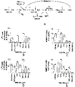
Figure 1 shows the fatty acid synthesis pathway, and the effect of
various fatty acid synthase inhibitors on fatty acid synthesis and tumor cell
growth.
Figure 2 shows malonyl CoA levels under various conditions.
CA 02391277 2002-05-10
WO 01/34145 6 PCT/US00/31067
Figure 3 shows the results of clonogenic assays and apoptosis assays
on breast cancer cells treated with various inhibitors.
Figure 4 shows various parameters in tumor cells and liver cells.
Figure 5 shows malonyl CoA levels in tumor cells and liver cells.
Figure 6 shows the pathway for cellular oxidation of fatty acids.
CPT-1 regulates oxidation of fatty acids in the mitochondrion by controlling
the
passage of long chain acyl CoA derivatives such as palmitoyl CoA through the
outer
mitochondria) membrane into the mitochondrion, thus preventing the futile
cycle of
oxidizing endogenously synthesized fatty acids.
Figure 7 shows the effect of Etomoxir on growth of MCF-7 cells with
and without C-75.
Figure 8 shows the effect of cerulenin on fatty acid oxidation in
MCF-7 cells.
Figure 9 shows the effect of Etomoxir, TOFA and cerulenin on CPT-
1 activity.
Figure 10 shows the effect of Etomoxir on fatty acid oxidation in
MCF-7 cells.
Figure 11 shows the effect of Etomoxir on growth of MCF-7 cells.
Figure 12 shows the results of clonogenic assays with MCF-7 cells
treated with both Etomoxir and TOFA.
Figure 13 shows the effect of Etomoxir and/or C-75 on growth of
MCF-7 cells.
Detailed Description of the Embodiments
If fatty acid starvation mediated the cytotoxic effects of cerulenin and
C75, then any other FA synthesis inhibitor of similar potency should produce
similar
effects. To test this idea, the inventors compared the effects on cancer cells
of
inhibition of acetyl-CoA carboxylase (ACC, E.C. 6.4.1.2), the rate limiting
enzyme
of fatty acid synthesis, with the effects of FAS inhibitors. The inventors
discovered
that inhibition of FAS leads to high levels of malonyl-CoA which occurs within
an
hour of C75 treatment. These superphysiological levels of malonyl-CoA, rather
than
merely low levels of endogenously synthesized fatty acids, are responsible for
breast
CA 02391277 2002-05-10
WO 01/34145 ,~ PCT/US00/31067
cancer cell apoptosis. In addition, this is a novel pathway which leads to
selective
apoptosis of cancer cells.
Figure 1A outlines the portion of the FA synthesis pathway
containing the target enzymes of the inhibitors used in this study. Inhibition
of fatty
acid synthase results in high levels of malonyl-CoA that contribute to the
cytotoxicity of against human breast cancer cells (ref). In addition to its
role as a
substrate for fatty acid synthesis, malonyl-CoA is a potent inhibitor of
carnitine
palmitoyltransferase-1 (CPT-1) the rate limiting enzyme of fatty acid
oxidation.
CPT-1 is an integral outer membrane protein of the mitochondrion that performs
a
trans-esterification of long chain fatty acyl CoA's to L-carnitine producing
acylcarnitine. Acylcarnitine is transported across the mitochondrial membranes
where it is esterified back to acyl-CoA by CPT-2. Physiologically, CPT-1
activity is
regulated through inhibition by malonyl-CoA, a substrate of fatty acid
synthesis.
Malonyl-CoA is the enzymatic product of acetyl-CoA carboxylase (ACC, E.C.
6.4.1.2), the pace-setting enzyme for fatty acid synthesis. Cytoplasmic
malonyl-
CoA levels are higher during fatty acid synthesis due to increased activity of
ACC.
The high levels of malonyl-CoA, in turn, inhibits CPT-1, and blocks entry of
long-
chain acyl-CoA's into the mitochondrion. This prevents the futile cycle of
simultaneous fatty acid synthesis and oxidation. In muscle, which is
essentially
devoid of FAS, ACC and malonyl-CoA regulate fatty acid oxidation, an important
fuel source for cardiac and skeletal muscle.
TOFA (5-(tetradecyloxy)-2-furoic acid) is an allosteric inhibitor of
acetyl-CoA carboxylase (ACC, E.C. 6.4.1.2), blocking the carboxylation of
acetyl-
CoA to malonyl-CoA. Once esterified to coenzyme-A, TOFA-CoA allosterically
inhibits ACC with a mechanism similar to long chain acyl-CoA's, the
physiological
end-product inhibitors of ACC (Halvorson, D. L. and McCune, S. A. Inhibition
of
fatty acid synthesis in isolated adipocytes by 5-(tetradecyloxy)-2-furoic
acid.,
Lipids. 19: 851-856, 1984). Both cerulenin (Funabashi, H., Kawaguchi, A.,
Tomoda, H., Omura, S., Okuda, S., and Iwasaki, S. Binding site of cerulenin in
fatty
acid synthetase., J. Biochem. 105: 751-755, 1989) and C75 (Pfizer, et al.,
1998) are
CA 02391277 2002-05-10
WO 01/34145 g PCT/US00/31067
inhibitors of FAS, preventing the condensation of malonyl-CoA and acetyl-CoA
into
fatty acids. Cerulenin is a suicide inhibitor, forming a covalent adduct with
FAS
(Moche, M., Schneider, G., Edwards, P., Dehesh, K., and Lindqvist, Y.
Structure of
the complex between the antibiotic cerulenin and its target, beta-ketoacyl
carrier
protein synthase., J Biol Chem. 274: 6031-6034, 1999), while C75 is likely a
slow-
binding inhibitor (Kuhajda, F.P., Pizer E.S., Mani, N.S., Pinn, M.L., Han
W.F.,
Chrest F.J., and CA.T., Synthesis and anti-tumor activity of a novel inhibitor
of fatty
acid synthase, Proceeding of the American Association for Cancer Research,
40:121,
1999). Using TOFA, the inventors have achieved FA synthesis inhibition in
human
breast cancer cell lines comparable to inhibition by cerulenin or C75.
Surprisingly,
however, TOFA was essentially non-cytotoxic in clonogenic assays of human
breast
cancer cells. These data indicate that fatty acid starvation is not a major
source of
cytotoxicity to cancer cells in serum supplemented culture. An alternative
effect of
FAS inhibition (high levels of the substrate, malonyl-CoA, resulting
specifically
from inhibition of FAS) appears to mediate cytotoxicity of cerulenin and C75.
Malonyl-CoA, the enzymatic product of acetyl-CoA carboxylase
(ACC, E.C. 6.4.1.2), is a key regulatory molecule in cellular metabolism. In
addition to its role as a substrate in fatty acid synthesis, malonyl-CoA
regulates (3-
oxidation of fatty acids through its interaction with carnitine
palmitoyltransferase-1
(CPT-1) at the outer membrane of the mitochondria. Carnitine
palmitoyltransferase
(CPT-1) is the rate limiting enzyme of mitochondria) fatty acid oxidation (See
Figure 6). It is an integral outer membrane protein of the mitochondrion that
performs a traps-esterification of long chain fatty acyl CoA's to L-carnitine
producing acylcarnitine. Acylcarnitine is transported across the mitochondria)
membranes where it is esterified back to acyl-CoA by CPT-2.
Many types of cancer cells have high levels of fatty acid synthesis.
As expected, cells with high levels of fatty acid synthesis have high steady
state
levels of malonyl-CoA, at least six times the levels in normal cells (see
Example 6).
Treatment of tumor cells with inhibitors of FAS will selectively and abruptly
raise
malonyl-CoA levels to superphysiological levels in cancer cells. This maneuver
CA 02391277 2002-05-10
WO 01/34145 PCT/US00/31067
9
raises malonyl-CoA levels by both blocking utilization of malonyl-CoA as a
substrate in fatty acid synthesis and concomitantly stimulating malonyl-CoA
synthesis by relieving fatty acyl-CoA inhibition of ACC (Figure 1A). Since FAS
is
preferentially expressed in cancer cells, the malonyl-CoA elevation is largely
restricted to tumors cells. This leads to cancer cell apoptosis and sparing of
normal
tissues as occurs in human cancer xenografts treated with FAS inhibitors (See
Example 5).
CPT-1 has two isoforms, liver-type (L-CPT-1) and muscle-type (M-
CPT-1) (Swanson, S. T., Foster, D. W., McGarry, J. D., and Brown, N. F. Roles
of
the N- and C-terminal domains of carnitine palmitoyltransferase I isoforms in
malonyl-CoA sensitivity of the enzymes: insights from expression of chimaeric
proteins and mutation of conserved histidine residues., Biochem . J. 335: 513-
S 19,
1998). These isoforms have widely different kinetic properties in their K",
for
carnitine (500 p.M for M-CPT-1 and ~30 p.M for L-CPT-1) and sensitivity to
malonyl-CoA inhibition (M-CPT-1 is 100-fold more sensitive, K,= 0.07 pM versus
7 ~M). While the regulatory site of malonyl-CoA resides in the N-terminal
region,
the exact binding site has not been elucidated (Swanson, et al., 1998).
Importantly,
etomoxir, a covalent inhibitor of CPT-1 that is used herein as an exemplary
CPT-1
inhibitor, binds at a site different than that of malonyl-CoA.
CPT-1 has not been studied in human cancer cells. Hence, the
isoform expressed in human cancer cells is unknown. Conceptually, the liver
isoform should be expressed in tumors of epithelial differentiation which
includes all
carcinomas, while the muscle isoform would be expressed in non-epithelial
tumors
such as sarcomas. However, studies of ACC liver and muscle isoforms have found
that either or both isoforms can be expressed in human breast cancer cells
(Witters,
L., Widmer, J., King, A., Fassihi, K., and Kuhajda, F. Identification of human
acetyl-CoA carboxylase isozymes in tissue and in breast cancer cells.,
International
Journal of Biochemistry. 26: 589-594, 1994). Similarly, human carcinoma cells
may have the ability to express either or both CPT-1 isoforms.
CA 02391277 2002-05-10
WO 01/34145 10 PCT/US00/31067
Recently, inhibition of CPT-1 was shown to sensitize cells to fatty
acid induced apoptosis (Paumen, M. B., Ishida, Y., Muramatsu, M., Yamamoto,
M.,
and Honjo, T. Inhibition of carnitine palmitoyltransferase I augments
sphingolipid
synthesis and palmitate-induced apoptosis., J. Biol. Chem. 272: 3324-3329,
1997).
Moreover, increased malonyl-CoA levels induced by the inhibition of fatty acid
synthase (FAS) are cytotoxic to human cancer cells (see Examples 4 and 5).
Taken
together, these data suggest that human cancer cells are susceptible to
induction of
apoptosis via alterations in fatty acid metabolism. CPT-1 has also been shown
to
interact directly with BCL-2, the anti-apoptosis protein, at the outer
mitochondrial
membrane (Paumen, M. B., Ishisa, Y., Han, H., Muramatsu, M., Eguchi, Y.,
Tsujimoto, Y., and Honjo, T. Direct interaction of the mitochondrial membrane
protein carnitine palmitoyltransferase I with Bcl-2, Biochem Biophys Res
Commun.
231: 523-525, 1997). Potentially, the interaction of CPT-1 with BCL-2 may
provide
a down-stream mechanism leading to apoptosis by modulating the anti-apoptotic
1 S effects of BCL-2.
In addition to its role as a substrate for FAS, malonyl-CoA acts at the
outer mitochondrial membrane to regulate fatty acid oxidation by inhibition of
carnitine palmitoyltransferase 1 (CPT-1). Inhibition of CPT-1 has been shown
to
sensitize cells to fatty acid induced apoptosis; CPT-1 may also interact
directly with
BCL-2, the anti-apoptosis protein, at the mitochondria. FAS inhibition leads
to high
levels of malonyl-CoA inhibiting CPT-1 which induces cancer cell apoptosis.
Since
most proliferating and non-proliferating normal cells do not have high levels
of
FAS, they will not be affected by this therapeutic strategy.
Malonyl CoA levels may be manipulated using a variety of methods
and target enzymes. The Examples demonstrate elevation of malonyl CoA levels
through reduced utilization and simultaneous enhanced production. Acute
increase
in malonyl CoA levels lead to the selective destruction of cancer cells via
apoptosis
leaving normal cells unaffected. Methods for inducing apoptosis according to
this
invention fall into two broad categories: direct induction of acute increase
in
malonyl-CoA (e.g., by inhibiting FAS) and use of combination therapy to
inhibit
CA 02391277 2002-05-10
WO 01/34145 11 PCT/US00/31067
both fatty acid oxidation and fatty acid synthesis (e.g., through a non-FAS
inhibitory
mode). This therapeutic strategy identifies potential new targets and
strategies for
cancer chemotherapy based upon alteration of fatty acid metabolism.
Fatty acid oxidation may be inhibited via CPT-1 inhibition directly
by inhibitory agents, such as etomoxir. Specific inhibitors to CPT-1 isoforms
may
also be developed. Alternatively, one could manipulate carnitine levels to
reduce
CPT-1 activity by reducing its substrate. Also, one may reduce CPT-1
expression
levels either through genetic manipulation or by reducing exogenous fatty
acids.
Example 7 below is an example of the method of directly inhibiting CPT-1 using
etomoxir in human breast cancer cells.
Other strategies for inhibiting fatty acid synthesis and oxidation
include any method to increase malonyl-CoA levels from increased synthesis,
decreased degradation, or preferably both. Malonyl-CoA levels may be
manipulated
using a variety of methods and target enzymes. Examples 4-S demonstrate
elevation
of malonyl-CoA levels through reduced utilization and simultaneous enhanced
production. Acute increase in malonyl-CoA levels leads to the selective
destruction
of cancer cells via apoptosis leaving normal cells unaffected. Other examples
demonstrate additional ways to cause cancer cell growth inhibition or death.
Preferably, manipulation of fatty acid metabolism according to this
invention is accomplished by administering a composition (or multiple
compositions) to an organism in need thereof. The composition administered to
the
organism will contain an agent having at least one biological effect on fatty
acid
metabolic pathways, for example by raising intracellular malonyl-CoA levels.
Typically, the organism will be a mammal, such as a mouse, rat, rabbit, guinea
pig,
cat dog, horse, cow, sheep, goat, pig, or a primate, such as a chimpanzee,
baboon, or
preferably a human. Usually, the organism will contain neoplastic (malignant)
cells.
The method of this invention is directed to selectively affecting malignant
cells, and
having less effect (or more preferably no effect) on normal (non-malignant)
cells.
CA 02391277 2002-05-10
WO 01/34145 12 PCT/US00/31067
The agent in the composition administered to the organism will
preferably raise the intracellular malonyl CoA levels in at least a portion of
the
malignant cells in the organism. Preferably the malonyl CoA level will be
raised at
least 2-fold, more preferably at least 5-fold. Preferably, the agent will
raise the
intracellular malonyl-CoA concentration in the malignant cells to a level
higher than
the level in surrounding normal cells.
Suitable agents may raise the malonyl CoA level by any of a number
of methods (see alternative mechanisms listed below). Preferred agents
typically
induce a sudden or abrupt rise in malonyl CoA level. In some embodiments, two
or
more agents are administered, and some or all of these agents may affect
malonyl
CoA level by a different mechanism. Alternatively, a combination of agents may
be
used to lower fatty acid synthesis and simultaneously lower fatty acid
oxidation.
Preferably, the levels of fatty acid synthesis and oxidation will be lowered
to levels
comparable to those achieved by cytotoxic treatment with cerulenin. Agents
acting
by any of the modes of the following list may be used in compositions and
methods
of this invention. Assays for the following activities are available in the
literature,
and determination of whether a particular agent exhibits one of these
activities is
within the skill in the art.
Increasing malonyl-CoA production:
Acetyl-CoA carboxylase (ACC) effectors: Agents which increase
ACC activity, reduce ACC inhibition, or increase the mass of active ACC enzyme
will lead to increased levels of malonyl-CoA.
5'-AMP protein kinase effectors: S'-AMP protein kinase inhibits
ACC by phosphorylation leading to acute reduction of malonyl-CoA. Inhibitors
of
this kinase would lead to acutely increased levels of malonyl-CoA by releasing
inhibition of ACC.
CA 02391277 2002-05-10
WO 01/34145 13 PCT/US00/31067
Citrate synthase effectors: Increasing mitochondrial citrate would
provide substrate for fatty acid synthesis, and citrate also acts as a "feed-
forward"
activator of ACC causing increase malonyl-CoA synthesis.
Acyl-CoA synthase effectors: Inhibition of acyl-CoA synthase would
reduce cellular fatty acyl-CoA concentration releasing inhibition of ACC. This
would result in increased ACC activity and malonyl-CoA levels.
Decreasing malonyl-CoA utilization:
Malonyl-CoA decarboxylase (MCD) effectors: This enzyme
catalyzes an ATP dependent decarboxylation of malonyl-CoA back to acetyl-CoA.
Inhibition of MCD would acutely raise malonyl-CoA levels.
Simultaneously decreased malonyl-CoA utilization and increased production:
Fatty acid synthase (FAS) effectors: Inhibition of FAS leads to
decreased utilization of malonyl-CoA by blocking its incorporation into fatty
acids.
FAS inhibition also leads to reduced fatty acyl-CoA levels which will activate
ACC.
Exemplary FAS inhibitors may be obtained as described in U.S. Patent Nos.
5,759,837 and 5,981,575, incorporated herein by reference.
These strategies for modifying fatty acid metabolism, and especially
for acutely increasing malonyl-CoA levels, may be used together or in concert
with
other drugs to enhance apoptosis of cancer cells. Preferably, at least one
agent in the
compositions of this invention raises the level of malonyl-CoA by a mechanism
other than inhibiting FAS.
ADMINISTRATION OF THE COMPONENTS
Therapeutic agents according to this invention are preferably
formulated in pharmaceutical compositions containing the agent and a
pharmaceutically acceptable carrier. The pharmaceutical composition may
contain
other components so long as the other components do not reduce the
effectiveness of
CA 02391277 2002-05-10
WO 01/34145 14 PCT/US00/31067
the agent according to this invention so much that the therapy is negated.
Pharmaceutically acceptable Garners are well known, and one skilled in the
pharmaceutical art can easily select carriers suitable for particular routes
of
administration (see e.g., Remington's Pharmaceutical Sciences, Mack Publishing
Co., Easton, PA, 1985).
The pharmaceutical compositions containing any of the agents of this
invention may be administered by parenteral (subcutaneously, intramuscularly,
intravenously, intraperitoneally, intrapleurally, intravesicularly or
intrathecally),
topical, oral, rectal, or nasal route, as necessitated by choice of drug. The
concentrations of the active agent in pharmaceutically acceptable carriers may
range
from 0.01 mM to 1 M or higher, so long as the concentration does not exceed an
acceptable level of toxicity at the point of administration.
Dose and duration of therapy will depend on a variety of factors,
including the therapeutic index of the drugs, disease type, patient age,
patient
weight, and tolerance of toxicity. Dose will generally be chosen to achieve
serum
concentrations from about 0.1 ~g/ml to about 100 pg/ml. Preferably, initial
dose
levels will be selected based on their ability to achieve ambient
concentrations
shown to be effective in in-vitro models, such as those described herein, and
in-vivo
models and in clinical trials, up to maximum tolerated levels. Standard
clinical
procedure prefers that chemotherapy be tailored to the individual patient and
the
systemic concentration of the chemotherapeutic agent be monitored regularly.
The
dose of a particular drug and duration of therapy for a particular patient can
be
determined by the skilled clinician using standard pharmacological approaches
in
view of the above factors. The response to treatment may be monitored by
analysis
of blood or body fluid levels of the agent according to this invention,
measurement
of activity if the agent or its levels in relevant tissues or monitoring
disease state in
the patient. The skilled clinician will adjust the dose and duration of
therapy based
on the response to treatment revealed by these measurements.
CA 02391277 2002-05-10
WO 01/34145 15 PCT/US00/31067
EXAMPLES
In order to facilitate a more complete understanding of the invention,
a number of Examples are provided below. However, the scope of the invention
is
not limited to specific embodiments disclosed in these Examples, which are for
purposes of illustration only.
Example 1. Inhibition of FAS in cells irr vitro
TOFA, Cerulenin, and C75 all inhibited fatty acid synthesis in human
breast cancer cells. The human breast cancer cell lines, SKBR3 and MCF7 were
maintained in RPMI with 10% fetal bovine serum. Cells were screened
periodically
for Mycoplasma contamination (Gen-probe). All inhibitors were added as stock 5
mg/ml solutions in DMSO. For fatty acid synthesis activity determinations,
5x10⁴
cells/well in 24 well plates were pulse labeled with [U-14C]-acetate after
exposure to
drug, and lipids were extracted and quantified as described previously
(Pfizer, et al.,
1988). For MCF7 cells, pathway activity was determined after 2 hours of
inhibitor
exposure. SKBR3 cells demonstrated slower response to FAS inhibitors, possibly
because of their extremely high FAS content, so pathway activity was
determined
after 6 hours of inhibitor exposure.
In standard pulse labeling experiments in which breast cancer cell
lines, SKBR3 and MCF7 were labeled for 2 hours after exposure to FA synthesis
inhibitors, TOFA, C75, and cerulenin all inhibited [U~4C-acetate]
incorporation into
lipids to a similar extent (Figure 1 B and D). In numerous similar experiments
(not
shown), TOFA maximally inhibited FA synthesis in the 1 to Sp,g/ml dose range
in
all cell lines tested, and cerulenin and C75 maximally inhibited FA synthesis
in the
range of lOpg/ml.
Example 2. Effect of the same inhibitors on cell growth
TOFA, Cerulenin, and C75 all inhibited fatty acid synthesis in human
breast cancer cells, but showed differential cytotoxicity. Cells and
inhibitors were as
CA 02391277 2002-05-10
WO 01/34145 16 PCT/US00/31067
described for Example 1. For clonogenic assays, 4x10⁵ cells were plated in 25
cm3
flasks with inhibitors added for 6 hours in concentrations listed. Equal
numbers of
treated cells and controls were plated in 60mm dishes. Clones were stained and
counted after 7 to 10 days.
S Although all inhibitors reduced FA synthesis to a similar degree,
TOFA was non-toxic or stimulatory to the cancer cell growth in the dose range
for
ACC inhibition, as measured by clonogenic assays, while cerulenin and C75 were
significantly cytotoxic in the dose range for FAS inhibition (Figure 1 C and
E). The
profound difference between the cytotoxic effects of ACC and FAS inhibition
demonstrate that the acute reduction of fatty acid production per se is not
the major
source of cell injury after FAS inhibition.
Example 3. Measurement of malonyl-CoA.
The most obvious difference in the expected results of inhibiting
these two enzymes was that malonyl-CoA levels should fall after ACC
inhibition,
but should increase after FAS inhibition. Although not previously investigated
in
eukaryotes, recent data in E. coli have demonstrated elevated levels of
malonyl-CoA
resulting from exposure to cerulenin (Chohnan, et al., 1997, "Changes in the
size
and composition of intracellular pools of non-esterified coenzyme A and
coenzyme
A thioesters in aerobic and facultatively anaerobic bacteria," Applied and
Environmental Microbiology, 63:555-560). Malonyl-CoA levels were measured in
cells subjected to FAS inhibition and to inhibition by TOFA under conditions
described in Example 2.
Malonyl-CoA levels were measured in MCF-7 cells using the HPLC
method of Corkey, et al. (1988, "Analysis of acyl-coenzyme A esters in
biological
samples,"Methods in Enzymology, 166:55-70). Briefly, 2.5 x 10⁵ cells/well in
24
well plates were subjected to 1.2 ml of 10% TCA at 4° C after various
drug
treatments. The pellet mass was recorded and the supernatant was washed 6
times
with 1.2 ml of ether and reduced to dryness using vacuum centrifugation at
25° C.
CA 02391277 2002-05-10
WO 01/34145 17 PCT/US00/31067
Coenzyme-A esters were separated and quantitated using reversed phase HPLC on
a
p Supelco C18 column with a Waters HPLC system running Millenium32 software
monitoring 254nm as the maximum absorbance for coenzyme-A. The following
gradients and buffers were utilized: Buffer A: 0.1 M potassium phosphate, pH
5.0,
S Buffer B: 0.1 M potassium phosphate, pH 5.0, with 40% acetonitrile.
Following a
20 min. isocratic run with 92% A, 8% B at 0.4 ml/min, flow was increased to
0.8
ml/min over one minute whereupon a linear gradient to 10% B was run until 24
min.
then held at 10% B until 50 min. where a linear gradient was run to 100% B at
55
min., completing at 60 min. The following coenzyme-A esters (Sigma) were run
as
standards: malonyl-CoA, acetyl-CoA, glutathione-CoA, succinyl-CoA, HMG-CoA,
and free CoA. Samples and standards were dissolved in 50 ~1 of buffer A.
Coenzyme-A esters eluted sequentially as follows: malonyl-CoA, glutathione-
CoA,
free CoA, succinyl-CoA, HMG-CoA, and acetyl-CoA. Quantitation of coenzyme-A
esters was performed by the Millenium32 software.
Direct measurement of coenzyme-A derivatives in MCF-7 cells by
reversed phase HPLC of acid soluble extracts from drug treated cells confirmed
that
both cerulenin and C75 caused a rapid increase in malonyl-CoA levels while
TOFA
reduced malonyl-CoA levels. Figure 2A is a representative chromatograph
demonstrating the separation and identification of coenzyme-A derivatives
important in cellular metabolism. Malonyl-CoA is the first of these to elute,
with a
column retention time of 19-22 minutes. The overlay of chromatographs in
Figure
2B shows that cerulenin treatment lead to a marked increase in malonyl-CoA
over
the control while TOFA caused a significant reduction. The chemical identity
of the
malonyl-CoA was independently confirmed by spiking samples with standards (not
shown).
Malonyl-CoA levels were markedly increased with FAS inhibition
and reduced by TOFA. Analysis of multiple experiments in Figure 2C
demonstrated
that following a 1 hour exposure to cerulenin or C75 at. 10 p.g/ml, malonyl-
CoA
levels increased by 930% and 370% respectively, over controls, while TOFA
treatment (20 pg/ml) led to a 60% reduction of malonyl-CoA levels. The
CA 02391277 2002-05-10
WO 01/34145 1 g PCT/US00/31067
concentration of TOFA required for maximal reduction of malonyl-CoA levels was
4 fold higher than the dose for pathway inhibition in Figure 1B and D.
However,
optimal cultures for extraction of CoA derivatives had 5 fold higher cell
density than
the cultures used in the other biochemical and viability assays presented.
S The remarkable increase in malonyl-CoA after FAS inhibition can be
attributed in part to the release of long-chain fatty acyl-CoA inhibition of
ACC
leading to an increase in ACC activity (Figure 1A). Moreover, the cerulenin-
induced increase in malonyl-CoA levels occurred within 30 minutes of treatment
(930 +/-15% increase over control, not shown), within the time frame of FA
synthesis inhibition, and well before the onset of DNA synthesis inhibition or
early
apoptotic events Thus, high levels of malonyl-CoA were a characteristic effect
of
FAS inhibitors and temporally preceded the other cellular responses, including
apoptosis.
The levels of cerulenin or C75 which induce high levels of malonyl-
CoA are cytotoxic to human breast cancer cells as measured by clonogenic
assays
and flow-cytometric analysis of apoptosis using merocyanin 450 staining. FAS
inhibition causes high malonyl-CoA levels by inhibiting its consumption
through
FAS inhibition, with concomitant stimulation of synthesis by relieving the
inhibitory
effect of long-chain acyl-CoA's upon ACC activity (Figure 2).
Example 4. TOFA rescue of FAS inhibition
TOFA rescue of FAS inhibition demonstrates that high levels of
malonyl-CoA are responsible for cancer cell cytotoxicity. If the elevated
levels of
malonyl-CoA resulting from FAS inhibition were responsible for cytotoxicity,
then
it should be possible to rescue cells from FAS inhibition by reducing malonyl-
CoA
accumulation with TOFA. Co-administration of TOFA and cerulenin to SKBR3
cells (Figure 3A) abrogated the cytotoxic effect of cerulenin alone in
clonogenic
assays performed as described in Example 2. In MCF7 cells (Figure 3C), TOFA
CA 02391277 2002-05-10
WO 01/34145 19 PCT/US00/31067
produced a modest rescue of both cerulenin and C75 under similar experimental
conditions.
Representative flow cytometric analyses of SKBR3 cells (Figure 3B)
and MCF7 (Figure 3D) substantiated these findings, since TOFA rescued cells
from
cerulenin induced apoptosis. Apoptosis was measured by multiparameter flow
cytometry using a FACStarP~°S flow cytometer equipped with argon and
krypton
lasers (Becton Dickinson). Apoptosis was quantified using merocyanine 540
staining (Sigma), which detects altered plasma membrane phospholipid packing
that
occurs early in apoptosis, added directly to cells from culture (Pfizer, et
al., 1998;
Mower, et al., 1994, "Decreased membrane pospholipid packing and decreased
cell
size precede DNA cleavage in mature mouse B cell apoptosis, J. Immunol.,
152:4832-4842). In some experiments, chromatin conformational changes of
apoptosis were simultaneously measured as decreased staining with LDS-751
(Exciton) (Frey, et al., 1995, "Nucleic acid dyes for detection of apoptosis
in live
cells," Cytometry, 21:265-274). Merocyanine 540 [10~g/ml] was added as a
1 mg/ml stock in water. Cells were stained with LDS-751 at a final
concentration of
100nM from a 1mM stock in DMSO. The merocyanine 540-positive cells were
marked by an increase in red fluorescence, collected at 575 +/- 20 nm, 0.5 to
2 logs
over merocyanine 540-negative cells. Similarly, the LDS-751 dim cells
demonstrated a reduction in fluorescence of 0.5 to 1.5 logs relative to normal
cells,
collected at 660 nm with a DF20 band pass filter. Data were collected and
analyzed
using CellQuest software (Becton Dickinson).
In these experiments, all LDS-751 dim cells were merocyanine 540
bright, however a population of merocyanine 540 bright cells were detected
that
were not yet LDS-751 dim. All merocyanine 540 bright cells were classified as
apoptotic. These experiments also confirmed the differential cytotoxicity
between
TOFA (<5% increase in apoptosis; no reduction in clonogenicity) compared to
cerulenin (>85% apoptosis; 70% reduction in clonogenicity). Taken together,
these
studies show that high malonyl-CoA levels play a role in the cytotoxic effect
of FAS
inhibitors on cancer cells.
CA 02391277 2002-05-10
WO 01/34145 20 PCT/US00/31067
Example 5. Effect of FAS inhibitors on tumor cell growth in vivo
To determine if the effects of FAS inhibition seen in vitro would
translate to an in vivo setting requiring systemic activity, C75 was tested
against
subcutaneous MCF-7 xenografts in athymic nude mice, to quantitate effects on
FA
synthesis and the growth of established solid tumor. Previous studies have
demonstrated local efficacy of cerulenin against a human cancer xenograft
(Pfizer, et
al., 1996, "Inhibition of fatty acid synthesis delays disease progression in a
xenograft
model of ovarian cancer," Cancer Res., 56: 1189-1193), but were limited by the
failure of cerulenin to act systemically. The similar responses of breast
cancer cells
to cerulenin and C75 in vitro suggested that C75 might be effective in vivo
against
xenografted breast cancer cells.
Subcutaneous flank xenografts of the human breast cancer cell line,
MCF-7 in nu/nu female mice (Harlan) were used to study the anti-tumor effects
of
C75 in vivo. All animal experiments complied with institutional animal care
guidelines. All mice received a 90-day slow-release subcutaneous estrogen
pellet
(Innovative Research) in the anterior flank 7 days before tumor inoculation.
10'
MCF-7 cells were xenografted from culture in DMEM supplemented with 10% FBS
and insulin 10 ~g/ml.
Treatment began when measurable tumors developed about 10 days
after inoculation. Eleven mice (divided among two separate experiments of 5
and 6
mice each) were treated intraperitoneally with weekly doses of C75 at 30 mg/kg
in
0.1 ml RPMI. Dosing was based on a single dose LD~o determination of 40 mg/kg
in BALB/c mice; 30 mg/kg has been well tolerated in outbred nude mice. Eleven
control mice (divided in the same way as the treatment groups) received RPMI
alone. Tumor volume was measured with calipers in three dimensions. Experiment
was terminated when controls reached the surrogate endpoint.
In a parallel experiment to determine fatty acid synthesis activity in
treated and control tumors, a group of MCF-7 xenografted mice were treated
with
CA 02391277 2002-05-10
WO 01/34145 21 PCT/US00/31067
C75 or vehicle at above doses and sacrificed after 3 hours. Tumor and liver
tissue
were ex vivo labeled with [Ul4-C] acetate, lipids were extracted and counted
as
described (Pfizer, et al, 1996).
In an additional parallel experiment to histologically examine treated
and control tumors, 6 C75 treated and 6 vehicle control mice were sacrificed 6
hours
after treatment. Tumor and normal tissues were fixed in neutral-buffered
formalin,
processed for routine histology, and immunohistochemistry for FAS was
performed.
Immunohistochemistry for FAS was performed on the MCF-7 xenografts using a
mouse monoclonal anti-FAS antibody (Alo, et al., 1996) at 1:2000 on the Dako
Immunostainer using the LSAB2 detection kit.
Fatty acid synthesis pathway activity in tissues of xenografted mice
was determined by ex vivo pulse labeling with [U~4C]-acetate. The tumor
xenografts
had 10-fold higher FA synthesis activity than liver, highlighting the
difference in
pathway activity between benign and malignant tissues (Figure 4A). FAS
expression
in the MCF-7 xenograft paralleled the high level of FA synthesis activity
(Figure
4B). Intraperitoneal injections of C75 at 30 mg/kg reduced fatty acid
synthesis in ex
vivo labeled liver by 76% and in the MCF-7 xenografts by 70% within 3 hours
(Figure 4A). These changes in FA synthesis preceded histological evidence of
cytotoxicity in the xenograft, which became evident 6 hours after treatment
(Figures
4 C and 4D). The C75 treated xenografts showed numerous apoptotic bodies
throughout the tumor tissue, which were not seen in vehicle treated tumors.
Histological analysis of liver and other host tissues following C75 treatment
showed
no evidence of any short or long term toxicity (not shown).
C75 treatment of the xenografts leads to cytotoxicity and reduction in
tumor growth without injury to normal tissues. Tumor histology 6 hours
following a
mg/kg dose of C75 demonstrates significant cytotoxicity compared to control
tumor (Figures 4 C and 4D, attached preprint). Note the evidence of apoptotic
bodies in the C75 treated xenograft while examination of liver and other
organs
show no evidence of tissue injury (data not shown). Weekly intraperitoneal C75
CA 02391277 2002-05-10
WO 01/34145 22 PCT/US00/31067
treatment retarded the growth of established subcutaneous MCF-7 tumors
compared
to vehicle controls, demonstrating a systemic anti-tumor effect (Figure 4E).
After
32 days of weekly treatments, there was a greater than eight-fold difference
in tumor
growth in the treatment group compared to vehicle controls. Similar to
cerulenin,
transient reversible weight loss was the only toxicity noted (Pfizer, et al.,
1996).
The systemic pharmacologic activity of C75 provided the first
analysis of the outcome of systemic FAS inhibitor treatment. The significant
anti-
tumor effect of C75 on a human breast cancer xenograft in the setting of
physiological levels of ambient fatty acids was similar to the in vitro result
in serum
supplemented culture, and was consistent with a cytotoxic mechanism
independent
of fatty acid starvation.
Example 6. Human cancer cells have high steady state levels of malonyl-CoA
in vivo.
The result in Example 5 suggested that malonyl-CoA accumulation
1 S may not be a significant problem in normal tissues, possibly because FA
synthesis
pathway activity is normally low, even in lipogenic organs such as the liver.
It is of
further interest that, while malonyl-CoA was the predominant low molecular
weight
CoA conjugate detected in breast cancer cells in these experiments, other
studies
have reported predominantly succinyl-CoA and acetyl-CoA in cultured
hepatocytes
(Corkey, 1988). The high level of malonyl-CoA in the tumor tissues reflects
the
high level of fatty acid synthesis in the tumor cells compared to liver
(Pfizer, et al.,
1996).
Using the MCF7 human breast cancer xenograft model of Example S,
malonyl-CoA levels were measured in the tumor xenograft and liver from the
same
animal using high-performance liquid chromatography. Figure 3 below shows high
levels of malonyl-CoA in the tumor tissue compared to the liver. In addition,
the
distribution of other CoA derivatives are markedly altered. For example, while
liver
has about 10 fold less malonyl-CoA compared to the xenograft, it has about 10
fold
higher levels of acetyl-CoA, and higher levels of other CoA derivatives,
particularly
CA 02391277 2002-05-10
WO 01/34145 23 PCTNS00/31067
succinyl-CoA. Differences in CoA derivative profiles may be indicative of
larger
differences in energy metabolism between cancer cells and hepatocytes.
Example 7. Cell Growth Inhibition by CPT-1 Inhibitors
Carnitine palmitoyltransferase-1 is inhibited by etomoxir (Paumen,
M. B., Ishida, Y., Muramatsu, M., Yamamoto, M., and Honjo, T. Inhibition of
carnitine palmitoyltransferase I augments sphingolipid synthesis and palmitate-
induced apoptosis., J. Biol. Chem. 272: 3324-3329, 1997; Ratheiser, K.,
Schneeweib, B., Waldhausl, W., Fasching, P., Korn, A., Nowotny, P., Rohac, M.,
and Wolf, H. P. O. Inhibition of etomoxir of carnitine palmitoyltransferse I
reduces
hepatic glucose production and plasma lipids in non-insulin-dependent diabetes
mellitus., Metabolism. 40: 1185-1190, 1991).
O
Etomoxir Cl ~ ~ O-(CH2) ~C-COO- Na+ 320.74
Figure 7A illustrates that etomoxir alone caused a significant growth
inhibitory effect greater than C75nm. C75 indirectly inhibits CPT-1 by
increasing
1 S malonyl-CoA. Figure 7B shows that etomoxir inhibition of growth of MCF-7
cells
is additive with C75. In panel 7A, Etomoxir produces a dose dependent growth
inhibition of MCF-7 cells over 72 h greater than that of C75 at 5 p.g/ml. In
panel
7B, etomoxir and C75 have a greater growth inhibitory effect than either
alone.
5x10⁴ MCF-7 cells were plated in 24-well plates treated with inhibitors at the
concentrations in the figure 18 h after plating. Cells are fixed with ethanol,
stained
with crystal violet, solubilized with SDS and read at 490 . Importantly, the
concentration of etomoxir is similar to that used in isolated hepatocytes to
inhibit
CPT-1; non-specific effects were identified at doses > 400 pM in vitro
(Paumen, et
al, 1997). When combined, etomoxir and C75 produced an additive growth
inhibitory effect. Since malonyl-CoA and etomoxir are both CPT-1 inhibitors,
and
have different binding sites on CPT-1, the potentiating effect of etomoxir and
C75 is
CA 02391277 2002-05-10
WO 01/34145 24 PCT/US00/31067
not surprising. Etomoxir has been used to treat diabetes in humans without
significant toxicity or weight loss (Ratheiser, et al., 1991). With this
history, CPT-1
may provide a means to move this work more rapidly into the clinic.
Example 8. Cerulenin Inhibits of Fatty Acid Oxidation.
Since increased levels of malonyl-CoA resulting from FAS inhibition
have been shown to be cytotoxic in human breast cancer cells, we sought to
determine if CPT-1 inhibition by malonyl-CoA also plays a role in the
mechanism
of cancer cell death.
MCF-7 human breast cancer cells were treated with cerulenin, a
known FAS inhibitor, to determine if cerulenin causes decreased fatty acid
oxidation
at doses known to induce apoptosis in MCF-7 cells, but before the onset of
actual
apoptosis. Fatty acid oxidation was measured by trapping and counting the
~4C02
released from the oxidation of ['4C]palmitate in base.
1 x 10⁶ MCF-7 cells were plated in T-25 flasks in triplicate and
incubated overnight at 37°C. The test compound (cerulenin) was then
added as
indicated diluted from 5 mg/ml stock in DMSO. After 2 hours, medium with drugs
was removed and cells were preincubated for 30 minutes with 1.5 ml of the
following buffer: 114 mM NaCI, 4.7 mM KCI, 1.2 mM KHZP04, 1.2 mM MgS04,
glucose 11 mM. After preincubation, 200 u1 of assay buffer was added
containing:
114 mM NaCI, 4.7 mM KCI, 1.2 mM KHZP04, 1.2 mM MgS04, glucose 11 mM,
2.5 mM palmitate (containing with 100 ~Ci of [1-'4C]palmitate) bound to
albumin,
0.4 mM L-carnitine, and cells were incubated at 37°C for 2 hours.
Following the
incubation, 400 ~,l of benzothonium hydrochloride was added to the center well
to
collect released ~4C02. Immediately, the reaction was stopped by adding 500 ~l
of
7% perchloric acid to the cells. The flasks with wells were then incubated for
2
hours at 37°C after which the benzothonium hydrochloride was removed
and
counted for 14C. Blanks were prepared by adding 500 ~,1 of 7% perchloric acid
to
the cells prior to the incubation with the assay buffer for 2 hours.
CA 02391277 2002-05-10
WO 01/34145 25 PCT/US00/31067
Figure 8 shows fatty acid oxidation in MCF-7 cells treated with
cerulenin at the indicated doses for 2 hours, well before the onset of
apoptosis in this
system.
Cerulenin causes a dose-responsive inhibition of fatty acid oxidation
in MCF-7 cells. At a dose of 10 ~g/ml, which is known to cause nearly a nine-
fold
increase in malonyl-CoA and >50% reduction in fatty acid synthesis within 2
hours,
cerulenin causes approximately a 50% reduction in fatty acid oxidation
compared to
control (p=0.0007; 2-tailed t-test)
Example 9. Inhibition of Carnitine Palmitoyltransferase-1.
Cerulenin is known to induce an increase in malonyl CoA levels in
cells when fatty acid synthase (FAS) is inhibited, and malonyl CoA is known to
inhibit fatty acid oxidation through its effect on carnitine
palmitoyltransferase-1
(CPT1). CPT-1 mediates the transfer of long-chain fatty acids into the
mitochondria
for (3-oxidation. It performs a trans-esterification of long chain fatty acyl
CoA's to
L-carnitine producing acylcarnitine. Through this reaction, the water-soluble
L-
carnitine becomes organically soluble after esterification to the fatty acid.
To test if
the cerulenin-induced reduction in fatty acid oxidation is due to increased
malonyl-
CoA or through a direct inhibition of cerulenin on CPT-1, cerulenin was
compared
to other inhibitory compounds in a CPT-1 assay in MCF-7 cells.
Carnitine Palmitoyltransferase-1 (CPT-1) Assay: MCF-7 cells
were plated in RPMI 1640 with 10% fetal bovine serum at lx 106 cells in six-
well
plates in triplicate. Following overnight incubation at 37°C, medium
was removed
and replaced with 700 ~1 of assay medium consisting of 50 mM imidazole, 70 mM
KCI, 80mM sucrose, 1 mM EGTA, 2 mM MgCl2, 1 mM DTT, 1 mM KCN, 1 mM
ATP, 0.1 % fatty acid free bovine serum albumin, 70 ~M palmitoyl-CoA, 0.25 pCi
[methyl-14C]L-carnitine, 40 p.g digitonin with or without 20 pM malonyl-CoA or
other indicated inhibitors.
CA 02391277 2002-05-10
WO 01/34145 26 PCT/US00/31067
After incubation for 3 or 6 minutes at 37°C, the reaction was
stopped
by the addition of 500 p1 of ice-cold 4 M perchloric acid. Cells were then
harvested
and centrifuged at 10,000 x g for 5 min. The pellet was washed with 500 ~.1
ice cold
perchloric acid and centrifuged again. The resulting pellet was resuspended in
800
~l dH20 and extracted with 150 p1 of butanol. The butanol phase was counted by
liquid scintillation and represents the acylcarnitine derivative.
Figure 9 shows the effect of three compounds on CPT-1: Etomoxir (a
known inhibitor of CPT-1), TOFA (known to inhibit fatty acid synthesis by
inhibiting acetyl CoA carboxylase, an enzyme in the fatty acid synthesis
pathway)
and cerulenin.
Figure 9 shows that cerulenin does not inhibit CPT-1 directly in
MCF-7 cells. In fact, at 10 ~.g/ml, cerulenin causes a slight, but not
statistically
significant increase in CPT-1 activity above vehicle control. Thus, the
decrease in
fatty acid oxidation induced by cerulenin is likely due to the concurrent
increase in
malonyl-CoA rather than from a direct effect of cerulenin on CPT-1.
Example 10. Effect CPT-1 Inhibition on Cell Growth.
Since cerulenin causes an increase in malonyl-CoA and decreased
fatty acid oxidation, tests were devised to see if CPT-1 inhibition was
involved in
triggering apoptosis. For that purpose, MCF-7 cells were treated with
Etomoxir, a
known direct inhibitor of CPT-1, and fatty acid oxidation by the cells was
measured
as described in Example 8. Figure 10. shows that Etomoxir causes inhibition of
fatty acid oxidation in MCF-7 cells.
At a dose of 50 p.g/ml fatty acid oxidation is decreased by >50% over
control (p=0.012; 2-tailed t-test). (Figure 9 demonstrates that Etomoxir
directly
inhibits CPT-1, with a dose of 10 pg/ml causing a 75% reduction in CPT-1
activity,
p=0.023; 2-tailed t-test.)
CA 02391277 2002-05-10
WO 01/34145 27 PCT/US00/31067
Cell growth inhibition assay: Although Etomoxir is a potent
inhibitor of CPT-1, when MCF-7 cells are treated with doses of Etomoxir known
to
inhibit CPT-1 and fatty acid oxidation, there is no significant growth
inhibition or
cytotoxicity. MCF-7 cells were plated in 24-well plates at 5x10⁴ cells per
well in
RPMI 1640 with 10% fetal bovine serum (Hyclone). After overnight incubation at
37°C, Etomoxir was added from stock 5 mg/ml solutions in DMSO. The
final
concentration of DMSO in the cultures was at or below 0.2%. After either 48 or
72
h, medium was removed, and wells were washed thrice with Hank's buffered
saline.
Wells were stained with crystal violet, then dried, and solubilized in 10%
SDS. 100
~1 aliquots were transferred to a 96-well plate and read on a Molecular
Dynamics
plate reader at 490 nm. Data are presented as absorbance units with error bars
showing standard error of the mean. Statistics and graphing were performed in
Prism 2.0 (Graph Pad).
Figure 11 shows the effect of Etomoxir on growth inhibition in MCF-
7 cells.
Only the 200 ~g/ml dose caused a significant reduction in growth
(p=0.006, two-tailed t-test). Thus, CPT-1 inhibition alone is significantly
growth
inhibitory to human breast cancer cells.
During cerulenin treatment, however, CPT-1 is inhibited and fatty
acid oxidation is reduced during fatty acid synthesis inhibition; this is a
non-
physiologic response. Physiologically, when fatty acid synthesis is reduced,
malonyl-CoA levels fall, relieving the inhibition of CPT-1 causing an increase
in
fatty acid oxidation. Thus, it is possible that CPT-1 inhibition also induces
cytotoxicity in the setting of fatty acid synthesis inhibition.
CA 02391277 2002-05-10
WO 01/34145 2g PCT/US00/31067
Example 11. Cytotoxic Effect of CPT-1 Inhibition and Fatty Acid Synthesis
Inhibition in Combination.
TOFA is an inhibitor of acetyl-CoA carboxylase (ACC), the rate
limiting enzyme in fatty acid synthesis. TOFA inhibition of ACC causes a
reduction
S in malonyl-CoA and subsequent inhibition of fatty acid synthesis. While both
TOFA and cerulenin cause inhibition of fatty acid synthesis, cerulenin
inhibits FAS
that leads to an increase in malonyl-CoA while TOFA inhibits ACC which causes
a
decrease in malonyl-CoA.
In this Example, cells were treated with TOFA to inhibit fatty acid
synthesis and Etomoxir to inhibit fatty acid oxidation. The effect of this
combined
inhibition on cytotoxicity was measured in a clonogenic assay.
Clonogenic Assay: After overnight incubation at 37°C, 1x10⁶ MCF-
7 cells were exposed to drugs as indicated for 6 h, washed, detached by
trypsin
digestion, counted and plated at 1000 or S00 cells/ 60-mm plate in triplicate.
Colonies were stained with crystal violet and counted 4-6 days after plating.
Controls consisted of cells incubated with DMSO without drugs. Error bars
represent standard error of the mean.
Figure 12 shows a clonogenic assay with MCF-7 cells treated with
both Etomoxir and TOFA.
Treatment of the cells with TOFA at 5 pg/ml is not significantly
cytotoxic; this is similar to our previously published studies (ref). Figure 9
also
shows that TOFA does not cause CPT-1 inhibition, nor does TOFA cause
significant
changes in fatty acid oxidation (data not shown). Etomoxir treatment is also
not
significantly cytotoxic complementing the growth inhibition studies in Figure
11.
However, the combination of TOFA and Etomoxir is significantly more cytotoxic
than TOFA alone (p=0.004, two-tailed t-test) or Etomoxir alone (p=0.002, two-
tailed
t-test).
CA 02391277 2002-05-10
WO 01/34145 PCT/US00/31067
29
These data indicate that CPT-1 inhibition is toxic to cancer cells
during fatty acid synthesis inhibition. Therefore. CPT-1 inhibitors could be
used in
conjunction with fatty acid synthesis inhibitors to increase anti-tumor
response.
Example 12. Additive effects on Cytotoxicity of a Fatty Acid Synthase
Inhibitor and a CPT-1 Inhibitor.
In growth inhibition assays using the procedure describe in Example
C, MCF-7 cells were treated with C75, an FAS inhibitor, alone or with Etomoxir
and analyzed 48 hours after treatment. Both Etomoxir and C75 caused
significant
growth inhibition over control (p=0.0001, p=0.005, two-tailed t-test). Figure
13
below shows that etomoxir can also enhance the cytotoxic effect of FAS
inhibition.
The combination of Etomoxir and C75 caused more significant
growth inhibition than Etomoxir alone (p=0.004, two-tailed t-test) and a
strong trend
toward increased growth inhibition than C75 alone (p=0.054 two-tailed t-test).
These data suggest that CPT-1 inhibition may also enhance the anti-tumor
effect of
FAS inhibitors.
For purposes of clarity of understanding, the foregoing invention has
been described in some detail by way of illustration and example in
conjunction with
specific embodiments, although other aspects, advantages and modifications
will be
apparent to those skilled in the art to which the invention pertains. The
foregoing
description and examples are intended to illustrate, but not limit the scope
of the
invention. Modifications of the above-described modes for carrying out the
invention that are apparent to persons of skill in medicine, biochemistry,
pharmacology, and/or related fields are intended to be within the scope of the
invention, which is limited only by the appended claims.
All publications and patent applications mentioned in this specification
are indicative of the level of skill of those skilled in the art to which this
invention
pertains. All publications and patent applications are herein incorporated by
CA 02391277 2002-05-10
WO 01/34145 3o PCT/US00/31067
reference to the same extent as if each individual publication or patent
application
was specifically and individually indicated to be incorporated by reference.