Note: Descriptions are shown in the official language in which they were submitted.
CA 02392313 2002-05-21
WO 01/43825 PCT/US00/33052
ILLUMINATING DEVICE FOR TREATING EYE DISEASE
Field of the Invention
This invention relates generally to a light therapy device for activation of
photoreactive agents at one or more treatment sites within a patient's eye,
and
S more specifically, to photodynamic therapy (PDT) devices adapted to use a
non-
coherent light source to activate photoreactive agents for treating macular
degeneration and other ocular diseases.
Background of the Invention
Macular degeneration is an eye disease that it is the leading cause of
blindness for those aged 55 and older in the United States, affecting more
than 10
million Americans. The macula is located at the center of the retina, and is
responsible for the fine detailed vision required for reading, driving a car,
and
recognizing objects and colors. While peripheral vision is not affected, the
loss of
visual acuity has a significant impact on the quality of life of the person
afflicted.
Two types of macular degeneration are known. The "dry" type represents
85% to 90% of the cases of macular degeneration and is most closely associated
with the aging process. The "dry" type of macular degeneration is
characterized
by the thinning and drying out of the macula, and the formation of small
yellow
deposits, known as drusen, under the macula. The amount of retinal thinning
caused by the drusen directly affects the loss of central visual acuity.
While the "dry" type of macular degeneration is significantly more
common than the second type of macular degeneration, the "wet" type can be
more devastating. The "wet" type of degeneration progresses extremely rapidly,
whereas the "dry" type progresses much more gradually. ' The "wet" type of
macular degeneration is characterized by the formation of abnormal blood
vessels
(known as subretinal neovascularization), which grow under the retina and
macula. Leakage of blood and other fluids from these abnormal vessels cause
the
macula to bulge or lift up, thus distorting or destroying central vision. Scar
tissue
CA 02392313 2002-05-21
WO 01/43825 PCT/US00/33052
-2-
frequently forms, resulting in a permanent loss of vision. Such permanent
vision
loss can occur in a matter of weeks or months.
While the "wet" type of macular degeneration is less common than the
"dry" type, it is significant to note that the "wet" type accounts for 90% of
all
cases of legal blindness.
If this disease is detected sufficiently early, immediate laser surgery can
reduce the severity of vision loss associated with the "wet" type of macular
degeneration. In surgically treating the problem, a laser is focused on the
abnormal blood vessels and used to destroy them, thus sealing the tissue to
prevent blood leakage into the eye and to prevent any additional damage to the
macula. Already damaged macular tissue cannot be repaired, and the success of
such laser treatments depends on destroying the abnormal vascular before
excessive damage to the macular tissue has occurred.
However, laser surgery can also lead to the scarring of the macula, and
additional vision loss. The abnormal blood vessels are often difficult to
precisely
target without causing damage to adjacent normal tissue. Various techniques
are
being investigated to enable more precise targeting of the abnormal blood
vessels,
and thereby, to reduce collateral damage to healthy tissue. One method uses a
high-speed scanning pulsed laser to rapidly acquire sequences of images of the
blood vessels underlying the retina, and to identify individual feeder
vessels,
which can then be accurately targeted for micro-laser coagulation. While this
procedure offers the potential for higher precision laser targeting (thus
minimizing
the amount of unnecessary damage to surrounding healthy tissue), the required
equipment is relatively expensive.
Indocyanine green dye has been used to pinpoint abnormal
neovascularization beneath the macula. The dye targets and sensitizes the
abnormal vessels to help focus laser energy used in some types of eye surgery.
However, the intensity of the laser light employed in the process can still
cause
damage to non-target normal tissue.
PDT techniques show significant potential in treating these eye diseases.
In PDT, a light activated compound is administered to the patient and tends to
concentrate in the areas of neovascularization. This absorbed compound is then
activated by directing a low-power laser light into the patient's eye and onto
the
neovascularization areas. When activated, the compound undergoes a chemical
change, producing free radicals and/or other products that destroy the
abnormal
tissue. Miravant of Santa Barbara, CA is testing a PDT drug called PURLYTINTM
CA 02392313 2002-05-21
WO 01/43825 PCT/US00/33052
-3-
PAGE MISSING AT TIME OF THE PUBLICATION
CA 02392313 2002-05-21
P.'O 01/~i38?5 PCT/US00/3.3il: 2
-4-
portion that blocks a waveband of light emitted by the light source, and a
second
portion that transmits the waveband of light emitted by the light source. The
filter
is disposed between the light source and the focusing lens. The size and shape
of
an area illuminated by the light that is filtered and directed on a diseased
treatment
site corresponds to the size and shape of the second portion of the filter.
The size
and shape of this area of light can be selectively varied by selecting a
filter with
an appropriately sized and shaped second portion.
In one embodiment, either or both the first and second portions change
from a first state in which the portion transmits the waveband of light, to a
second
state in which the waveband of light from the light source is blocked. This
change
of state is responsive to an electrical stimulus. A liquid crystal material or
a
piezoelectric ceramic material is preferably used to fabricate the filter.
The location within the eye of the focal point of light emitted by the light
source is selectively varied by changing the position of either the light
source or
IS the focusing lens. In an embodiment that includes a plurality of focusing
lenses of
different focal lengths, the disposition of the focal point within the eye is
varied
by selecting an appropriate one of the plurality of focusing lenses.
In an embodiment in which the focusing lens is fabricated from a
deformable material, the focal point is selectively adjusted by means that
deform
the focusing lens, disposed adjacent to a periphery of the focusing lens.
Means
such as a mechanical actuator, a hydraulic actuator, or an electrical actuator
are
employed. The location within the eye of the focal point is thus varied.
Yet another embodiment includes a filter that transmits specific
wavebands of light generally corresponding to the activation wavebands of the
PDT drug that has been administered. Such a filter is selectively moveable
between a first position in which it is outside an optical path of the light
source,
and a second position in which it is in the optical path.
A further embodiment includes a plurality of light sources. A first light
source emits a wavelength of light that does not activate the PDT drug that
has
been administered, and a second light source emits a wavelength that does.
Preferably, the first and second light sources are disposed such that a first
focal
point of the first light source substantially overlaps a second focal point of
the
second light source. Thus, the position of the first focal point can be
targeted at
the diseased treatment site in the eye without activating the PDT drug, and
the
second light source can then be energized to activate the PDT drug at the
diseased
treatment site, without activating any PDT drug located in other sites of the
eye.
CA 02392313 2002-05-21
WO 01/43825 PCT/US00/33052
-5-
In this manner, the first light source is used to help pinpoint the target
zone of the
second light source to prevent collateral damage.
Another embodiment includes a first light source that emits light in a
waveband characterized by not penetrating deep into tissue, and a second light
source that emits light in a waveband that characteristically penetrates
substantially deeper, into tissue.
Brief Description of the Drawing Figures
The foregoing aspects and many of the attendant advantages of this
invention will become more readily appreciated as the same becomes better
understood by reference to the following detailed description, when taken in
conjunction with the accompanying drawings, wherein:
FIGURE 1 is a schematic view of an eye, illustrating several regions of the
eye that relate to the present invention;
FIGURE 2 is a schematic view of a first embodiment in accord with the
present invention being used to deliver non-coherent light to a treatment site
in the
eye;
FIGURE 3 illustrates how a focal point of the light delivered by the first
embodiment can be shifted to a different treatment site in the eye by changing
the
position of the device;
FIGURE 4 is a schematic view of a second embodiment of the present
invention that incorporates a TIR lens being used to deliver non-coherent
light to a
treatment site in the eye;
FIGURE 5 is a schematic view of a third embodiment of the present
invention that incorporates a plurality of TIR lenses, and the different focal
points
associated with each lens;
FIGURE 6 is a schematic view of a fourth embodiment of the present
invention that incorporates a plurality of TIR lenses, and the different focal
points
associated with each lens;
FIGURE 7 is a schematic view of a fifth embodiment of the present
invention that incorporates a complex lens and a filter which changes the size
and
shape of the focal point being used to deliver non-coherent light to a
treatment site
in the eye;
FIGURES 7A and 7B illustrate filters that each result in a different size
and shape of the focal point for use in conjunction with the fifth embodiment
of
the present invention as illustrated in FIGURE 7;
CA 02392313 2002-05-21
WO 01/43825 PCT/US00/33052
-6-
FIGURE 8 is a schematic view of a sixth embodiment of the present
invention that incorporates two complex lenses and a filter, which changes the
size and shape of the focal point in response to an electrical stimulus, being
used
to deliver non-coherent light to a treatment site in the eye;
S FIGURE 9 is a schematic view of a seventh embodiment of the present
invention that incorporates a deformable lens, which changes the size and
shape of
the focal point in response to a deforming force, being used to deliver non-
coherent light to a treatment site in the eye;
FIGURE 9A is an enlarged cross-sectional view of the focal point
delivered by the seventh embodiment of the present invention when the
deformable lens is not effected by, a deforming force;
FIGURE 10 is a schematic view of the seventh embodiment of the present
invention in which a force has been applied to the deformable lens resulting
in a
change in the size and shape of the focal point being used to deliver non-
coherent
light to a treatment site in the eye;
FIGURE 10A is an enlarged cross-sectional view of the focal point
delivered by the seventh embodiment of the present invention when the
deformable lens is effected by a deforming force;
FIGURE 1 1A is a schematic view of an eighth embodiment of the present
invention that incorporates a plurality of light sources being used to deliver
non
coherent light, whose wavelength will not activate a photoactive agent, to a
treatment site in the eye;
FIGURE 11B is a schematic view of the eighth embodiment of the present
invention being used to deliver non-coherent light, whose wavelength will
activate
a photoactive agent, to a treatment site in the eye;
FIGURE 11C is a schematic view of a ninth embodiment of the present
invention that incorporates a plurality of filters, which can be used to
modify the
wavelength of light emitted by the light source, being used to deliver non-
coherent
light to a treatment site in the eye;
FIGURE 12 is a schematic view of a headset that incorporates one of the
embodiments of the present invention;
FIGURE 13 is a schematic view of the headset of FIGURE 12 being used
to deliver non-coherent light to a treatment site in the eye;
FIGURE 14 is a schematic view of another type of headset that
incorporates one of the embodiments of the present invention;
CA 02392313 2002-05-21
WO 01/43825 PCT/US00/33052
FIGURE 15 is a schematic view of yet another type of headset that
incorporates one of the embodiments of the present invention;
FIGURE 16 is a schematic view of a slit lamp that incorporates one of the
embodiments of the present invention being used to deliver non-coherent light
to a
treatment site in the eye;
FIGURE 17 is a schematic view of a method of delivering non-coherent
light to a treatment site in the eye using a direct approach that results in
the light
entering the eye through the lens of the eye; and
FIGURE 18 is a schematic view of methods of delivering non-coherent
light to a treatment site in the eye using indirect approaches that result in
the light
entering the eye transcutaneously..
Description of the Preferred Embodiment
In FIGURE 1, an eye 10 is schematically illustrated; the Figure is not
intended to show all of the anatomical structures of an eye, but rather to
illustrate
only the structures of interest related to PDT in accord with the present
invention.
The eye includes a lens 14, as well as a normal vasculature 16, a macula 18,
and
an optic nerve 12. As noted above in the Background of the Invention,
age-related macular degeneration is an eye disease that occurs in two
variants: the
wet type and the dry type. The dry type is characterized by a thinning of
macula 18, and the formation of drusen, a yellow material that forms in the
macula. The wet type is characterized by the growth of abnormal vascular
within
the macula. This abnormal vascular growth damages the tissue of the macula and
typically results in a loss of vision. The wet type variant is amenable to
treatment
using PDT. Studies have shown that photodynamic drugs such as verteporfin
(developed by QLT Phototherapeutics Inc. and CIBA Vision Corporation) and
purlytin (developed by Miravant of Santa Barbara, California) may be useful in
treating eye diseases such as age-related macular degeneration when the drugs
are
activated with non-thermal laser light.
It is believed that non-coherent light sources may also be used to activate
PDT compounds to treat diseases of the eye. The use of a non-coherent light
source has the potential of reducing the cost of the associated treatment
apparatus,
and reducing the risk of damage to non-target tissue in the macula that may
occur
when even low-powered lasers are used.
Accordingly, a PDT device 20 is illustrated in FIGURE 2 that includes a
non-coherent light source 22. PDT device 20 also includes a convergent lens
26,
a light emitting diode (LED) non-coherent light source 22, and a concave
CA 02392313 2002-05-21
WO 01/43825 F~CT/US00/33052
_g_
reflector 24. However, other types of non-coherent light sources may be used,
such as incandescent bulbs. The non-coherent light source emits light having a
waveband corresponding to the absorption or activation waveband of the PDT
drug being used. The PDT drug that is employed is selected because of its
characteristic of being concentrated in the abnormal vascular of the macular
region in eye 10. Referring once again to FIGURE 2, light rays 28 are shown
passing through lens 14 and converging at a focal point 19 within macula 18.
Focal point 19 may be targeted at a desired location within macula 18 (or some
other region of interest within the eye) by manipulating the position and/or
orientation or focus of PDT device 20.
FIGURE 3 illustrates a PDT device 20 which has been shifted by a lateral
adjustment 30. Focal point 19 has experienced a corresponding lateral shift
30a to
a new position 19a. Thus, by manipulating the position and/or orientation,
and/or
focus of PDT device 20, a practitioner can selectively target discrete
locations
within macula 18 or other places within the eye. Furthermore, it should be
noted
that because light source 22 is non-coherent and of relatively low power,
compared to coherent laser sources, there is very little danger of damage to
non-target tissue. Not only is light source 22 of such low power as to be
incapable
of causing tissue damage in and of itself, the PDT drug that is preferably
used in
association with PDT device 20 can be selected for its characteristic of
selectively
concentrating within the abnormal vascular of the macular region.
Alternatively,
it is contemplated that targeted PDT drugs may be produced that preferably
link to
specific types of cells or cell components so as to ensure that the PDT drug
is
primarily concentrated in the abnormal tissue to be destroyed by PDT. In any
case, there will be little PDT drug within other structures of the eye that
might be
activated by light source 22, and damage to other structure in the eye that
are
normal should not occur.
FIGURE 4 illustrates a second embodiment. A PDT device 40 is shown
administering light rays 28 to a focal point 54 in macula 18. PDT device 40
incorporates a TIR lens 52 in place of the convergent lens that was used in
the
first embodiment discussed above. As noted previously, light source 22 is
preferably an LED, although other types of non-coherent light sources may be
used. A reflector 24 can optionally be included in PDT device 40. As described
above with respect to PDT device 20, PDT device 40 can be similarly
manipulated to change the position of focal point 54 to which the light
emitted by
light source 22 is directed.
CA 02392313 2002-05-21
WO 01/43825 PCT/US00/33052
-9-
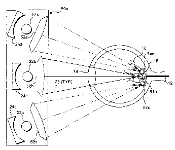
FIGURE 5 illustrates a PDT device 50 in which the focal point can be
adjusted without repositioning the PDT device. PDT device 50 incorporates a
plurality of TIR lenses 52a, 52b, and 52c, each of which provides a different
focal
point. It should be noted that the convergent lens of PDT device 20 could also
be
incorporated in PDT device 50. Also it should be noted that the number of
lenses
that can be incorporated into PDT device 50 is not limited to only three. More
lenses arranged in a two-dimensional array, for example, could be used to
provide
greater flexibility in selecting a particular focal point without
repositioning PDT
device 50. PDT device 50 incorporates a light source 22 and a reflector 24. As
noted with regard to PDT device 40, reflector 24 is optional and can be
omitted.
In one embodiment of PDT device 50, TIR lenses 52a, 52b, and 52c are
moveable, and can be selectively positioned in front of light source 22. TIR
lens 52a is has a focal point 54a, while TIR lens 52b has a focal point 54b,
and
TIR lens 52c has a focal point 54c. Thus, without repositioning PDT device 50,
a
plurality of different focal points can be achieved by moving an appropriate
one of
the TIR lenses in front of light source 22.
A second embodiment of a PDT device 50a that provides a plurality of
different focal points without requiring repositioning of the PDT device is
illustrated in FIGURE 6. In PDT device 50a, TIR lenses 52a, 52b, and 52c are
not
moveable. Instead, PDT device 52a incorporates a plurality of light sources
22a,
22b, and 22c, one for each TIR lens 52a, 52b, and 52c. Reflectors 24a, 24b,
and 24c can optionally be included, one for each of the light sources. In PDT
device 50a, focal point 54a, 54b, or 54c is selected by energizing light
source 22a, 22b, or 22c that is associated with the desired focal point. As
illustrated in FIGURE 6, light source 22b has been energized, resulting in
focal
point 54b being achieved. While not shown, it is envisioned that a similar
device
can be constructed using a plurality of TIR lenses in association with a
single light
source and a plurality of reflectors that enable light rays from the light
source to
be selectively directed through the TIR lens that corresponds to the desired
focal
point.
In FIGURE 7, a PDT device 60 incorporates a light source 22d, a filter 63,
which includes an opaque region 62 and a transparent region 64, and a
diverging
lens 66. Light rays from light source 22d are blocked by opaque region 62,
casting a correspondingly shaped shadow 68 on the treatment site within the
eye.
Light not blocked by the opaque region is transmitted through transparent
region 64 and into the eye. Those of ordinary skill in the art will readily
CA 02392313 2002-05-21
WO 01/43825 _ 10_ PCT/US00/33052
understand that the size of shadow 68 relative to transparent region 64 can be
varied by selecting an appropriate focus for diverging lens 66. As discussed
above, the position of the focal point and of shadow 68 relative to macula 18
can
be adjusted by repositioning PDT device 60. Light source 22d is preferably an
LED and can include, if desired, a reflector (not shown) similar to the
reflectors
described in the preceding embodiments.
FIGURES 7A and 7B illustrate filters 63a and 63b, which incorporate
different shapes of transparent regions 64a and 64b and opaque regions 62a
and 62b. Thus, by replacing filter 63 with a different filter, the shape of
the
shadow at the focal point of the PDT device can readily be varied to treat
different
shapes of abnormal tissue at the treatment site within the patient's eye.
FIGURE 8 illustrates a PDT device 70 in which the shape of the focal
point can be varied without replacing the filter element. PDT device 70
includes a
light source 22, a diverging lens 72, a filter element 75, which includes a
transparent region 76 and an opaque region 74, and a diverging lens 78. PDT
device 70 also includes an electrical stimulus controller 80. As noted above,
the
choice of divergent lens 72 and divergent lens 78 can provide a desired focal
point 82, which is useful in treating diseases on macula 18. Filter element 75
is
preferably fabricated from a material that changes its index of refraction or
its
transparency with an applied heat or in response to an electrical stimulus.
The
techniques used to produce liquid crystal displays (LCDs) can also be used to
create a mask that can selectively be controlled to block light transmission
through a portion of the filter element. The mask can be made so that the
desired
shape of light or of the shadow at the focal point is achieved when the mask
is
energized to block some of the light going through the lens. The mask can also
include a plurality of pixels or regions that are electrically controlled so
as to alter
the pattern of light transmission through the mask as is typically done in a
LCD.
In this manner, the shape or pattern of the light blocking pixels can be
changed as
desired in order to create different light/shadow patterns at the focal point
of the
PDT device. Ceramic materials (referred to as PZT materials) are also readily
available that can be used to create a light mask. These ceramic materials
appear
transparent to light until an electrical stimulation is applied, at which
time, they
are rendered partially or completely opaque. The electrical stimulation can be
applied through a transparent, indium oxide conductive electrode that is
applied to
the surface of the PZT material in a predefined electrode pattern similar to
that
employed for the LCD mask pattern.
CA 02392313 2002-05-21
WO 01/43825 PCT/US00/33052
FIGURES 9, 9A, 10, and 1 OA illustrate a PDT device 81, which changes
the shape of the focal point using a different property. PDT device 81
incorporates a light source 22a and a convergent lens 83. Convergent lens 83
is
fabricated from a material that can be deformed sufficiently without damaging
the
lens. Lens 83 is selectively deformable, to change the pattern of light
passing
through the lens, when focused on a treatment site or to alter the focal
point. As
lens 83 is deformed, the light traveling through the lens is redirected to
form an
elongate oval pattern at the focus of the lens. Pressure can be exerted on the
outer
periphery of the lens to cause the lens deformation resulting in a desired
light
pattern. Alternatively, mechanical forces can be applied to the outer surface
of
lens 83 to cause the lens to distort or deform, thereby producing the desired
light
pattern on a treatment site inside the eye. This mechanical force can be
applied to
the lens by activating piezoelectric crystals, or by miniature
pneumatic/hydraulic
cylinders, or simply by use of electrically actuated (motor driven) screws,
clamps,
I S or lever devices. In FIGURE 9, lens 83 is undistorted and a focal point 84
represents a circular light pattern. It should also be noted that the position
of focal
points 84 and 86 can be manipulated by moving PDT device 81. Furthermore,
reflective elements similar to reflectors 24, as illustrated in the previously
discussed Figures, can be included in PDT device 81.
FIGURES 11 A, 11 B, and 1 I C illustrate PDT devices that allow a
practitioner to use a non-activating wavelength of light to verify the
position of
the focal point relative to the macula, and then to selectively transmit a
desired
wavelength of light to the focal point. PDT drugs are known that are activated
using either blue or red light of a wavelength corresponding to the absorption
waveband of the photoreactive agent drug. Blue light sensitive PDT drugs
typically have relatively large absorption peaks, which enable far less drug
to be
given to achieve the same therapeutic effect. The reduced dosage of the PDT
drug
in turn reduces side effects, such as elevated dermal photosensitivity.
Another
advantage of using PDT drugs that are activated by blue light is that light of
this
waveband penetrates only a short distance into normal tissues surrounding the
treatment site. PDT drugs activated by red light are preferred if the
treatment site
is obscured by tissue or physiological structures, since the longer wavelength
light
more readily penetrates such tissue to a greater depth.
With reference to FIGURES 1 IA and I IB, a PDT device 90 includes a
non-PDT drug activating light source 22a, a PDT drug activating light source
22b,
and a PDT drug activating light source 22c having a different wavelength than
CA 02392313 2002-05-21
WO 01/43825 PCT/US00/33052
-12-
light source 22b. PDT device 90 also includes a TIR lens 52, and optionally
includes a reflector 24. It should be noted that convergent lenses as
discussed
with respect to other embodiments could also be used instead of TIR lens 52 as
shown. In FIGURE 11A, light source 22a is energized and the practitioner can
observe the condition of the eye at a focal point 91 a, to determine how best
to
render PDT, and to select a focus for the PDT that will be provided when the
other
light sources are activated. Because light source 22a produces a wavelength
that
does not activate the PDT drug that has been administered to the patient, the
practitioner is able to position PDT treatment device 90 such that the
position of
focal point 91 a corresponds to the desired treatment area within macula 18
(or
other region of interest). Because light source 22a is non-coherent, and does
not
activate the PDT drug, continued illumination with light source 22a while the
PDT is ongoing has no adverse effect on the patient. In FIGURE 11B, light
source 22a has been de-energized, and light sources 22b and 22c have been
energized. The wavelengths of light rays 28b overlap the absorption or
activation
waveband of the PDT drug administered to the patient. Accordingly, the light
converging at focal point 91b activates the PDT drug and initiates the
treatment
process. Light sources 22b and 22c are selectively energizable, such that
either or
both can be energized as desired. As discussed above, light sources 22b and
22c
preferably are LEDs, which generate light in the blue waveband and red
wavebands, respectively. The various light sources are moved into the position
behind TIR lens 52 when activated to provide light on the treatment site.
FIGURE 11 C illustrates a PDT device 92 that also enables a practitioner to
use a non-PDT drug activating wavelength of light to insure that focal point
91a is
located corresponding to the target of interest, and then to selectively
transmit
wavelengths of light which activate the PDT drug. PDT device 92 incorporates a
light source 22d, which emits a waveband of light that activates the PDT drug,
as
well as a waveband of light that does not. PDT device 92 also includes TIR
lens 52, and optionally includes a reflector 24. As noted above, other types
of
lenses may be beneficially provided in PDT device 92. PDT device 92 also
includes a filter 94. Filter 94 is selected to modify the wavelength of light
emitted
by light source 22d, such that the light passing through TIR lens 52 and
converging at focal point 91 a has been filtered to exclude the waveband that
activates the PDT drug. As illustrated in FIGURE 11C, filter 94 is positioned
to
filter the light emitted by light source 22d. Thus, focal point 91 a does not
include
the wavelength of light that activates the PDT drug, and in this
configuration,
CA 02392313 2002-05-21
WO 01/43825 _ 13_ PCT/US00/33052
PDT treatment device 92 can be focused at the desired target of interest
without
activating the PDT drug. When filter 94 is moved out of the optical path
between
light source 22d and TIR lens 52, the wavelengths of light converging at focal
point 91 a include the wavelengths corresponding to the activation waveband of
the PDT drug, and treatment is rendered. As noted above, filter 94 preferably
modifies the wavelength of light emitted by light source 22d to eliminate
light in
the red and blue spectrums.
It is envisioned that the PDT devices described in the above embodiments
can be beneficially incorporated into a lightweight headset that can be worn
by a
patient, or incorporated into a more traditional slit lamp similar to those
used by
optometrists. FIGURES 12, 13, 14, and 15 illustrate different embodiments of a
headset that can be worn by a patient. FIGURE 16 illustrates how the PDT
devices described above are incorporated into a traditional, permanently
fixtured
slit lamp.
FIGURE 12 illustrates a PDT headset 100. PDT headset 100 appears
much like a conventional pair of wraparound eyeglasses, but includes PDT
devices 106a and 106b disposed on the front of the lenses, adjacent to the
patient's
eyes. A frame 102 is preferably fabricated from metal, and if desired, can be
coated with a plastic material as is commonly done for prescription
eyeglasses, or
can be fabricated entirely of plastic. A lens 104 is preferably an opaque,
shatter
resistant plastic material. While lens 104 could be transparent, use of an
opaque
material for lens 104 enables the patient's eyes to dilate by reducing
incident light
on the eyes and helps to prevent untargeted light rays from reaching the
patient's
eyes. It should be noted that PDT devices 106a and 106b are not obstructed by
opaque lens 104, but instead are either fitted over a window (not shown) in
the
lenses or the portion of lens 104 covered by PDT devices 106a and 106b are
transparent and not opaque.
Control cables 108a and 108b connect PDT devices 106a and 106b,
respectively, to a control unit 110. Control unit 110 enables the practitioner
to
selectively activate the light sources in either or both PDT devices 106a and
106b.
FIGURE 13 illustrates a side view of PDT headset 100. PDT device 106a has
been selectively energized, and is being focused on the macula of eye 10. It
is
envisioned that PDT device 106a and 106b can beneficially include the elements
of any of the embodiments described above. However, PDT devices 106a
and 106b preferably enable the focal point of the light being administered to
the
treatment site within the eye to be adjusted without requiring movement of PDT
CA 02392313 2002-05-21
WO 01/43825 PCT/US00/33052
-14-
headset 100, and those embodiments of the PDT devices discussed above that
permit such functionality are preferred. While PDT headset 100 can be
manipulated somewhat while being worn by a patient, it is more convenient if
the
light sources in PDT devices 106a and 106b can be focused on the treatment
site
in the patient's eyes without requiring such manipulation.
FIGURES 14 and 15 illustrate different embodiments of a PDT
headset 112 and 114, respectively. These devices are identical in operation to
PDT headset 100, and illustrate that a plurality of different headset designs
are
possible within the scope of the present invention. Preferably, PDT
headsets 100, 112, and 114 are constructed of lightweight and durable
materials,
not prone to breakage, and have a. shape and size selected so that the
headsets are
comfortable when worn by a patient. As much as possible, the elements required
for the operation of PDT devices 106a and 106b are incorporated into control
unit 110 to reduce the weight of the headset. Plus power supplies will be
incorporated into control unit 110, rather than in PDT devices 106a and 106b.
It
is envisioned that a further reduction in weight could be achieved by locating
the
light sources within control unit I 10 and using one or more fiber optic
cables (not
shown) to convey the light emitted by the light sources to PDT devices 106a
and 106b. However, this option may not result in a significant decrease in
weight,
since it is envisioned that the light sources used will be relatively
lightweight
LEDs. Another contemplated embodiment would provide for running control
cables 108a and 108b alongside the left and right earpieces of the headset and
disposing control unit 110 at the rear of the headset. While this modification
would increase the weight of the headset, it would result in a headset in
which
control cables 108a or 108b are less likely to become tangled, disconnected,
or
broken.
FIGURE 16 illustrates a slit lamp 120 that has been modified to include a
PDT treatment unit 130, corresponding to any of the embodiments of the PDT
devices described above (except the headsets). In general, a slit lamp is a
table
mounted unit that includes a chin rest and head rests to hold a patient's head
stationary so that precise targeting of light or examination within a
patient's eyes
may be achieved. In FIGURE 16, PDT treatment unit 130 is targeted at
macula 18 of eye 10. It is envisioned that any of the PDT devices described
above
with respect to FIGURES 2-1 1 C could be used for PDT treatment unit 130.
Light having a wavelength that corresponds to the absorption waveband of
the photoreactive agent can be directed at the treatment site or target region
in the
CA 02392313 2002-05-21
WO 01/43825 PCT/US00/33052
- I 5-
macula (or at any other desired target region) either directly through the
lens and
cornea of the eye or indirectly, along paths that do not pass through the
eye's lens
or cornea. FIGURE 17 schematically illustrates the direct approach. In
FIGURE 17, light of the appropriate waveband is directed at the treatment
region
from the front of the eye, as indicated by an arrow 138. The light passes
through
lens 14 in the eye and proceeds to a target area, preferably in macula 18.
FIGURE 18 illustrates several indirect paths for targeting the macula (or
other region of interest in the eye). These indirect paths include a lateral
orbital
approach 142, a superior orbital approach 144, and an inferior orbital
approach 146. Light following each of these indirect paths passes into the eye
transcutaneously, passing through an orbital wall. When following such an
indirect path, the light illuminates the area of pathology in a more diffuse
manner
than when administered along the direct path. It is envisioned that light
administered along an indirect path be administered from a light source
included
in a PDT device on a headset, generally like those described above. Although
the
headsets described above are adapted to administer light into the patient's
eyes)
along a direct path, as illustrated in FIGURE 17, PDT devices attached to
headsets
can readily be configured to provide light administered along the inferior,
superior, and lateral approaches illustrated in FIGURE 18. For instance, a
headset
for administering light from PDT devices utilizing superior approach 144 could
be
fabricated in the form of a headband, in which the PDT devices are disposed on
the patient's forehead above each eye. A PDT headset utilizing lateral orbital
approach 142 could be provided by mounting PDT devices on the arm or ear
pieces of the headset, generally between the patient's ears and the front
lenses of
the headset. Inferior orbital approach 146 can be achieved by mounting the PDT
devices below the lenses of the headset, such that the PDT devices rest on the
patient's cheeks.
The Incorporation of Lenses into the Preferred Embodiments
It should be noted that a plurality of different lenses can be beneficially
employed to selectively focus the light emitted from a light source. It is
anticipated that lens configurations such as convergent lenses, totally
internally
reflective lenses, divergent lenses, and a plurality of lenses in combination
can be
employed in conjunction with the present invention. Those of ordinary skill in
the
art will readily understand how these types of lenses can be incorporated in
the
above described embodiments.
CA 02392313 2002-05-21
WO 01/43825 _ 16_ PCT/US00/33052
Targeted PDT Therapy_
The effects of the light therapy on normal tissue that has absorbed the
photoreactive agent may range from mild reddening of the tissue to severe
tissue
damage, depending on the amount of light delivered to the normal tissue, and
the
amount of the photoreactive agent absorbed by the normal tissue. One method to
reduce the risk of incidental damage to non target tissue is to provide a
highly
focused pattern of light that irradiates only the target tissue (such as
abnormal
neovascular). Various of the embodiments described above can be beneficially
employed to provide such a highly focused light pattern. Another method to
minimize damage to the normal tissue is to substantially reduce the extent to
which normal tissue absorbs the photoreactive agent.
One approach developed to ensure that the photoreactive agent is
preferentially absorbed by the target tissue, rather than by normal tissue, is
to bind
antibodies to a photoreactive agent, such that the antibodies are targeted to
the
abnormal cells at a treatment site. When a photoreactive agent conjugated with
an
antibody is administered to a patient, the antibodies will tend to bind the
photoreactive agent to the abnormal tissue, but not to normal tissue, thereby
improving the specificity of the PDT and avoiding harm to the normal tissue.
Those of ordinary skill in the art will readily recognize that an antibody
specific to
abnormal target tissue within the eye can be conjugated with a selected
photoreactive agent, to provide for the preferential absorption of the
photoreactive
agent by the abnormal tissue, thereby minimizing the risk of damage to non
target
tissue.
Although the present invention has been described in connection with
several preferred forms of practicing it and modifications thereto, those of
ordinary skill in the art will understand that many other modifications can be
made to the invention within the scope of the claims that follow. Accordingly,
it
is not intended that the scope of the invention in any way be limited by the
above
description, but instead be determined entirely by reference to the claims
that
follow.